A potential role for the gut microbiota in the specialisation of - bioRxiv
←
→
Page content transcription
If your browser does not render page correctly, please read the page content below
bioRxiv preprint first posted online Jan. 22, 2019; doi: http://dx.doi.org/10.1101/526517. The copyright holder for this preprint
 (which was not peer-reviewed) is the author/funder, who has granted bioRxiv a license to display the preprint in perpetuity.
                              It is made available under a CC-BY-NC-ND 4.0 International license.

                                                                                                                                    1

     A potential role for the gut microbiota in the specialisation of
     Drosophila sechellia to its toxic host noni (Morinda citrifolia)

     Heys C1,2, Fisher AM1, Dewhurst AD1, Lewis Z1* and Lizé A1,3

     1Institute    of Integrative Biology/School of Life Sciences, University of Liverpool,
     Liverpool, L69 7ZB, UK
     2Institute    of Biodiversity, Animal Health, and Comparative Medicine, University of
     Glasgow, Glasgow, G12 8QQ, UK
     3UMR      CNRS 6553 ECOBIO, University of Rennes 1, 35042, Rennes, FRANCE

     *Author for correspondence: Z.Lewis@liverpool.ac.uk

     Abstract

     Adaptation to a novel food source can have significant evolutionary advantages. The
     fruit fly, Drosophila sechellia, is a specialist of the toxic plant noni (Morinda citrifolia).
     Little is known as to how D. sechellia has become resistant to the toxins in the fruit -
     comprised predominantly of octanoic acid - but to date, the behavioural preferences
     for the fruit and genetic architecture underlying them, have been well studied. Here,
     we examine whether the gut microbiota could have played a role in adaptation to the
     fruit. In the first series of experiments, we examine the gut microbiota of wild-type,
     laboratory reared flies and characterise the gut microbiota when reared on the natural
     host plant, versus a standard Drosophila diet. We show a rapid transition in the core
     bacterial diversity and abundance within this species and discover sole precedence of
     Lactobacillus plantarum when reared on M. citrifolia. We also discover that flies reared
     on a laboratory diet are more likely to carry bacterial pathogens such as Bacillus
     cereus, although their function in Drosophila is unknown. Flies reared on a laboratory
     diet have a significantly reduced weight but with no impact on the risk of death before
     adulthood, when compared to the wild noni diet. In the second series of experiments,
bioRxiv preprint first posted online Jan. 22, 2019; doi: http://dx.doi.org/10.1101/526517. The copyright holder for this preprint
 (which was not peer-reviewed) is the author/funder, who has granted bioRxiv a license to display the preprint in perpetuity.
                              It is made available under a CC-BY-NC-ND 4.0 International license.

                                                                                                                                    2

     we examine the potential role of the gut microbiota in adaptation to octanoic acid
     resistance in this species and its sister species, Drosophila melanogaster, to which the
     fruit is usually fatal. We use a combination of methods to analyse resistance to
     octanoic acid by conducting life history analysis, behavioural assays and bacterial
     analysis in both D. sechellia and D. melanogaster. We find that by creating
     experimental evolution lines of D. melanogaster supplemented with gut microbiota
     from D. sechellia, we can decrease D. melanogaster aversion to octanoic acid, with
     the flies even preferring to feed on food supplemented with the acid. We suggest this
     represents the first step in the evolutionary and ecological specialisation of D. sechellia
     to its toxic host plant, and that the gut microbiota, Lactobacillus plantarum in particular,
     may have played a key role in host specialisation.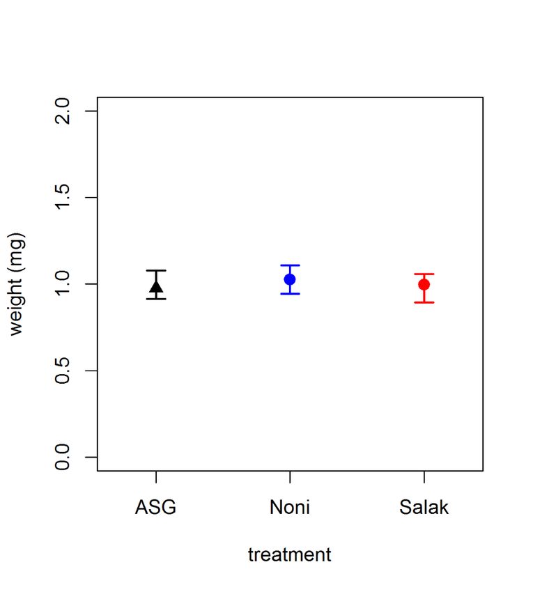
bioRxiv preprint first posted online Jan. 22, 2019; doi: http://dx.doi.org/10.1101/526517. The copyright holder for this preprint
 (which was not peer-reviewed) is the author/funder, who has granted bioRxiv a license to display the preprint in perpetuity.
                              It is made available under a CC-BY-NC-ND 4.0 International license.

                                                                                                                                    3

     Introduction

     Many studies have examined the complex relationships between animals and plants
     (reviewed in Herrera and Pellmyr, 2009). The dynamic ecological and evolutionary
     interactions between an animal and its host plant can take many forms. In some cases,
     insects have even become adapted to living on a toxic plant host, such as Pierinae
     butterflies that feed on toxic Brassicales (e.g. Edger et al., 2015). Developing
     resistance to a toxic host plant through specialisation can have a multitude of
     advantages, including the ability to exploit an otherwise unutilised resource, and there
     are many ways that insects can overcome the toxins in the host plant. Behavioural and
     physiological adaptations to exploit toxic resources can drive speciation of insects or
     other animals (Matsuo et al., 2007). However, it is argued that specialisation to a toxic
     host does not necessarily result in speciation itself, and therefore the role of ecological
     specialisation in speciation remains to be proven (Matsuo et al., 2007).

     The way in which these specialists adapt to life in a novel environment can occur
     through a number of different ways, and one is potentially through the gut microbiota
     (e.g. Bolnick et al., 2014; Morrow et al., 2015). In insects, for example, members of the
     order Hemiptera have evolved to feed on plant phloem sap - a nutritionally poor diet
     due to the grossly unbalanced amino acid composition (e.g. Douglas, 1993; Sandström
     and Moran, 1999; Sandström, 2000). A number of studies have demonstrated that all
     phloem feeders within this order possess certain symbiotic bacteria that mitigate the
     effects of this nutritionally poor diet (Buchner 1965; Gündüz and Douglas, 2009). For
     example, two specialist species of Lepidoptera, Hyles euphorbiae and Brithys crini,
     feed exclusively on latex-rich Euphorbia sp. and alkaloid-rich Pancratium maritimum,
     respectively (Vilanova et al., 2016). Metagenomic sequencing has identified that the
     primary microbiota within the gut is Entereococcus sp., which it is predicted to be
     responsible for mitigating the effects of these nutrient-poor diets to the host.

     The participation of the gut microbiota in the transformation of toxic plant chemicals is
     an important aspect to be considered when studying insect-plant interactions, and one
     that is gaining attention (e.g. Genta et al., 2006; Ceja-Navarro et al., 2015). Yet due to
     the complexity and diversity of gut microorganisms, there is little experimental
     evidence to support this idea. It was suggested by Douglas (1992) that a possible role
     of the midgut microbiota is in detoxification of toxic compounds and a study by Genta
bioRxiv preprint first posted online Jan. 22, 2019; doi: http://dx.doi.org/10.1101/526517. The copyright holder for this preprint
(which was not peer-reviewed) is the author/funder, who has granted bioRxiv a license to display the preprint in perpetuity.
It is made available under a CC-BY-NC-ND 4.0 International license.
4
et al., (2006) in Tenebrio molitor highlighted the role of gut microbiota in detoxifying
the cell walls of fungi and bacteria that typically inhabit their food source. Similarly, the
coffee berry borer (Hypothenemus hampei), the most devastating insect pest of coffee
crops worldwide, has adapted to metabolise caffeine – a toxic alkaloid present within
coffee plants (Ceja-Navarro et al., 2015). Caffeine is shown to be degraded in the gut
via Pseudomonas species, which subsist on caffeine as a sole source of carbon and
nitrogen (Ceja-Navarro et al., 2015).
The majority of species within the Drosophila melanogaster species-complex are food
generalists and saprophagous, meaning they feed on a variety of decaying plant
matter (Rohlfs and Kürschner, 2010). Studies have shown the importance of a diverse
diet in creating and maintaining a diverse gut microbiota within D. melanogaster, with
a diverse gut microbiota increasing survival and reducing development time (Rohlfs
and Kürschner, 2010). In comparison, several species within the D. melanogaster
species-complex have evolved some form of diet specialisation (e.g. Lachaise et al.,
1988). One species within this group, Drosophila sechellia, is a specialist of ripe
Morinda citrifolia fruit – a toxic fruit commonly known as the Tahitian noni (Jones,
2005). D. sechellia’s closely related species, Drosophila simulans, Drosophila
mauritiana and D. melanogaster are notably repelled by the pungent scent of the fruit
and even die upon contact (Legal et al., 1994; Legal et al., 1999). Resistance of D.
melanogaster to the fruit is shown to be dependent on strain (Legal et al., 1992).
The primary toxins found within noni are octanoic and hexanoic acids (Farine et al.,
1996), and numerous studies have focussed on the underlying genetic and molecular
mechanisms that have enabled D. sechellia to adapt to the fruit (e.g. Harada et al.,
2008, 2012; Labeur et al., 2002; Matsuo et al., 2007. However, whether the gut
microbiota has played a role in this specialisation has not yet been investigated.
Interestingly, Chandler et al. (2011) characterised the microbiota of wild D. sechellia
found feeding on the noni and discovered that the gut is dominated by a single
Lactobacillales OTU (84%). This demonstrates the very low bacterial community
richness and diversity within this species, particularly when it is compared to its sister
species, D. melanogaster, which exhibit greater diversity and carry the bacterial
genera Lactobacillus, Acetobacter and Enterococcus (Ryu et al., 2008; Brummel et al.,
2004; Ren et al., 2007; Cox and Gilmore, 2007). Further, the composition of an
individual’s gut microbiota is known to be influenced by pH (see Overend et al., 2016).bioRxiv preprint first posted online Jan. 22, 2019; doi: http://dx.doi.org/10.1101/526517. The copyright holder for this preprint
(which was not peer-reviewed) is the author/funder, who has granted bioRxiv a license to display the preprint in perpetuity.
It is made available under a CC-BY-NC-ND 4.0 International license.
5
Overend et al. (2016) demonstrated that decreasing the pH in certain regions of the
Drosophila gut can lead to an increased abundance of key members of the gut
microbiota – Lactobacillus and Acetobacter. This raises the question whether the very
low bacterial richness found in the D. sechellia gut when it is feeding on its acidic
natural host diet, M. citrifolia, is due to the pH determining the microbiota?
Alternatively, potentially the almost exclusive prevalence of this Lactobacillales is
caused by the specialism of D. sechellia to the host plant, and thus the Lactobacillales
acts as a form of detoxifying agent by metabolising the toxic acids found within the
noni.
In this study we investigated the role of the gut microbiota on host specialisation in D.
sechellia. This study is separated into two sections. In the first, we determined the
effect that rearing D. sechellia on a standard laboratory diet has on the diversity and
richness of the gut microbiota. D. sechellia are widely kept in the laboratory, but little
attention has been paid to the effect that feeding this specialist species a generalist
diet has on the resulting gut microbiota. Flies were first reared on a standard
Drosophila diet (ASG), then moved onto noni, before being transferred back onto ASG.
At each stage, the diversity and abundance of the gut bacteria was measured. We also
disentangled the role of pH on shaping the gut microbiota, from the toxic compounds
present in the noni. We introduced a new dietary treatment, salak fruit (Salacca
zalacca), with similar nutritional and acidic properties to noni but lacking in the toxic
compounds - octanoic and hexanoic acid. We determined the effect that these diets -
a standard laboratory diet, noni and salak fruit - had on a series of life history traits,
including larval, pupal and adult weight and the risk of death before adulthood. We
predicted that the gut microbiota would become more simplified on the noni diet, but
not the salak or ASG, which would indicate that the gut microbiota plays a role in
specialisation to this diet.
In the second part of this experiment, we investigated the effects of differing
concentrations of the toxic compound octanoic acid, the main acidic constituent of D.
sechellia’s natural host plant, M. citrifolia, on weight, development time, survival,
bacterial load and diversity in D. sechellia. We also investigated the same effects in D.
sechellia’s sister species, D. melanogaster, to determine any differences between a
fruit specialist and a fruit generalist species. We analysed both inbred and outbred
lines in D. melanogaster in order to test the effect of strain on ability to withstandbioRxiv preprint first posted online Jan. 22, 2019; doi: http://dx.doi.org/10.1101/526517. The copyright holder for this preprint
(which was not peer-reviewed) is the author/funder, who has granted bioRxiv a license to display the preprint in perpetuity.
It is made available under a CC-BY-NC-ND 4.0 International license.
6
octanoic acid exposure. We predicted that D. melanogaster would have a reduced
weight, development time and survival ability compared to D. sechellia, particularly in
the inbred strain in which genetic diversity is low. We also predicted that we would
observe differences in the diversity and abundance of the gut microbiota of D. sechellia
compared to D. melanogaster, due to D. sechellia possessing a more specialised gut
microbiota that enable them to withstand high concentrations of octanoic acid. We
further examined the role of the gut microbiota in this specialisation by creating
experimental evolution lines of D. melanogaster supplemented with D. sechellia gut
microbes. As we hypothesised the gut microbiota may have played a key role in the
specialisation in D. sechellia, we predicted that after ten generations D. melanogaster
would be less averse to exposure to octanoic acid.
Materials and methods
Flies used and general maintenance
D. sechellia stocks were obtained from the National Drosophila Species Stock Center
located in San Diego. Three lines of outbred flies were utilised (lines 0.21, 0.07 and
0.08), that were collected on Cousin Island, Seychelles in 1980 and maintained in the
laboratory ever since. All flies were kept and reared at 25°C on a 12:12hour light-dark
cycle. Flies were kept in standard 75x25mm Drosophila vials containing 25ml of
standard Drosophila food composed of yeast/agar/maize/sugar. Flies were moved to
new vials every 4 days.
Experiment 1: The changing gut microbiota of D. sechellia
Experimental treatments
Newly emerged, virgin adult flies were obtained from the stock population and
transferred to a new vial containing 25ml of a standard Drosophila dietary media
composed of yeast/sugar/agar/maize (hereon known as ASG). Flies were left to
mature on this media for two days before being transferred to a new vial containing
the same media (N=30). After one week, two males and two females from different
vials from each stock line were isolated using carbon dioxide gas anaesthesia. ThebioRxiv preprint first posted online Jan. 22, 2019; doi: http://dx.doi.org/10.1101/526517. The copyright holder for this preprint
(which was not peer-reviewed) is the author/funder, who has granted bioRxiv a license to display the preprint in perpetuity.
It is made available under a CC-BY-NC-ND 4.0 International license.
7
protocol below detailing the bacterial analysis was then followed for these individuals,
to determine the gut bacterial load and diversity of flies reared on this diet. These flies
formed the “ASG 1” treatment.
The remaining flies were then gently aspirated into fresh vials containing 25g of noni
and left for one week (N=30). After this time, two males and two females from different
vials from each stock line were again isolated and the same bacterial analysis protocol
was followed. This enabled us to determine any changes in the gut microbiota in the
same population of flies, that were first reared on a different diet. These flies formed
the “Noni” treatment.
Similarly, the remaining flies were again, gently aspirated into fresh vials containing
25ml of ASG and left for one week (N=30). After this time, two males and two females
from different vials from each stock line were again isolated and the same bacterial
analysis protocol was followed. This enabled us to determine any further changes in
the gut microbiota, when flies from the same population were transferred between two
different diets in a short period of time. These flies formed the “ASG 2” treatment.
pH and diet type on life history traits
In order to test the effect of acidity on D. sechellia life history traits, we constructed
three different diets of varying pH. Flies were reared on one of three diets: ASG (for 1l
of water: 85g of sugar, 60g of corn, 20g of yeast, 10g of agar and 25ml of nipagin),
noni, or salak fruit. Morinda citrifolia is the diet of wild D. sechellia and has a low, acidic
pH of 3.86 due to the high concentrations of both octanoic and hexanoic acids (Legal
et al., 1994). The salak fruit diet was used as an alternative diet to the noni, as it also
has a low, acidic pH at 3.59, but does not contain the toxic octanoic and hexanoic
acids that are present in noni. These two diet types were compared to the typical
laboratory diet of ASG, that has a higher, more alkaline pH than the fruit diets at 5.97.
Risk of death before adulthood
The number of days was measured from day of female oviposition to day of adult
emergence. Vials were checked at three time points within each day – 9am, 12pm and
5pm – and the cumulative number of adults emerged from each time point was scored
(NASG = 76; NNoni = 60; NSalak = 125).
Weight at different life stagesbioRxiv preprint first posted online Jan. 22, 2019; doi: http://dx.doi.org/10.1101/526517. The copyright holder for this preprint
(which was not peer-reviewed) is the author/funder, who has granted bioRxiv a license to display the preprint in perpetuity.
It is made available under a CC-BY-NC-ND 4.0 International license.
8
In order to accurately determine the effect of acidity on life history, the weights of three
different life stages were measured. For larval weight, vials were checked daily during
the morning and any third instar larvae present were removed and washed with
distilled water in order to remove any excess food. Larvae were grouped according to
treatment and placed into the freezer at -18°C for two hours. Later, larvae were
removed and weighed using an Ohaus five place balance and their weight was
recorded (in mg) to four decimal places (NASG = 50; NNoni = 50; NSalak = 50).
For the pupal and adult weights, vials were similarly checked daily at three time points
– 9am, 12pm and 5pm – to check for any freshly pupated or newly emerged individuals.
For the pupae, care was taken to remove pupae from the vials without damaging them
(NASG = 50; NNoni = 50; NSalak = 50). The adult flies were isolated as virgins and
separated according to sex. Adults were placed into vials at a standard density of ten
per vial and left for two hours to allow their wings to dry out and inflate. Two hours
later, vials were placed into the freezer at -18°C and left overnight. Pupae were
grouped according to treatment and placed into the freezer at -18°C for two hours.
Later, the pupae and adults were removed and weighed using an Ohaus five place
balance and their weight was recorded (in mg) to four decimal places. In the adults,
male and female measurements for each treatment were recorded and analysed
separately (females: NASG = 53; NNoni = 50; NSalak = 50; males: NASG = 45; NNoni = 45;
NSalak = 50). This method of determining adult weight was used for both sections of the
experiments.
Experiment 2: Octanoic acid resistance in D. sechellia and D. melanogaster
Survival rate
To determine resistance to the toxic octanoic acid compound, the survival of the three
different strains, outbred D. melanogaster, inbred D. melanogaster and D. sechellia,
to exposure of differing concentrations of octanoic acid was measured. Newly
emerged, virgin adults were isolated from the stock populations and transferred to
fresh vials containing 25ml standard Drosophila media and left for 3 days to mature.
During this time, ≥99% octanoic acid (purchased from Sigma-Aldrich) was diluted in
distilled water to the following concentrations: 0% (distilled water), 1%, 5%, 10%, 25%,
50%. 30μl of the acid solute was pipetted onto the surface of a fresh vial containingbioRxiv preprint first posted online Jan. 22, 2019; doi: http://dx.doi.org/10.1101/526517. The copyright holder for this preprint
(which was not peer-reviewed) is the author/funder, who has granted bioRxiv a license to display the preprint in perpetuity.
It is made available under a CC-BY-NC-ND 4.0 International license.
9
25ml standard Drosophila media. The vial was tipped to the side to ensure the acid
covered the entire surface of the food media and left to dry for 2 hours. After this time,
males and females were separated according to sex and placed at a standard density
of 10 individuals per vial. The number of dead individuals was counted every 24hours
for a total period of 168hours and the survival rate calculated (N InbredDmel = 50;
NOutbredDmel = 50; NDsechellia = 50 for each acid concentration).
Development time
Mated adults at a density of five females and five males were placed onto fresh vials
containing the following concentrations of octanoic acid: 0% (distilled water), 1%, 5%,
10%, 25%, 50%, to determine the effects of different concentrations of octanoic acid
on development time. The pairs were left to oviposit. Development time was measured
as the number of days from female oviposition to day of adult emergence. Vials were
checked at three time points within each day – 9am, 12pm and 5pm – and the
cumulative number of adults emerged from each time point was scored. Emergent
adult flies from each time point were removed from the vial and placed into a fresh vial
containing 25ml of standard Drosophila food (sample sizes are documented below.
Experimental evolution of D. melanogaster lines exposed to D. sechellia gut microbiota
To determine whether it is the gut microbiota that enables D. sechellia to become
attracted to and feed on a diet that contains high levels of the toxic compound, octanoic
acid, we created a series of experimental evolution lines. Using the stock population
of outbred D. melanogaster, we added D. sechellia gut solute to the dietary media over
several generations. Here, stock population D. sechellia that were continually reared
on a noni diet, were first surface sterilised in 70% ethanol, rinsed in distilled water and
air dried. The head was then removed. Two guts were dissected into each Eppendorf
containing 250μl of sterile LB (Lysogeny Broth) broth. An equal number of males and
females were used to ensure there were no sex-specific differences in the bacterial
content. Gut tissue was homogenised with a sterile plastic pestle. 30μl of gut isolate
was then pipetted onto the surface of a vial containing 25ml standard Drosophila
media. The vial was tipped to the side to ensure the solute covered the entire surface
of the food media and left to dry for 20minutes. After this time, newly emerged, virgin
males and females were isolated from the stock population of outbred D. melanogaster
and placed at a standard density of 10 males and 10 females per vial. After pupaebioRxiv preprint first posted online Jan. 22, 2019; doi: http://dx.doi.org/10.1101/526517. The copyright holder for this preprint
(which was not peer-reviewed) is the author/funder, who has granted bioRxiv a license to display the preprint in perpetuity.
It is made available under a CC-BY-NC-ND 4.0 International license.
10
were seen in all vials, the adult flies were removed so the offspring and adults did not
interbreed. Once the offspring had emerged as adults, a sub-sample of the first
generation were isolated. Some of these individuals were harvested and their gut
bacterial content and diversity analysed. The rest of the sub-sample of the first
generation were placed into octanoic acid aversion trials (see protocol below). The
remaining flies were placed onto a fresh vial similarly containing 25ml standard
Drosophila media and 30μl of gut isolate. This process was repeated until the offspring
reached the 10th generation. Newly emerged adult offspring were harvested as before.
The remaining adults were placed into octanoic acid aversion trials. Thus, the aversion
trials were conducted on unselected stock individuals for each species, and the first
and tenth generations of the experimental evolution lines.
Octanoic acid aversion trials
In order to test whether experimental evolution of outbred D. melanogaster reared on
a diet supplemented with D. sechellia gut microbiota reduces aversion to the toxic
octanoic acid, aversion trials were performed, using a similar methodology to that
utilised previously (e.g. Dekker et al., 2006). Newly emerged, virgin adults were
isolated from the D. sechellia and outbred D. melanogaster stock populations,
separated according to sex and placed into fresh vials containing 25ml standard
Drosophila media. Similarly, newly emerged, virgin adults were isolated from the first
generation (hereon known as Dmel 1) and 10th generation (hereon known as Dmel 10)
experimental evolution lines described above, separated according to sex and placed
into fresh vials containing 25ml standard Drosophila media. Flies were left to mature
for 3 days before being placed into an aversion arena (Figure 1). The aversion arena
consisted of a standard petri dish (measuring 100mm x 15mm) containing two pieces
(10g) of standard Drosophila food located at opposite ends, with a marked line half-
way across clearly showing the two separate sides. 10ul of concentrated octanoic acid
was pipetted on to one of these pieces of food. An individual fly was gently aspirated
into the centre of the arena and left to acclimatise for five minutes. After this time, the
side on which the fly was located was scored as its preference, i.e., either the side with
food containing octanoic acid, or without (female NDmel = 50, male NDmel = 50; female
NDsech = 50, male NDsech = 50; female NDmel1 = 51, male NDmel1 = 50; female NDmel10 =
49, NDmel10 = 48).bioRxiv preprint first posted online Jan. 22, 2019; doi: http://dx.doi.org/10.1101/526517. The copyright holder for this preprint
(which was not peer-reviewed) is the author/funder, who has granted bioRxiv a license to display the preprint in perpetuity.
It is made available under a CC-BY-NC-ND 4.0 International license.
11
Bacterial analysis for all experiments
Collected flies were first surface sterilised in 70% ethanol, rinsed in distilled water and
air dried. The head was then removed. Two guts were dissected into each Eppendorf
containing 250μl of sterile LB (Lysogeny Broth) broth (Bertani, 2004). An equal number
of males and females were used to ensure there were no sex-specific differences in
the bacterial content. Gut tissue was homogenised with a sterile plastic pestle. 100μl
of gut homogenate was pipetted onto BHI (Brain, Heart Infusion) agar (Atlas, 2004)
and spread-plated using a sterile glass loop. BHI media was used as it was found to
favour greater colony growth. Plates were left to air dry aseptically, before being closed
and sealed with parafilm. Plates were incubated at 25°C for 72 hours, and bacterial
load was quantified by performing CFU (Colony Forming Unit) counts.
Single colonies were isolated using a sterile 1μl loop and placed into an Eppendorf
with 10μl sterile water. PCR amplification was performed in a 25μl reaction volume
consisting of 10μl nuclease-free water, 13μl Taq green master mix, 0.5μl of forward
primer 27F (5’- AGAGTTTGATCMTGGCTCAG-3’) and reverse primer 1492R (5′-
GGTTACCTTGTTACGACTT-3’) and 1μl of template DNA. Thermal cycling was
performed for 90 seconds at 95°C as initial denaturation, followed by 35 cycles of 30
sec at 95°C for denaturation, 30 sec at 55 °C as annealing, 90 sec at 72 °C for
extension, and final extension at 72 °C for 5 min. 1500 bp 16S PCR products were
purified with Ampure beads and subjected to Sanger sequencing. The resulting
sequences were identified using NCBI BLAST against the nt database (Altschul et al.,
1990).
Statistical analysis
All data were analysed using R (version 3.3.0; R Core Team, 2016). Larval, pupal and
adult weight were analysed using separate General Linear Models (GLM). Adult weight
was first analysed with both sexes grouped, before further analysis separated
according to sex were performed. The aversion data was analysed using a binomial
GLM. Variation in development time, survival response to octanoic acid and the risk of
death before adulthood was analysed via Cox Proportional-Hazard Regressions.bioRxiv preprint first posted online Jan. 22, 2019; doi: http://dx.doi.org/10.1101/526517. The copyright holder for this preprint
(which was not peer-reviewed) is the author/funder, who has granted bioRxiv a license to display the preprint in perpetuity.
It is made available under a CC-BY-NC-ND 4.0 International license.
12
Development failure of flies was used as the ‘event’ for the risk of death before
adulthood data. The Survdiff function was used to assess differences between two or
more survival curves according to treatment. The coxph function was used to assess
differences between treatments. This allowed treatments to be compared in a pairwise
fashion, to ascertain whether all treatments differed, or whether any significant
differences observed were derived from a single treatment.
Results
Experiment 1: The changing gut microbiota of D. sechellia
Bacterial colony growth was observed in all treatments, with both greater diversity and
greater abundance of bacteria found in the ASG 1 and ASG 2 flies (Table 1). Flies
analysed from these treatments were found to have Lactobacillus plantarum,
Paenibacillus sp. and Bacillus cereus species present. In nearly all of the noni flies
only L. plantarum was observed, with the exception of minor colony growth in two of
the male replicates. Little difference was observed between the three different strains
of D. sechellia, or between sexes.bioRxiv preprint first posted online Jan. 22, 2019; doi: http://dx.doi.org/10.1101/526517. The copyright holder for this preprint
(which was not peer-reviewed) is the author/funder, who has granted bioRxiv a license to display the preprint in perpetuity.
It is made available under a CC-BY-NC-ND 4.0 International license.
13
Table 1. Number of bacterial colonies isolated from the midgut of adult flies. Flies
were first reared on ASG (represented by ASG 1), then moved onto noni, before being
transferred back onto ASG (ASG 2).
L. Paenibacillus Bacillus
Diet Strain Replicate Sex plantarum sp. cereus
ASG 1 0.21 1 F 3.12x10^2 0.20x10^1 0
ASG 1 0.21 2 F 1.82x10^2 1.40x10^1 0
ASG 1 0.21 1 M 1.58x10^2 0 0.10x10^1
ASG 1 0.21 2 M 2.50x10^1 0.90x10^1 0
ASG 1 0.07 1 F 5.11x10^3 1.81x10^2 0.50x10^1
ASG 1 0.07 2 F 5.94x10^3 1.23x10^2 0.20x10^1
ASG 1 0.07 1 M 4.88x10^3 5.40x10^1 0.70x10^1
ASG 1 0.07 2 M 3.58x10^3 1.75x10^ 2.20x10^1
ASG 1 0.08 1 F 6.25x10^3 2.02x10^2 2.70x10^1
ASG 1 0.08 2 F 4.09x10^3 1.96x10^2 1.50x10^1
ASG 1 0.08 1 M 3.17x10^3 2.70x10^1 0.40x10^1
ASG 1 0.08 2 M 2.89x10^3 8.80x10^1 0
Noni 0.21 1 F 2.72x10^3 0 0
Noni 0.21 2 F 1.78x10^3 0 0
Noni 0.21 1 M 1.62x10^2 0 0
Noni 0.21 2 M 1.59x10^2 0 0
Noni 0.07 1 F 1.43x10^3 0 0
Noni 0.07 2 F 1.34x10^3 0 0
Noni 0.07 1 M 2.55x10^2 1.50x10^1 0
Noni 0.07 2 M 1.92x10^2 0.70x10^1 0
Noni 0.08 1 F 4.5x10^2 0 0
Noni 0.08 2 F 1.81x10^2 0 0
Noni 0.08 1 M 8.10x10^1 0.50x10^1 0
Noni 0.08 2 M 7.20x10^1 0 0
ASG 2 0.21 1 F 1.21x10^3 2.40x10^1 0
ASG 2 0.21 2 F 1.45x10^3 1.20x10^1 0.10x10^1
ASG 2 0.21 1 M 1.22x10^2 0.20x10^1 0.10x10^1
ASG 2 0.21 2 M 2.31x10^2 0.50x10^1 0.20x10^1
ASG 2 0.07 1 F 4.51x10^2 1.50x10^2 0.70x10^1
ASG 2 0.07 2 F 5.22x10^2 8.90x10^2 0.20x10^1
ASG 2 0.07 1 M 2.09x10^3 2.90x10^1 0.60x10^1
ASG 2 0.07 2 M 2.87x10^3 4.60x10^1 1.30x10^1
ASG 2 0.08 1 F 3.22x10^3 1.98x10^2 2.90x10^1
ASG 2 0.08 2 F 2.46x10^3 2.51x10^2 2.20x10^1
ASG 2 0.08 1 M 1.78x10^3 1.12x10^2 0.50x10^1
ASG 2 0.08 2 M 2.34x10^3 1.43x10^2 0.70x10^1bioRxiv preprint first posted online Jan. 22, 2019; doi: http://dx.doi.org/10.1101/526517. The copyright holder for this preprint
(which was not peer-reviewed) is the author/funder, who has granted bioRxiv a license to display the preprint in perpetuity.
It is made available under a CC-BY-NC-ND 4.0 International license.
14
Risk of death before adulthood
No significant difference was observed in the risk of death before adulthood between
the ASG or noni treatments (Z2=0.373, P=0.709) (Figure 1). However, flies reared on
salak had a significantly lower risk of death before adulthood than flies reared on noni
(Z2=-2.187, P=0.028). A trend was observed in the risk of death before adulthood
between ASG and salak treatments, with salak individuals exhibiting a higher risk of
death before adulthood than ASG flies (Z2=-1.905 P=0.056).
Figure 1. Development time failure, measured in days as the risk to die before
adulthood, of D. sechellia. Eggs were reared under one of three different dietary
treatments - either ASG, noni or salak diets.bioRxiv preprint first posted online Jan. 22, 2019; doi: http://dx.doi.org/10.1101/526517. The copyright holder for this preprint
(which was not peer-reviewed) is the author/funder, who has granted bioRxiv a license to display the preprint in perpetuity.
It is made available under a CC-BY-NC-ND 4.0 International license.
15
Larval weight
No difference was observed in larval weight in any pairwise comparisons across all
three treatments: ASG and noni (T2=0.850, P=0.397), ASG and salak (T2=0.335,
P=0.738), noni and salak (T2=-0.515, P=0.608) (Figure 2).
Figure 2. Weight (mg) of third instar D. sechellia larvae reared on one of three different
diet types - either ASG, noni or salak. No significant differences were found.
Pupal weight
Pupae collected from the noni treatment weighed significantly less than pupae from
the ASG treatment (T2=-8.961, PbioRxiv preprint first posted online Jan. 22, 2019; doi: http://dx.doi.org/10.1101/526517. The copyright holder for this preprint
(which was not peer-reviewed) is the author/funder, who has granted bioRxiv a license to display the preprint in perpetuity.
It is made available under a CC-BY-NC-ND 4.0 International license.
16
***
Figure 3. Weights (mg) of D. sechellia pupae reared on one of three different dietary
treatments - either ASG, noni or salak. Vials were checked at various time points for
freshly pupated flies. Significant results are shown with *.
Adult weight
Adult male flies were always found to weigh less than females (PbioRxiv preprint first posted online Jan. 22, 2019; doi: http://dx.doi.org/10.1101/526517. The copyright holder for this preprint
(which was not peer-reviewed) is the author/funder, who has granted bioRxiv a license to display the preprint in perpetuity.
It is made available under a CC-BY-NC-ND 4.0 International license.
17
***
Figure 4. Weights (mg) of both male and female, newly emerged adult D. sechellia
flies reared on one of three different diet types - either ASG, noni or salak. Newly
emerged adults were collected at various time points and allowed two hours for wing
inflation. Male flies are shown here using the blue plots, whilst females are depicted
in the black plots. Significant results are marked with a *.
Bacterial analysis
Bacterial colony growth was observed in all treatments, with a greater diversity of
bacteria found in the flies that were reared on the ASG diet (Table 2). In all flies that
were reared on the noni and salak treatments, only one kind of bacterial colony formed.
Sanger sequencing data identified this as L. plantarum. In the ASG diet flies, L.
plantarum was similarly observed, with Paenibacillus sp. and Bacillus cereus also
found. Little difference was observed between the bacterial load of noni and salak
reared flies, or between the different sexes.bioRxiv preprint first posted online Jan. 22, 2019; doi: http://dx.doi.org/10.1101/526517. The copyright holder for this preprint
(which was not peer-reviewed) is the author/funder, who has granted bioRxiv a license to display the preprint in perpetuity.
It is made available under a CC-BY-NC-ND 4.0 International license.
18
Table 2. Number of bacterial colonies isolated from the midgut of adult flies that were
reared on one of three diets – ASG, noni or salak. Males and females were quantified
separately.
Paenibacillus
Diet Replicate Sex L. plantarum sp. B. cereus
ASG 1 F 2.52x10^2 1.30x10^1 0.20x10^1
2 F 2.91x10^2 1.90x10^1 0
1 M 1.89x10^2 2.30x10^1 0
2 M 2.34x10^2 1.80x10^1 0.60x10^1
Noni 1 F 2.85x10^2 0 0
2 F 3.19x10^2 0 0
1 M 1.51x10^2 0 0
2 M 1.99x10^2 0 0
Salak 1 F 1.31x10^2 0 0
2 F 1.66x10^2 0 0
1 M 2.12x10^2 0 0
2 M 2.45x10^2 0 0
Experiment 2: Octanoic acid resistance in D. sechellia and D. melanogaster
Survival rate
D. sechellia females reared on 0% octanoic acid exhibited significantly higher survival
than females reared on the 1% concentration (Z5=3.340; PbioRxiv preprint first posted online Jan. 22, 2019; doi: http://dx.doi.org/10.1101/526517. The copyright holder for this preprint
(which was not peer-reviewed) is the author/funder, who has granted bioRxiv a license to display the preprint in perpetuity.
It is made available under a CC-BY-NC-ND 4.0 International license.
19
higher concentrations: 5% (Z5=3.340; P20
A B C
Figure 5. the proportion of survived female flies when exposed to standard Drosophila food containing differing concentrations of octanoic acid –
0%, 1%, 5%, 10%, 25%, 50%. The vials were checked every 24 hours with the last time point 168 hours, and the number of dead flies recorded.
The graphs are arranged according to species: D. sechellia (A), inbred D. melanogaster (B) and outbred D. melanogaster (C).bioRxiv preprint first posted online Jan. 22, 2019; doi: http://dx.doi.org/10.1101/526517. The copyright holder for this preprint
(which was not peer-reviewed) is the author/funder, who has granted bioRxiv a license to display the preprint in perpetuity.
It is made available under a CC-BY-NC-ND 4.0 International license.
21
In contrast to female survival rates, in male D. sechellia, flies reared on the 0% acid
treatment had equal survival rates to all other treatments, with no difference found
compared to 1% reared flies (Z5=0.941; P=0.357), 5% (Z5=1.341; P=0.180), 10%
(Z5=-1.448; P=0.148), 25% (Z5=0.808; P=0.419) or 50% (Z5=-0.045; P=0.964) (Figure
6). Similar to the female D. sechellia, male flies reared on the 10% treatment were
significantly more able to survive than those reared on 1% treatment (Z5=-2.239;
P=0.019), the 5% treatment (Z5=-2.691; P=0.007) and the 25% treatment (Z5=2.207;
P=0.027).
In male inbred D. melanogaster flies, 0% reared flies had a significantly higher survival
ability than 25% flies (Z5=7.830; PbioRxiv preprint first posted online Jan. 22, 2019; doi: http://dx.doi.org/10.1101/526517. The copyright holder for this preprint
(which was not peer-reviewed) is the author/funder, who has granted bioRxiv a license to display the preprint in perpetuity.
It is made available under a CC-BY-NC-ND 4.0 International license.
22
When comparing the proportions of survived males across species, there is a vast
difference in the final proportion of survived D. sechellia males compared to inbred D.
melanogaster (Figure 6). Male inbred D. melanogaster are less able to survive than D.
sechellia males, particularly when reared at high concentrations (e.g. 25% and 50%).
The survival ability of outbred D. melanogaster flies is better than that of inbred flies
with a higher proportion survived at the end of the trial, even when males were placed
at high concentrations, but the proportion was less than that of D. sechellia.23 Figure 6. the proportion of survived male flies when exposed to standard Drosophila food containing differing concentrations of octanoic acid – 0%, 1%, 5%, 10%, 25%, 50%. The vials were checked every 24 hours with the last time point 168 hours, and the number of dead flies recorded. The graphs are arranged according to species: D. sechellia (A), inbred D. melanogaster (B) and outbred D. melanogaster (C).
bioRxiv preprint first posted online Jan. 22, 2019; doi: http://dx.doi.org/10.1101/526517. The copyright holder for this preprint
 (which was not peer-reviewed) is the author/funder, who has granted bioRxiv a license to display the preprint in perpetuity.
                              It is made available under a CC-BY-NC-ND 4.0 International license.

                                                                                                                                    24

     Development time

     In D. sechellia, no significant differences were found in the time taken to develop from
     egg to adulthood between any of the acid treatments (Figure 4). In particular, no
     significant difference was observed between the 0% and the 50% treatments
     (F5=0.740; P=0.459). However, in the inbred line of D. melanogaster, significant
     differences were observed between all treatments with flies reared on higher
     concentrations of acid taking longer to develop than those reared on smaller
     concentrations (Figure 7). Similarly, variation was seen in the time taken for outbred
     D. melanogaster individuals to develop when reared on different concentrations of acid
     (Figure 4). Flies reared on 0% acid concentration took significantly less time to develop
     than those reared on 1% treatment (F5=-7.930; P
25
B C
A
Figure 7. Development time failure measured in days, as the risk to die before adulthood. Eggs were reared on standard Drosophila media that
was supplemented with differing concentrations of octanoic acid – 0%, 1%, 5%, 10%, 25%, 50%. The graphs are arranged according to species:
D. sechellia (A), inbred D. melanogaster (B) and outbred D. melanogaster (C).bioRxiv preprint first posted online Jan. 22, 2019; doi: http://dx.doi.org/10.1101/526517. The copyright holder for this preprint
(which was not peer-reviewed) is the author/funder, who has granted bioRxiv a license to display the preprint in perpetuity.
It is made available under a CC-BY-NC-ND 4.0 International license.
26
Female offspring adult weight
In D. sechellia, female offspring reared on a 0% acid concentration weighed
significantly more than females reared on 1% (T5=-4.244; PbioRxiv preprint first posted online Jan. 22, 2019; doi: http://dx.doi.org/10.1101/526517. The copyright holder for this preprint
(which was not peer-reviewed) is the author/funder, who has granted bioRxiv a license to display the preprint in perpetuity.
It is made available under a CC-BY-NC-ND 4.0 International license.
27
In male inbred D. melanogaster offspring, unlike female flies, 0% flies weighed
significantly more than all other treatments: 1% (T5=-9.574; P28
B C
A
Figure 8. Weight of adult offspring (mg) of newly emerged adults, when their parents were reared on different concentrations of octanoic acid –
0%, 1%, 5%, 10%, 25%, 50%. The graphs are arranged according to species: D. sechellia (A), inbred D. melanogaster (B) and outbred D.
melanogaster (C). Females are shown here by the pink dots and males represented by the blue.bioRxiv preprint first posted online Jan. 22, 2019; doi: http://dx.doi.org/10.1101/526517. The copyright holder for this preprint
(which was not peer-reviewed) is the author/funder, who has granted bioRxiv a license to display the preprint in perpetuity.
It is made available under a CC-BY-NC-ND 4.0 International license.
29
Bacterial analysis
Bacterial colony growth was observed in all treatments, with both greater diversity and
greater abundance of bacteria found in the D. melanogaster inbred and outbred flies
(Table 3). Sanger sequencing data identifies colony 1 as Lactobacillus plantarum;
colony 2 as Paenibacillus sp. and colony 3 as Bacillus cereus. In all the D. sechellia
adult flies and almost all the outbred D. melanogaster flies, only L. plantarum growth
was observed. This is in comparison to the adult inbred D. melanogaster flies, which
exhibited substantial growth of both L. plantarum, Paenibacillus sp. and B. cereus
bacteria. Paenibacillus sp. and B. cereus colonies appear to be present in higher
numbers when the D. melanogaster strains are reared at higher concentrations of the
octanoic acid. The number of colonies identified of each bacterial species, from each
Drosophila species, is included within the appendix (Table 4).
Table 3. Presence or absence of bacterial species detected in the midgut of adult D.
sechellia and both outbred and inbred D. melanogaster flies, when reared on diets
containing different concentrations of octanoic acid. Presence of a certain bacterial
species is denoted with a tick (✓) and absence is with a cross ().
Species Concentration L. plantarum Paenibacillus sp. B. cereus
D. sechellia 0% ✓
D. sechellia 1% ✓
D. sechellia 5% ✓
D. sechellia 10% ✓
D. sechellia 25% ✓
D. sechellia 50% ✓
Outbred D. melanogaster 0% ✓
Outbred D. melanogaster 1% ✓
Outbred D. melanogaster 5% ✓
Outbred D. melanogaster 10% ✓
Outbred D. melanogaster 25% ✓
Outbred D. melanogaster 50% ✓ ✓
Inbred D. melanogaster 0% ✓
Inbred D. melanogaster 1% ✓
Inbred D. melanogaster 5% ✓ ✓ ✓
Inbred D. melanogaster 10% ✓ ✓ ✓
Inbred D. melanogaster 25% ✓ ✓ ✓
Inbred D. melanogaster 50% ✓ ✓ ✓
Octanoic acid aversion trialsbioRxiv preprint first posted online Jan. 22, 2019; doi: http://dx.doi.org/10.1101/526517. The copyright holder for this preprint
(which was not peer-reviewed) is the author/funder, who has granted bioRxiv a license to display the preprint in perpetuity.
It is made available under a CC-BY-NC-ND 4.0 International license.
30
Males and females were tested separately to determine if there was a difference in
aversion rate according to sex. No difference was observed so subsequent analysis
was performed with both sexes grouped together and separated according to species.
Unselected D. sechellia (hereon known as Dsech ST) were found to prefer the food
containing octanoic acid, in comparison to the unselected D. melanogaster stock
population (hereon known as Dmel ST), which were significantly more averse (F 5=-
2.124; P=0.027). First generation D. melanogaster (Dmel 1) flies that had been reared
on a diet supplemented with D. sechellia gut microbiota, were significantly more averse
to the food containing octanoic acid than Dsech ST flies (F5=-2.541; P=0.011). There
was no difference in aversion of octanoic acid found between Dsech ST and tenth
generation D. melanogaster (Dmel 10) flies that had been reared on a diet
supplemented with D. sechellia gut microbiota (F5=1.371; P=0.170). Dmel 10 flies were
also found to be significantly less averse to octanoic acid than Dmel ST (F 5=2.889;
P=0.003); with no significant difference shown between Dmel ST and Dmel 1 flies (F5=-
0.973; P=0.330). Notably, Dmel 1 were found to be significantly more averse to the
food containing octanoic acid than Dmel 10 flies (F5=3.774; PbioRxiv preprint first posted online Jan. 22, 2019; doi: http://dx.doi.org/10.1101/526517. The copyright holder for this preprint
(which was not peer-reviewed) is the author/funder, who has granted bioRxiv a license to display the preprint in perpetuity.
It is made available under a CC-BY-NC-ND 4.0 International license.
31
diet similar in nutritional properties to its natural host plant but without the toxins, we
observe a microbiota of very similar diversity. We then showed the effect that altering
the gut microbiota via diet has on subsequent life history traits, larval, pupal and adult
weight, with little difference observed between larvae and pupae of all diet types. The
only difference was that adults reared on the standard laboratory diet (ASG) weighed
significantly less than noni reared flies. Noni flies had significantly higher development
failure than salak flies, but they also weighed more at adult emergence, suggesting
they have greater fitness.
As predicted, differences in survival ability, development time and resulting offspring
adult weight were shown across the three different study groups – D. sechellia, inbred
D. melanogaster and outbred D. melanogaster – when exposed to increasing
concentrations of toxic, octanoic acid. D. sechellia generally exhibited higher survival
rates in both males and females, when exposed to higher concentrations of octanoic
acid. Inbred and outbred D. melanogaster exhibited similar survival abilities, with
higher mortality seen at higher acid concentrations. The development time of offspring
whose parents were reared on differing concentrations differed according to species,
with D. sechellia and outbred D. melanogaster exhibiting a similar development time,
and with more variance seen in inbred D. melanogaster. Offspring weight was also
more variable in inbred and outbred D. melanogaster compared to D. sechellia, with
both strains of D. melanogaster weighing more than D. sechellia at lower
concentrations, but this effect was reversed at higher concentrations. Similar to
previous results (Chandler et al., 2011), when reared on a standard Drosophila diet
supplemented with octanoic acid, D. sechellia gut microbiota was found to be
consistent to those isolated from the natural host plant, M. citrifolia. The bacteria
isolated from D. sechellia was characterised as L. plantarum. This was also present in
the gut of both inbred and outbred D. melanogaster, with B. cereus and Paenibacillus
sp. also identified.
Little attention has been paid to the gut microbiota of D. sechellia, with the focus
instead on genetic adaptation to its toxic host plant. Chandler et al. (2011)
characterised the gut bacteria of wild D. sechellia found feeding on noni and
determined that the natural gut microbiota of this species is dominated by a single
Lactobacillales. This is in stark contrast to other fruit-feeding, closely related species
of Drosophila that exhibit considerably greater bacterial diversity, such as wild D.bioRxiv preprint first posted online Jan. 22, 2019; doi: http://dx.doi.org/10.1101/526517. The copyright holder for this preprint
(which was not peer-reviewed) is the author/funder, who has granted bioRxiv a license to display the preprint in perpetuity.
It is made available under a CC-BY-NC-ND 4.0 International license.
32
melanogaster which host a number of bacterial genera, including Enterobacteriales,
Burkholderiales and Pseudomonadales. Here, we show that the gut microbiota of wild-
type D. sechellia is diverse when individuals are kept under laboratory conditions on a
formulated diet. We determined that both males and female guts contain Paenibacillus
sp. and Bacillus cereus. Although evidence has shown that the gut microbiota of
laboratory reared species is considerably less diverse than their wild counterparts
(Brummel et al., 2004; Roh et al., 2008), some studies do show deviation from the
typically found bacterial genera of Lactobacillus, Acetobacters and Enterobacter in
laboratory reared flies (e.g. Ren et al., 2007). It could therefore be argued that these
genera of bacteria are present in wild populations of D. sechellia, but only thrive in
great enough numbers for detection when placed onto a diet that encourages their
growth. A bacterial pathogen, Paenibacillus species are known to be present in
honeybee larvae and are responsible for colony collapse by causing American
Foulbrood (e.g. Genersch, 2010).
Both Paenibacillus nanensis and B. cereus have been discovered in wild populations
of Drosophila ananassae (Maji et al., 2013), although their function or effect on the
host is as yet unknown. Presence of these pathogens in both strains on D.
melanogaster may be due to the natural inability and avoidance of D. melanogaster to
tolerate the octanoic acid. Although, previous studies have reported death on contact
with either the noni or octanoic acid (R’Kha et al., 1991; Dekker et al., 2006), our study
shows D. melanogaster are able to survive on moderate to high concentrations of the
acid for a considerable period. This may be due to the strains of D. melanogaster used.
Susceptibility to bacterial pathogens may be unwanted side effect of this survival
ability, due to a weakened immune system. This may particularly be the case for the
inbred strain of D. melanogaster, where a lack of genetic diversity may render
individuals more susceptible to pathogen colonisation (e.g. Alarco et al., 2004).
When individuals are then transferred onto the natural host noni, the gut microbiota
simplifies to a single species - Lactobacillus plantarum – as similarly shown in previous
studies (Chandler et al., 2011). It could be suggested that colonies of Lactobacillus
plantarum dominate when individuals are transferred onto noni, due to this bacterium
acting as a detoxifying agent by metabolising the toxic compounds present in noni. In
humans, L. plantarum is responsible for protecting the urogenital and intestinal tracts
from infection from pathogenic bacteria (Reid and Burton, 2002). In Drosophila, L.bioRxiv preprint first posted online Jan. 22, 2019; doi: http://dx.doi.org/10.1101/526517. The copyright holder for this preprint
(which was not peer-reviewed) is the author/funder, who has granted bioRxiv a license to display the preprint in perpetuity.
It is made available under a CC-BY-NC-ND 4.0 International license.
33
plantarum has similarly been shown to protect against colonisation of pathogens in the
gut (Ryu et al., 2008), by digesting sugars to produce lactic acid, which inhibits the
growth of non-commensal organisms and promotes the growth of Lactobacilli that
thrive in low pH conditions (e.g. Kleerebezem et al., 2003). It is also responsible for
promoting larval growth when nutrients are scarce (Storelli et al., 2011), and plays a
role in mating preferences (e.g. Sharon et al., 2010). Despite the clear role that L.
plantarum plays on D. sechellia host physiology and likely role in digestion of toxic
compounds, high levels of L. plantarum were also found when flies were reared on
salak. Therefore, the dominance of L. plantarum may simply be due to the acidic
conditions provided by both fruits. Further work is needed to elucidate the links
between these two components.
The weight of individuals at different life stages greatly varied depending on the diet
on which they were reared. At the larval stage, no difference in weights was observed
across any of the treatments, yet at the pupal stage, ASG pupae weighed significantly
more than those reared on noni or salak. As such high abundances of Lactobacillus
plantarum were found in the gut across all treatments; it could be that L. plantarum,
which is known to promote larval growth under conditions where nutrients are scarce,
is compensating for the host developing on this laboratory formulated diet. In contrast,
at adulthood, both male and female ASG reared flies weighed significantly less than
both noni and salak flies. One reason for this difference in weights at adulthood may
be due to the presence of B. cereus and Paenibacillus sp. in the adult ASG flies that
are not present in individuals reared on noni or salak. B. cereus and Paenibacillus sp.
have been reported in wild populations of D. ananassae (Maji et al., 2013), with some
studies showing that the immune responses produced by D. melanogaster individuals
in defence of the pathogen B. cereus, can have detrimental effects on life span (Ma et
al., 2012; Ma et al., 2013). Therefore, it could be that the immune responses elicited
by D. sechellia when individuals are reared on the less–preferred diet of ASG override
the beneficial effects of L. plantarum to negatively affect adult weight.
As predicted, both female and male D. sechellia were able to survive on a diet
containing the highest concentration of octanoic acid (50%) with around 50% of
individuals still alive at the end of the assay. Little differences were observed between
any of the different acid concentrations, with the main difference that males reared on
diets supplemented with 10% acid displayed a higher survival than those reared on abioRxiv preprint first posted online Jan. 22, 2019; doi: http://dx.doi.org/10.1101/526517. The copyright holder for this preprint
(which was not peer-reviewed) is the author/funder, who has granted bioRxiv a license to display the preprint in perpetuity.
It is made available under a CC-BY-NC-ND 4.0 International license.
34
diet containing 0% octanoic acid. This suggests that addition of octanoic acid to D.
sechellia is beneficial to survival, at least at lower concentrations. The ability of D.
sechellia to survive at high concentrations of octanoic acid is somewhat to be expected
as M. citrifolia’s, main toxic constituent is octanoic acid, although there is some
variation in the natural concentrations found, with some studies reporting 58% (Farine
et al., 1996) and others 70% (Pino et al., 2010).
Males and females from both the inbred and outbred lines of D. melanogaster were
able to survive when placed onto a diet containing all concentrations of octanoic acid
(Legal et al., 1994; Legal et al., 1999), however survival at high concentrations was
lower than D. sechellia flies. This result is somewhat surprising as previous studies
have noted that D. melanogaster dies upon contact with M. citrifolia, with most doing
so within one hour (Legal et al., 1994; Legal et al., 1999). Survival of both sexes in the
inbred D. melanogaster strain was significantly reduced at higher concentrations
compared to a non-acidic diet, with nearly all individuals recorded dead at the end of
the time period. The survival ability of outbred D. melanogaster was substantially better
than the inbred strain, with only around 50% of females and 30% of males recorded
as dead when reared at 50% acid concentration, at the end of the study. The outbred
D. melanogaster strain may have a better ability to survive on the octanoic acid due to
it being a wild-type strain and maintaining genetic diversity. Further, as D. sechellia is
a sister species of D. melanogaster, the outbred or wild-type strain are more likely to
share more genetic information than the inbred strain, including genes that underpin
resistance to M. citrifolia. Animals with increased genetic diversity are also known to
adapt to stress better than those with reduced genetic diversity (e.g. Bell, 2013).
The development time of offspring that emerged from adults that were reared on
different concentrations of octanoic acid was measured. In D. sechellia, no differences
were observed in the development time of offspring across any of the octanoic acid
concentrations. Similarly, the outbred strain of D. melanogaster also exhibits little
variation in development time between all octanoic acid concentrations, but flies reared
at 0% took the shortest time to develop. This is in comparison to the inbred D.
melanogaster, in which flies reared at the 0% acid concentration took significantly
longer to develop than flies reared at higher concentrations. It is surprising that outbred
D. melanogaster flies displayed a similar development time to D. sechellia. This is likely
due to the conserved genetic diversity in the outbred line. Similar studies have shownYou can also read