FKBP39 controls nutrient dependent Nprl3 expression and TORC1 activity in Drosophila
←
→
Page content transcription
If your browser does not render page correctly, please read the page content below
Zhou et al. Cell Death and Disease (2021)12:571
                                    https://doi.org/10.1038/s41419-021-03860-z                                                                                                             Cell Death & Disease

                                     ARTICLE                                                                                                                                                    Open Access

                                    FKBP39 controls nutrient dependent Nprl3
                                    expression and TORC1 activity in Drosophila
                                    Ying Zhou1,2, Jian Guo1,2, Xinyu Wang1,2, Yang Cheng1,2, Jianwen Guan1,2, Priyam Barman2, Ming-An Sun3,
                                    Yuanyuan Fu1,2, Wanhong Wei2, Congjing Feng4, Mary A. Lilly 5 and Youheng Wei 1,2

                                      Abstract
                                      Target of Rapamycin Complex 1 (TORC1) is a master regulator that coordinates nutrient status with cell metabolism.
                                      The GTPase-activating protein towards Rags complex 1 (GATOR1) inhibits TORC1 activity and protects cells from
                                      damage during periods of stress. Here we characterize multiple pathways that regulate the expression of the GATOR1
                                      component Nprl3 in Drosophila. We determine that the stability of Nprl3 is impacted by the Unassembled Soluble
                                      Complex Proteins Degradation (USPD) pathway. In addition, we find that FK506 binding protein 39 (FKBP39)-
                                      dependent proteolytic destruction maintains Nprl3 at low levels in nutrient replete conditions. Nutrient starvation
                                      abrogates the degradation of the Nprl3 protein and rapidly promotes Nprl3 accumulation. Consistent with a role in
                                      promoting the stability of a TORC1 inhibitor, mutations in fkbp39 decrease TORC1 activity and increase autophagy.
                                      Finally, we show that the 5′UTR of nprl3 transcripts contain a functional upstream open reading frame (uORF) that
                                      inhibits main ORF translation. In summary, our work has uncovered novel mechanisms of Nprl3 regulation and
                                      identifies an important role for FKBP39 in the control of cellular metabolism.
                  1234567890():,;
1234567890():,;
                  1234567890():,;
1234567890():,;

                                    Introduction                                                                                          Some stress signals, such as nutrient scarcity or growth
                                      In eukaryotes, Target of Rapamycin Complex 1                                                      factor shortage, reduce TORC1 activity and inhibit the
                                    (TORC1), an evolutionarily conserved serine/threonine                                               initiation of cap-dependent translation6,7. Cap-dependent
                                    kinase, coordinates nutrient and energy status with cell                                            translation begins with the formation of the 43S pre-
                                    growth1–3. When nutrients are sufficient, TORC1 activity                                             initiation complex (PIC) that moves along the 5′ UTR and
                                    promotes protein synthesis by phosphorylating multiple                                              searches for an AUG start codon, where the 80S ribosome
                                    substrates including, but not limited to, S6 kinases (S6Ks)                                         is formed to initiate translation until arriving at the stop
                                    and 4E-binding proteins (4E-BPs)4. S6Ks phosphorylate                                               codon for termination8,9. However, in eukaryotes, some
                                    multiple translation factors, while 4E-BPs bind to and                                              transcripts contain upstream open reading frames
                                    inhibit the cytoplasmic cap-binding subunit involved in                                             (uORFs), short translated sequences defined by a start
                                    translation initiation5.                                                                            codon AUG and an in-frame stop codon, within their 5′
                                                                                                                                        UTR10–12. Ribosome profiling, a method based on deep
                                                                                                                                        sequencing of ribosome-protected mRNA fragments
                                                                                                                                        (RPFs), is widely used to identify functional uORFs, which
                                    Correspondence: Mary A. Lilly (lillym@mail.nih.gov) or                                              appear in ribo-seq data together with the actively trans-
                                    Youheng Wei (yhwei@yzu.edu.cn)
                                    1
                                                                                                                                        lated main ORF13,14. Typically, functional uORFs act as
                                     Joint International Research Laboratory of Agriculture and Agri-Product Safety,
                                                                                                                                        translation inhibitors of their downstream main ORF8,10.
                                    the Ministry of Education of China, Yangzhou University, Yangzhou 225009, China
                                    2
                                     Animal Physiology Group, College of Bioscience and Biotechnology, Yangzhou                           Amino acids control TORC1 activity by modulating the
                                    University, Yangzhou 225009, China                                                                  guanine nucleotide-binding status of heterodimeric Rags
                                    Full list of author information is available at the end of the article
                                                                                                                                        GTPase RagA/B and RagC/D15,16. The Rag GTPase pro-
                                    These authors contributed equally: Ying Zhou, Jian Guo, Xinyu Wang,
                                    Yang Cheng                                                                                          motes TORC1 activity when RagA/B is bound to GTP and
                                    Edited by E. Baehrecke

                                    © The Author(s) 2021
                                                        Open Access This article is licensed under a Creative Commons Attribution 4.0 International License, which permits use, sharing, adaptation, distribution and reproduction
                                                        in any medium or format, as long as you give appropriate credit to the original author(s) and the source, provide a link to the Creative Commons license, and indicate if
                                    changes were made. The images or other third party material in this article are included in the article’s Creative Commons license, unless indicated otherwise in a credit line to the material. If
                                    material is not included in the article’s Creative Commons license and your intended use is not permitted by statutory regulation or exceeds the permitted use, you will need to obtain
                                    permission directly from the copyright holder. To view a copy of this license, visit http://creativecommons.org/licenses/by/4.0/.

                                    Official journal of the Cell Death Differentiation Association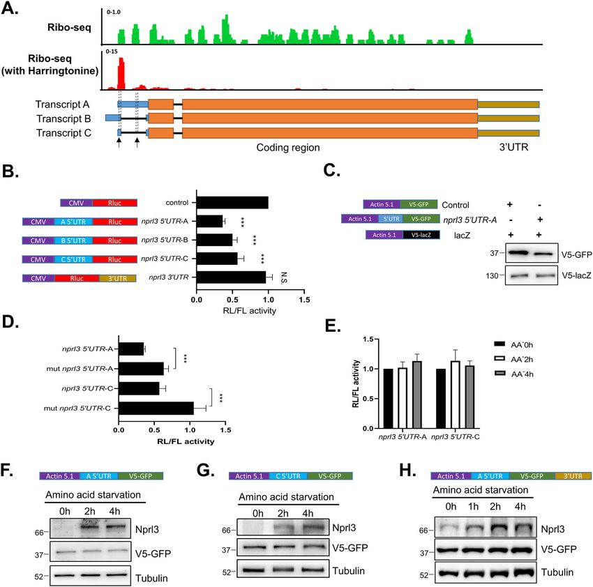
Zhou et al. Cell Death and Disease (2021)12:571                                                               Page 2 of 15

RagC/D is bound to GDP. Conversely, the Rag GTPase              The Tsc1 RNAi (BDSC#35144), MTD-Gal4 (BDSC#31
functions to inhibit TORC1 activity when RagA/B is in           777) stocks were obtained from the Bloomington Droso-
the GDP state and Rag C/D is GTP bound17,18. The                phila Stock Center. The Tub-GFP-LAMP1 was provided
GTPase-activating protein (GAP) towards Rags (GATOR)            by Helmut Kramer (UT Southwestern)28. The Nos-Gal4
complex, comprised of two subcomplexes GATOR1 and               was provided by Sharon Bickel (Dartmouth College)29.
GATOR2, mediate cellular amino acid levels and TORC1            The nprl2 RNAi, nprl3 RNAi, UAS-HA-Nprl2, UAS-HA-
activity19. The GATOR1 complex, composed of Nprl2,              Nprl3, UAS-V5-Nprl3, nprl21, nprl31 were described
Nprl3 and Iml1/DEPDC5, displays GAP activity that               previously30–32. All fly stocks were maintained on BDSC
hydrolyzes the RagA/B bound GTP to GDP and releases             standard cornmeal medium at 25 °C.
TORC1 from the lysosome to inhibit its activity19. The
GATOR2 complex physically interacts with and inhibits           Cell culture and amino acid starvation
the GATOR1 complex and functions to promote TORC1                 Drosophila S2 cells (Thermo Fisher, #R69007) were
activity15,20. Cytoplasmic amino acids control TORC1            cultured in Schneider’s Drosophila medium (Merck,
activity through amino acid sensors that bind to GATOR1         #S9895) supplemented with 10% fetal bovine serum (FBS,
or GATOR2. Upon amino acid starvation, Sestrin2 and             Thermo Fisher, #A3160802). Amino acid starvation
CASTOR1 bind and antagonize GATOR2 function,                    medium was prepared based on the formulation of
whereas SAMTOR and KCSTOR bind and promote                      Schneider’s medium without the indicated amino acids.
GATOR1 function21–24. Interestingly, although the               Specifically, the amino acid starvation media contain
GATOR and TORC1 are evolutionally conserved in                  CaCl2 (5.40 mM), MgSO4 (15.06 mM), KCl (21.33 mM),
eukaryotes25, some amino acid mediators are absent in           KH2PO4 (3.31 mM), NaHCO3 (4.76 mM), NaCl
invertebrates. For example, the KICSTOR and the CAS-            (36.21 mM), Na2HPO4 (4.94 mM), Alpha-Ketoglutaric
TOR1 complex are not encoded in the genome of Dro-              acid (1.37 mM), D-Glucose (11.11 mM), Fumaric acid
sophila melanogaster and Caenorhabditis elegans21,23.           (0.86 mM), Malic acid (0.75 mM), Succinic acid
  In mammalian cells, amino acids control the protein           (0.85 mM), Trehalose (5.85 mM). For amino acid starva-
stability of the GATOR1 component DEPDC5, the                   tion, cultured S2 cells or dissected Drosophila ovaries
homolog of Iml126. When nutrients are sufficient,                were incubated in the amino acid starvation media for the
CUL3–KLHL22 E3 ligase promotes the ubiquitination               indicated time.
and degradation of the DEPDC5 via the proteasome.
Upon amino acid starvation, KLHL22 is bound and                 Plasmids and cell transfection
trapped by 14-3-3 proteins in the nucleus, resulting in a         The plasmids were constructed using a recombination
dramatic increase in DEPDC5 protein stability26. Inter-         kit according to the manufacturer’s instruction (Vazyme,
estingly, depletion of DEPDC5 results in the decreased          #C112-01). The nprl3, GFP, and FKBP39 coding regions
expression of the other two GATOR1 components                   were amplified using specific primers and then inserted
NPRL2 and NPRL323,27. We reasoned that under amino              into a pAC5.1-His-V5 vector (Invitrogen, #V411020) to
acid starvation the expression levels of Nprl2 and Nprl3        construct the pAct-Nprl3-His-V5, pAct-GFP-His-V5,
might also be regulated, especially in the invertebrates        and pAct-FKBP39-His-V5 plasmids. The 5′UTRs and 3′
that have no orthologs of KICSTOR and CASTOR1.                  UTR of nprl3 were amplified using specific primers and
  Here we identified multiple pathways that control              inserted into pAct-GFP-His-V5 to construct a series of
Drosophila Nprl3 expression. We found that amino acid           GFP reporter plasmids. The 5′UTRs, 3′UTR, and muta-
starvation enhanced Nprl3 expression, by preventing the         ted 5′UTRs of nprl3 were amplified using specific pri-
FKBP39 dependent destruction of the Nprl3 protein via           mers and inserted into psiCKECK2 (Promega, #C8021)
the proteasome. In addition, we showed that the Nprl3           to construct a series of Renilla luciferase reporter plas-
protein is unstable in the absence of the other GATOR1          mids. The nprl3 and FKBP39 coding regions were
components, Nprl2 and Iml1. Finally, we determined that         amplified using specific primers and then inserted into a
the uORF in the 5’UTR of the nprl3 transcript is a func-        pENTR-1A vector (Invitrogen, #A10462). The pENTR-
tional cis-element that acts to reduce Nprl3 translation.       Nprl3 and pENTR-FKBP39 plasmids were recombined
Our data support the model that the regulation of Nprl3         into pAHW (DGRC) to generate pAct-HA-Npr3 and
expression functions as a key regulator of GATOR1               pAct-HA-FKBP39 plasmids using Gateway LR Clonase
activity.                                                       II Enzyme (Invitrogen, #11791020). Primer sequences
                                                                used to generate plasmids are listed in Supplementary
Materials and methods                                           Table 3. S2 cells grown in six-well plates were trans-
Drosophila strains                                              fected with 0.2 μg plasmids using Effectene Transfection
  The stocks fkbp39 RNAi (THU#0803), huwe1 RNAi                 Reagent according to the manufacturer’s instruction
(THU#1805) were obtained from Tsinghua Fly Center.              (Qiagen, #301427).

Official journal of the Cell Death Differentiation Association
Zhou et al. Cell Death and Disease (2021)12:571                                                                Page 3 of 15

dsRNA and RNAi                                                  inhibit mRNA translation. At the indicated time points
  To generate double-stranded RNA(dsRNA), DNA                   after the addition of CHX, cells were harvested and the
templates were PCR amplified to include a 5′ T7 RNA              protein expression was detected using western blot. The
polymerase binding site. PCR products were used as              α-Tubulin protein levels were used for normalization. The
templates to produce dsRNA using the MEGAscript                 remaining protein was determined by comparison with
RNAi kit according to the manufacturer’s instruction            the expression level at the time point when CHX
(Invitrogen, #AM1626). Primer sequences used to gen-            was added.
erate DNA templates for synthesizing double-strand RNA
(dsRNA) and plasmids are listed in Supplementary Table          Generation of fkbp39 mutant lines
3. For RNAi, S2 cells were incubated with 15 μg dsRNA in           fkbp39 mutant fly was generated similarly as described
six-well plates contained 0.5 mL Schneider’s medium             previously32. Primers used for cloning and detection are
without FBS for 1 h, and then 1.5 mL Schneider’s medium         listed in Supplemental Table 1.
supplemented with 10% FBS was added and incubated for
3 days.                                                         LysoTracker staining
                                                                  The Drosophila ovaries were dissected in Schneider’s
Tandem affinity purification and mass spectrometry                medium supplemental with 10% FBS. The tissues were
  Ovaries from the transgenic flies that stably express          incubated with LysoTracker Red (1:2000, Thermo Fisher,
HA-FLAG tagged Nprl3 proteins and from control yw               #L7528) and Hoechst (1:10,000, Thermo Fisher, #62245)
flies were homogenized and perform analysis as pre-              for 5 min, and then washed and mounted using Schnei-
viously described30.                                            der’s medium supplemental with 10% FBS. Images were
                                                                acquired using a Zeiss LSM 880 confocal microscope.
Immunoprecipitation (IP) and western blot analysis
  For IPs, the S2 cells transfected with specific plasmids       Ribosome profiling data analysis
were lysed using 1 ml RIPA buffer (Merck, #20-188)                Ribo-Seq data from the previous study were retrieved
containing complete protease inhibitor (Roche,                  from NCBI Sequence Read Archive database under
#1697498001) and phosphatase inhibitor (Roche,                  accession SRP06754213. The reads were trimmed with
#4906845001). The protein extracts were incubated with          Trim Galore (https://www.bioinformatics.babraham.ac.
30 μL HA antibody-coated magnet beads for 6 h at room           uk/projects/trim_galore/) to remove adapter sequences
temperature. The beads were collected and washed six            and bad quality bases, and then aligned to D. melanoga-
times with lysis buffer, and then detected by western blot.     ster reference genome (release dm6) using STAR with
For western blot, antibodies were used at the following         default settings34. After excluding reads aligned to rRNAs,
concentrations for western blot: mouse anti-α-tubulin at        tRNAs, and snoRNAs, remaining aligned reads between
1:10,000 (Beyotime, #AF0001), mouse anti-V5 antibody at         27 and 34 nt were considered as RPFs for further analysis.
1:4000 (Invitrogen, # R960-25), rabbit anti-pS6K at 1:1000      Using the count function of igv tools, the alignment files
(Cell Signaling Technology, #9209), rabbit anti-HA anti-        were converted to TDF format for visualization in IGV35.
body at 1:1000 (Beyotime, #AF0039), guinea pig anti-
Nprl3 (generated at this work) at 1:2000.                       Statistical analyses
                                                                  Data are reported as mean ± SD of at least three inde-
RNA isolation and mRNA analysis                                 pendent experiments and analyzed using a two-tailed t-
  RNA isolation and qRT-PCR were performed as previously        test.
described33 using the primers list in Supplemental Table.
                                                                Results
Luciferase reporter assay                                       Nprl3 protein stability is regulated by the unassembled
  S2 cells transfected with pSiCHECK-2 plasmids that            soluble complex protein degradation (USPD) pathway
contained 5′UTR or 3′UTR of nprl3 transcripts were lysed          GATOR1 promotes the conversion of RagA/B-GTP to
and the firefly luciferase (FL) and Renilla luciferase (RL)       RagA/B- GDP, which results in the release of TORC1
activities were measured using a dual luciferase assay          from lysosomes and an accompanying loss of TORC1
system kit according to the manufacturer’s instruction          activity19. To detect endogenous GATOR1 expression, we
(Promega, #E1910). The RL activity was normalized to the        generated a guinea pig anti-Nprl3 antibody that specifi-
activity of FL.                                                 cally recognizes Drosophila Nprl3 (Supplemental Fig. 1).
                                                                Interestingly, in mammalian cells mutations in the
CHX chase studies for protein stability measurement             GATOR1 component DEPDC5 decrease the expression of
  For protein stability assays, S2 cells were incubated with    the other two GATOR1 components Nprl2 and
10 μg/ml cycloheximide (CHX, Millipore, #66819) to              Nprl323,27. Consistent with this interdependence, we

Official journal of the Cell Death Differentiation Association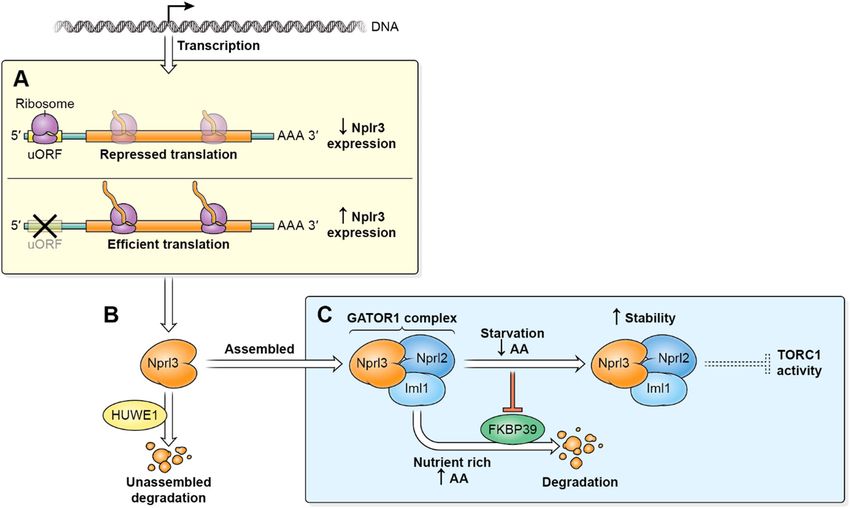
Zhou et al. Cell Death and Disease (2021)12:571                                                                                                Page 4 of 15

  Fig. 1 Nprl3 and Nprl2 expression levels are interdependent. A Western blot analysis of Nprl3 protein in the indicated dsRNA treated S2 cells.
  B Western blot analysis of Nprl3 protein in the ovaries of MTD > mCherry RNAi (control), MTD > nprl2 RNAi, MTD > nprl3 RNAi and MTD > Tsc1 RNAi flies.
  C Western blot analysis of Nprl3 protein in the ovaries of yw (control), nprl21 and nprl31. D Western blot analysis of Nprl3 protein in the ovaries of yw,
  TorA948V/+ and TorA948V/+; iml11. E Western blot analysis of HA-Nprl3 protein in the ovaries of MTD > HA-Nprl3 and MTD > HA-Nprl3, nprl2 RNAi flies.
  F Western blot analysis of the HA-Nprl2 protein in the ovaries of MTD > HA-Nprl2 and MTD > HA-Nprl2, nprl3 RNAi flies. G Western blot analysis of Nprl3
  protein in the ovaries of nos > mCherry RNAi (control), nos > nprl2 RNAi, nos > nprl2 RNAi, huwe1 RNAi and nos > huwe1 RNAi flies. α-Tubulin was used
  as a loading control. Similar western blot results were observed in more than three independent experiments for each group.

determined that knocking down nprl2 decreased Nprl3                              domain-containing E3 ubiquitin-protein ligase 1
expression in Drosophila S2 cells (Fig. 1A). Similarly, the                      (HUWE1) was reported to interact with many multi-
levels of Nprl3 protein were decreased in the ovaries of                         protein complexes to promote the USPD process36.
nprl2 mutants or in the ovaries expressing an nprl2 RNAi                         Interestingly, depletion of HUWE1 rescued the expression
using a germline-specific MTD-GAL4 driver (Fig. 1B, C).                           defect of Nprl3 in the Drosophila nprl2 RNAi ovaries (Fig.
In contrast, knocking down Tsc1, an independent inhi-                            1G). These data strongly suggest that the expression of
bitor of TORC1, did not decrease Nprl3 expression, which                         the GATOR1 components are interdependent and con-
suggested that the decreased Nprl3 expression was not                            trolled by the USPD pathway.
caused by TORC1 hyperactivation (Fig. 1A, B). Next, we
detected the levels of Nprl3 protein in the mutant of iml1,                      Amino acid starvation increases Nprl3 protein levels
homologue of DEPDC5 in Drosophila. Consistently, the                               Previously, we demonstrated that the GATOR1 com-
levels of Nprl3 protein were decreased in the ovaries of                         plex is required for Drosophila viability and young egg
iml11 mutant with TorA948V heterozygous, which rescued                           chamber survival under nutrient stress conditions31,32.
the iml11 lethality32 (Fig. 1D). In eukaryotes, the USPD                         Based on our findings that the expression of GATOR1
pathway is thought to prevent protein aggregation36,37.                          components are interdependent and that DEPDC5/Iml1
Thus, we reasoned that the decreased expression of Nprl3                         expression is sensitive to nutrient status in mammals26,
in nprl2 depletion cells might reflect the activation of the                      we reasoned that the levels of the GATOR1 component
USPD pathway. To test this model, we detected the                                Nprl3 might also be sensitive to amino acid starvation.
expression of exogenous HA-tagged Nprl3 and found that                           First, we examined whether the expression of Nprl3 is
nprl2 depletions decreased the levels of ectopically                             affected by nutrient status in Drosophila. Interestingly,
expressed HA-Nprl3 protein in the Drosophila ovary (Fig.                         amino acid starvation significantly increased the expres-
1E). Conversely, knocking down nprl3 transcript levels                           sion of Nprl3 protein in Drosophila S2 cells (Fig. 2A). By
decreased HA-tagged Nprl2 expression in the Drosophila                           contrast, exposure of S2 cells to amino acid starvation did
ovary (Fig. 1F). Recently, the HECT, UBA and WWE                                 not change nprl3 mRNA levels significantly, as assessed

Official journal of the Cell Death Differentiation Association
Zhou et al. Cell Death and Disease (2021)12:571                 Page 5 of 15

  Fig. 2 (See legend on next page.)

Official journal of the Cell Death Differentiation Association
Zhou et al. Cell Death and Disease (2021)12:571                                                                                          Page 6 of 15

  (see figure on previous page)
  Fig. 2 Amino acid starvation enhances Nprl3 protein stability. A Western blot analysis of Nprl3 protein in S2 cells under amino acid starvation.
  B qRT-PCR analysis of nprl3 mRNA in S2 cells under amino acid starvation. Error bars represent standard error values from three independent
  experiments. C Western blot analysis of the Nprl3 protein in S2 cells treated with actinomycin D together with amino acid starvation. D Ovaries from
  yw flies were dissected and cultured in amino-acid-free Schneider’s medium (AA-) for 2 h. Nprl3 was detected using Western blot. E S2 cells were
  transfected with the pAC5.1 construct inserted with nprl3 5’UTR, V5-tagged Nprl3 coding region, and nprl3 3′UTR. The cells were starved for the
  indicated time and the V5-Nprl3 levels were measured by western blot. F S2 cells were transfected with the pAC5.1 construct inserted with V5-tagged
  Nprl3 coding region. The cells were starved for the indicated time and the V5-Nprl3 levels were measured by western blot. G S2 cells were
  transfected with the pAC5.1 construct inserted with V5-tagged Nprl3 coding region. The cells were treated with CHX alone or CHX together with
  amino acid starvation medium for the indicated time. The V5-Nprl3 levels were measured by western blot. The remaining protein levels at each time
  point were calculated, taken the expression level of V5-Nprl3 at 0 h as 1. Error bars represent standard error values from three independent
  experiments. **P < 0.01; ***P < 0.001. H S2 cells were transfected with the pAC5.1 construct inserted with V5-tagged Nprl3 coding region. The cells
  were treated with CHX alone or CHX together with MG132 for the indicated time. The V5-Nprl3 levels were measured by western blot. I Western blot
  analysis of endogenous Nprl3 protein in S2 cells under amino acid starvation and MG132 treatment. Rp49 was used for normalization for qPCR. α-
  Tubulin was used as a loading control for western blot. Similar western blot results were observed in more than three independent experiments for
  each group.

by RT-qPCR amplification (Fig. 2B). In addition, actino-                       test this model, we examined Nprl3 protein stability in
mycin D, an inhibitor of transcription, did not prevent the                   the presence of the translational inhibitor cycloheximide
increase in Nprl3 levels observed under amino acid star-                      (CHX). Because the endogenous Nprl3 protein is too low
vation (Fig. 2C). The Drosophila ovary has a high level of                    under amino acid sufficient condition to detect in S2
Nprl3 expression (Supplemental Fig. 2). To determine                          cells, we followed the ectopically expressed V5-tagged
whether Nprl3 protein level is induced by nutrient                            Nprl3 protein. Notably, amino acid starvation sig-
depletion in vivo, we dissected and incubated Drosophila                      nificantly decreased the degradation rate of V5-Nprl3
ovaries in amino acid starvation media for 2 h and found                      protein (Fig. 2G). To determine whether Nprl3 protein
that Nprl3 levels increased significantly in dissected                         degradation is mediated by the proteasome, we used
ovaries after amino acid starvation (Fig. 2D). Consistent                     MG132 to inhibit the proteolytic activity of the 26S
with these observations, when we incubated female flies in                     proteasome complex. We found that MG132 treatment
20% sucrose media with no amino acids for 1 day, we                           significantly inhibited V5-Nprl3 protein degradation
found that Nprl3 protein levels significantly increased in                     (Fig. 2H). Moreover, similar to amino acid starvation
ovaries (Supplemental Fig. 3). To confirm that the                             treatment, MG132 treatment increased endogenous
increased Nprl3 expression observed under amino acid                          Nprl3 levels in non-starvation conditions (Fig. 2I).
starvation is independent of transcription, we constructed                    Importantly, subjecting cells to MG132 treatment and
a plasmid that contains the 5’UTR, V5-tagged coding                           amino acid starvation did not significantly increase the
region, and 3’UTR of the nprl3 mRNA under the control                         levels of Nprl3 protein above either single treatment
of the actin promoter in pAC5.1 vector. We found that                         (Fig. 2I). These data suggest that MG132 treatment and
even when Nprl3 was expressed from the actin promoter,                        amino acid starvation likely impact the same pathway.
amino acid starvation significantly increased V5-tagged                        Taken together our data strongly suggest that amino
Nprl3 protein levels in S2 cells (Fig. 2E). Thus, the                         acid starvation increases Nprl3 protein stability at least
increase in Nprl3 protein expression upon amino acid                          in part by inhibiting proteasome-dependent degradation
starvation is driven by a posttranscriptional mechanism.                      in nutrient replete conditions.
Taken together our data indicate that the increase in
Nprl3 protein expression observed upon amino acid                             FKBP39 associates with Nprl3 and regulates its stability
starvation in both S2 cells and the Drosophila ovary is                         To elucidate mechanistically why Nprl3 stability
driven by a posttranscriptional mechanism.                                    increases upon amino acid starvation, we used immuno-
  Next, we wanted to determine if the increased Nprl3                         precipitation followed by quantitative mass spectrometry
protein observed upon amino acid starvation mapped to                         to identify proteins that physically interact with the Nprl3
the coding region of Nprl3, suggesting a possible change                      protein. Immunoprecipitations were performed using
in protein stability. To answer this question, we trans-                      lysates generated from control and HA-FLAG-Nprl3
fected S2 cells with a construct that contained the V5-                       expressing (MTD-GAL4 driver) Drosophila ovaries. The
tagged Nprl3 coding region but not the 3′ or 5′ UTRs                          immunoprecipitates were subject to mass spectrometry
and found that amino acid starvation still induced                            analysis for quantitation. As anticipated, additional
increased V5-Nprl3 levels in starved cells (Fig. 2F).                         GATOR complex components including Nprl2, Iml1,
These data are consistent with the idea that nutrient                         Seh1, and Mio were specifically pulled down from the
starvation impacts the stability of the Nprl3 protein. To                     HA-FLAG-Nprl3 lysate (Supplemental Table 1). Notably,

Official journal of the Cell Death Differentiation Association
Zhou et al. Cell Death and Disease (2021)12:571                                                             Page 7 of 15

  Fig. 3 (See legend on next page.)

we found that the FK506 binding protein 39 KD (FKBP39)          beads. The immunoprecipitation of HA-tagged Nprl3 co-
was also enriched in the HA-FLAG-Nprl3 ovary lysate. To         precipitated V5-tagged FKBP39, but not the negative
verify the interaction between Nprl3 and FKBP39, we co-         control V5-tagged LacZ (Fig. 3A). Next, we detected
expressed HA-tagged Nprl3 and V5-tagged FKBP39                  whether the interaction is affected by nutrient status.
in Drosophila S2 cells and carried out co-                      Amino acid starvation did not significantly affect the
immunoprecipitation assays using HA antibody-coated             physical interaction between FKBP39 and Nprl3 in S2

Official journal of the Cell Death Differentiation Association
Zhou et al. Cell Death and Disease (2021)12:571                                                                                              Page 8 of 15

  (see figure on previous page)
  Fig. 3 FKBP39 associates with Nprl3 and promotes Nprl3 degradation. A S2 cells were co-transfected with HA-tagged Nprl3 and V5-tagged
  FKBP39 or lacZ (control) plasmids. Cells were lysed and immunoprecipitated using beads coated with anti-HA antibody. Cell lysates (inputs) and
  immunoprecipitates (IP) were detected by western blot. B S2 cells were co-transfected with HA-tagged Nprl3 and V5-tagged FKBP39 or lacZ (control)
  plasmids. Cells were cultured in Schneider’s medium plus 10% FBS (AA+) or amino-acid-free Schneider’s medium (AA−) for 2 h. The cells were lysed
  and immunoprecipitated using beads coated with an anti-HA antibody. Cell lysates (inputs) and immunoprecipitates (IP) were detected by western
  blot. C S2 cells were transfected with GFP (control) or fkbp39 dsRNA. Cells were treated with amino acid starvation for 2 h. The levels of Nprl3 were
  detected by western blot. D S2 cells were co-transfected with the GFP (control) or fkbp39 dsRNA, constructs expressed HA-tagged Nprl3 and V5
  tagged lacZ. The levels of HA-Nprl3 and V5-lacZ were detected by western blot. V5-lacZ was used as a loading control for western blot. E S2 cells
  were co-transfected with the GFP (control) or fkbp39 dsRNA and construct expressed V5-tagged Nprl3. Cells were treated with amino acid starvation
  for the indicated hours. The levels of V5-Nprl3 were detected by western blot. F S2 cells were co-transfected with the GFP (control) or fkbp39 dsRNA
  and construct expressed V5-tagged Nprl3. The cells were treated with CHX for the indicated time and the V5-Nprl3 levels were measured by western
  blot. The remaining protein levels at each time point were calculated, taken the expression level of V5-Nprl3 at 0 h as 1. Error bars represent standard
  error values from three independent experiments. *P < 0.05. α-Tubulin was used as a loading control for western blot. Similar western blot results
  were observed in more than three independent experiments for each group.

cells, suggesting the association between these two pro-                        Compared to control cells, the expression level of HA-
teins is amino acid insensitive (Fig. 3B).                                      Nprl2 protein was much higher in fkbp39 RNAi cells (Fig.
  To evaluate the effect of FKBP39 on Nprl3 protein                             4C). These results indicate that FKBP39 regulates the
levels, the endogenous Nprl3 protein in fkbp39 RNAi cells                       stability of multiple GATOR1 components.
was detected using western blot. Knocking down FKBP39
increased Nprl3 levels and these levels could not be                            FKBP39 promotes TORC1 activity in vivo
increased further by amino acid starvation (Fig. 3C). We                          To confirm the function of FKBP39 in regulating Nprl3
speculated that the FKBP39 protein destabilizes the Nprl3                       protein expression in vivo, we generated fkbp39 mutant
protein under amino acid sufficient condition and this                           flies, in which the coding region of the fkbp39 gene was
destabilization is alleviated by amino acid starvation. To                      removed using the CRISPR/Cas9 method. The mutant,
test this hypothesis, the ectopically expressed HA-tagged                       named fkbp391, has more than 90% deletion in the coding
Nprl3 protein was detected in fkbp39 RNAi cells. Com-                           region (Supplemental Fig. 4). Interestingly, fkbp391 was
pared to the control cells, the expression level of HA-                         viable and had no obvious defects. We detected the Nprl3
Nprl3 protein was much higher in fkbp39 RNAi cells (Fig.                        expression in fkbp39 mutant and RNAi flies. The
3D). Moreover, amino acid starvation could not increase                         expression level of Nprl3 protein in fkbp39 mutant or
the levels of ectopically expressed V5-Nprl3 in fkbp39                          fkbp39 RNAi ovaries was much higher than that in control
RNAi cells (Fig. 3E). Next, we detected the effect of                           (Fig. 5A, B). Next, we dissected ovaries from wild-type or
FKBP39 on the stability of Nprl3 protein and found that                         fkbp391 females and cultured them in amino acid star-
the decay rate of the V5-tagged Nprl3 protein was much                          vation media. The expression level of Nprl3 in fkbp391
slower in fkbp39 RNAi cells (Fig. 3F). In summary, our                          ovary was higher than control and could not be increased
data strongly suggest that FKBP39 associates with the                           by amino acid starvation (Fig. 5C). To determine there are
Nprl3 protein and promotes its degradation in nutrient-                         stage-specific differences in the accumulation of Nprl3
replete conditions.                                                             protein during oogenesis, we examined the distribution of
                                                                                Nprl3 in the Drosophila ovary using immunofluorescence.
FKBP39 associates with Nprl2 and regulates its expression                       As we previously reported, Nprl3 localizes to lysosomes
  Next, we determine whether amino acid starvation and                          and autolysosomes that appear as bright puncta in the
FKBP39 regulates the expression levels of another                               cytoplasm31. We found that both starvation and fkbp39
GATOR1 component, Nprl2 in Drosophila. Due to the                               mutation increased Nprl3 puncta brightness mainly in the
lack of a suitable anti-Nprl2 antibody, we measured the                         germanium and young egg chambers (supplemental Fig.
expression level of ectopically expressed HA-tagged                             5). In summary, these results indicate that FKBP39 inhi-
Nprl2. Interestingly, the levels of HA-Nprl2 increased                          bits Nprl3 accumulation and this effect can be alleviated
upon amino acid starvation (Fig. 4A). We co-expressed                           by nutrient starvation in vivo.
HA-tagged Nprl2 and V5-tagged FKBP in Drosophila S2                               As Nprl3 is an inhibitor of TORC1, FKBP39 might
cells and carried out co-immunoprecipitation assays using                       promote TORC1 activity through decreasing Nprl3 levels.
HA antibody-coated beads. The immunoprecipitation of                            We assayed TORC1 activity by measuring the levels of
HA-tagged Nprl2 co-precipitated V5-tagged FKBP39, but                           phosphorylated S6K, a downstream effector of TORC1.
not the negative control V5-tagged LacZ (Fig. 4B). Next,                        Notably, the TORC1 activity significantly decreased in
we examined the effect of FKBP39 on Nprl2 expression.                           FKBP39 knockdown S2 cells and fkbp39 mutant ovaries

Official journal of the Cell Death Differentiation Association
Zhou et al. Cell Death and Disease (2021)12:571 Page 9 of 15 Fig. 4 FKBP39 associates with Nprl2 and decreases its expression. A S2 cells were transfected with the indicated construct inserted with HA- tagged Nprl2 coding region. The cells were starved for the indicated time and the HA-Nprl2 levels were measured by western blot. α-Tubulin was used as a loading control for western blot. B S2 cells were co-transfected with HA-tagged Nprl2 and V5-tagged FKBP39 or lacZ (control) plasmids. Cells were lysed and immunoprecipitated using beads coated with anti-HA antibody. Cell lysates (inputs) and immunoprecipitates (IP) were detected by western blot. C S2 cells were co-transfected with the GFP (control) or fkbp39 dsRNA, constructs expressed HA-tagged Nprl2 and V5-tagged lacZ. The levels of HA-Nprl2 and V5-lacZ were detected by western blot. V5-lacZ was used as a loading control for western blot. Similar western blot results were observed in more than three independent experiments for each group. (Fig. 5D, E). To confirm the effect of FKBP39 on TORC1 Nutrient status and the USPD pathway independently is mediated by the GATOR1 complex, we detected the control Nprl3 stability phosphorylated S6K in ovaries with germline knockdowns Both nutrient status and the USPD pathway controls the of both FKBP39 and Nprl2. As expected, the effect of stability of GATOR1 components. To address whether FKBP39 knockdown on decreasing TORC1 activity was the regulation of Nprl3 expression by nutrient signals is eliminated by depletion of GATOR1 component Nprl2 dependent on the USPD pathway, we examined the (Fig. 5F). These results strongly suggest that FKBP39 changes of Nprl3 expression upon amino acid starvation maintains TORC1 activity by decreasing GATOR1 in nprl2 RNAi cells by western blot. Interestingly, amino expression. acid starvation still significantly increased Nprl3 levels in As a master mediator of nutrient status and cell meta- nprl2 RNAi S2 cells (Fig. 6A). In addition, amino acid bolism, TORC1 controls cell growth and autophagy. starvation increased Nprl3 levels in nprl2 mutant ovaries Autophagy is a catabolic process that uses lysosomal (Fig. 6B). Finally, amino acid starvation increased exo- degradation to eliminate damaged proteins and provide genous HA-tagged Nprl3 levels in nprl2 RNAi ovaries nutrients for cell survival under stress conditions38. (Fig. 6C). Next, we examined Nprl3 expression in cells Nutrient starvation induces autophagy through inhibition and animals in which the USPD component HUWE1 was of TORC139. To confirm the decreased TORC1 activity in depleted. Consistent with the above results, amino acid fkbp39 mutant, we stained the Drosophila ovaries with starvation increased Nprl3 levels in HUWE1 RNAi cells LysoTracker, which has been commonly used to examine both in vitro and in vivo (Fig. 6D, E). These results autophagy for it stains acidic lysosomes and autolyso- strongly suggest that nutrient status controls Nprl3 somes40. In fed conditions, the young egg chambers from expression independent of the USPD pathway. To deter- the wild-type flies had few LysoTracker-positive puncta mine whether FKBP39 acts in the USPD to regulate the (Fig. 5G). In contrast, the egg chambers from fkbp39 levels of GATOR1 complex components, we detected mutant flies contained more and larger LysoTracker- Nprl3 expression in nprl2 and FKBP39 double knock- positive puncta (Fig. 5H). LAMP1 is a widely used lyso- down cells. Unlike HUWE1 depletion, FKBP39 depletion some and autolysosome marker for autophagy detec- could not rescue Nprl3 levels in nprl2 RNAi cells (Fig. 6F). tion40. Consistent with the LysoTracker staining, the This result suggests that nutrient signal machinery com- GFP–LAMP1 positive puncta in fkbp39 mutants were ponent FKBP39 has no effect on USPD of the GATOR1 more and larger than in control flies (Supplemental Fig. complex. In summary, our results suggest that nutrient 6). Decreased TORC1 promotes lifespan and starvation signaling and the USPD pathway independently control resistance in Drosophila41. Interestingly, fkbp39 mutant GATOR1 expression. had increased longevity under starvation, consistent with the low TORC1 activity in fkbp39 mutant animals (Fig. The nprl3 5′UTR contains functional uORFs 5I). These results indicate that FKBP39 promotes TORC1 Eukaryotic gene expression is regulated at multiple activity and regulates cell metabolism in vivo. steps, including transcription, RNA processing, mRNA Official journal of the Cell Death Differentiation Association
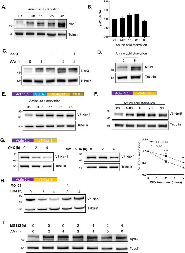
Zhou et al. Cell Death and Disease (2021)12:571                                                                                          Page 10 of 15

  Fig. 5 FKBP39 increases TORC1 activity in vivo. A Western blot analysis of Nprl3 protein in the ovaries of yw (control) and fkbp391. B Western blot
  analysis of the Nprl3 protein expression in the ovaries of MTD > mCherry RNAi (control) and MTD > fkbp39 RNAi flies. C The ovaries from yw (control)
  and fkbp391 were dissected and cultured in amino-acid-free Schneider’s medium (AA-) for 2 h. The levels of Nprl3 were detected by western blot. D
  Western blot analysis of phosphorylated S6K in the GFP (control) or fkbp39 dsRNA treated S2 cells. E Western blot analysis of phosphorylated S6K in
  the ovaries of yw (control) and fkbp391. F Western blot analysis of phosphorylated S6K in the ovaries of nos > mCherry RNAi (control), nos > fkbp39
  RNAi, nos > nprl2 RNAi and nos > fkbp39 RNAi, nprl2 RNAi flies. G–H The ovaries from yw (control) and fkbp391 were dissected in cell culture media and
  stained with Hoechst and LysoTracker Red. Bar, 20 μm. I Three days old yw (n = 30) and fkbp391 (n = 44) flies were cultured with complete starvation
  media (1% Agar in PBS). Survivors were counted at the indicated time points. Pairwise comparisons by the Mantel-Cox log rank test showed P < 0.001.
  α-Tubulin was used as a loading control for western blot. Similar western blot results were observed in more than three independent experiments for
  each group.

translation and protein stability. To elucidate whether                       a translational protein synthesis inhibitor, suggested the
nprl3 mRNA contains cis-elements that respond to amino                        uORF might be translated and function to inhibit down-
acid starvation, we examined the effect of the 5′UTR and                      stream Nprl3 translation (Fig. 7A). To determine if the
3′UTR of the nprl3 mRNA on translation. The nprl3 gene                        predicted uORF impacts the translation of the down-
encodes three transcripts that contain the same coding                        stream nprl3 coding region, we introduced the 5’UTR of
region and 3′UTR (Fig. 7A). The only difference between                       the nprl3 transcripts into the psiCHECK-2 vector in front
the three transcripts is 5′UTR. Interestingly, in all three                   of the renilla luciferase coding sequence. Intriguingly, the
mRNA isoforms, the 5′UTR sequence contains a 15 bp                            5′UTR of all three nprl3 transcripts decreased RL activity
uORF sequence with the transcript A 5′UTR containing                          significantly, comparing to no inserted (Fig. 7B). Fur-
one additional uORF sequence (Fig. 7A). Re-analyzing the                      thermore, placing the nprl3 transcript A 5′UTR upstream
published ribosome profiling data in S2 cells13, we                            of the GFP reporter in the pAC5.1 vector, resulted in
observed that while the ribosome-protected fragments                          decreased GFP protein expression (Fig. 7C). These data
(RPF) were present across the entire coding sequence                          strongly suggest that the nprl3 5′UTR inhibits the trans-
(CDS) of the nprl3 transcript, a strong peak was also                         lation of the downstream coding region. To validate if the
present within the uORF (Fig. 7A). The highest RPF peaks                      ribosome-protected uORFs in 5′UTR is the cis-element
around the nprl3 uORF in the treatment of harringtonine,                      that inhibits the downstream main ORF translation, the

Official journal of the Cell Death Differentiation Association
Zhou et al. Cell Death and Disease (2021)12:571                                                                                             Page 11 of 15

  Fig. 6 Nutrient signal and USPD independently controls Nprl3 stability. A S2 cells were transfected with GFP (control) or nprl2 dsRNA. Cells were
  treated with amino acid starvation for 2 h. The levels of Nprl3 were detected by western blot. B The ovaries from yw (control) and nprl21 were
  dissected and cultured in amino-acid-free Schneider’s medium (AA−) for 2 h. The levels of Nprl3 were detected by western blot. C. The ovaries from
  MTD > Nprl3-HA (control) and MTD > Nprl3-HA, nprl2 RNAi were dissected and cultured in amino-acid-free Schneider’s medium (AA−) for 2 h. The
  levels of HA-Nprl3 were detected by western blot. D S2 cells were transfected with GFP (control) or huwe1 dsRNA. Cells were treated with amino acid
  starvation for 2 h. The levels of Nprl3 were detected by western blot. E The ovaries from nos > mCherry RNAi (control) and nos > huwe1 RNAi were
  dissected and cultured in amino-acid-free Schneider’s medium (AA-) for 2 h. The levels of Nprl3 were detected by western blot. F Western blot
  analysis of Nprl3 protein in the ovaries of Nos > mCherry RNAi (control), Nos > nprl2 RNAi, Nos > nprl2 RNAi, fkbp39 RNAi and Nos > fkbp39 RNAi flies. α-
  Tubulin was used as a loading control. Similar western blot results were observed in more than three independent experiments for each group.

start codon ATG was mutated to GTG in the uORF.                                 located in the nprl3 5′UTR inhibits Nprl3 translation but
Critically, this mutation significantly increased the                            is not sensitive to amino acid starvation.
downstream reporter expression, supporting the transla-
tion inhibitory function of the uORFs (Fig. 7D).                                Discussion
  Some functional uORFs play essential roles in mod-                              The GATOR1 complex is a highly conserved inhibitor
ulating the translation of CDS, by decreasing their inhi-                       of TORC1 activity. Mutations in GATOR1 components
bition on main ORF protein translation under stress                             are associated with numerous pathologies including
conditions8. To determine if nutrient depletion attenuates                      multiple cancers and epilepsy19,42,43. Here we report the
the inhibition of nprl3 uORFs on downstream translation,                        characterization of multiple novel pathways that regulate
S2 cells that ectopically expressed Renilla luciferase (RL)                     the expression of the essential GATOR1 component
under the control of nprl3 5’UTR were starved in amino                          Nprl3 in Drosophila (Fig. 8). While the uORFs located at
acid depletion media. Amino acid starvation did not                             5′UTR of nprl3 transcripts inhibit its translation, the
enhance RL activity above basal levels (Fig. 7E). Con-                          USPD and nutrient signaling pathways control Nprl3
sistent with this observation, amino acid starvation did                        protein stability. In addition, we demonstrate that the
not increase the levels of ectopically expressed GFP under                      HUWE1-associated USPD pathway and FKBP39-
the control of nprl3 5′UTR, despite the strong increase in                      mediated nutrient signaling independently regulate
endogenous Nprl3 protein (Fig. 7F–H). Given the RL                              Nprl3 protein stability to control Nprl3 expression.
activity under the control of transcript A 5’UTR is less
than that of transcript B or C 5’UTR (Fig. 7B), the ratio of                    FKBP39 regulates Nprl3 and Nprl2 protein stability in
different transcripts might contribute to the Nprl3 protein                     response to nutrient status
expression during amino acid starvation. We detected the                          In metazoans, the TORC1 pathway senses multiple
transcripts using two pairs of primers that generate PCR                        nutrient signals and controls protein synthesis and cell
products at different sizes between transcript A and B/C.                       proliferation. Under amino acid depletion, the GATOR1
The ratio of different PCR product amounts were not                             complex inhibits TORC1 activity and protects cell survi-
significantly changed upon amino acid starvation (Sup-                           val31. In mammalian cells, the GATOR1 complex com-
plemental Fig. 7). These findings indicate that the uORF                         ponent DEPDC5/Iml1 has increased protein stability

Official journal of the Cell Death Differentiation Association
Zhou et al. Cell Death and Disease (2021)12:571                                                                                            Page 12 of 15

  Fig. 7 The nprl3 5’UTR contains functional uORFs. A Ribosome-protected fragment (RPF) reads mapped to the nprl3 transcripts. The green is RPF
  from S2 cells without treatment, and the red is RPF from S2 cells treated with harringtonine. The three transcripts of nprl3 are depicted. The location
  of the uORF that exists in all three transcripts is denoted by an arrow, and the main ORF is marked in orange. B S2 cells were transfected with the
  indicated constructs that inserted nprl3 5′UTR and 3′UTR to control renilla luciferase (RL) expression. The psiCHECK-2 constructs contain firefly
  luciferase (FL), which were used as for normalization. Values are expressed as fold change relative to the empty construct (Control). Error bars
  represent standard error values from three independent experiments. ***P < 0.001. C S2 cells were co-transfected with the V5-tagged GFP reporter
  construct inserted with nprl3 5’UTR and V5-tagged lacZ construct. The levels of V5-GFP and V5-lacZ were detected by western blot. V5-lacZ was used
  as a loading control for western blot. D S2 cells were transfected with the indicated constructs that inserted wild-type nprl3 5’ UTR or the mutant
  nprl3 5’UTR with the ATG in the uORF changed to GTG. Values are expressed as fold change relative to the empty construct (Control). Error bars
  represent standard error values from three independent experiments. ***P < 0.001. E S2 cells were transfected with the indicated constructs that
  inserted wild-type nprl3 5’ UTR. Cells were treated with amino acid starvation (AA−) for indicated hours. Values are expressed as fold change relative
  to the value before starvation. F–H S2 cells were transfected with the indicated constructs inserted with nprl3 5′UTR or 3′UTR to control GFP reporter
  expression. The cells were starved for the indicated time, and the V5-GFP was measured with western blot. α-Tubulin was used as a loading control.
  Similar western blot results were observed in more than three independent experiments for each group.

Official journal of the Cell Death Differentiation Association
Zhou et al. Cell Death and Disease (2021)12:571                                                                                   Page 13 of 15

  Fig. 8 Multiple pathways regulate the levels of the TORC1 inhibitor Nprl3. A The nprl3 mRNA contains a functional uORF that reduces Nprl3
  translation. B Nprl3 forms the trimeric GATOR1 complex with the proteins Nprl2 and Iml1. When not assembled into the GATOR1 complex, Nprl3 is
  degraded via a HUWEI dependent pathway. C In nutrient-replete conditions, FKBP39 associates with Nprl3 and promotes its degradation. Upon
  amino acid starvation, the FKBP39 dependent destruction of Nprl3 is blocked, and the increased levels of GATOR1 resulting in decreased TORC1
  activity.

under conditions of amino acid starvation while the other                 nutrient status. Notably, we also demonstrate that
two GATOR1 components, Nprl2 and Nprl3, are stable                        FKBP39 regulates the expression of another GATOR1
and unaffected by nutrient status26. Here we find that in                  component Nprl2, with fkbp39 mutants exhibiting
Drosophila the Nprl3 and Nprl2 proteins exhibit                           increased levels of Nprl2 protein relative to wild type in
increased protein stability under conditions of amino acid                fed conditions. Thus, in Drosophila, the FKBP39 protein
starvation. Notably, the Drosophila genome lacks mam-                     functions to restrict the accumulation of both Nprl3 and
malian orthologues of some amino acid sensors that                        Nprl2 in nutrient-replete conditions. Thus, the amino
regulate GATOR1 activity including KICSTOR and                            acid-sensitive FKBP39 pathway may promote the sto-
CASTOR121,23. Thus, the different responses of Nprl3                      chiometric increase in GATOR1 components upon amino
and Nprl2 expression upon starvation between Droso-                       acid starvation.
phila and mammals might reflect different strategies of                      Consistent with a role in the regulation of TORC1, we
GATOR1 regulation.                                                        find that mutants in fkbp39, which have increased levels of
   Here we find that FKBP39 interacts and promotes Nprl3                   the TORC1 inhibitor Nprl3, have decreased TORC1
degradation under nutrient sufficient conditions. FKBP39                   activity and increased autophagy. Thus, it is tempting to
is the first member of the FK506-binding protein (FKBPs)                   speculate that FKBP39, by preventing the accumulation of
family characterized in Drosophila44. FKBPs possess a cis-                Nprl2 and Nprl3, blocks the inappropriate activation of
trans peptidyl-prolyl isomerase (PPIase) activity to aid                  GATOR1 and the downregulation of TORC1 activity
protein folding as chaperons and perform some cellular                    when nutrients are plentiful. Previous results have shown
functions, such as apoptosis and protein trafficking44,45.                 that overexpression of FKBP39 inhibits fat body cell
Interestingly, the physical interaction between FKBP39                    growth and autophagy during the transition from larvae
and Nprl3 is not affected by nutrient status, which sug-                  to pupae in Drosophila46. High TORC1 activity inhibits
gests that FKBP39 might be one component of a larger                      the activation of autophagy pathways. Thus, the inhibition
pathway that regulates Nprl3 stability in response to                     of autophagy in FKBP39 overexpression is consistent with

Official journal of the Cell Death Differentiation Association
Zhou et al. Cell Death and Disease (2021)12:571                                                                                                                   Page 14 of 15

our findings that FKBP39 increased TORC1 activity.                                           Ethics statement
However, the inhibition of cell size in FKBP39 over-                                        In this study, we use the invertebrate model system Drosophila melanogaster,
                                                                                            which is not necessarily for ethics approval.
expression is not consistent with our findings that
FKBP39 increased TORC1 activity. Thus, FKBP39 might                                         Conflict of interest
regulate additional pathways that impact cell size. FKBP39                                  The authors declare no competing interests.
is also reported to be a component of a multi-protein
complex that regulates the ecdysone and juvenile hor-                                       Publisher’s note
mone signal transduction pathways47. Whether these                                          Springer Nature remains neutral with regard to jurisdictional claims in
physiological functions of FKBP39 are associated with its                                   published maps and institutional affiliations.
roles in promoting Nprl3 degradation is an interesting
                                                                                            Supplementary information The online version contains supplementary
question for future study.                                                                  material available at https://doi.org/10.1038/s41419-021-03860-z.

An upstream uORF inhibits the translation of nprl3                                          Received: 20 January 2021 Revised: 20 May 2021 Accepted: 21 May 2021
  In eukaryotic genomes, the sequences of uORFs are
widespread. Normally, functional uORFs inhibit their
downstream main ORF. Some uORFs located in special
mRNAs 5′UTR decrease their inhibition on main ORF                                           References
protein translation under stress conditions8. One of the                                     1. Liu, G. Y. & Sabatini, D. M. mTOR at the nexus of nutrition, growth, ageing and
                                                                                                 disease. Nat. Rev. Mol. Cell Biol. 21, 183–203 (2020).
best-known examples is activating transcription factor 4
                                                                                             2. Kim, J. & Guan, K. L. mTOR as a central hub of nutrient signalling and cell
(ATF4), a transcriptional factor that functions in the                                           growth. Nat. Cell Biol. 21, 63–71 (2019).
integrated stress response48,49. We have determined that                                     3. Raught, B., Gingras, A. C. & Sonenberg, N. The target of rapamycin (TOR)
                                                                                                 proteins. Proc. Natl Acad. Sci. USA 98, 7037–7044 (2001).
nprl3 mRNA contains a functional uORF through re-
                                                                                             4. Hay, N. & Sonenberg, N. Upstream and downstream of mTOR. Genes Dev. 18,
analyzing ribosome profile data13. We demonstrate that                                            1926–1945 (2004).
the functional uORF downregulates the translation of the                                     5. Fingar, D. C., Salama, S., Tsou, C., Harlow, E. & Blenis, J. Mammalian cell size is
                                                                                                 controlled by mTOR and its downstream targets S6K1 and 4EBP1/eIF4E. Genes
main nprl3 ORF. However, the uORF in the nprl3 5′UTR
                                                                                                 Dev. 16, 1472–1487 (2002).
is not responsive to amino acid starvation. The identifi-                                     6. Heberle, A. M. et al. Molecular mechanisms of mTOR regulation by stress. Mol.
cation of the upstream stress or other stimuli that alter                                        Cell Oncol. 2, e970489 (2015).
                                                                                             7. Lee, C. H. et al. Constitutive mTOR activation in TSC mutants sensitizes cells to
Nprl3 translation through the uORF will be an interesting
                                                                                                 energy starvation and genomic damage via p53. EMBO J. 26, 4812–4823
area for future studies.                                                                         (2007).
                                                                                             8. Spriggs, K. A., Bushell, M. & Willis, A. E. Translational regulation of gene
Acknowledgements                                                                                 expression during conditions of cell stress. Mol. Cell 40, 228–237 (2010).
We thank members of the Animal Physiology Group in Yangzhou University                       9. Haimov, O. & Sinvani, H. Dikstein R. Cap-dependent, scanning-free translation
for comments on the manuscript. We thank Sharon Bickel and Helmut Kramer                         initiation mechanisms. Bba-Gene Regul. Mech. 1849, 1313–1318 (2015).
for providing reagents.                                                                     10. Zhang, H., Wang, Y. & Lu, J. Function and evolution of upstream ORFs in
                                                                                                eukaryotes. Trends Biochem. Sci. 44, 782–794 (2019).
                                                                                            11. Chikashige, Y. et al. Gcn2 eIF2 alpha kinase mediates combinatorial transla-
Author details                                                                                  tional regulation through nucleotide motifs and uORFs in target mRNAs.
1
 Joint International Research Laboratory of Agriculture and Agri-Product Safety,                Nucleic Acids Res. 48, 8977–8992 (2020).
the Ministry of Education of China, Yangzhou University, Yangzhou 225009,                   12. Medenbach, J., Seiler, M. & Hentze, M. W. Translational control via protein-
China. 2Animal Physiology Group, College of Bioscience and Biotechnology,                       regulated upstream open reading frames. Cell 145, 902–913 (2011).
Yangzhou University, Yangzhou 225009, China. 3Institute of Comparative                      13. Zhang, H. et al. Genome-wide maps of ribosomal occupancy provide insights
Medicine, College of Veterinary Medicine, Yangzhou University, Yangzhou                         into adaptive evolution and regulatory roles of uORFs during Drosophila
225009, China. 4College of Horticulture and Plant Protection, Yangzhou University,              development. PLoS Biol. 16, e2003903 (2018).
Yangzhou 225009, China. 5Eunice Kennedy Shriver National Institute of Child                 14. Darnell, A. M., Subramaniam, A. R. & O’Shea, E. K. Translational control through
Health and Human Development, National Institutes of Health, Bethesda, MD                       differential ribosome pausing during amino acid limitation in mammalian
20892, USA                                                                                      cells. Mol. Cell 71, 229 (2018).
                                                                                            15. Bar-Peled, L. & Sabatini, D. M. Regulation of mTORC1 by amino acids. Trends
                                                                                                Cell Biol. 24, 400–406 (2014).
Author contributions
                                                                                            16. Kim, E., Goraksha-Hicks, P., Li, L., Neufeld, T. P. & Guan, K. L. Regulation of TORC1
Conceptualization, Y.Z., Y.W., M.A.L., W.W., and C.F.; performed experiments and
                                                                                                by Rag GTPases in nutrient response. Nat. Cell Biol. 10, 935–945 (2008).
analyzed data, Y.Z., J.G., X.W., Y.C., J.G., P.B., Y.F., and M.A.S.; data interpretation,
                                                                                            17. Yang, S. et al. The rag GTPase regulates the dynamic behavior of TSC
Y.W., M.A.L., M.A.S., W.W., and C.F.; paper writing, Y.W. and M.A.L.; supervision,
                                                                                                downstream of both amino acid and growth factor restriction. Dev. Cell 55,
Y.W. and M.A.L.
                                                                                                272–288.e275 (2020).
                                                                                            18. Demetriades, C., Doumpas, N. & Teleman, A. A. Regulation of TORC1 in
Funding                                                                                         response to amino acid starvation via lysosomal recruitment of TSC2. Cell 156,
National Natural Science Foundation of China (31872287 to Y.W. and 31900375                     786–799 (2014).
to Y.C.); Natural Science Foundation of Jiangsu Province (BK20181456 to Y.W.                19. Bar-Peled, L. et al. A tumor suppressor complex with GAP activity for the rag
and BK20190882 to Y.C.); Postdoctoral Science Foundation of China                               GTPases that signal amino acid sufficiency to mTORC1. Science 340,
(2019M650124 to Y.C.); Six talent peaks project in Jiangsu Province (SWYY-146                   1100–1106 (2013).
to Y.W.); and Eunice Kennedy Shriver National Institute of Child Health and                 20. Wei, Y. et al. TORC1 regulators Iml1/GATOR1 and GATOR2 control meiotic
Human Development (NICHD) Intramural Research program (No. HD00163 16,                          entry and oocyte development in Drosophila. Proc. Natl Acad. Sci. USA 111,
to M.A.L.).                                                                                     E5670–E5677 (2014).

Official journal of the Cell Death Differentiation Association
Zhou et al. Cell Death and Disease (2021)12:571 Page 15 of 15
21. Chantranupong, L. et al. The CASTOR proteins are arginine sensors for the 36. Xu, Y., Anderson, D. E. & Ye, Y. H. The HECT domain ubiquitin ligase HUWE1
mTORC1 pathway. Cell 165, 153–164 (2016). targets unassembled soluble proteins for degradation. Cell Discov 2, 16040
22. Gu, X. et al. SAMTOR is an S-adenosylmethionine sensor for the mTORC1 (2016).
pathway. Science 358, 813–818 (2017). 37. Juszkiewicz, S. & Hegde, R. S. Quality control of orphaned proteins. Mol. Cell 71,
23. Wolfson, R. L. et al. KICSTOR recruits GATOR1 to the lysosome and is necessary 443–457 (2018).
for nutrients to regulate mTORC1. Nature 543, 438 (2017). 38. Mizushima, N. Autophagy: process and function. Genes Dev. 21, 2861–2873
24. Wolfson, R. L. et al. METABOLISM Sestrin2 is a leucine sensor for the mTORC1 (2007).
pathway. Science 351, 43–48 (2016). 39. Rabanal-Ruiz, Y., Otten, E. G. & Korolchuk, V. I. mTORC1 as the main gateway to
25. Dokudovskaya, S. & Rout, M. P. SEA you later alli-GATOR - a dynamic regulator autophagy. Essays Biochem. 61, 565–584 (2017).
of the TORC1 stress response pathway. J. Cell Sci. 128, 2219–2228 (2015). 40. Mauvezin, C., Ayala, C., Braden, C. R., Kim, J. & Neufeld, T. P. Assays to monitor
26. Chen, J. et al. KLHL22 activates amino-acid-dependent mTORC1 signalling to autophagy in Drosophila. Methods 68, 134–139 (2014).
promote tumorigenesis and ageing. Nature 557, 585–589 (2018). 41. Bjedov, I. et al. Mechanisms of life span extension by rapamycin in the fruit fly
27. Yuskaitis, C. J. et al. Chronic mTORC1 inhibition rescues behavioral and bio- Drosophila melanogaster. Cell Metab. 11, 35–46 (2010).
chemical deficits resulting from neuronal Depdc5 loss in mice. Hum. Mol. 42. Sim, J. C. et al. Familial cortical dysplasia caused by mutation in the mTOR
Genet. 28, 2952–2964 (2019). regulator NPRL3. Annals Neurol 79, 132–137 (2015).
28. Akbar, M. A., Ray, S. & Kramer, H. The SM protein Car/Vps33A regulates SNARE- 43. Baulac, S. mTOR signaling pathway genes in focal epilepsies. Prog. brain Res.
mediated trafficking to lysosomes and lysosome-related organelles. Mol. Biol. 226, 61–79 (2016).
Cell 20, 1705–1714 (2009). 44. Theopold, U., Dal Zotto, L. & Hultmark, D. FKBP39, a Drosophila member of a
29. Van Doren, M., Williamson, A. L. & Lehmann, R. Regulation of zygotic gene family of proteins that bind the immunosuppressive drug FK506. Gene 156,
expression in Drosophila primordial germ cells. Curr. Biol. 8, 243–246 (1998). 247–251 (1995).
30. Cai, W. L., Wei, Y. H., Jarnik, M., Reich, J. & Lilly, M. A. The GATOR2 component 45. Ghartey-Kwansah, G. et al. Comparative analysis of FKBP family protein: eva-
Wdr24 regulates TORC1 activity and lysosome function. PLoS Genet. 12, (2016). luation, structure, and function in mammals and Drosophila melanogaster.
31. Wei, Y. & Lilly, M. A. The TORC1 inhibitors Nprl2 and Nprl3 mediate an adaptive Bmc Dev. Biol. 18, 7 (2018).
response to amino-acid starvation in Drosophila. Cell Death Differ. 21, 46. Juhasz, G. et al. Gene expression profiling identifies FKBP39 as an inhibitor of
1460–1468 (2014). autophagy in larval Drosophila fat body. Cell Death Differ. 14, 1181–1190
32. Wei, Y., Reveal, B., Cai, W. & Lilly, M. A. The GATOR1 complex regulates (2007).
metabolic homeostasis and the response to nutrient stress in Drosophila 47. Li, Y., Zhang, Z., Robinson, G. E. & Palli, S. R. Identification and characterization
melanogaster. G3 (Bethesda) 6, 3859–3867 (2016). of a juvenile hormone response element and its binding proteins. J. Biol.
33. Wei, Y. et al. Differential regulation of mRNA stability controls the transient Chem. 282, 37605–37617 (2007).
expression of genes encoding Drosophila antimicrobial peptide with distinct 48. Harding, H. P. et al. Regulated translation initiation controls stress-
immune response characteristics. Nucleic Acids Res. 37, 6550–6561 (2009). induced gene expression in mammalian cells. Mol. Cell 6, 1099–1108
34. Dobin, A. et al. STAR: ultrafast universal RNA-seq aligner. Bioinformatics 29, (2000).
15–21 (2013). 49. Park, Y., Reyna-Neyra, A., Philippe, L. & Thoreen, C. C. mTORC1 balances cellular
35. Robinson, J. T. et al. Integrative genomics viewer. Nat. Biotechnol. 29, 24–26 amino acid supply with demand for protein synthesis through post-
(2011). transcriptional control of ATF4. Cell Rep. 19, 1083–1090 (2017).
Official journal of the Cell Death Differentiation AssociationYou can also read