MCU-induced mitochondrial calcium uptake promotes mitochondrial biogenesis and colorectal cancer growth - Nature
←
→
Page content transcription
If your browser does not render page correctly, please read the page content below
Signal Transduction and Targeted Therapy                                                                                                         www.nature.com/sigtrans

                  ARTICLE                  OPEN

                  MCU-induced mitochondrial calcium uptake promotes
                  mitochondrial biogenesis and colorectal cancer growth
                  Yang Liu1,2, Mingpeng Jin1, Yaya Wang1,3, Jianjun Zhu4, Rui Tan5, Jing Zhao1, Xiaoying Ji1, Chao Jin6, Yongfeng Jia2, Tingting Ren7 and
                  Jinliang Xing1

                      Mitochondrial calcium uniporter (MCU) has an important role in regulating mitochondrial calcium (Ca2+) homeostasis.
                      Dysregulation of mitochondrial Ca2+ homeostasis has been implicated in various cancers. However, it remains unclear whether
                      MCU regulates mitochondrial Ca2+ uptake to promote cell growth in colorectal cancer (CRC). Therefore, in the present study the
                      expression of MCU in CRC tissues and its clinical significance were examined. Following which, the biological function of MCU-
                      mediated mitochondrial Ca2+ uptake in CRC cell growth and the underlying mechanisms were systematically evaluated using in
                      in vitro and in vivo assays, which included western blotting, cell viability and apoptosis assays, as well as xenograft nude mice
                      models. Our results demonstrated that MCU was markedly upregulated in CRC tissues at both the mRNA and protein levels.
                      Upregulated MCU was associated with poor prognosis in patients with CRC. Our data reported that upregulation of MCU enhanced
                      the mitochondrial Ca2+ uptake to promote mitochondrial biogenesis, which in turn facilitated CRC cell growth in vitro and in vivo.
                      In terms of the underlying mechanism, it was identified that MCU-mediated mitochondrial Ca2+ uptake inhibited the
1234567890();,:

                      phosphorylation of transcription factor A, mitochondrial (TFAM), and thus enhanced its stability to promote mitochondrial
                      biogenesis. Furthermore, our data indicated that increased mitochondrial Ca2+ uptake led to increased mitochondrial production of
                      ROS via the upregulation of mitochondrial biogenesis, which subsequently activated NF-κB signaling to accelerate CRC growth. In
                      conclusion, the results indicated that MCU-induced mitochondrial Ca2+ uptake promotes mitochondrial biogenesis by suppressing
                      phosphorylation of TFAM, thus contributing to CRC cell growth. Our findings reveal a novel mechanism underlying mitochondrial
                      Ca2+-mediated CRC cell growth and may provide a potential pharmacological target for CRC treatment.

                      Signal Transduction and Targeted Therapy (2020)5:59                                       ; https://doi.org/10.1038/s41392-020-0155-5

                  INTRODUCTION                                                                               signaling is often detrimental and has been associated with each
                  Colorectal cancer (CRC) represents a huge public health burden                             of the “cancer hallmarks.”4
                  worldwide and has higher rates of incidence in developed                                      Owing to its Ca2+ buffering capacity, the mitochondrion is an
                  countries.1 Every year, CRC leads to the death of nearly 700,000                           important organelle responsible for maintaining intracellular Ca2+
                  individuals, making it one of the most deadly cancers.1 Although                           homeostasis. Ca2+ influx into mitochondria, which is primarily
                  there has been progress in the early diagnosis and treatment of                            regulated by the mitochondrial calcium uniporter (MCU) complex, is
                  CRC, the mechanism underlying the pathogenesis of CRC remains                              a pleiotropic signal that controls a broad spectrum of cellular
                  to be elucidated. Thus, studies that explore the molecular                                 functions, including vital metabolic pathways, production of reactive
                  mechanisms contributing to the growth of CRC cells are urgently                            oxygen species (ROS), and the life/death decisions of cells.5 The
                  needed in order to develop novel therapeutic strategies.                                   understanding of the MCU complex has rapidly increased due to a
                    Intracellular calcium (Ca2+), which is a ubiquitous second                               myriad of recent studies that have identified the pore-forming
                  messenger, plays important roles in various types of biological                            molecule MCU and its regulatory subunits, including essential MCU
                  events. Owing to the significance of Ca2+ in signaling pathways,                            regulator (EMRE), MCU regulator 1 (MCUR1), MCU-dominant-
                  the level of Ca2+ in cells is strictly controlled. Altered Ca2+                            negative β-subunit (MCUb), mitochondrial calcium uptake (MICU)
                  homeostasis may lead to different pathological conditions,                                 1, MICU2, and MICU3.6 Abnormal changes in the expression levels
                  depending on the type of cell involved.2 For instance, it has been                         or functional role of one or more members of the MCU complex
                  well documented that Ca2+ signaling is a key regulator in a wide                           have been associated with cancer-related phenotypes in different
                  range of cellular processes, including tumor growth, progression,                          types of cancers, such as hepatocellular carcinoma, breast cancer,
                  and metastasis.3 This demonstrates that dysregulated Ca2+                                  colon cancer, and pancreatic cancer.7

                  1
                   State Key Laboratory of Cancer Biology and Department of Physiology and Pathophysiology, Fourth Military Medical University, Xi’an, China; 2Department of Pathology, Basic
                  Medical College, Inner Mongolia Medical University, Huhhot, China; 3College of Chemistry and Chemical Engineering, Xi’an University of Science and Technology, Xi’an, China;
                  4
                   Department of Cell Biology and Genetics, Basic Medicine College, Shanxi Medical University, Taiyuan, China; 5Department of Orthopedics, Xijing Hospital, Fourth Military Medical
                  University, Xi’an, China; 6Department of General Surgery, Tangdu Hospital, Fourth Military Medical University, Xi’an, China and 7State Key Laboratory of Cancer Biology and
                  Experimental Teaching Center of Basic Medicine, Fourth Military Medical University, Xi’an, China
                  Correspondence: Tingting Ren (rtt419@fmmu.edu.cn) or Jinliang Xing (xingjl@fmmu.edu.cn)
                  These authors contributed equally: Yang Liu, Mingpeng Jin, Yaya Wang

                  Received: 11 December 2019 Revised: 30 March 2020 Accepted: 3 April 2020

                  © The Author(s) 2020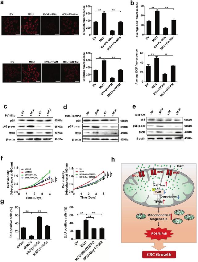
MCU-induced mitochondrial calcium uptake promotes mitochondrial. . .
Liu et al.
2
In recent years, an increasing number of studies are beginning which exhibited a median expression level of MCU (Fig. 2a).
to pay close attention to the functional role of MCU, a key Furthermore, the overexpression and knockdown of MCU in
component in the MCU complex, in different diseases, especially LS174T and HCT-8 cells were confirmed by western blotting
in cancers. Growing evidence has demonstrated that MCU (Fig. 2b and Supplementary Fig. S2a). As shown in Fig. 2c,
possesses pivotal roles in different types of cancers.8–10 For knockdown of MCU in LS174T cells resulted in a marked reduction
example, it has been reported that the expression of MCU in the basal level of mitochondrial Ca2+ ([Ca2+]m) compared with
elevated in basal-like and estrogen receptor-negative breast control cells, whereas MCU overexpression considerably increased
cancers, and the depletion of MCU promotes caspase- basal [Ca2+]m. We also found that the cells treated with the [Ca2+]
independent apoptosis in breast cancer cells.9 Similarly, our m buffering protein parvalbumin (PV) using PV-Mito showed
previous study demonstrated that MCU is upregulated in HCC decreased mitochondrial Ca2+ levels and thus reversed the MCU-
cells and promotes HCC cell survival via the ROS/AKT/MDM2 induced mitochondrial Ca2+ uptake (Fig. 2c). Similar findings were
pathway.11 Furthermore, Tosatto et al.12 have reported that MCU is obtained in HCT-8 cells (Supplementary Fig. S2b). Histamine, an
instrumental for the growth of triple-negative breast cancer. One InsP3-linked agonist, was employed to examine the effect of MCU
recent study also indicated that high-mitochondrial Ca2+ on the ability of mitochondrial Ca2+ uptake by rapidly increasing
mediated by MCU increases prostate cancer cell proliferation by the intracellular Ca2+ ([Ca2+]c), and subsequently promoting the
inhibiting mitochondrial permeability transition pore (mPTP).13 mitochondrial Ca2+ uptake. The capability of mitochondrial Ca2+
Although the biological role of MCU in the progression of several uptake was suppressed in MCU-knockdown CRC cells, whereas
cancer types has been extensively studied, it remains unclear overexpression of MCU in CRC cells increased mitochondrial Ca2+
whether MCU is involved in CRC cell growth via the regulation of uptake (Fig. 2d). Similar findings were obtained in HCT-8 cells
mitochondrial Ca2+ uptake. (Supplementary Fig. S2c). In conclusion, these data clearly
To further investigate the potential role of MCU and mitochon- indicated that upregulation of MCU enhances the mitochondrial
drial Ca2+ in CRC growth, we investigated the expression level of Ca2+ uptake in CRC cells.
MCU and the biological role of MCU-mediated mitochondrial Ca2+ To investigate whether MCU overexpression results in mito-
homeostasis in CRC cell growth. To the best of our knowledge, this chondrial Ca2+ overload, we further investigated the effect of
is the first study to demonstrate the functional significance of MCU overexpression on mPTP opening using both mitochondrial
MCU-mediated mitochondrial Ca2+ homeostasis in CRC and reveal swelling and calcein release assays (Supplementary Fig. S2d, e).
a novel underlying mechanism, thus providing a potential Our data indicated that treatment with both MCU overexpression
therapeutic strategy for patients with CRC. and cyclosporine A (CSA) had no effect on mitochondrial swelling
and calcein release in CRC cells. Thus, suggesting that MCU-
induced mitochondrial Ca2+ uptake and mitochondrial ROS
RESULTS production may be in the normal range and therefore, the
Upregulation of MCU is associated with poor prognosis in patients functional status of mPTP is not affected.
with CRC
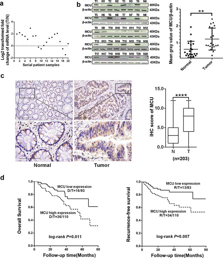
To determine the biological role of MCU in tumorigenesis of CRC, MCU-induced mitochondrial Ca2+ uptake promotes mitochondrial
reverse transcription-quantitative PCR (RT-qPCR) and western biogenesis in CRC cells
blotting assays were performed to examine the expression level Several recent studies have reported the biological role of
of MCU in 20 paired CRC and adjacent normal tissues. Our data increased mitochondrial biogenesis in CRC tumorigenesis.14,15
revealed that MCU was markedly upregulated in the majority of Therefore, we further explored the correlation between the
CRC tissues at both the mRNA and protein levels compared with expression of MCU and mitochondrial biogenesis in CRC cells. As
paired non-malignant tissues (Fig. 1a, b). In addition, we evaluated shown in Fig. 3a–d, LS174T MCU-knockdown cells exhibited a
the expression level of other MCU complex subunits in human decrease in the relative mitochondrial content, relative mtDNA
CRC tissues based on public RNA-seq data from The Cancer copy number, expression levels of oxidative phosphorylation
Genome Atlas (TCGA) database and the RT-qPCR analysis of our (OXPHOS)-related proteins, and ATP production when compared
samples. TCGA data analysis from 41 paired samples showed that with control cells. Similar results were obtained in LS174T cells
MICU1 was significantly downregulated at the mRNA level in CRC treated with the [Ca2+]m buffering using PV-Mito. In contrast, the
tissues compared with the adjacent tissues, while no significant opposite results were observed in MCU-overexpressing
differences were observed in the mRNA expression of MICU2, LS174T cells. Moreover, our data indicated that the [Ca2+]m
MCUb, MCUR1, and EMRE (Supplementary Fig. S1a). Furthermore, buffering by PV-Mito reversed the effects caused by MCU
RT-qPCR analysis of 20 paired tissue samples showed similar overexpression. IHC analysis also provided further supporting
results, indicating that MICU1 mRNA expression is downregulated data, indicating that the protein expression level of MCU was
in CRC tissues (Supplementary Fig. S1b). positively correlated with protein expression level of cytochrome
Our result was further validated by immunohistochemical (IHC) c oxidase subunit 4 (COX4), mtDNA copy number, and mitochon-
analysis in 159 of 203 (78%) paired CRC and adjacent normal drial content in CRC tissues (Fig. 3e–g). All these findings suggest
tissues, indicating that CRC tissues had a significantly higher that MCU promotes mitochondrial biogenesis in CRC cells
median MCU IHC score compared with adjacent non-tumor tissues primarily by regulating the level of mitochondrial Ca2+.
(P < 0.001, Fig. 1c). Moreover, MCU expression was stratified into
two groups based on the median value of the IHC score. MCU-induced mitochondrial Ca2+ uptake promotes CRC growth
Kaplan–Meier analysis showed that patients with CRC that had a in vitro
high expression of MCU, displayed notably shorter overall survival To determine the function of MCU-mediated mitochondrial Ca2+
(OS) and recurrence-free survival (RFS) compared with those who uptake in CRC growth, we first established CRC cell lines with
had low MCU expression (log-rank P = 0.011 and 0.007, respec- stable low or high expression of MCU by transfecting plasmids
tively) (Fig. 1d). expressing MCU and its small-hairpin RNA (shRNA). As presented
in Fig. 4a and Supplementary Fig. S3a, the cell viability assay
Upregulation of MCU enhances the mitochondrial Ca2+ uptake in showed that the decreased expression of MCU inhibited the
CRC cells growth of CRC cells compared with controls, whereas the opposite
As MCU has a critical role in maintaining mitochondrial Ca2+ result was obtained when MCU expression was upregulated.
homeostasis,6 we first determined whether MCU exerted an effect Moreover, [Ca2+]m buffering by PV-Mito significantly inhibited the
on the level of mitochondrial Ca2+ in LS174T and HCT-8 cells, growth of CRC cells and notably reversed the growth-promoting
Signal Transduction and Targeted Therapy (2020)5:59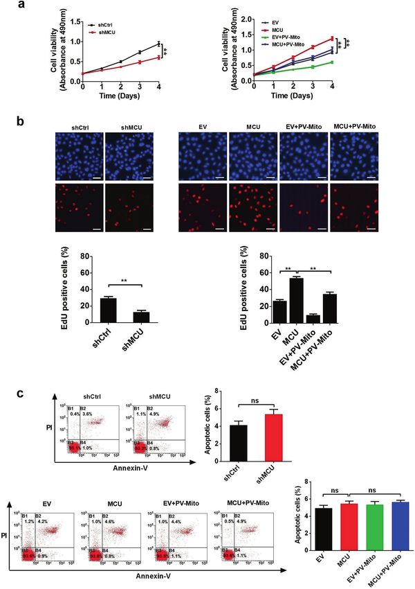
MCU-induced mitochondrial calcium uptake promotes mitochondrial. . .
                                                                         Liu et al.
                                                                                                                                                 3

Fig. 1 Upregulated MCU is associated with poor prognosis in patients with CRC. a Reverse transcription-quantitative PCR was performed to
measure the relative mRNA expression of MCU in 20 paired fresh tissues. The ratio of relative mRNA expression between tumor and normal
tissues was log2-transformed. b Western blotting analysis to measure protein expression level of MCU in 20 paired fresh tissues. Mean gray
values of MCU and β-actin (internal control) expression were determined by Quantity One software. c Representative IHC staining images
(Left) and IHC score (Right) of MCU in 203 paired tissues, which includes the aforementioned 20 paired tissues. d Kaplan–Meier plot of overall
and recurrence-free survival of patients with CRC depending on MCU expression. *P < 0.05; **P < 0.01. MCU mitochondrial calcium uniporter,
CRC colorectal cancer, T tumor, N normal, IHC immunohistochemical

effect of MCU. Furthermore, the Edu incorporation assay indicated        MCU-induced mitochondrial Ca2+ uptake promotes CRC growth
that MCU-knockdown cells had a lower percentage of proliferation         in vivo
compared with controls, whereas the opposite effects were                The effect of MCU-mediated mitochondrial Ca2+ uptake on CRC
observed in MCU-overexpressing cells (Fig. 4b and Supplementary          cell growth was further studied in vivo by generating a CRC
Fig. S3b). Consistently, PV-Mito played a similar role in this assay.    xenograft nude mice model. As shown in Fig. 5a, CRC xenografts
Moreover, flow cytometry analysis indicated that both MCU                 with MCU knockdown showed a slower growth rate compared
expression and [Ca2+]m buffering by PV-Mito had no effect on CRC         with the controls, whereas those overexpressing MCU exhibited a
cell apoptosis (Fig. 4c). Taken together, these data indicated that      faster growth compared with the corresponding controls. PV-Mito
MCU-mediated mitochondrial Ca2+ uptake facilitates CRC cell              treatment suppressed CRC growth and notably reversed the
proliferation in vitro.                                                  growth-promoting effect of MCU. Furthermore, IHC analysis

Signal Transduction and Targeted Therapy (2020)5:59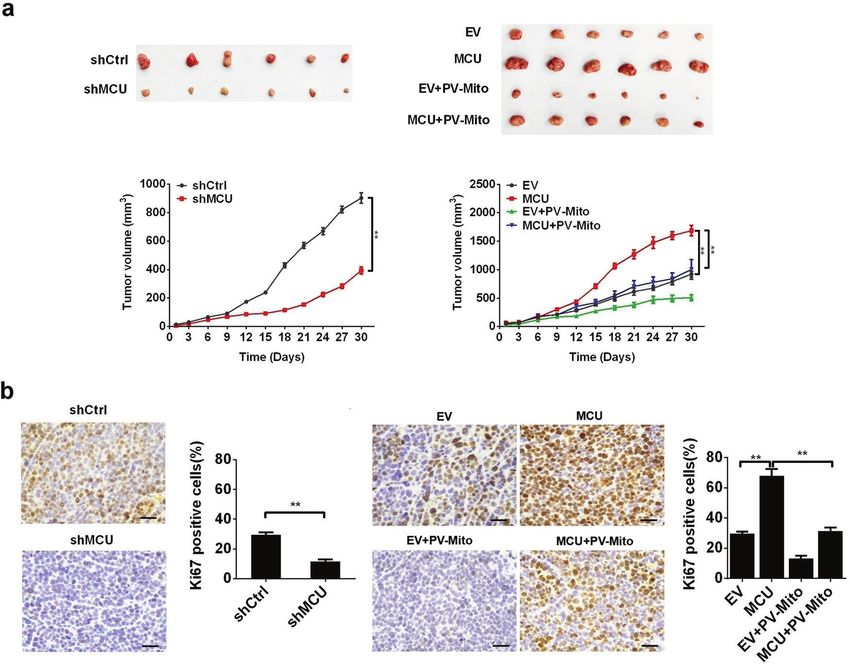
MCU-induced mitochondrial calcium uptake promotes mitochondrial. . .
                                                              Liu et al.
4

    Fig. 2 Upregulation of MCU increases mitochondrial Ca2+ uptake in CRC cells. a Reverse transcription-quantitative PCR and western blotting
    analyses for mRNA and protein expression levels of MCU in different CRC cell lines (COLO205, LS174T, LoVo, HCT-8, CaCo-2, SW620, DLD-1, and
    HT-29). b Western blotting analysis to measure MCU protein expression in LS174T cells, treated as indicated. c Confocal microscope images of
    [Ca2+]m using MitoPericam (Green) to label mitochondria in LS174T cells, treated as indicated. Scale bar, 5 µm. d Confocal microscope analysis
    of mitochondrial [Ca2+]m in CRC cells with a panel of treatment in response to 10 µM histamine. Ruthenium 360 (10 µM) was used to inhibit
    MCU activity. *P < 0.05; **P < 0.01. MCU mitochondrial calcium uniporter, CRC colorectal cancer, shCtrl control shRNA, shMCU shRNA against
    MCU, EV empty vector, [Ca2+]m mitochondrial Ca2+ levels, PV-Mito expression vector encoding parvalbumin with mitochondria target
    sequence, Ca2+ calcium

    (Fig. 5b) showed that the CRC xenografts with MCU knockdown or           the protein expression of TFAM in CRC tissues (Fig. 6b). Moreover, it
    PV-Mito treatment exhibited a significantly lower percentage of           has been reported that protein level of TFAM is regulated by the
    Ki67-positive cells compared with the corresponding controls. In         post-translational modification of phosphorylation, which leads to
    contrast, CRC xenografts overexpressing MCU had a significantly           its degradation.19 Thus, we sought to determine whether
    higher percentage of Ki67-positive cells compared with controls.         mitochondrial Ca2+ exerts an effect on the phosphorylation of
    These results support the conclusion that MCU-mediated mito-             TFAM. Our data revealed that downregulation of MCU enhanced
    chondrial Ca2+ uptake accelerates CRC growth in vivo by                  the phosphorylation of TFAM, whereas overexpression of MCU
    promoting CRC cell growth.                                               promoted dephosphorylation of TFAM in CRC cells (Fig. 6a). As
                                                                             expected, treatment with PV-Mito notably reversed the effect of
    Mitochondrial Ca2+ uptake promotes the dephosphorylation of              MCU overexpression on TFAM expression and its phosphorylation
    TFAM to enhance the mitochondrial biogenesis                             (Fig. 6a). Furthermore, a site-directed mutagenesis assay of serine
    Previous studies have demonstrated that TFAM is a crucial                residues indicated that phosphorylation of TFAM was inhibited
    regulator of mitochondrial biogenesis.16–18 Therefore, we                when serine-55 was mutated to alanine in MCU-knockdown CRC
    attempted to examine whether TFAM is involved in the                     cells, whereas the phosphorylation state of TFAM was not affected
    mitochondrial Ca2+-mediated mitochondrial biogenesis. The                when serine-160 or −170 was mutated to alanine (Fig. 6c),
    mRNA and protein expression levels of TFAM in MCU-knockdown              indicating that serine-55 of TFAM was the primary phosphorylation
    or MCU-overexpressing CRC cells with MCU knockdown and                   site. Additionally, a previous study has reported that mutation of
    overexpression were examined. We found that MCU expression               serine-55 to aspartate can mimic the sustained phosphorylation of
    had no effect on mRNA transcription of TFAM in CRC cells                 TFAM because the aspartate residue is negatively charged.19
    (Supplementary Fig. S4a). The expression of TFAM was down-               Consistently, our western blotting analysis clearly demonstrated
    regulated in MCU-knockdown CRC cells compared with control               the phosphomimics of TFAM in MCU-overexpressing CRC cells
    cells, whereas the opposite result was observed in MCU-                  when serine-55 was mutated to an aspartate residue. In contrast,
    overexpressing CRC cells (Fig. 6a). IHC analysis also indicated that     phosphorylation of TFAM was not affected in MCU-overexpressing
    the protein expression level of MCU was positively correlated with       CRC cells when serine-160 or -177 were mutated to aspartate

                                                                                Signal Transduction and Targeted Therapy (2020)5:59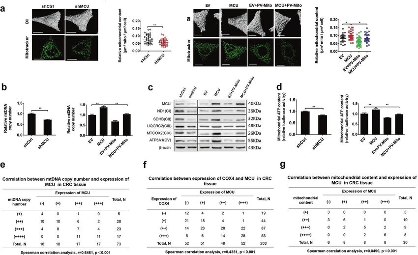
MCU-induced mitochondrial calcium uptake promotes mitochondrial. . .
                                                                          Liu et al.
                                                                                                                                                  5

Fig. 3 MCU-induced mitochondrial Ca2+ uptake promotes mitochondrial biogenesis in CRC cells. a Relative mitochondrial content (µm3
mitochondria per µm3 cell) was determined based on a mitochondrial three-dimensional model of confocal microscope images in
LS174T cells, treated as indicated. MitoTracker (Green) was used to label mitochondria. b Relative mtDNA copy number was determined by RT-
qPCR in LS174T cells, treated as indicated. c Western blotting analysis to measure the expression of oxidative phosphorylation related proteins
in LS174 cells treated as indicated. d Mitochondrial ATP levels were determined using the ATP Determination Kit in LS174T cells, treated as
indicated. e Correlation between relative mtDNA copy number and relative MCU expression in CRC tissue. The mtDNA copy number was
divided into four levels based on the quartering of RT-qPCR results. As the quartiles are 3.21 (upper quartile), 4.12 (median) and 5.22 (lower
quartile), RT-qPCR value 5.22 were defined as (+), (++), (+++), and (++++), respectively. f Correlation
between relative cytochrome c oxidase subunit 4 expression and relative MCU expression in CRC tissue. g Correlation between relative
mitochondrial content and relative MCU expression in CRC tissue. Mitochondrial content was divided into four groups based on the
quartering of the relative mitochondrial content levels. As the quartiles are 0.15 (upper quartile), 0.21 (median) and 0.25 (lower quartile),
relative mitochondrial content level 0.25 were defined as (+), (++), (+++), and(++++), respectively. *P <
0.05; **P < 0.01. MCU mitochondrial calcium uniporter, CRC colorectal cancer, mtDNA mitochondrial DNA, RT-qPCR reverse transcription-
quantitative PCR, Ca2+ calcium

(Fig. 6c). Altogether, our data indicated that MCU-mediated               Ca2+-mediated mitochondrial biogenesis. In summary, our data
mitochondrial Ca2+ uptake regulates the phosphorylation of TFAM           revealed that mitochondrial Ca2+ may promote mitochondrial
primarily via serine-55, but not -160 and -177 (Fig. 6c). Collectively,   biogenesis primarily by regulating the phosphorylation of TFAM at
our results suggested that mitochondrial Ca2+ may play a vital role       serine-55 to affect the stability of TFAM in CRC cells.
in modulating the phosphorylation of TFAM.                                   Considering the close link between mitochondrial Ca2+ home-
   Next, we examined whether MCU-mediated mitochondrial Ca2+              ostasis and mitochondrial dynamics, we also investigated the
uptake promoted mitochondrial biogenesis by regulating the                effect of MCU-mediated mitochondrial Ca2+ uptake on the
phosphorylation of TFAM. As shown in Fig. 6d–f and Supplementary          expression of proteins associated with mitochondrial dynamics.
Fig. S4b, c, TFAM overexpression resulted in a significant increase of     As shown in Supplementary Fig. S4d, CRC cells with MCU
the relative mtDNA copy number, relative mitochondrial content,           knockdown exhibited a decreased expression level of dynamin
expression levels of OXPHOS-related proteins and ATP production           related protein 1 (Drp1) and phosphorylated Drp1 on Ser616 and
compared with the corresponding controls in MCU-knockdown or              an increased expression of OPA1 mitochondrial dynamin like
MCU-overexpressing CRC cells. This indicated that the protein             GTPase. Similar results were obtained in CRC cells treated with PV-
expression of TFAM is essential for MCU-mediated mitochondrial            Mito. In contrast, the opposite results were observed in MCU-
biogenesis. Moreover, we found that the overexpression of                 overexpressing CRC cells. Our data suggested that MCU-mediated
TFAMS55D resulted in a lower relative mtDNA copy number, relative         mitochondrial Ca2+ uptake may promote mitochondrial fission
mitochondrial content, expression levels of OXPHOS-related                and inhibit mitochondrial fusion, which is consistent with the
proteins and ATP production in MCU-knockdown or MCU-                      findings of several previous reports.20,21
overexpressing CRC cells compared with the overexpression of
wild type TFAM. In contrast, the overexpression of TFAMS55A               Mitochondrial Ca2+-mediated mitochondrial biogenesis promotes
exhibited the opposite effect on mitochondrial biogenesis in MCU-         CRC growth via ROS/NF-κB signaling
knockdown or MCU-overexpressing CRC cells (Fig. 6d–f and                  Previous studies have demonstrated that [Ca2+]m influences the
Supplementary Fig. S4b, c). These findings supported the notion            production of ROS.22 Therefore, we explored whether MCU-
that TFAM phosphorylation plays a critical role in mitochondrial          mediated mitochondrial Ca2+ uptake would have an effect on ROS

Signal Transduction and Targeted Therapy (2020)5:59
MCU-induced mitochondrial calcium uptake promotes mitochondrial. . .
                                                              Liu et al.
6

    Fig. 4 MCU-induced mitochondrial Ca2+ uptake promotes CRC growth in vitro. a MTS assays of LS174T cells, treated as indicated.
    b Representative images (upper panel) of EdU incorporation assays for cell proliferation in LS174T cells, treated as indicated and percentage of
    EDU-positive cells (lower panel), treated as indicated. c Flow cytometry analysis of cell apoptosis by Annexin V (an indicator of apoptosis) and
    PI staining in LS174T cells, treated as indicated. *P < 0.05; **P < 0.01. MCU mitochondrial calcium uniporter, CRC colorectal cancer, Ca2+ calcium

                                                                                  Signal Transduction and Targeted Therapy (2020)5:59
MCU-induced mitochondrial calcium uptake promotes mitochondrial. . .
                                                                      Liu et al.
                                                                                                                                             7

Fig. 5 MCU-induced mitochondrial Ca2+ uptake promotes CRC growth in vivo. a Dissected tumors from sacrificed mice and tumor growth
curves of subcutaneous xenograft tumor developed from LS174T cells, treated as indicated. b Representative immunohistochemistry staining
images of Ki67 in xenograft tumors developed from LS174T cells, treated as indicated. *P < 0.05; **P < 0.01. MCU mitochondrial calcium
uniporter, CRC colorectal cancer, Ca2+ calcium

generation by TFAM-regulated mitochondrial biogenesis and thus        DISCUSSION
promote CRC cell growth. As shown in Fig. 7a, b, when compared        Previous studies indicated that mitochondrial Ca2+ entry regu-
with controls, overexpression of MCU increased the mitochondrial      lated by the MCU complex is closely associated with cancer
and total ROS level in CRC cells, which was reversed by PV-Mito-      progression, with remarkably different underlying mechanisms
mediated [Ca2+]m buffering or TFAM knockdown. This finding             depending on the type and stage of cancer. However, to date, the
indicated that mitochondrial biogenesis possesses a vital role in     biological function of MCU in CRC remains unknown. In our study,
mitochondrial Ca2+-mediated generation of ROS. It has been well       we have obtained two major findings. First, we have demon-
established that the ROS-activated NF-κB signaling pathway has a      strated that upregulation of MCU promotes CRC cell growth via
key role in various types of cancers.23,24 Thus, we aimed to          ROS/NF-κB signaling. More importantly, to the best of our
examine whether the ROS/NF-κB pathway is involved in mito-            knowledge, we have for the first time established a link between
chondrial Ca2+-regulated CRC cell growth. Western blotting data       the upregulation of MCU and mitochondrial biogenesis by
implied that MCU overexpression increased the protein expression      mitochondrial Ca2+-mediated dephosphorylation of TFAM, which
of phosphorylated p65, a key member of NF-κB signaling pathway,       provides a novel insight into the regulation of mitochondrial
compared with the control (Fig. 7c–e). This effect of MCU             functions.
overexpression on the activation of NF-κB signaling was reversed         A number of studies have demonstrated that deregulation of
by PV-Mito mediated [Ca2+]m buffering or mito-TEMPO, a                MCU is associated with different types of cancer.7 For instance, it
mitochondrial ROS scavenger, or TFAM knockdown (Fig. 7c–e).           has been reported that the expression of MCU is elevated in
Furthermore, treatment with H2O2 increased cell viability and the     various types of cancer, including breast cancer25 and hepatocel-
percentage of EDU-positive cells and clearly reversed the effect of   lular carcinoma.11 Consistently, our data indicated that the mRNA
MCU knockdown in CRC cells, whereas both the ROS scavenging           and protein expression levels of MCU were frequently upregulated
by Mito-TEMPO and NF-κB-specific inhibitor Bay11–7082                  in CRC cells and tissues, which then contributed to poorer OS of
decreased cell viability and the percentage of EDU-positive cells,    patients with CRC. We further confirmed our results in the Human
clearly reversed the effect of MCU overexpression in CRC cells        Protein Atlas database,26 indicating that MCU is overexpressed at
(Fig. 7f, g and Supplementary Fig. S5). Taken together, these data    both the mRNA and protein level in CRC. However, Marchi et al.27
supported the notion that mitochondrial Ca2+-mediated mito-           reported an inconsistent result, indicating that protein expression
chondrial biogenesis enhances CRC growth primarily via ROS/NF-        of MCU is decreased in CRC. This discrepancy may be caused by
κB signaling.                                                         selection bias of samples such as different stages of CRC and

Signal Transduction and Targeted Therapy (2020)5:59
MCU-induced mitochondrial calcium uptake promotes mitochondrial. . .
Liu et al.
8
Fig. 6 Mitochondrial Ca2+ promotes dephosphorylation of TFAM to enhance the mitochondrial biogenesis. a Western blotting analysis to
measure protein expression levels of MCU, TFAM, and phosphorylated TFAM in LS174 cells, treated as indicated. b Correlation between
relative TFAM expression and relative MCU expression in CRC tissue. c Western blotting analysis to measure protein expression levels of MCU,
TFAM, and phosphorylated TFAM in LS174 cells, treated as indicated. TFAM S55A, mutation of serine-55 to alanine. Band intensity were
quantified by densitometry and represented as the fold change of corresponding control value. d Reverse transcription-quantitative PCR
analysis of relative mitochondrial DNA copy number in LS174T cells, treated as indicated. e, f Relative mitochondrial content and
mitochondrial ATP levels were determined as mentioned above in LS174T cells treated as indicated. *P < 0.05; **P < 0.01. MCU mitochondrial
calcium uniporter, CRC colorectal cancer, TFAM transcription factor A, mitochondrial, Ca2+, calcium
Signal Transduction and Targeted Therapy (2020)5:59MCU-induced mitochondrial calcium uptake promotes mitochondrial. . .
Liu et al.
9
Fig. 7 Mitochondrial Ca2+-mediated mitochondrial biogenesis promotes CRC proliferation by ROS/NF-kB signaling. a Immunofluorescence
images (Left) of mitochondrial ROS (mROS) and MitoSOX fluorescence intensity (Right) in LS174T cells, treated as indicated. b Average DCF
fluorescence intensity in LS174T cells treated as indicated. Western blotting analysis to measure the expression levels of p65 and pi-p65 in
LS17T cells with MCU overexpression or treated with c PV-Mito, d Mito-TEMPO, or e siTFAM. f MTS assay to measure cell viability and g EdU
incorporation assays to measure cell proliferation in LS174T cells, treated as indicated. h Schematic representation showing the underlying
mechanism of MCU-mediated mitochondrial Ca2+ uptake in the promotion of CRC growth. *P < 0.05; **P < 0.01. MCU mitochondrial calcium
uniporter, CRC colorectal cancer, TFAM transcription factor A, mitochondrial, ROS reactive oxygen species, PV-Mito expression vector
encoding parvalbumin with mitochondria target sequence, si small interfering, Ca2+ calcium
Signal Transduction and Targeted Therapy (2020)5:59MCU-induced mitochondrial calcium uptake promotes mitochondrial. . .
Liu et al.
10
treatment procedures or use of a different antibody for IHC, which provide further understanding of the molecular mechanism
needs to be clarified in future studies. A recent study has underlying mitochondrial Ca2+-mediated mitochondrial biogen-
demonstrated that alternations in the stoichiometry of MCU esis. Several studies have reported the increase of mitochondrial
complexes causes a change in the Ca2+ influx into the biogenesis in CRC cells. For example, Witherspoon et al.14
mitochondria.28 Furthermore, Julia et al. reported that the MICU1 indicated that the constitutive expression of ETHE1 increases
functions as a gatekeeper to inhibit mitochondrial Ca2+ over- aerobic glycolysis (“Warburg effect”), oxidative phosphorylation,
load.29 Our data indicated that the mRNA expression of MICU1, and mitochondrial biogenesis in colorectal cancer (CRC) cell
but not other members of the MCU complex is downregulated in lines.35 Yang and colleagues36 suggested that mitochondrial
CRC tissues, suggesting that MICU1 may function together with biogenesis and maintenance may play an important part in tumor
MCU to regulate the level of mitochondrial Ca2+ in CRC cells. cell survival during CRC progression. In our study, our data showed
Previous studies indicated that the MCU complex possesses that upregulated MCU promotes mitochondrial biogenesis in CRC
distinct functions in different types of cancer. For instance, Tosatto cells. Very similarly, Zeng et al.33 also reported that basal and
et al.12 reported that silencing MCU in breast cancer cells results in maximal respirations are significantly higher in MCU-
decreased ROS production and expression of hypoxia-inducible overexpressing HT-29 cells than that in controls.
factor-1α (HIF-1α), which then leads to reduced tumor growth. A It is generally well accepted that dividing cells, including cancer
recent study also revealed that downregulation of MCU sup- cells, meet their energy demands by reprogramming their cell
pressed cell motility and reduced tumor growth in triple-negative metabolism such as altering mitochondrial dynamics.37,38 Our data
breast cancer by regulating store-operated Ca2+ entry.30 More- suggested that MCU-mediated mitochondrial Ca2+ uptake may
over, a study from our group revealed that MCU promotes the promote mitochondrial fission and inhibit mitochondrial fusion,
metastasis of HCC cells via the nicotinamide adenine dinucleotide which is in agreement with several previous reports.20,21 Over the
+ /sirtuin 3/superoxide dismutase 2 signaling pathway.11 To the past decades, multiple studies have reported that mitochondrial
best of our knowledge, the present study is the first to provide biogenesis and quality control are often upregulated in various
evidence that MCU-mediated mitochondrial Ca2+ uptake is crucial types of cancer, including breast cancer, lung cancer, and
for CRC cell growth by increasing mitochondrial biogenesis and hepatocellular carcinoma.39–42 However, the mechanism under-
ROS/NF-κB signaling. Previous studies have indicated that lying enhanced mitochondrial biogenesis in CRC remains obscure.
mitochondrial Ca2+ overload is closely involved in ROS production In the present study, we showed that MCU-mediated Ca2+ uptake
and the decrease of mitochondrial membrane potential, thus played an essential role in mitochondrial biogenesis via depho-
leading to cell death.31 In comparison, accumulating evidence sphorylation of TFAM. To the best of our knowledge, our work
indicates that mitochondrial Ca2+ uptake mediated by upregu- provides the first mechanistic insights into mitochondrial
lated MCU significantly promotes cell proliferation in cancers. Li Ca2+-mediated mitochondrial biogenesis in CRC.
et al32. showed that MCU overexpression is associated with the Growing evidence suggests that accumulation of Ca2+ in
glioblastoma cell proliferation but not the induction of cell death. mitochondria leads to ROS production.43,44 In the present study,
Curry et al.9 demonstrated that MCU overexpression is a feature of we showed that the forced expression of MCU in CRC cells greatly
breast cancers and offers a survival advantage against cell death enhanced mitochondrial Ca2+ uptake and a markedly increased
pathways. Zeng et al.33 reported that RIPK1 interacts with MCU to the production of ROS. Moreover, growing evidence has demon-
promote colorectal cancer cell proliferation by increasing mito- strated that there is an association between ROS and NF-κB
chondrial Ca2+ uptake and energy metabolism. Consistent with signaling.45 For instance, Takada et al.46 demonstrated that ROS
these reports, our data indicated that treatment with MCU influences the activation of the NF-κB pathway primarily by
overexpression had no effect on mitochondrial swelling and inhibiting the phosphorylation of nuclear factor of kappa light
calcein release in CRC cells, suggesting that MCU-induced polypeptide gene enhancer in B-cells inhibitor, alpha,46 and that
mitochondrial Ca2+ uptake and mitochondrial ROS production ROS also results in S-glutathionylation of IκB kinase β (IKKβ) on
may be in a normal range, and thus the functional status of mPTP cysteine-179 and then suppresses IKKβ activity.47 Consistently, our
is not affected. Our data have also provided further support in data revealed that increased mitochondrial biogenesis promoted
showing that MCU overexpression had no effect on the apoptosis ROS production to activate NF-κB signaling, which in turn
of CRC cells. These findings further suggested that the functional facilitated CRC growth in vitro and in vivo.
role of MCU-mediated mitochondrial Ca2+ uptake may be context Several studies have reported that Ru360 is a highly potent
dependent or cancer cell type specific. and selective MCU inhibitor.48 In our previous publication,11
Our data showed that MCU-mediated mitochondrial Ca2+ blocking of MCU activity by Ru360 has been verified in HCC cells.
uptake notably enhanced mitochondrial biogenesis by regulating Moreover, our data reported that MCU-mediated mitochondrial
the dephosphorylation of TFAM and thus increasing its stability. Ca2+ uptake is effectively blocked by Ru360 in CRC cells. These
Furthermore, it has been recently reported that the stability of results suggested a potential application as an antineoplastic
TFAM in mitochondria is mediated by post-translational phos- compound. However, other studies have also demonstrated that
phorylation of serine-55.19 Consistently, our site-directed muta- its general applicability is limited by several shortcomings.49 For
genesis analysis indicated that TFAM was phosphorylated at example, this compound is impermeant to the plasma mem-
serine-55. Furthermore, our data revealed that phosphorylation of brane in specific cell lines. Furthermore, its synthesis is
TFAM led to its degradation, while dephosphorylation of TFAM via challenging and low-yielding. Fortunately, recently the structural
the mutation of serine-55 to alanine greatly enhanced TFAM data of MCU has become available,50 which is beneficial to
stability, which is consistent with previous studies.19,34 It has been elucidate the structural basis of Ru360 blocking and could be
reported34 that phosphodiesterase 2A is involved in the regulation used to improve its potency and selectivity by developing its
of the protein kinase A activity, which is responsible for the structural analogs.
phosphorylation of TFAM in a number of cases.19 Therefore, we In conclusion, we systematically investigated the functional role
hypothesized that both enzymes may be related to the depho- of MCU-mediated mitochondrial Ca2+ homeostasis in promoting
sphorylation of TFAM induced by mitochondrial Ca2+, which CRC cell growth in vitro and in vivo. We demonstrated that MCU-
warrants further investigation in the future. Our data further mediated mitochondrial Ca2+ uptake significantly promotes
demonstrates that mitochondrial Ca2+ uptake increases mito- mitochondrial biogenesis by promoting the dephosphorylation
chondrial biogenesis by promoting dephosphorylation of TFAM. of TFAM. Furthermore, we also demonstrated that increased
This is consistent with previous findings, indicating that TFAM is mitochondrial Ca2+ uptake promotes CRC growth by activating
an essential regulator of mitochondrial biogenesis.18 Our results NF-κB signaling via ROS. Our findings identify a novel mechanism
Signal Transduction and Targeted Therapy (2020)5:59MCU-induced mitochondrial calcium uptake promotes mitochondrial. . .
Liu et al.
11
underlying MCU-mediated mitochondrial Ca2+ uptake in facilitat- Immunohistochemical (IHC) staining
ing CRC cell growth. IHC analysis and quantification of IHC staining score was evaluated
as previously described.24 The expression of proteins was
determined by two independent pathologists who were blinded
MATERIALS AND METHODS to the clinical characteristics of the patients.
Cell culture and tissue collection
Human CRC cell lines COLO205, LS174T, LoVo, HCT-8, Caco-2, Cell viability and apoptosis assays
SW620, DLD-1, and HT-29 and were purchased from The American Cell viability and apoptosis experiments were carried out as
Type Culture Collection (USA) and routinely cultured at 37 ˚C and previously described.24
5% CO2 in Dulbecco’s modified Eagle’s medium (DMEM) or RPMI-
1640 medium, which was supplemented with 10% fetal bovine Cell proliferation assay
serum. Tumor tissues and paired adjacent non-tumor tissues were Cell-LightTM EdU DNA Cell Proliferation Kit (Guangzhou Ribobio
collected from 203 patients who had undergone surgery and had Co., Ltd, China) was used to detect cell proliferation. As previously
no treatment before collection of tissue samples at Tangdu stated,51 EdU reagent was employed to treat cells 48 h after
Hospital affiliated with Fourth Military Medical University. Supple- transfection, followed by treatment with a permeabilization and
mentary Table S1 lists the distribution of clinical characteristics of phosphate-buffered saline (PBS) buffer. Cells were then visualized
203 patients with CRC. This study was performed with approval under a fluorescence microscope after staining with Apollo
from the Ethics Committee of Fourth Military Medical University reagent for 30 min.
(Permission number: KY20173189–1; Date issued: 2017-03-06).
Site-directed mutagenesis
Knockdown and overexpression of target genes Site-directed mutagenesis was carried out using the mutagenesis
To generate shRNA expression vectors, a shRNA targeting the kit (Beyotime Institute of Biotechnology, China) according to the
human MCU mRNA sequence and a control shRNA were cloned manufacturer’s instructions. In brief, the PCR primers (Supplemen-
into the pSilencer™ 3.1-H1 puro vector (Ambion; Thermo Fisher tary Table S2) containing desired mutation sites were used to
Scientific, Inc., USA). The shRNA sequences are listed in establish of plasmid constructs expressing mutant TFAM, which
Supplementary Table S2. To overexpress the target genes, were referred to as pcDNA-TFAMS55D and pcDNA-TFAMS55A. The
complementary DNA (cDNA) derived from LS174T cells was used plasmid pcDNA3.1-TFAM was used as the DNA template for PCR
as a DNA template to amplify MCU and TFAM using the primers amplification, according to the reaction conditions recommended
listed in Supplementary Table S2. The target genes were then by the mutagenesis kit. The resulting constructs, pcDNA-TFAMS55D
cloned into a pcDNA3.1(+) vector (Invitrogen; Thermo Fisher and pcDNA-TFAMS55A, were confirmed by DNA sequencing
Scientific, Inc., USA). The expression plasmid of PV-Mito encoding (Shanghai Shenggong, Biology Engineering Technology Service,
mitochondrial Ca2+-binding protein was provided by Dr. Atsushi Ltd., China). Proteins resulting from these plasmids are referred to
Miyawaki (RIKEN Brain Science Institute, Japan). Cells were seeded as TFAMS55A and TFAMS55D.
in 6-well plates until they reached 60% to 80% confluence for
transfection of vectors or small-interfering RNAs (siRNAs) with Measurement of mitochondrial Ca2+
Lipofectamine® 2000 reagent (Thermo Fisher Scientific, Inc., USA) The amount of Ca2+ in mitochondria was determined as described
according to the manufacturer’s protocols. All siRNAs were previously.24 In brief, cells were transfected with a plasmid carrying
synthesized by Shanghai GenePharma Co., Ltd., (China) and their mitochondrial matrix-targeted fluorescent tagged inverse pericam,
sequences are listed in Supplementary Table S2. which was referred to as mitopericam. A confocal laser scanning
microscope FV1000 (Olympus Corporation, Japan) was then
Measurement of mtDNA content by RT-qPCR employed to detect the cells. To measure dynamic mitochondrial
E.Z.N.A Tissue DNA Kit (Omega Bio-Tek, Inc., USA) was used to Ca2+, histamine (10 µM) was added after 30 s of baseline recording,
extract genomic DNA, according to the manufacturer’s instruc- 380 nm and 490 nm excitation filters were utilized in combination
tions. An RT-qPCR-based method was employed to measure the with 540 nm emission filter and photos were recorded every 3 s.
relative mtDNA copy number, as previously described.37
Detection of mitochondrial swelling
Quantitative reverse transcription PCR (RT-qPCR) analyses Freshly isolated mitochondria were added to buffer containing
Total RNA was isolated from cultured CRC cells or human CRC 120 mM KCl, 10 mM Tris, 20 mM MOPS, and 5 mM KH2PO4 (pH 7.4)
tissues and reversely transcribed. RT-qPCR was performed as to achieve a final concentration of 0.25 mg/mL mitochondria.
previously described.21 Relative mRNA expression levels were Mitochondrial swelling was detected by monitoring the decrease
quantified by the 2−ΔΔCt method. Experiments were performed in in light scattering. The absorbance at 530 nm was monitored
triplicate and GAPDH was used as an internal control. Supplemen- every 30 s for 10 min. In addition, the relative absorbance was also
tary Table S2 lists the primer sequences used for RT-qPCR analysis. compared for samples at 0 min and 60 min, respectively. In order
to determine PTP-dependent mitochondrial swelling, CSA
Western blotting analysis (200 nM), which is a cis-trans isomerase activity inhibitor of
Western blotting assays were performed based on regular cyclophilin D (CypD), was used to inhibit PTP opening, while FCCP
procedures. Briefly, total proteins from lysed cell samples were (5 µM), which causes depolarization of mitochondrial inner
analyzed on SDS-polyacrylamide gel electrophoresis (SDS-PAGE) membrane, was used to promote PTP opening.
and electrophoretically separated proteins were subsequently
transferred to a polyvinylidene fluoride (PVDF) membrane. The Calcein release assay
membrane with transferred proteins was then probed with a Cells were suspended in complete medium at a final concentra-
specific primary antibody overnight at 4 ˚C and incubated with a tion of 1 × 10⁶/mL, and then transferred to a 96-well plate (100 µL/
horseradish peroxidase-conjugated-secondary antibody for 2 h at well) for 4 h incubation with 5 µM fluorescent dye calcein-AM
room temperature. β-actin was used as a loading control in (Thermo Fisher Scientific, Inc., USA) in a CO2 incubator at 37 ˚C.
western blotting analysis. The membrane was then visualized using Cells were then washed three times (5 min each time) by PBS in
the enhanced chemiluminescence system (Pierce; Thermo Fisher presence of CoCl2 (1 mM) to quench the cytosolic compartments.
Scientific, Inc., USA). The antibody dilutions used are listed in Finally, cells were re-suspended in 100 μL PBS for data collection.
Supplementary Table S3. A microplate reader (Bio-Rad Laboratories, Inc., USA) was
Signal Transduction and Targeted Therapy (2020)5:59MCU-induced mitochondrial calcium uptake promotes mitochondrial. . .
Liu et al.
12
employed to measure calcein fluorescence every 5 min over statistically significant difference. For prognosis analysis, MCU
25 min. In addition, the relative calcein fluorescence was expression was first categorized into high or low level by the
measured at 0 and 60 min, respectively. median value of the IHC score. The OS and RFS in patients with
CRC who had high or low MCU expression were compared using
Measurement of mitochondrial mass Kaplan–Meier survival curves and log-rank test.
A three-dimensional (3D) model of mitochondria was constructed
using the method described by Cheverollier et al.52 Briefly, 100 nM
MitotrackerTM Green was used to label mitochondria, and DMEM ACKNOWLEDGEMENTS
without red phenol was employed to wash the cells. For imaging, We would like to thank Jiaojiao Wang and Feng Zhou (Experimental Teaching Center
coverslips were mounted in an incubation chamber placed on the of Basic Medicine) for their ongoing support and discussions. This work was
stage of an inverted microscope. An inverted wide-field Leica supported by the National Natural Science Foundation of China (grants 81872302
and 81902513), and Science and Technology Co-ordinate Innovation Project of
microscope equipped with a high-sensitivity CCD camera was
Shaanxi Province, China (grants 2016TZC-S-18-1).
employed to obtain images. An average of 30 image planes were
obtained along the z-axis at 0.2 µm increments. Three-dimensional
data processing and morphometric analysis was performed using
AUTHOR CONTRIBUTIONS
the Imaris 7.1.1 software (Bitplane, USA). Y.L., M.J., Y.J., T.R., and J.X. designed the study and supervised the project. Y.L., M.J.,
Y.W., and J.X. analyzed the data and wrote the manuscript. J.Z., T.R., and X.J. collected
Mitochondrial content analyses and processed colorectal cancer tissue samples and performed experiments.
Mitochondrial content was determined using the method
described previously.24 Briefly, glutaraldehyde was used to fix
human CRC tissues. The specimens were then postfixed with ADDITIONAL INFORMATION
osmium tetroxide, dehydrated with alcohol and embedded in The online version of this article (https://doi.org/10.1038/s41392-020-0155-5)
araldite. Uranyl acetate and lead citrate were used to stain the thin contains supplementary material, which is available to authorized users.
sections, followed by visualization under a Tecnai G2 electron
microscope (FEI; Thermo Fisher Scientific, Inc., USA). The area of Competing interests: The authors declare no competing interests.
mitochondria and CRC cells were determined using FIJI software
(NIH, USA). Transmission electron microscopy images were
examined by two independent pathologists, who were blinded REFERENCES
to clinical data. The mitochondrial content was calculated by the 1. Brody, H. Colorectal cancer. Nature 521, S1 (2015).
2. Duchen, M. R., Verkhratsky, A. & Muallem, S. Mitochondria and calcium in health
ratio of mitochondrial area to CRC cell area.
and disease. Cell Calcium 44, 1–5 (2008).
3. Stewart, T. A., Yapa, K. T. & Monteith, G. R. Altered calcium signaling in cancer
Detection of reactive oxygen species cells. Biochim. Biophys. Acta 1848, 2502–2511 (2015).
Cellular ROS and mitochondrial reactive species (mitoROS) were 4. Monteith, G. R., Prevarskaya, N. & Roberts-Thomson, S. J. The calcium-cancer
detected using the fluorescence probe DCFH-DA (Beyotime signalling nexus. Nat. Rev. Cancer 17, 367–380 (2017).
Institute of Biotechnology, China) and mitoSOX fluorescence 5. Granatiero, V., De Stefani, D. & Rizzuto, R. Mitochondrial calcium handling in
probe (Invitrogen; Thermo Fisher Scientific, Inc., USA), respectively, physiology and disease. Adv. Exp. Med. Biol. 982, 25–47 (2017).
following the protocols described previously.10 ImagePro image 6. Yamamoto, T. et al. Functional analysis of coiled-coil domains of MCU in
analysis software (Media Cybernetics, Inc., USA) was used to mitochondrial calcium uptake. Biochim. Biophys. Acta Bioenerg. 1860, 148061
(2019).
capture and process the images.
7. Vultur, A., Gibhardt, C. S., Stanisz, H. & Bogeski, I. The role of the mitochondrial
calcium uniporter (MCU) complex in cancer. Pflug. Arch. 470, 1149–1163 (2018).
In vivo subcutaneous xenograft models 8. Yu, C. et al. Mitochondrial calcium uniporter as a target of microRNA-340 and
The dorsal right flank of five-week-old Balb/c nude mice (six per promoter of metastasis via enhancing the Warburg effect. Oncotarget 8,
group) was subcutaneously injected with LS174T cells with low or 83831–83844 (2017).
high expression levels of MCU. The tumor volume was measured 9. Curry, M. C. et al. Mitochondrial calcium uniporter silencing potentiates caspase-
every three days for four weeks. Then mice were sacrificed to independent cell death in MDA-MB-231 breast cancer cells. Biochem. Biophys. Res.
measure the wet weight of the excised tumors. All animal Commun. 434, 695–700 (2013).
experiments were conducted according to the guidelines of the 10. Jin, M. et al. MCUR1 facilitates epithelial-mesenchymal transition and metastasis
via the mitochondrial calcium dependent ROS/Nrf2/Notch pathway in hepato-
Institutional Animal Care and Use Committee of the Fourth
cellular carcinoma. J. Exp. Clin. Cancer Res. 38, 136 (2019).
Military Medical University (Permission number: IACUC-20170105; 11. Ren, T. et al. MCU-dependent mitochondrial Ca(2+) inhibits NAD(+)/SIRT3/SOD2
Date issued: 2017-01-01). pathway to promote ROS production and metastasis of HCC cells. Oncogene 36,
5897–5909 (2017).
Immunoprecipitation assay 12. Tosatto, A. et al. The mitochondrial calcium uniporter regulates breast cancer
The phosphorylation of TFAM was assayed using the immuno- progression via HIF-1alpha. EMBO Mol. Med. 8, 569–585 (2016).
precipitation assay. Cells were lysed and incubated with 200 μL 13. Marchi, S., Vitto, V. A. M., Patergnani, S. & Pinton, P. High mitochondrial Ca(2+)
Protein A beads (Santa Cruz Biotechnology) supplemented with content increases cancer cell proliferation upon inhibition of mitochondrial
50 μg anti-TFAM antibody overnight. Normalized amounts of total permeability transition pore (mPTP). Cell Cycle 18, 914–916 (2019).
14. Witherspoon, M. et al. ETHE1 overexpression promotes SIRT1 and PGC1alpha
lysates or immunoprecipitated samples were analyzed by SDS-
mediated aerobic glycolysis, oxidative phosphorylation, mitochondrial biogenesis
PAGE and western blotting with phosphorylated antibody.19 and colorectal cancer. Oncotarget 10, 4004–4017 (2019).
15. Cruz, M. D. et al. Metabolic reprogramming of the premalignant colonic mucosa
Statistical analysis is an early event in carcinogenesis. Oncotarget 8, 20543–20557 (2017).
All statistical analyses were performed using SPSS 17.0 software 16. Kelly, D. P. & Scarpulla, R. C. Transcriptional regulatory circuits controlling mito-
(SPSS, Inc., USA). Data are shown as the mean ± SD from three chondrial biogenesis and function. Genes Dev. 18, 357–368 (2004).
independent experiments, where appropriate. Student’s t-test was 17. Yao, K. et al. Carvedilol promotes mitochondrial biogenesis by regulating the
employed to analyze the significant differences between two PGC-1/TFAM pathway in human umbilical vein endothelial cells (HUVECs). Bio-
groups. Paired two-tailed t-tests were employed to compare the chem. Biophys. Res. Commun. 470, 961–966 (2016).
18. Picca, A. & Lezza, A. M. Regulation of mitochondrial biogenesis through TFAM-
differences between tumor tissue and adjacent non-tumor
mitochondrial DNA interactions: Useful insights from aging and calorie restriction
mucosa. The correlations between measured variables were studies. Mitochondrion 25, 67–75 (2015).
analyzed by Spearman’s correlation. P > 0.05 was considered a
Signal Transduction and Targeted Therapy (2020)5:59MCU-induced mitochondrial calcium uptake promotes mitochondrial. . .
Liu et al.
13
19. Lu, B. et al. Phosphorylation of human TFAM in mitochondria impairs DNA 40. Tohme, S. et al. Hypoxia mediates mitochondrial biogenesis in hepatocellular
binding and promotes degradation by the AAA+ Lon protease. Mol. Cell. 49, carcinoma to promote tumor growth through HMGB1 and TLR9 interaction.
121–132 (2013). Hepatology 66, 182–197 (2017).
20. Kaddour-Djebbar, I. et al. Specific mitochondrial calcium overload induces 41. LeBleu, V. S. et al. PGC-1alpha mediates mitochondrial biogenesis and oxidative
mitochondrial fission in prostate cancer cells. Int. J. Oncol. 36, 1437–1444 (2010). phosphorylation in cancer cells to promote metastasis. Nat. Cell Biol. 16,
21. Guan, L. et al. MCU Up-regulation contributes to myocardial ischemia-reperfusion 992–1003 (2014). 1001-1015.
Injury through calpain/OPA-1-mediated mitochondrial fusion/mitophagy Inhibi- 42. Bellance, N. et al. Bioenergetics of lung tumors: alteration of mitochondrial bio-
tion. J. Cell Mol. Med. 23, 7830–7843 (2019). genesis and respiratory capacity. Int. J. Biochem Cell Biol. 41, 2566–2577 (2009).
22. Santo-Domingo, J., Wiederkehr, A. & De Marchi, U. Modulation of the matrix 43. Ma, W. et al. Cardiotoxicity of sorafenib is mediated through elevation of ROS
redox signaling by mitochondrial Ca(2.). World J. Biol. Chem. 6, 310–323 (2015). level and CaMKII activity and dysregulation of calcium homoeostasis. Basic Clin.
23. Yang, H. L. et al. Anti-EMT properties of CoQ0 attributed to PI3K/AKT/NFKB/MMP- Pharm. Toxicol. 126, 166–180 (2020).
9 signaling pathway through ROS-mediated apoptosis. J. Exp. Clin. Cancer Res. 38, 44. Yang, Z. et al. Important role of sarcoplasmic reticulum Ca(2+) release via rya-
186 (2019). nodine receptor-2 channel in hypoxia-induced rieske iron-sulfur protein-medi-
24. Huang, Q. et al. Increased mitochondrial fission promotes autophagy and ated mitochondrial reactive oxygen species generation in pulmonary artery
hepatocellular carcinoma cell survival through the ROS-modulated coordinated smooth muscle cells. Antioxid. Redox Signal. 32, 447–462 (2020).
regulation of the NFKB and TP53 pathways. Autophagy 12, 999–1014 (2016). 45. Zhang, J. et al. ROS and ROS-mediated cellular signaling. Oxid. Med. Cell Longev.
25. Hall, D. D. et al. Mitochondrial calcium uniporter activity is dispensable for MDA- 2016, 4350965 (2016).
MB-231 breast carcinoma cell survival. PLoS ONE 9, e96866 (2014). 46. Takada, Y. et al. Hydrogen peroxide activates NF-kappa B through tyrosine
26. Uhlen, M. et al. Proteomics. Tissue-based map of the human proteome. Science phosphorylation of I kappa B alpha and serine phosphorylation of p65: evidence
347, 1260419 (2015). for the involvement of I kappa B alpha kinase and Syk protein-tyrosine kinase. J.
27. Marchi, S. et al. Downregulation of the mitochondrial calcium uniporter by Biol. Chem. 278, 24233–24241 (2003).
cancer-related miR-25. Curr. Biol. 23, 58–63 (2013). 47. Reynaert, N. L. et al. Dynamic redox control of NF-kappaB through glutaredoxin-
28. Chapoy-Villanueva, H. et al. Changes in the stoichiometry of uniplex decrease regulated S-glutathionylation of inhibitory kappaB kinase beta. Proc. Natl Acad.
mitochondrial calcium overload and contribute to tolerance of cardiac ischemia/ Sci. USA 103, 13086–13091 (2006).
reperfusion injury in hypothyroidism. Thyroid 29, 1755–1764 (2019). 48. Ying, W. L., Emerson, J., Clarke, M. J. & Sanadi, D. R. Inhibition of mitochondrial
29. Liu, J. C. et al. MICU1 serves as a molecular gatekeeper to prevent in vivo calcium ion transport by an oxo-bridged dinuclear ruthenium ammine complex.
mitochondrial calcium overload. Cell Rep. 16, 1561–1573 (2016). Biochemistry 30, 4949–4952 (1991).
30. Tang, S. et al. Mitochondrial Ca(2)(+) uniporter is critical for store-operated Ca(2) 49. Woods, J. J. et al. A selective and cell-permeable mitochondrial calcium uniporter
(+) entry-dependent breast cancer cell migration. Biochem. Biophys. Res. Com- (MCU) inhibitor preserves mitochondrial bioenergetics after hypoxia/reox-
mun. 458, 186–193 (2015). ygenation injury. ACS Cent. Sci. 5, 153–166 (2019).
31. Liao, Y. et al. Mitochondrial calcium uniporter protein MCU is involved in oxi- 50. Nguyen, N. X. et al. Cryo-EM structure of a fungal mitochondrial calcium uni-
dative stress-induced cell death. Protein Cell. 6, 434–442 (2015). porter. Nature 559, 570–574 (2018).
32. Li, X. et al. Mechanisms of malignancy in glioblastoma cells are linked to mito- 51. Chen, L. et al. Positive feedback loop between mitochondrial fission and Notch
chondrial Ca(2) (+) uniporter upregulation and higher intracellular Ca(2+) levels. signaling promotes survivin-mediated survival of TNBC cells. Cell Death Dis. 9,
J. Cell Sci. 133, 563–579 (2020). 1050 (2018).
33. Zeng, F. et al. RIPK1 Binds MCU to mediate induction of mitochondrial Ca(2+) 52. Chevrollier, A. et al. Standardized mitochondrial analysis gives new insights into
uptake and promotes colorectal oncogenesis. Cancer Res. 78, 2876–2885 (2018). mitochondrial dynamics and OPA1 function. Int. J. Biochem. Cell Biol. 44, 980–988
34. Zhang, F. et al. The cAMP phosphodiesterase Prune localizes to the mitochondrial (2012).
matrix and promotes mtDNA replication by stabilizing TFAM. EMBO Rep. 16,
520–527 (2015).
35. Witherspoon, M. et al. ETHE1 overexpression promotes SIRT1 and PGC1alpha Open Access This article is licensed under a Creative Commons
mediated aerobic glycolysis, oxidative phosphorylation, mitochondrial biogenesis Attribution 4.0 International License, which permits use, sharing,
and colorectal cancer. Oncotarget 10, 4004–4017 (2019). adaptation, distribution and reproduction in any medium or format, as long as you give
36. Yang, Y. et al. SSBP1 Upregulation In Colorectal Cancer Regulates Mitochondrial appropriate credit to the original author(s) and the source, provide a link to the Creative
Mass. Cancer Manag. Res. 11, 10093–10106 (2019). Commons license, and indicate if changes were made. The images or other third party
37. Porat-Shliom, N. et al. Mitochondrial populations exhibit differential dynamic material in this article are included in the article’s Creative Commons license, unless
responses to increased energy demand during exocytosis in vivo. iScience 11, indicated otherwise in a credit line to the material. If material is not included in the
440–449 (2019). article’s Creative Commons license and your intended use is not permitted by statutory
38. Vaupel, P., Schmidberger, H. & Mayer, A. The Warburg effect: essential part of regulation or exceeds the permitted use, you will need to obtain permission directly
metabolic reprogramming and central contributor to cancer progression. Int. J. from the copyright holder. To view a copy of this license, visit http://creativecommons.
Radiat. Biol. 95, 912–919 (2019). org/licenses/by/4.0/.
39. Zong, W. X., Rabinowitz, J. D. & White, E. Mitochondria and cancer. Mol. Cell. 61,
667–676 (2016).
© The Author(s) 2020
Signal Transduction and Targeted Therapy (2020)5:59You can also read


