Dandelion Root Extract Induces Intracellular Ca2+ Increases in HEK293 Cells - MDPI
←
→
Page content transcription
If your browser does not render page correctly, please read the page content below
International Journal of
           Molecular Sciences

Article
Dandelion Root Extract Induces Intracellular Ca2+
Increases in HEK293 Cells
Andrea Gerbino 1, * ID , Daniela Russo 2 , Matilde Colella 1 ID , Giuseppe Procino 1         ID
                                                                                                  ,
Maria Svelto 1 , Luigi Milella 2 ID and Monica Carmosino 2, *
 1    Department of Biosciences, Biotechnologies and Biopharmaceutics, University of Bari, 70126 Bari, Italy;
      matilde.colella@uniba.it (M.C.); giuseppe.procino@uniba.it (G.P.); maria.svelto@uniba.it (M.S.)
 2    Department of Sciences, University of Basilicata, 85100 Potenza, Italy; daniela.russo@unibas.it (D.R.);
      luigi.milella@unibas.it (L.M.)
 *    Correspondence: andrea.gerbino@uniba.it (A.G.); monica.carmosino@unibas.it (M.C.);
      Tel.: +39-080-544-3334 (A.G.); +39-335-6302642 (M.C.)

 Received: 6 March 2018; Accepted: 4 April 2018; Published: 7 April 2018

 Abstract: Dandelion (Taraxacum officinale Weber ex F.H.Wigg.) has been used for centuries as an
 ethnomedical remedy. Nonetheless, the extensive use of different kinds of dandelion extracts and
 preparations is based on empirical findings. Some of the tissue-specific effects reported for diverse
 dandelion extracts may result from their action on intracellular signaling cascades. Therefore, the
 aim of this study was to evaluate the effects of an ethanolic dandelion root extract (DRE) on Ca2+
 signaling in human embryonic kidney (HEK) 293 cells. The cytotoxicity of increasing doses of
 crude DRE was determined by the Calcein viability assay. Fura-2 and the fluorescence resonance
 energy transfer (FRET)-based probe ERD1 were used to measure cytoplasmic and intraluminal
 endoplasmic reticulum (ER) Ca2+ levels, respectively. Furthermore, a green fluorescent protein
 (GFP)-based probe was used to monitor phospholipase C (PLC) activation (pleckstrin homology
 [PH]–PLCδ–GFP). DRE (10–400 µg/mL) exposure, in the presence of external Ca2+ , dose-dependently
 increased intracellular Ca2+ levels. The DRE-induced Ca2+ increase was significantly reduced in
 the absence of extracellular Ca2+ . In addition, DRE caused a significant Ca2+ release from the ER of
 intact cells and a concomitant translocation of PH–PLCδ–GFP. In conclusion, DRE directly activates
 both the release of Ca2+ from internal stores and a significant Ca2+ influx at the plasma membrane.
 The resulting high Ca2+ levels within the cell seem to directly stimulate PLC activity.

 Keywords: Ca2+ signaling; Ca2+ influx; plasma membrane; endoplasmic reticulum; phospholipase C;
 Fura-2; Ca2+ fluorescent sensors; herbal extract; bioactive compounds

1. Introduction
      To date, natural products from medicinal plants, microorganisms, marine organisms (e.g., Acmella
oleracea [1], Sclerocarya birrea [2,3], Agelas clathrodes [4], and Tedania ignis [5]) are in the spotlight of
many research and industrial laboratories for (i) biomedical applications, (ii) the recovery of bioactives,
and (iii) the development of pharmacological drugs. Taraxacum officinale Weber, known as dandelion,
is a perennial weed that has been used for hundreds of years (starting from the 10th and 11th centuries)
as a traditional medical remedy for several diseases [6]. As a matter of fact, dandelion is nowadays
commercialized as a healthy food because of its health-promoting ethno-medical properties, including
anti-inflammatory, anti-rheumatic, anti-oxidant, anti-carcinogenic, diuretic, choleretic and cholagogue,
laxative, and hypoglycemic activities [6,7]. However, only some of these empirical effects have been
validated by a proper scientific investigation [8], therefore more experimental evidence is needed to
justify such extensive use of dandelion as a natural therapeutic remedy.

Int. J. Mol. Sci. 2018, 19, 1112; doi:10.3390/ijms19041112                           www.mdpi.com/journal/ijms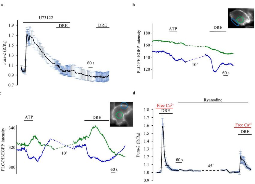
Int. J. Mol. Sci. 2018, 19, 1112                                                                    2 of 17

      A possible explanation of such a wide panel of physiological effects might be found in
the chemical composition of the different dandelion preparations. Dandelion’s extract might be
composed by the whole plant or by different parts of it (e.g., roots, leaves, stem, flowers), alone
or in combination. In addition, its chemical composition strictly depends on both the extraction
protocol and the solvents used (ethanol, acetone, water, or methanol) that have to be properly
designed and identified to efficiently produce extracts containing the desired bioactive compounds.
For example, the phytochemical composition of dandelion roots extracts (DRE) reported the presence
of sesquiterpenes, various triterpenes, phytosterols, and phenolic compounds [6]. Among the phenolic
compounds, hydroxycinnamic acid derivatives (chlorogenic, caffeic, 4-coumaric, 3-coumaric, ferulic
acids) are the main represented class, whereas a small amount of flavonoids and hydroxybenzoic
acid derivatives is generally reported [9–11]. Compared to the roots, dandelion leaves and flowers
are more enriched in flavonoids (luteolin and its glycoside derivatives, chrysoeriol) and coumarins
(cichoriin and aesculin), but also hydroxycinnamic acid derivatives (caffeic, chlorogenic, chicoric,
and monocaffeoyltartaric acids) were reported to be present [10,12].
      These various chemical components may act individually, additively, or in synergy to regulate
different tissue-specific functions. Still, the surprising amount of physiological functions that are
somehow related to the medical use of T. officinale suggests the ability of cells to respond to its herbal
extracts through a basic and widespread intracellular mechanism.
      Therefore, in this study, we evaluated whether the acute exposure of HEK293 cells to an ethanolic
dandelion root extract (DRE) might impact intracellular Ca2+ homeostasis. Among the more general
transduction pathways of a cell we decided to analyze specifically Ca2+ signaling for two main reasons.
First, even though there is no information available in the literature about the effects of dandelion
extracts on intracellular Ca2+ dynamics, numerous reports regarding the effects of single bioactive
components, especially hydroxycinnamic acid derivatives, have been published. Caffeic acid (CA)
and its derivatives are natural phenolic compounds that affect cellular Ca2+ homeostasis in different
experimental models, such as human gastric cancer (SCM1) cells [13], T lymphocytes [14], and Jurkat
cells [15]. Chlorogenic (CGA) and ferulic acids (FA) are polyphenols that protect different cell models
(human umbilical vein endothelial cells and rat cortical neurons) from Ca2+ -mediated insults by
reducing, through cell-specific mechanisms, Ca2+ entry at the plasma membrane [16–18].
      Second, most of the physiological functions that are regulated by dandelion extracts are finely
modulated by intracellular Ca2+ events (e.g., inflammation, proliferation, diuresis, just to name a few).
This implies either that the two players (namely, Ca2+ and DRE) share common cellular targets or that
Ca2+ is a dandelion-induced mediator within the cells.
      Therefore, we used Fura-2 and the fluorescence resonance energy transfer (FRET)-based probe
ERD1 [19] to measure cytoplasmic and intraluminal Ca2+ levels within the endoplasmic reticulum,
respectively, in HEK293 cells in response to acute exposure to DRE. In addition, a green fluorescent
protein (GFP)-based probe [20] was used to monitor the time course of phospholipase C (PLC) activation
(pleckstrin homology [PH]–PLCδ–GFP). All the experiments performed depict a scenario in which DRE
induced a rapid and reversible increase in intracellular Ca2+ levels. Such rise in cytosolic Ca2+ levels
was due to both Ca2+ release from the endoplasmic reticulum and Ca2+ entry at the plasma membrane,
likely via store-operated channels. DRE exposure also induced the activation of a downstream signaling
element such as PLC, which is elicited, even though not exclusively, by G-protein-coupled receptors [21].
      The precise dissection of DRE-induced intracellular Ca2+ events at the cellular and molecular
level will pave the way for the proper use of this promising extract as a natural pharmaceutical tool.

2. Results

2.1. High-Performance Liquid Chromatography (HPLC) Analysis of the Ethanolic DRE and Its
Cytotoxic Activity
    Dandelion dried roots were extracted with ethanol by exhaustive maceration. Ethanol is an
appropriate solvent for both phenols and other bioactive compounds because of its intermediate
Int. J. Mol. Sci. 2018, 19, 1112                                                                                 3 of 17

polarity. It is well known that solvents affect the extraction of bioactive compounds and the
phytochemical profiles of extracts from various plant parts [22]. Consequently, the phytochemical
constituents and their amount in different plant extracts reflect different biological properties of a plant.
     The chemical composition of our dandelion ethanolic root extract (DRE) was evaluated by
reversed-phase high-performance liquid chromatography (RP-HPLC). Figure 1a shows the HPLC
profile of the ethanolic DRE. The results of the quali–quantitative analysis reported the presence of
hydroxybenzoic acid derivatives and hydroxycinnamic acids. In particular, chlorogenic acid (retention
time, hereafter Rt: 7.75 min) and caffeic acid (Rt: 10.17 min) were identified in the extract as the main
components (see Table 1).

      Table 1. HPLC quantitative analysis of the ethanolic extract from dandelion root. The values are expressed
      as mean ± standard deviation (SD) of milligram (mg) of compound per gram (g) of extract (n = 3).

                                   Compound           Rt (min)       Average ± SD (mg/g)
                                  Gallic acid (1)        4.72             0.164 ± 0.018
                               Chlorogenic acid (2)      7.75             1.256 ± 0.012
                                  Caffeic acid (3)      10.17             0.776 ± 0.063
                                 Vanillic acid (4)      10.83             0.148 ± 0.012
                                 Syringic acid (5)      11.32             0.234 ± 0.009
                               p-Coumaric acid (6)      14.72             0.041 ± 0.005
                                  Ferulic acid (7)      17.71             0.273 ± 0.070

      We also identified, although present in lower amount, gallic acid (Rt: 4.72 min), vanillic acid
(Rt: 10.83 min), syringic acid (Rt: 11.32 min), p-coumaric acid (Rt: 14.72 min), and ferulic acid
(Rt: 17.71 min). All compounds were identified comparing their retention time and UV spectrum with
those of reference standards. The other peaks of the extract could not be identified because of a lack of
validated reference standards to compare with. All these compounds had already been identified in
dandelion root extracts [9]. In this study we first aimed at identifying the non-toxic concentration of
the DRE by performing a Calcein cytotoxic assay in HEK293 cells. As shown in Figure 1b, compared
with vehicle control (ethanol), DRE induced significant cell death only at 750 µg/mL.

      Figure 1. HPLC analysis of dandelion root extract (DRE) and evaluation of its cytotoxicity.
      (a) Chromatogram obtained by HPLC analysis of a Taraxacum officinale root extract at 320 nm.
      The compounds identified are gallic acid (1), chlorogenic acid (2), caffeic acid (3), vanillic acid (4),
      syringic acid (5), p-coumaric (6), and ferulic acid (7). (b) Viability of HEK293 cells cultured in the
      absence (ethanol 0.6% as control, CTRL) and presence of diverse doses of T. officinale root extract for 24 h
      by Calcein viability assay. The reported values are means ± SEM of three independent experiments
      (** p < 0.01 vs. CTRL).

      On the basis of this result, in the following functional studies, we used DRE at 400 µg/mL (or lower).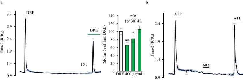
Int. J. Mol. Sci. 2018, 19, 1112                                                                            4 of 17

2.2. Exposure to DRE Increased Intracellular Ca2+ Levels
     The results collected in Figure 2 show that DRE causes an increase of intracellular Ca2+ levels
in HEK293 cells. In the presence of 1.2 mM extracellular Ca2+ , 400 µg/mL DRE elicited cytosolic
Ca2+ transients that were not significantly larger (118.88 ± 7.96% of control peak; p = 0.4) than those
produced by a saturating dose of the Ca2+ -mobilizing agonist ATP (100 µM) in the same cells (Figure 2a;
n = 5 experiments, HEK293 cells).

      Figure 2. DRE increases intracellular Ca2+ levels, while caffeic and chlorogenic acids do not,
      as measured by Fura-2 in single HEK293 cells. (a) Response of Fura-2-loaded HEK293 cells to the
      Ca2+ -mobilizing agonist ATP (100 µM) compared with the Ca2+ increase elicited by DRE (400 µg/mL).
      The reported values are means ± SE from all the responsive cells on a single coverslip. (b) Changes
      in the Fura-2 ratio in Ca2+ -containing solutions in response to increasing doses of DRE (10, 50, 200,
      and 400 µg/mL) normalized to the response induced by ATP. The reported values are means ± SE
      from all the responsive cells of all experiments performed (** p < 0.01 vs. ATP, * p < 0.05 vs. ATP).
      (c) Number of DRE-treated (10, 50, 200, and 400 µg/mL) Ca2+ -responsive cells versus the total number
      of cells analyzed. The bar graph summarizes the number of cells of all the experiments performed
      for each concentration. (d) Response of Fura-2-loaded HEK293 cells to caffeic acid (CA, 150 µM),
      chlorogenic acid (CGA, 200 µM), and DRE (400 µg/mL) compared with the effect induced by the
      Ca2+ -mobilizing agonist ATP (100 µM). The reported values are means ± SE from all the responsive
      cells on a single coverslip. (e) Response of Fura-2-loaded HEK293 cells to the simultaneous exposure to
      caffeic (CA, 150 µM) and chlorogenic acid (CGA, 200 µM) compared with the effect induced by the
      Ca2+ -mobilizing agonist ATP (100 µM). The reported values are means ± SE from all the responsive
      cells on a single coverslip.

     The Ca2+ response induced by DRE was dose-dependent. The smallest, albeit significant, increase
in intracellular Ca2+ was detected at a concentration of DRE as low as 10 µg/mL (32.7 ± 12.6% of
control peak; n = 3, m = 149 cells, p < 0.01). Concentrations of 50 and 200 µg/mL DRE induced linear
increases in intracellular Ca2+ that were, however, significantly smaller than those induced by ATP
(Figure 2b, 62.4 ± 8.60% of control peak; n = 3, m = 129 cells, p < 0.01 and 79.4 ± 4.75% of control peak;
n = 4, m = 187 cells, p < 0.05, respectively). In addition, the higher was the concentration of DRE used,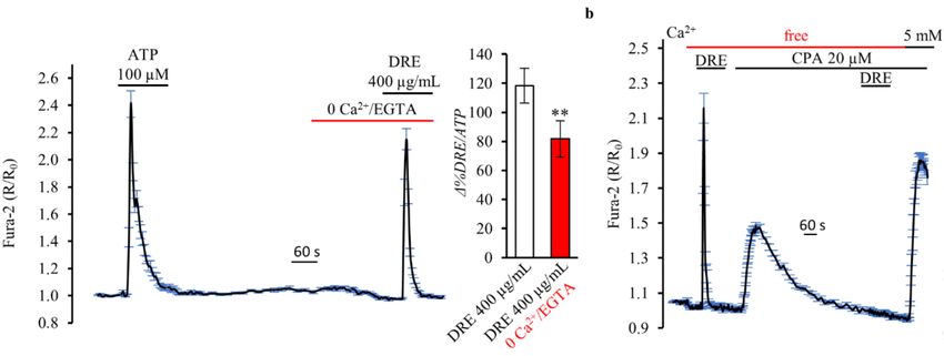
Int. J. Mol. Sci. 2018, 19, 1112                                                                            5 of 17

the larger was the number of responsive cells on a single coverslip (Figure 2c), clearly indicating the
activation of a cellular mechanism that was strictly dependent on DRE concentration.
      Were these Ca2+ transients induced by a specific component of the DRE? To answer this question,
we analyzed the effects of both caffeic and chlorogenic acids (CA and CGA, respectively) on the
intracellular Ca2+ levels. Both compounds, enriched in our DRE (see Table 1), are known to impact
in vitro Ca2+ homeostasis [13–17]. We exposed HEK293 cells to different concentrations of CA and
CGA, starting with the concentration at which these compounds were present in our extract, but no
intracellular Ca2+ changes were recorded (data not shown). Also, when we exposed HEK293 cells to
the highest investigated concentrations of either caffeic acid (150 µM) or chlorogenic acid (200 µM) we
did not record intracellular Ca2+ changes (Figure 2d). On the other hand, the exposure of cells on the
same coverslip to DRE induced a significant increase in the intracellular Ca2+ levels that was similar to
that induced by ATP.
      The same effect, namely, no intracellular Ca2+ increase, was also obtained when both components
were perfused at the same time (Figure 2e), indicating that the response elicited by DRE is induced by
other components (even though present in a smaller dose) or by the extract as a whole.
      The Ca2+ -mediated response induced by 400 µg/mL DRE was only slightly, but significantly,
decreased, when compared to ATP, in the absence of external Ca2+ (Figure 3a; 81.69 ± 7.17% of control
response to ATP; n = 5, m = 235 cells, p < 0.001). These results suggest that DRE was able to release
Ca2+ from internal stores. Indeed, Figure 3b shows that, when the cells were treated with the reversible
sarco/endoplasmic reticulum Ca2+ -ATPase (SERCA) pump inhibitor cyclopiazonic acid (CPA) in free
extracellular Ca2+ to deplete intracellular Ca2+ stores, the Ca2+ response induced by DRE in control
conditions was completely prevented. Note that this experimental approach also revealed that the
acute exposure to 400 µg/mL DRE did not interfere with the mechanisms underlining CPA-induced
endoplasmic reticulum (ER) Ca2+ release and with the capacitative Ca2+ entry induced by perfusion
with 5 mM Ca2+ after store emptying. T. officinale has been usually associated with diuretic effects,
thus renal cells represent putative physiological targets. In separate experiments, we showed that
exposure to DRE (400 µg/mL) induced Ca2+ transients similar to those obtained in HEK293 cells either
in the presence or absence of extracellular Ca2+ also in a renal model of epithelial cells (MCD4, mouse
collecting duct cells) (data not shown).

      Figure 3. DRE increases intracellular Ca2+ levels in a Ca2+ -free extracellular solution as measured by
      Fura-2 in single HEK293 cells. (a) Response of Fura-2-loaded HEK293 cells to the Ca2+ -mobilizing
      agonist ATP (100 µM) compared with the Ca2+ increase elicited by DRE (400 µg/mL) in the absence
      of extracellular Ca2+ . The reported values are means ± SE from all the responsive cells on a single
      coverslip. Right inset: Changes in the Fura-2 ratio in response to DRE (400 µg/mL) in the presence or
      absence of extracellular Ca2+ , normalized to the response induced by ATP. The reported values are
      means ± SE from all the responsive cells of all experiments performed (** p < 0.01 vs. DRE with Ca2+ ).
      (b) Real-time measurements of intracellular Ca2+ levels in response to DRE (400 µg/mL) before and
      after application of 20 µM cyclopiazonic acid (CPA) in the absence or presence of extracellular Ca2+
      (5.0 mM) in Fura-2-loaded HEK293. The reported values are means ± SE from all the responsive cells
      on a single coverslip.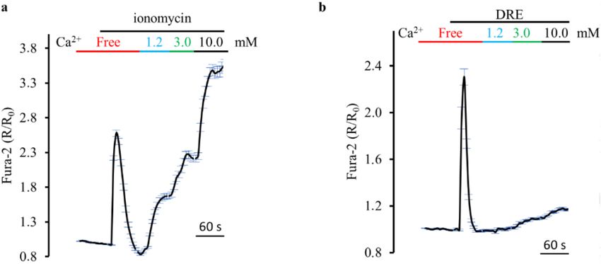
Int. J. Mol. Sci. 2018, 19, 1112                                                                             6 of 17

2.3. DRE Exposure Induced Ca2+ Release from CPA-Sensitive Stores
      We next examined the ER Ca2+ release process in more detail in intact HEK293 cells transfected
with the FRET-based probe ERD1. Figure 4a shows that exposure to 400 µg/mL DRE in extracellular
Ca2+ -free solution for 4 min decreased the intraluminal free Ca2+ levels by 46.12 ± 6.6% (n = 4,
m = 32 cells, p < 0.001). DRE-induced intraluminal reduction was normalized with respect to the
maximal ER-emptying effect elicited by 5 µM ionomycin in the absence of Ca2+ . Exposure of cells to the
Ca2+ -mediated agonist ATP (Figure 4b) was able to induce a decrease in ER Ca2+ levels, corresponding
to 29.84 ± 8.1% of the maximal ionomycin response (n = 4, m = 41 cells, p < 0.001).
      It is widely recognized that the emptying of the endoplasmic reticulum activates store-operated
   2+
Ca entry at the plasma membrane [23]. As showed in Figure 4c, re-addition of 2 mM extracellular
Ca2+ in a Ringer’s solution nominally free of Ca2+ , in the continuous presence of DRE, induced an
increase in the intracellular Ca2+ levels likely through store-operated channels (SOCs). Comparable
results were obtained when we used, in the same experimental protocol, ATP instead of DRE.

      Figure 4. DRE induces both Ca2+ release from the endoplasmic reticulum and Ca2+ entry at the plasma
      membrane. Response of ERD1-transfected HEK-293 cells to (a) DRE (400 µg/L) or (b) ATP (100 µM)
      in extracellular Ca2+ -free solution. Both responses were compared with the maximal ER Ca2+ release
      elicited by ionomycin 5 µM in extracellular Ca2+ -free solution. The reported values are means ± SE
      from all the responsive cells on a single coverslip (c) Response of Fura-2-loaded HEK293 cells to either
      DRE (400 µg/mL), in the absence or presence (2 mM) of extracellular Ca2+ , or ATP (100 µM), in the
      absence or presence (2 mM) of extracellular Ca2+ . The reported values are means ± SE from all the
      responsive cells on a single coverslip.

2.4. Dissecting DRE Effect on Ca2+ Signaling
     The results collected so far suggest that DRE can either cross the plasma membrane and exert
direct actions on internal Ca2+ stores (e.g., like a Ca2+ -ionophore such as ionomycin) or activate a
plasma membrane receptor coupled to a Gq alpha subunit (Gαq). However, when we used ionomycin
in a protocol similar to that depicted in Figure 4c, we found completely different results.
     Figure 5a shows that ionomycin elicited a rapid and reversible increase in cytosolic Ca2+ ,
considered in this protocol as the control response. The quantal increase (1.2, 3.0, 10 mM) of the
extracellular Ca2+ concentration rapidly raised the cytosolic amount of Ca2+ to about 160% in the
presence of 10 mM extracellular Ca2+ (164.84 ± 10.4%, n = 3, versus the control response). When we
repeated the same protocol with DRE instead of ionomycin, we recorded, in the presence of 10 mM
extracellular Ca2+ , smaller increases in cytosolic Ca2+ (Figure 5b, 20.15 ± 8.8%, n = 3, versus the control
response) that suggested a capacitative Ca2+ entry rather than a ionophore-mediated influx through
the plasma membrane.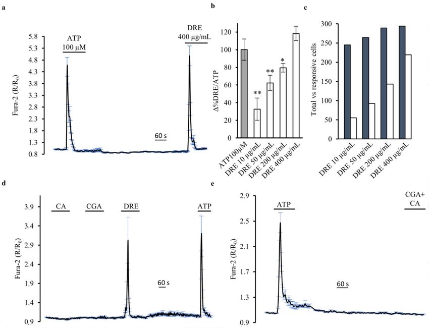
Int. J. Mol. Sci. 2018, 19, 1112                                                                              7 of 17

      Figure 5. DRE does not act as an ionophore-like compound such as ionomycin. (a) Response of
      Fura-2-loaded HEK293 cells to the ionophore ionomycin (5 µM) in the absence or presence (1.2, 3.0,
      and 10 mM) of extracellular Ca2+ . The reported values are means ± SE from all the responsive cells on
      a single coverslip. (b) Response of Fura-2-loaded HEK293 cells to DRE (400 µg/mL) in the absence or
      presence (1.2, 3.0, and 10 mM) of extracellular Ca2+ . The reported values are means ± SE from all the
      responsive cells on a single coverslip.

     Thus, is there a G-protein coupled receptor (GPCR) involved? It is pharmacologically complicated
to specifically block Gαq. This is the reason why we decided to characterize features that are generally
related to the increase in cytosolic Ca2+ mediated by GPCR.
     First, the Ca2+ response induced by DRE was significantly decreased when repeated, at the
same concentration, after 10–15 min of DRE washout (Figure 6a, 65.75 ± 7.17%, n = 4, p < 0.01).
When we increased the interval between two DRE stimulations, we found a significant recovery of the
second Ca2+ response that resulted non-significantly changed compared to the first one after 45 min
(105.70 ± 8.26%, n = 4, p = 0.19). One possible explanation could be that it takes time to refill the
ER that has been challenged after the first DRE stimulation. However, when we repeated the same
experimental protocol with ATP, we found a complete recovery of the Ca2+ response after 15–20 min
(Figure 6b). Indeed, these results can be explained considering the receptor desensitization of a putative
GPCR involved. Considering that in HEK293 cells (i) the cytosolic amount of Ca2+ recorded under
both agonists (DRE and ATP) stimulation is of similar extent and (ii) the Ca2+ homeostatic mechanisms
are the same for the two agonists, we conclude that the hypothesis that DRE is activating a GPCR has
to be taken into account.

      Figure 6. Does DRE activate plasma membrane receptors? (a) Response of Fura-2-loaded HEK293 cells
      to two repeated exposures to DRE (400 µg/mL) separated by a 15 min washout (w/o). The reported
      values are means ± SE from all the responsive cells on a single coverslip. Right inset: Changes in
      the Fura-2 ratio in Ca2+ -containing solutions in response to repeated exposures to DRE (400 µg/mL)
      separated by washouts of different durations (150 , 300 , and 450 ). The second response to DRE is
      normalized to the first one. The reported values are means ± SE from all the responsive cells of all
      experiments performed (** p < 0.01 vs. first DRE, * p < 0.05 vs. first DRE) (b) Response of Fura-2-loaded
      HEK293 cells to two repeated exposures to ATP (100 µM) separated by a 15 min washout (w/o).
      The reported values are means ± SE from all the responsive cells on a single coverslip.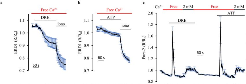
Int. J. Mol. Sci. 2018, 19, 1112                                                                          8 of 17

     Under this scenario, the Ca2+ release from the ER would have been the result of the activation
of the Gαq/PLC/IP3 pathway. Therefore, we used 10 µM U73122 in order to inhibit PLC. Figure 7a
shows that DRE (400 µg/mL) was unable to increase intracellular Ca2+ levels both in the presence of
U73122 and 10 min after its washout. However, these results are significantly affected by the fact that
U73122 likely increased intracellular Ca2+ levels by emptying the endoplasmic reticulum, as already
shown for baby hamster kidney (BHK21) cells [24]. This secondary and non-specific effect of U73122
fully explains its inhibitory action on the DRE-induced Ca2+ increase that we observed in our study.
Therefore, we cannot use these experiments to extrapolate information about the putative activation of
PLC following exposure to DRE.

      Figure 7. DRE exposure activates the downstream signaling effector phospholipase C. (a) Response
      of Fura-2-loaded HEK293 cells to DRE (400 µg/mL) in the presence of or after exposure to U73122
      (10 µM). The reported values are means ± SE from all the responsive cells on a single coverslip.
      (b,c) Representative traces of the measurement of PLC activation in response to ATP and DRE,
      as measured by translocation of PH–PLCδ–EGFP from the plasma membrane (blue trace) to the cytosol
      (green trace). Inset image: Fluorescence of a representative cell excited at 480 nm showing, at rest,
      PH–PLCδ–EGFP associated mostly at the plasma membrane (blue ROI) but also weakly present in the
      peripheral cytoplasm (green ROI). (d) Response of Fura-2-loaded HEK293 cells to DRE (400 µg/mL)
      in extracellular Ca2+ -free solution before and after a prolonged (about 50 min) exposure to 100 µM
      Ryanodine. The reported values are means ± SE from all the responsive cells on a single coverslip.

      Thus, as an alternative strategy, we used a GFP-based indicator, PH–PLCδ–EGFP [20,24] that
allowed us to directly examine in real time the action of DRE on PLC activation in single HEK293 cells.
In resting conditions, the PH domain of this probe interacts with PIP2 at the plasma membrane, so
that it is predominantly visible at the periphery of the cells. Upon PLC activation and PIP2 hydrolysis,
the PH domain translocates toward the cytosol with inositol trisphosphate (InsP3).
      The two typical patterns of response observed in single cell epifluorescence experiments in
PH–PLCδ–EGFP-expressing HEK293 cells upon stimulation with ATP and DRE are shown in
Figure 7b,c. Most probably because of the morphology and the large volume occupied by the nucleus
in HEK293 cells plated at high confluence, the expected antiparallel signal of PH–PLCδ–EGFP [9] was
Int. J. Mol. Sci. 2018, 19, 1112 9 of 17
not always appreciable, both upon agonist and upon DRE stimulation (n = 4, m = 63). In order to have
a semiquantitative appreciation of the responses, as for the previous experiments, we analyzed only
cells which clearly responded to ATP with either a decrease of the plasma membrane signal or the
classical antiparallel behavior of the membrane and cytosolic fluorescence intensity.
Figure 7b shows a representative trace in which, albeit only a small increase in the cytosolic
signal is appreciable upon agonist stimulation, a clear and reversible reduction of PLC fluorescence
intensity is apparent at the membrane, indicative of PIP2 hydrolysis. In the cells which showed this
behavior upon ATP stimulation, a clear response was also recordable upon DRE perfusion (36/36 cells).
Importantly, in half of the ATP responsive cells (18/36 cells), also a clear translocation of the probe to
the cytosol was appreciable upon ATP stimulation, and again an analogous behavior was recordable
with DRE (18/18 cells) (Figure 7c). All these data clearly indicated a similar mechanism of action of
DRE and ATP on PLC, as measured by PH–PLCδ–EGFP.
Thus, DRE-induced activation of PLC might induce Ca2+ release from the endoplasmic reticulum
through the formation of IP3 and the activation of IP3R. Ca2+ released from the ER might, in turn,
activate the ryanodine receptors (RyRs) showed to be expressed and functional in HEK293 cells [25].
Of note, as shown in Figure 7d, DRE-induced increase in intracellular Ca2+ was significantly
reduced (by about 60%, n = 3, p < 0.01) under prolonged (at least 50 min) RyRs blockade with high
concentrantions of ryanodine (100 µM), clearly indicating Ca2+ -induced Ca2+ release as an additional
mechanism in DRE-elicited ER Ca2+ release.
3. Discussion
The use of plant extracts to treat diseases is ancient, and popular observations regarding their use
and efficacy prompt the investigation of their therapeutic properties [26]. The identification of bioactive
compounds and their action mechanisms against diseases are the assumptions for the potential use
of plant preparations as healthy sources [3]. T. officinale (dandelion) is an ethnomedical herb used as
anti-inflammatory, antioxidant, diuretic, choleretic, laxative, and to treat arthritis and liver disorders [6].
However, despite dandelion incredible popularity, scientific research regarding the mechanisms of
action of its extracts is still limited [8].
In this study, we showed, for the first time, that acute exposure (2–5 min, 400 µg/mL) to an
ethanolic dandelion root extract (DRE) induced a dose-dependent and reversible Ca2+ increase in
HEK293 cells (Figure 2). DRE-mediated increase in intracellular Ca2+ was characterized by using both
classical experimental manoeuvres (e.g., removal of extracellular Ca2+ , inhibition of the SERCA pump,
blockers of mechanisms regulating Ca2+ homeostasis) and direct comparison with drugs (e.g., ATP
and ionomycin) that have a well-known action on Ca2+ homeostasis. We found that the rapid Ca2+
transient that follows DRE exposure results from a significant Ca2+ release from the endoplasmic
reticulum and Ca2+ entry at the plasma membrane probably through store-operated Ca2+ channels.
Of note, DRE stimulation induced a significant activation of PLC. This important observation was
obtained considering the subcellular localization of a GFP-tagged PH–PLCδ construct [20,24]. In the
absence of DRE, the probe was properly located at the plasma membrane whilst, in the presence
of DRE, the PH domain moved to the cytosol with InsP3 (IP3) [20,24], thus providing both a direct
indication of PLC activation and indirect measurements of IP3 formation [27].
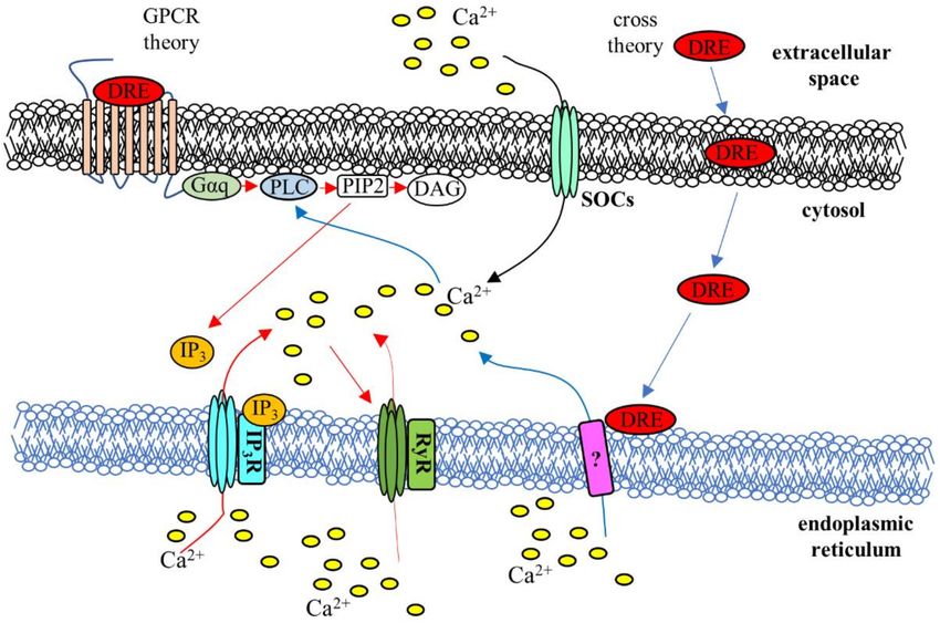
Collectively, these results draw two possible scenarios as to how the presence of the extract is
sensed and transduced by the cells (see Figure 8). In the first one, DRE (or one of its components)
crosses the plasma membrane (“cross theory”) leading to Ca2+ entry and “in situ” Ca2+ release from
the ER and activation of PLC that seconds the cytosolic Ca2+ increase. In the second scenario, DRE
(or one of its components) activates a G-protein coupled receptor at the plasma membrane and PLC
and IP3 production which, in turn, induces Ca2+ release from the endoplasmic reticulum potentiated
by Ca2+ -induced Ca2+ release from RyRs (Figure 7d) and the consequent store-operated Ca2+ entry.
In addition, the fact that the activity of DRE, in terms of activation of Ca2+ transients and number
of responsive cells, is strictly dose-dependent, is also in line with the “GPCR theory”. Finally, in theInt. J. Mol. Sci. 2018, 19, 1112 10 of 17
same direction goes the evidence that close, repeated stimulations with DRE seemed associated with
an apparent desensitization of the “putative” receptor, which, on the other hand, did not affect the
receptor for ATP.
Figure 8. Schematic illustration of the putative mechanisms for DRE-induced Ca2+ signaling. The “cross
theory” (blue arrows) and the “GPCR theory” (red arrows) have been proposed to explain the
mechanisms underlying the Ca2+ increase induced by exposure to DRE: See the text for a detailed
description. DRE, dandelion root extract; IP3, Inositol trisphosphate; IP3R, Inositol trisphosphate
receptor; RyR, Ryanodine receptor; DAG, diacylglycerol, PIP2, phosphatidylinositol 4,5-bisphosphate;
PLC, phospholipase C; Gαq, alpha subunit of Gq; SOCs, store-operated Ca2+ channels, “?” indicates
unknown mechanisms.
Under the same scenario, Chang et al. showed that 800 µM caffeic acid evoked Ca2+ release
from thapsigargin-sensitive stores in SCM1 human gastric cancer cells, speculating the activation of
plasma membrane receptors [13]. In addition, the authors proved that Ca2+ release was induced by
a phospholipase C-dependent mechanism, since it was blocked when phospholipase C activity was
inhibited by U73122. However, in our hands, caffeic acid did not activate Ca2+ transients neither in
HEK293 (Figure 3) nor in MCD4 cells (data not shown). This difference is likely related to the lower
concentration used in our Fura-2 experiment that, in turn, matched the range of concentration of caffeic
measured in the DRE (see Table 1). In our experimental protocols, also chlorogenic acid, previously
reported to regulate Ca2+ homeostasis even though via different cellular mechanisms (e.g., inhibition
of agonists-stimulated Ca2+ entry), was ineffective when used at a concentration (200 µM) proportional
to its presence in our DRE. Therefore, regardless of which theory is correct, it is currently unknown
if the Ca2+ effects measured in response to DRE are mediated by the whole extract or by individual
components diverse from caffeic and chlorogenic acids.
Whatever the specific mechanism of action at the plasma or intracellular membrane, the direct
observation that DRE impacts Ca2+ homeostasis at different subcellular locations is an important
piece of the puzzle for understanding the wide array of biological functions (of different organs
and systems) reported to be modulated by T. officinale. As a matter of fact, and as suggested
for Taraxacum [28–31], Ca2+ signaling finely regulates processes like cellular proliferation [32],
bile secretion [33], diuresis [1,34], inflammation [35], and oxidative stress [35], just to name a few.
Thus, being aware of this Taraxacum–Ca2+ interplay can be considered as a starting point to better
project and more precisely target the use of T. officinale extracts as pharmacological tools.Int. J. Mol. Sci. 2018, 19, 1112 11 of 17
Then, it is also important to note that both the composition and the effects induced by these
herbal extracts strictly depend on which part of the plant has been used to produce them. In the
last years, reports on the phytochemical composition of T. officinale extracts are increasing, and it
has been shown that dandelion root extracts are mainly rich in hydroxycinnamic acids, whereas
hydroxybenzoic acids derivatives and flavonoids have been reported in lower amounts. In this study,
the chemical composition of an ethanol extract of dandelion roots was investigated by RP-HPLC
analysis (Figure 1) and chlorogenic and caffeic acids were found to be the most abundant compounds
(Table 1). This result is in agreement with a previous study [9] where chlorogenic acid was the most
abundant chemical compound in a dandelion root extract, followed by caffeic acid. Also, ferulic,
syringic, vanillic, p-coumaric, and gallic acids were identified. Overall, the root extract of dandelion
can be considered a good natural product with antioxidant activity, immunostimulatory capacity,
anticancer, anti-inflammatory, and antimicrobial properties [7,36–39]. On the other hand, dandelion
leaves and flowers are more enriched in flavonoids (luteolin and its glycoside derivatives, chrysoeriol)
and coumarins (cichoriin and aesculin) but also contain hydroxycinnamic acid derivatives (caffeic,
chlorogenic, chicoric, and monocaffeoyltartaric acids) [8,10]. Because of this specific composition,
extracts produced using different portions of the plant have different biological effects. For example,
a dandelion leaves extract has been extensively used as diuretic [40], that is why in Italy it goes under
the name of “piscialetto” (bedwetter). Interestingly, we have in vitro evidence that argue against the
putative diuretic effect of our DRE. Accordingly, the British Herbal Medicine Association indicated the
leaves as the part of the Taraxacum associated with a diuretic action.
DRE is not the first herbal extract tested for an effect on the mechanisms underlying Ca2+
homeostasis. We recently showed that a methanolic extract of A. oleracea (flowers, leaves, and stems)
induced a significant increase in cytosolic Ca2+ levels in HEK293 and MCD4 cells. We proved that
spilanthol, the main component of the Acmella extract, acted like a potent and natural diuretic in
mouse by activating a mechanism based on Ca2+ -induced inhibition of cytosolic cyclic adenosine
monophosphate (cAMP) [1]. More examples are available in the literature about olive leaf extracts [41],
Scrophularia orientalis [42], and Echinacea extracts [43,44]. For this latter extract, it has been shown
that different bioactive components of Echinacea, such as alkamides (via activation of cannabinoid
receptors) [43] and unknown lipophilic fractions (via direct activation of IP3 receptor) [44], are able to
induce intracellular Ca2+ peaks.
In conclusion, this report highlights the effect of an ethanolic dandelion root extract on Ca2+
homeostasis in HEK293 cells. The mechanism proposed, although not completely characterized,
indicates Ca2+ signaling as the cellular mediator used by the cells to transduce the effects of the
exposure to the bioactive components of the T. officinale extract. Of note, this important evidence has a
number of perspectives. First, researchers know how the mechanisms underlying Ca2+ homeostasis
work, how to modulate them pharmacologically, and how they impact diverse biological activities.
Thus, it will be possible to finely tune T. officinale effects on Ca2+ -signaling in order to improve the
biological outcome. Second, Ca2+ signaling intersects the activity of a huge number of other signaling
molecules (e.g., cAMP [45]) and/or effectors (e.g., mitogen-activated protein (MAP) kinases [46]).
Thus, it is likely that T. officinale-induced Ca2+ signaling activates other pathways, which have to be
brought to light by tissue-specific research projects. Third, a deeper comparative analysis of Taraxacum
extracts (from roots, leaves, flowers) in terms of their relative composition of bioactive compounds
will help to link the presence of specific components to the activation of precise signaling mechanisms.
This latter perspective might have an enormous pharmaceutical impact.
4. Materials and Methods
4.1. Plant Extraction
T. officinale was collected in Basilicata region (40◦ 340 50.900 N 15◦ 490 38.200 E) in October 2017;
the roots were separated from the aerial parts and then dried at room temperature in the dark.Int. J. Mol. Sci. 2018, 19, 1112 12 of 17
The dried roots (15 g) were extracted by maceration with EtOH 95% at 1:10 w/v (plant
material–solvent ratio) under continuous stirring for 3 days, changing the solvent every 24 h.
The extract was filtered, and the solvent was removed in a rotary evaporator. The dried crude
extract was kept in the dark until use.
4.2. High-Performance Liquid Chromatography (HPLC) Analysis
The re-dissolved crude extract (100 mg/mL) was analyzed on an analytical HPLC–DAD
(Shimadzu Corp., Kyoto, Japan) unit using an EC 250/4.6 Nucleodur 100-5 C18ec column
(Macherey-nagel, Düren, Germany). The mobile phase involved two solvents: water–formic acid (5%)
(A) and acetonitrile (B). The gradient elution method has already been described by our group [47].
All peaks were collected in the range of 200–400 nm, and chromatograms were recorded at 280 nm
for hydroxybenzoic acids, 320 nm for hydroxycinnamic acids, and 350 nm for flavonoids. Standards,
such as caffeic acid, chlorogenic acid, syringic acid, ferulic acid, vanillic acid, p-coumaric acid, and gallic
acid, were used (Sigma Aldrich, Milan, Italy). The quantification of phenolic compounds was realized
by the comparison between chromatogram recorded absorbances and external calibration standards.
All experiments were carried out in triplicate.
4.3. Cell Culture
HEK293 cells were grown in Dulbecco’s modified Eagle medium (DMEM) Glutamax
supplemented with 10% fetal bovine serum and 1% penicillin/streptomycin (all these products were
from Thermo Fisher Scientific, Waltham, MA, USA) and were maintained in a humidified incubator
at 37◦ C in the presence of 5% CO2 /95% air. MCD4 cells, a clone of M-1 cells stably transfected with
human-Aquaporin (AQP) 2, were cultured as described elsewhere [48]. Trypsin was used to subculture
cells that were used for no longer than ten/twelve passages after thawing. The cells were seeded on
glass coverslips (at a density of 60–70%) and used 24/48 h later for imaging evaluations of intracellular
Ca2+ levels with Fura-2-AM (Thermo Fisher Scientific, Waltham, MA, USA). Alternatively, 24 h after
plating, the cells were transfected with ERD1 [19] or the PLC GFP-reporter [20] and used the next day
for fluorescence imaging experiments.
4.4. In Vitro Cytotoxic Assay
The cytotoxic effect of the crude DRE was determined by the Calcein-AM viability assay [2].
HEK293 and MCD4 cells were seeded into 96-well black plates for 24 h and thereafter exposed to
different concentrations of DRE (750, 400, 200, 100, 50, 10, 5 µg/mL) or vehicle (ethanol 0.6%) as a
control, at 37◦ C for 24 h. Afterward, the medium was removed, and 100 µL of 1µM Calcein-AM
(Thermo Fisher Scientific, Waltham, MA, USA) in phosphate buffered saline (PBS) was added for 30
min at 37◦ C. The fluorescence was detected by FLEX STATION 3 (Molecular Devices, San Jose, CA,
USA) plate reader using a blue filter (Ex 490 nm, Em 510–570 nm).
4.5. Intracellular Ca2+ Measurements
For cytosolic Ca2+ recordings, the cells were seeded on 25 (or 18) mm Ø glass coverslips. HEK293
cells were loaded with 2–4 µM Fura-2-AM (Thermo Fisher Scientific, Waltham, MA, USA) for 30
min at 37◦ C in DMEM Glutamax, followed by 15 min in an extracellular solution to allow Fura-2
de-esterification. The coverslips with dye-loaded cells were mounted in an open perfusion chamber
(a modified version of FCS2 Closed Chamber System, Biopthechs, Butler, PA, USA), and recordings
were carried out using an inverted Eclipse TE2000-S microscope (Nikon, Shinagawa, Tokyo, Japan)
equipped for single cell fluorescence evaluations and imaging analysis. Each Fura-2-AM loaded sample
was illuminated every 5 s through a 40× oil immersion objective (numerical aperture = 1.30) at 340 and
380 nm. The emitted fluorescence was passed through a dichroic mirror, filtered at 510 nm (Omega
Optical, Brattleboro, VT, USA), and captured by a cooled CCD CoolSNAP HQ camera (Photometrics,Int. J. Mol. Sci. 2018, 19, 1112 13 of 17
Tucson, AZ, USA). Additional technical information about the setup used in our imaging facility is
available in other publications from our group [34,49,50]. Fluorescence measurements were carried
out using the MetaFluor Fluorescence Ratio Imaging Software (Version 7.7.3.0, Molecular Devices, San
Jose, CA, USA). The ratio of the fluorescence signal acquired upon excitation at 340 and 380 nm was
normalized to the basal fluorescence ratio obtained in the absence of the stimulus (reported as R/R0).
Bar graphs show the averaged rate of fluorescence ratio changes normalized to those induced by
maximal ATP stimulation, the latter used in each experiment as an internal positive control. The data
from at least 30 cells were summarized in a single run and averaged in a plot ± SE; at least three
independent experiments were conducted. n indicates the number of experiments performed for each
protocol, m the number of cells analyzed in n experiments.
4.6. Measurement of Intraluminal ER Ca2+ Levels
The same imaging setup and perfusion apparatus were used for the measurements of Ca2+ levels
within the endoplasmic reticulum of HEK293 cells transfected with ERD1. Real-time FRET experiments
were carried out using the MetaFluor Fluorescence Ratio Imaging Software (Version 7.7.3.0, Molecular
Devices, San Jose, CA, USA). FRET from cyan fluorescent protein (CFP) to yellow fluorescent protein
(YFP) was evaluated by excitation of CFP (435 nm) and measurement of the fluorescence emitted by
YFP. The results are presented as the emission ratio 485/535 nm collected every 5 s in control conditions
and after stimulation with the agonists (DRE, ATP, or ionomycin). The emission ratio was normalized
to the maximal store emptying induced by 5 µM ionomycin in an extracellular Ca2+ -free solution
(R/R0). The data from 5 to 10 transfected cells were summarized in a single run and averaged in a
plot ± SE, and at least four independent experiments for each agonist were conducted. n indicates the
number of experiments performed for each protocol, m the number of cells analyzed in n experiments.
4.7. Fluorescence Imaging of GFP-Based Reporters
The synthesis of intracellular InsP3/PLC activation was evaluated with the GFP-tagged pleckstrin
homology domain of PLCδ1 (PH–PLCδ1–EGFP) [20,27,51]. HEK293 cells were transfected transiently
with the GFP-based indicator using Lipofectamine 2000 (Thermo Fisher Scientific, Waltham, MA, USA).
Fluorescence images of cells expressing the probe were acquired every 5 s using the MetaFluor
Fluorescence Ratio Imaging Software (Version 7.7.3.0, Molecular Devices, San Jose, CA, USA) and the
setup and the perfusion apparatus described in [52,53]. GFP was excited at 485 nm, and the emission
was collected above 530 nm. n indicates the number of experiments performed for each protocol, m the
number of cells analyzed in n experiments.
4.8. Solutions and Materials
Most of the chemicals were obtained from Sigma (Sigma-Aldrich, Saint Louis, MO, USA).
The experiments were carried out with an extracellular (Ringer’s) solution containing (in mmol/L):
140 NaCl, 5 KCl, 1 CaCl2 , 1 MgCl2 , 5 glucose, 10 HEPES, adjusted to pH 7.40 with NaOH.
Ionomycin and Fura-2-AM were from Thermo Fisher Scientific (Waltham, MA, USA).
When dimethyl sulfoxide or ethanol were used as a vehicle, the final solvent concentration was
always below 0.01% or 0.1%, respectively.
4.9. Data Analysis
Whenever possible, responses to DRE and ATP (as internal control) were compared in the same
cell (paired data), thus eliminating concerns about the variability of the starting Fura-2 ratio. Whenever
appropriate, paired data were assessed for statistical significance using the Student’s t test. The data
were expressed as means ± SE with n equal to the number of experimental runs. For Fura-2 and ERD1
ratio imaging experiments, p < 0.05 was considered statistically significant.Int. J. Mol. Sci. 2018, 19, 1112 14 of 17
Acknowledgments: The authors would like to thank Stephen Ferguson, University of Ottawa, Canada for
providing the pleckstrin-homology [PH]–PLCδ–GFP construct. The work of Andrea Gerbino and Daniela Russo
was supported by funding from the CLUSTER TECNOLOGICO REGIONALE “DICLIMAX” (project # MTJU9H8)
to Maria Svelto. This research was also supported by the Master in ”Structural Osteopathy” of the University of
Basilicata to Monica Carmosino. The founding sponsors had no role in the design of the study, in the collection,
analyses, or interpretation of data, in the writing of the manuscript, and in the decision to publish the results.
Andrea Gerbino would like to thank Giuseppe Cassano, University of Bari, for his comments and suggestions
regarding the Ca2+ -imaging experiments.
Author Contributions: Andrea Gerbino conceived and designed the experiments; Andrea Gerbino, Daniela Russo,
and Matilde Colella performed the experiments and analyzed the data; Daniela Russo and Luigi Milella collected,
extracted, and performed quali–quantitative HPLC analysis. Andrea Gerbino and Monica Carmosino wrote the
paper; Giuseppe Procino and Maria Svelto critically and substantively revised the paper; Andrea Gerbino and
Monica Carmosino supervised and managed the project.
Conflicts of Interest: The authors declare no conflict of interest.
Abbreviations
DRE Dandelion root extract
ATP Adenosine triphosphate
SERCA Sarco/endoplasmic reticulum Ca2+ -ATPase
CPA Cyclopiazonic acid
References
1. Gerbino, A.; Schena, G.; Milano, S.; Milella, L.; Barbosa, A.F.; Armentano, F.; Procino, G.; Svelto, M.;
Carmosino, M. Spilanthol from Acmella oleracea lowers the intracellular levels of camp impairing NKCC2
phosphorylation and water channel AQP2 membrane expression in mouse kidney. PLoS ONE 2016,
11, e0156021. [CrossRef] [PubMed]
2. Armentano, M.F.; Bisaccia, F.; Miglionico, R.; Russo, D.; Nolfi, N.; Carmosino, M.; Andrade, P.B.; Valentão, P.;
Diop, M.S.; Milella, L. Antioxidant and proapoptotic activities of Sclerocarya birrea [(A. Rich.) hochst.]
methanolic root extract on the hepatocellular carcinoma cell line HepG2. Biomed. Res. Int. 2015, 2015, 561589.
[CrossRef] [PubMed]
3. Russo, D.; Miglionico, R.; Carmosino, M.; Bisaccia, F.; Andrade, P.B.; Valentão, P.; Milella, L.; Armentano, M.F.
A comparative study on phytochemical profiles and biological activities of Sclerocarya birrea (A. Rich.) Hochst
leaf and bark extracts. Int. J. Mol. Sci. 2018, 19, 186. [CrossRef] [PubMed]
4. Costantino, V.; Fattorusso, E.; Imperatore, C.; Mangoni, A. Glycolipids from sponges. Part 17. Clathrosides
and isoclathrosides, unique glycolipids from the caribbean sponge Agelas clathrodes. J. Nat. Prod. 2006,
69, 73–78. [CrossRef] [PubMed]
5. Costantino, V.; Fattorusso, E.; Mangoni, A.; Perinu, C.; Teta, R.; Panza, E.; Ianaro, A. Tedarenes A and B:
Structural and stereochemical analysis of two new strained cyclic diarylheptanoids from the marine sponge
Tedania ignis. J. Org. Chem. 2012, 77, 6377–6383. [CrossRef] [PubMed]
6. Schütz, K.; Carle, R.; Schieber, A. Taraxacum—A review on its phytochemical and pharmacological profile.
J. Ethnopharmacol. 2006, 107, 313–323. [CrossRef] [PubMed]
7. Wirngo, F.E.; Lambert, M.N.; Jeppesen, P.B. The physiological effects of dandelion (Taraxacum officinale) in
type 2 diabetes. Rev. Diabet. Stud. 2016, 13, 113–131. [CrossRef] [PubMed]
8. Martinez, M.; Poirrier, P.; Chamy, R.; Prüfer, D.; Schulze-Gronover, C.; Jorquera, L.; Ruiz, G. Taraxacum
officinale and related species—An ethnopharmacological review and its potential as a commercial medicinal
plant. J. Ethnopharmacol. 2015, 169, 244–262. [CrossRef] [PubMed]
9. Kenny, O.; Smyth, T.; Hewage, C.; Brunton, N. Antioxidant properties and quantitative UPLC-MS/MS
analysis of phenolic compounds in dandelion (Taraxacum officinale) root extracts. Free Radic. Antioxid. 2014,
4, 7. [CrossRef]
10. Williams, C.A.; Goldstone, F.; Greenham, J. Flavonoids, cinnamic acids and coumarins from the different
tissues and medicinal preparations of Taraxacum officinale. Phytochemistry 1996, 42, 121–127. [CrossRef]
11. Ivanov, I. Polyphenols content and antioxidant activities of Taraxacum officinale F.H. Wigg (dandelion) leaves.
Int. J. Pharmacogn. Phytochem. Res. 2014, 6, 889–893.Int. J. Mol. Sci. 2018, 19, 1112 15 of 17
12. Budzianowski, J. Coumarins, caffeoyltartaric acids and their artifactual methyl esters from Taraxacum officinale
leaves. Planta Med. 1997, 63, 288. [CrossRef] [PubMed]
13. Chang, H.T.; Chen, I.L.; Chou, C.T.; Liang, W.Z.; Kuo, D.H.; Shieh, P.; Jan, C.R. Effect of caffeic acid on Ca2+
homeostasis and apoptosis in SCM1 human gastric cancer cells. Arch. Toxicol. 2013, 87, 2141–2150. [CrossRef]
[PubMed]
14. Nam, J.H.; Shin, D.H.; Zheng, H.; Kang, J.S.; Kim, W.K.; Kim, S.J. Inhibition of store-operated Ca2+ entry
channels and K+ channels by caffeic acid phenethylester in T lymphocytes. Eur. J. Pharmacol. 2009, 612, 153–160.
[CrossRef] [PubMed]
15. Bose, J.S.; Gangan, V.; Jain, S.K.; Manna, S.K. Novel caffeic acid ester derivative induces apoptosis by
expressing fasl and downregulating NF-KappaB: Potentiation of cell death mediated by chemotherapeutic
agents. J. Cell. Physiol. 2009, 218, 653–662. [CrossRef] [PubMed]
16. Mikami, Y.; Yamazawa, T. Chlorogenic acid, a polyphenol in coffee, protects neurons against glutamate
neurotoxicity. Life Sci. 2015, 139, 69–74. [CrossRef] [PubMed]
17. Jung, H.J.; Im, S.S.; Song, D.K.; Bae, J.H. Effects of chlorogenic acid on intracellular calcium regulation in
lysophosphatidylcholine-treated endothelial cells. BMB Rep. 2017, 50, 323–328. [CrossRef] [PubMed]
18. Lin, T.Y.; Lu, C.W.; Huang, S.K.; Wang, S.J. Ferulic acid suppresses glutamate release through inhibition
of voltage-dependent calcium entry in rat cerebrocortical nerve terminals. J. Med. Food 2013, 16, 112–119.
[CrossRef] [PubMed]
19. Palmer, A.E.; Jin, C.; Reed, J.C.; Tsien, R.Y. Bcl-2-mediated alterations in endoplasmic reticulum Ca2+ analyzed
with an improved genetically encoded fluorescent sensor. Proc. Natl. Acad. Sci. USA 2004, 101, 17404–17409.
[CrossRef] [PubMed]
20. Dale, L.B.; Babwah, A.V.; Bhattacharya, M.; Kelvin, D.J.; Ferguson, S.S. Spatial-temporal patterning of
metabotropic glutamate receptor-mediated inositol 1,4,5-triphosphate, calcium, and protein kinase C
oscillations: Protein kinase C-dependent receptor phosphorylation is not required. J. Biol. Chem. 2001,
276, 35900–35908. [CrossRef] [PubMed]
21. Berridge, M.J.; Lipp, P.; Bootman, M.D. The versatility and universality of calcium signalling. Nat. Rev. Mol.
Cell Biol. 2000, 1, 11–21. [CrossRef] [PubMed]
22. Altemimi, A.; Lakhssassi, N.; Baharlouei, A.; Watson, D.G.; Lightfoot, D.A. Phytochemicals: Extraction,
isolation, and identification of bioactive compounds from plant extracts. Plants 2017, 6, 42. [CrossRef]
[PubMed]
23. Hofer, A.M.; Fasolato, C.; Pozzan, T. Capacitative Ca2+ entry is closely linked to the filling state of internal
Ca2+ stores: A study using simultaneous measurements of ICRAC and intraluminal [Ca2+ ]. J. Cell Biol. 1998,
140, 325–334. [CrossRef] [PubMed]
24. Lau, B.W.; Colella, M.; Ruder, W.C.; Ranieri, M.; Curci, S.; Hofer, A.M. Deoxycholic acid activates protein
kinase C and phospholipase C via increased Ca2+ entry at plasma membrane. Gastroenterology 2005,
128, 695–707. [CrossRef] [PubMed]
25. Querfurth, H.W.; Haughey, N.J.; Greenway, S.C.; Yacono, P.W.; Golan, D.E.; Geiger, J.D. Expression of
ryanodine receptors in human embryonic kidney (HEK293) cells. Biochem. J. 1998, 334, 79–86. [CrossRef]
[PubMed]
26. Mezrag, A.; Malafronte, N.; Bouheroum, M.; Travaglino, C.; Russo, D.; Milella, L.; Severino, L.; De Tommasi, N.;
Braca, A.; Dal Piaz, F. Phytochemical and antioxidant activity studies on Ononis angustissima L. Aerial parts:
Isolation of two new flavonoids. Nat. Prod. Res. 2017, 31, 507–514. [CrossRef] [PubMed]
27. Okubo, Y.; Kakizawa, S.; Hirose, K.; Iino, M. Visualization of IP3 dynamics reveals a novel AMPA
receptor-triggered IP3 production pathway mediated by voltage-dependent Ca2+ influx in Purkinje cells.
Neuron 2001, 32, 113–122. [CrossRef]
28. Rácz-Kotilla, E.; Rácz, G.; Solomon, A. The action of Taraxacum officinale extracts on the body weight and
diuresis of laboratory animals. Planta Med. 1974, 26, 212–217. [CrossRef] [PubMed]
29. Yasukawa, K.; Akihisa, T.; Oinuma, H.; Kasahara, Y.; Kimura, Y.; Yamanouchi, S.; Kumaki, K.; Tamura, T.;
Takido, M. Inhibitory effect of Di- and trihydroxy triterpenes from the flowers of compositae on
12-O-tetradecanoylphorbol-13-acetate-induced inflammation in mice. Biol. Pharm. Bull. 1996, 19, 1329–1331.
[CrossRef] [PubMed]
30. Hu, C.; Kitts, D.D. Dandelion (Taraxacum officinale) flower extract suppresses both reactive oxygen species
and nitric oxide and prevents lipid oxidation in vitro. Phytomedicine 2005, 12, 588–597. [CrossRef] [PubMed]Int. J. Mol. Sci. 2018, 19, 1112 16 of 17
31. Takasaki, M.; Konoshima, T.; Tokuda, H.; Masuda, K.; Arai, Y.; Shiojima, K.; Ageta, H. Anti-carcinogenic
activity of Taraxacum plant. I. Biol. Pharm. Bull. 1999, 22, 602–605. [CrossRef] [PubMed]
32. Humeau, J.; Bravo-San Pedro, J.M.; Vitale, I.; Nuñez, L.; Villalobos, C.; Kroemer, G.; Senovilla, L. Calcium
signaling and cell cycle: Progression or death. Cell Calcium 2018, 70, 3–15. [CrossRef] [PubMed]
33. Amaya, M.J.; Nathanson, M.H. Calcium signaling and the secretory activity of bile duct epithelia. Cell Calcium
2014, 55, 317–324. [CrossRef] [PubMed]
34. Procino, G.; Gerbino, A.; Milano, S.; Nicoletti, M.C.; Mastrofrancesco, L.; Carmosino, M.; Svelto, M.
Rosiglitazone promotes AQP2 plasma membrane expression in renal cells via a Ca2+ -dependent/
cAMP-independent mechanism. Cell. Physiol. Biochem. 2015, 35, 1070–1085. [CrossRef] [PubMed]
35. Zierler, S.; Hampe, S.; Nadolni, W. TRPM channels as potential therapeutic targets against pro-inflammatory
diseases. Cell Calcium 2017, 67, 105–115. [CrossRef] [PubMed]
36. Dekdouk, N.; Malafronte, N.; Russo, D.; Faraone, I.; De Tommasi, N.; Ameddah, S.; Severino, L.; Milella, L.
Phenolic compounds from Olea europaea L. Possess antioxidant activity and inhibit carbohydrate metabolizing
enzymes in vitro. Evid. Based Complement. Altern. Med. 2015, 2015, 684925. [CrossRef] [PubMed]
37. Ou, S.; Kwok, K.-C. Ferulic acid: Pharmaceutical functions, preparation and applications in foods. J. Sci.
Food Agric. 2004, 84, 1261–1269. [CrossRef]
38. Chong, K.P.; Rossall, S.; Atong, M. In vitro antimicrobial activity and fungitoxicity of syringic acid, caffeic
acid and 4-hydroxybenzoic acid against Ganoderma boninense. J. Agric. Sci. 2009, 1, 6. [CrossRef]
39. Dos Santos, M.D.; Almeida, M.C.; Lopes, N.P.; de Souza, G.E. Evaluation of the anti-inflammatory, analgesic
and antipyretic activities of the natural polyphenol chlorogenic acid. Biol. Pharm. Bull. 2006, 29, 2236–2240.
[CrossRef] [PubMed]
40. Clare, B.A.; Conroy, R.S.; Spelman, K. The diuretic effect in human subjects of an extract of Taraxacum officinale
folium over a single day. J. Altern. Complement. Med. 2009, 15, 929–934. [CrossRef] [PubMed]
41. Marchetti, C.; Clericuzio, M.; Borghesi, B.; Cornara, L.; Ribulla, S.; Gosetti, F.; Marengo, E.; Burlando, B.
Oleuropein-enriched olive leaf extract affects calcium dynamics and impairs viability of malignant
mesothelioma cells. Evid. Based Complement. Altern. Med. 2015, 2015, 908493. [CrossRef] [PubMed]
42. Lange, I.; Moschny, J.; Tamanyan, K.; Khutsishvili, M.; Atha, D.; Borris, R.P.; Koomoa, D.L. Scrophularia
orientalis extract induces calcium signaling and apoptosis in neuroblastoma cells. Int. J. Oncol. 2016,
48, 1608–1616. [CrossRef] [PubMed]
43. Raduner, S.; Majewska, A.; Chen, J.Z.; Xie, X.Q.; Hamon, J.; Faller, B.; Altmann, K.H.; Gertsch, J. Alkylamides
from Echinacea are a new class of cannabinomimetics. Cannabinoid type 2 receptor-dependent and
-independent immunomodulatory effects. J. Biol. Chem. 2006, 281, 14192–14206. [CrossRef] [PubMed]
44. Wu, L.; Rowe, E.W.; Jeftinija, K.; Jeftinija, S.; Rizshsky, L.; Nikolau, B.J.; McKay, J.; Kohut, M.; Wurtele, E.S.
Echinacea-induced cytosolic Ca2+ elevation in HEK293. BMC Complement. Altern. Med. 2010, 10, 72.
[CrossRef] [PubMed]
45. Gerbino, A.; Ruder, W.C.; Curci, S.; Pozzan, T.; Zaccolo, M.; Hofer, A.M. Termination of cAMP signals by Ca2+
and Gαi via extracellular Ca2+ sensors: A link to intracellular Ca2+ oscillations. J. Cell Biol. 2005, 171, 303–312.
[CrossRef] [PubMed]
46. Colella, M.; Gerbino, A.; Hofer, A.M.; Curci, S. Recent advances in understanding the extracellular
calcium-sensing receptor. F1000Res 2016, 5. [CrossRef] [PubMed]
47. Russo, D.; Valentão, P.; Andrade, P.B.; Fernandez, E.C.; Milella, L. Evaluation of antioxidant, antidiabetic and
anticholinesterase activities of Smallanthus sonchifolius landraces and correlation with their phytochemical
profiles. Int. J. Mol. Sci. 2015, 16, 17696–17718. [CrossRef] [PubMed]
48. Procino, G.; Barbieri, C.; Tamma, G.; De Benedictis, L.; Pessin, J.E.; Svelto, M.; Valenti, G. AQP2 exocytosis in
the renal collecting duct—Involvement of SNARE isoforms and the regulatory role of Munc18b. J. Cell Sci.
2008, 121, 2097–2106. [CrossRef] [PubMed]
49. Carmosino, M.; Gerbino, A.; Hendy, G.N.; Torretta, S.; Rizzo, F.; Debellis, L.; Procino, G.; Svelto, M. NKCC2
activity is inhibited by the bartter’s syndrome type 5 gain-of-function CaR-A843E mutant in renal cells.
Biol. Cell 2015, 107, 98–110. [CrossRef] [PubMed]
50. Carmosino, M.; Gerbino, A.; Schena, G.; Procino, G.; Miglionico, R.; Forleo, C.; Favale, S.; Svelto, M.
The expression of Lamin A mutant R321X leads to endoplasmic reticulum stress with aberrant Ca2+ . J. Cell.
Mol. Med. 2016, 20, 2194–2207. [CrossRef] [PubMed]You can also read