Printability, Durability, Contractility and Vascular Network Formation in 3D Bioprinted Cardiac Endothelial Cells Using Alginate-Gelatin Hydrogels
←
→
Page content transcription
If your browser does not render page correctly, please read the page content below
ORIGINAL RESEARCH
                                                                                                                                             published: 26 February 2021
                                                                                                                                         doi: 10.3389/fbioe.2021.636257

                                              Printability, Durability, Contractility
                                              and Vascular Network Formation in
                                              3D Bioprinted Cardiac Endothelial
                                              Cells Using Alginate–Gelatin
                                              Hydrogels
                                              Christopher David Roche 1,2 , Poonam Sharma 1,2,3 , Anthony Wayne Ashton 1 ,
                                              Chris Jackson 1 , Meilang Xue 1 and Carmine Gentile 1,2*
                                              1
                                               Northern Clinical School, Faculty of Medicine and Health, The University of Sydney, Sydney, NSW, Australia, 2 School of
                                              Biomedical Engineering, Faculty of Engineering and IT, University of Technology Sydney, Sydney, NSW, Australia, 3 Faculty of
                                              Health and Medicine, The University of Newcastle, Callaghan, NSW, Australia

                                              Background: 3D bioprinting cardiac patches for epicardial transplantation are a
                                              promising approach for myocardial regeneration. Challenges remain such as quantifying
                                              printability, determining the ideal moment to transplant, and promoting vascularisation
                                              within bioprinted patches. We aimed to evaluate 3D bioprinted cardiac patches
                                              for printability, durability in culture, cell viability, and endothelial cell structural self-
                           Edited by:         organisation into networks.
                         João Conde,
    New University of Lisbon, Portugal        Methods: We evaluated 3D-bioprinted double-layer patches using alginate/gelatine
                      Reviewed by:            (AlgGel) hydrogels and three extrusion bioprinters (REGEMAT3D, INVIVO, BIO X). Bioink
                   PaYaM ZarrinTaj,
          Oklahoma State University,
                                              contained either neonatal mouse cardiac cell spheroids or free (not-in-spheroid) human
                       United States          coronary artery endothelial cells with fibroblasts, mixed with AlgGel. To test the effects on
                    Heungsoo Shin,
                                              durability, some patches were bioprinted as a single layer only, cultured under minimal
     Hanyang University, South Korea
                                              movement conditions or had added fibroblast-derived extracellular matrix hydrogel
                  *Correspondence:
                     Carmine Gentile          (AlloECM). Controls included acellular AlgGel and gelatin methacryloyl (GELMA) patches.
           carmine.gentile@uts.edu.au
                                              Results: Printability was similar across bioprinters. For AlgGel compared to GELMA:
                    Specialty section:        resolutions were similar (200–700 µm line diameters), printing accuracy was 45 and
         This article was submitted to
                                              25%, respectively (AlgGel was 1.7x more accurate; p < 0.05), and shape fidelity was
                          Biomaterials,
                a section of the journal      92% (AlgGel) and 96% (GELMA); p = 0.36. For durability, AlgGel patch median survival
       Frontiers in Bioengineering and        in culture was 14 days (IQR:10–27) overall which was not significantly affected by
                        Biotechnology
                                              bioprinting system or cellular content in patches. We identified three factors which
       Received: 01 December 2020
        Accepted: 01 February 2021
                                              reduced durability in culture: (1) bioprinting one layer depth patches (instead of two
       Published: 26 February 2021            layers); (2) movement disturbance to patches in media; and (3) the addition of AlloECM
                                Citation:     to AlgGel. Cells were viable after bioprinting followed by 28 days in culture, and all BIO
    Roche CD, Sharma P, Ashton AW,
                                              X-bioprinted mouse cardiac cell spheroid patches presented contractile activity starting
     Jackson C, Xue M and Gentile C
           (2021) Printability, Durability,   between day 7 and 13 after bioprinting. At day 28, endothelial cells in hydrogel displayed
    Contractility and Vascular Network        organisation into endothelial network-like structures.
   Formation in 3D Bioprinted Cardiac
                 Endothelial Cells Using      Conclusion: AlgGel-based 3D bioprinted heart patches permit cardiomyocyte
            Alginate–Gelatin Hydrogels.
 Front. Bioeng. Biotechnol. 9:636257.
                                              contractility and endothelial cell structural self-organisation. After bioprinting, a period
     doi: 10.3389/fbioe.2021.636257           of 2 weeks maturation in culture prior to transplantation may be optimal, allowing for a

Frontiers in Bioengineering and Biotechnology | www.frontiersin.org                  1                                          February 2021 | Volume 9 | Article 636257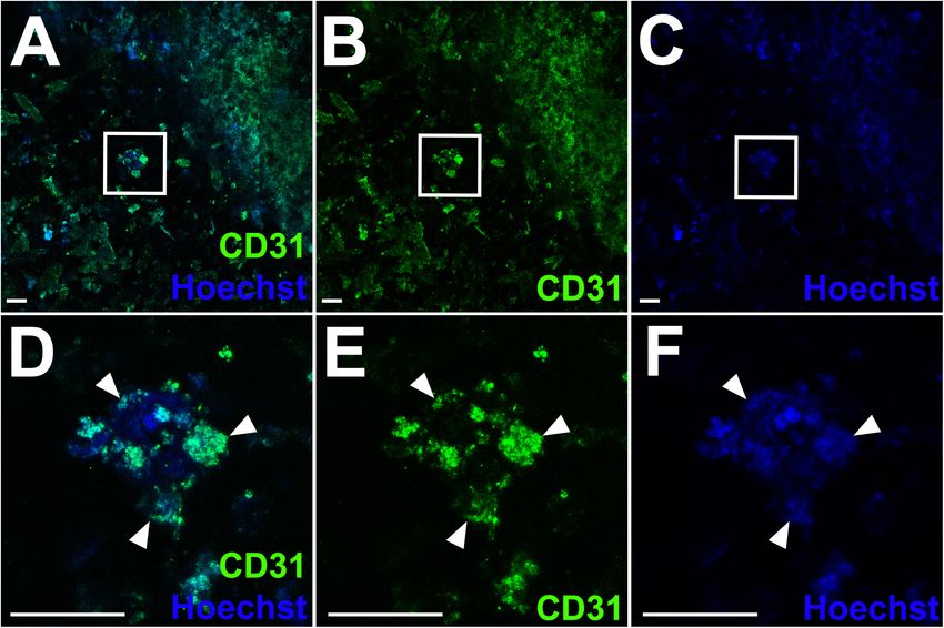
Roche et al.                                                                                                                 3D Bioprinted Cardiac Endothelial Cells

                                            degree of tissue maturation but before many patches start to lose integrity. We quantify
                                            AlgGel printability and present novel factors which reduce AlgGel patch durability (layer
                                            number, movement, and the addition of AlloECM) and factors which had minimal effect
                                            on durability (bioprinting system and cellular patch content).
                                            Keywords: 3D bioprinting, spheroids, hydrogel, bioink, durability, printability, alginate, gelatin

INTRODUCTION                                                                         the tissue can be cultured to allow for a period of tissue
                                                                                     maturation before transplantation (Roche and Gentile, 2020).
The latest developments in three-dimensional (3D) bioprinting                        During this post-printing phase, the bioink can promote tissue
technology have led to the hope that viable 3D bioprinted                            maturation, with durability in culture being an important
cardiac tissues could be generated to promote myocardial                             characteristic to predict hydrogel disintegration (Bishop et al.,
regeneration (Noor et al., 2019; Roche et al., 2020). Extrusion                      2017; Roche and Gentile, 2020), although the optimal moment
3D bioprinters have been widely used as a versatile tool to                          to transplant after a period in culture has not previously
deposit different cells as ‘bio-inks’ to generate complex 3D                         been confirmed. During this phase, cardiomyocyte contractility
tissues, including cardiac tissues (Zhang et al., 2016; Ong et al.,                  should be permitted and endothelial cells within the bioprinted
2017a; Maiullari et al., 2018; Noor et al., 2019). This technology                   tissue should be permitted to organise into networks, as one
promises a safe, precise, automatable and cost-effective method                      of the major challenges in 3D bioprinting of cardiac tissues
to generate myocardial tissue (Noor et al., 2019; Roche et al.,                      is the fabrication of a hierarchical vascular system within
2020). Extrusion 3D-bioprinters using cell-permissive pressures                      tissues (Gentile, 2016; Ong et al., 2017b; Polonchuk et al.,
can extrude myocardial cells without prohibiting their ability                       2017; Cui et al., 2019; Polley et al., 2020; Roche et al., 2020;
to live, mature and function in a physiological environment                          Xu et al., 2020).
(Blaeser et al., 2016). These 3D bioprinters extrude bioinks                             Here, we present a promising approach to generate 3D
which can be made from hydrogels (Cattelan et al., 2020;                             bioprinted cardiac patches presenting a structural endothelial
Roche et al., 2020).                                                                 cell network using alginate/gelatin (AlgGel) hydrogels – a mix
    The bioink formulation is critical to determine printability (a                  of alginate (to provide an ionically cross-linkable structure
function of the bioink’s rheological properties which determines                     for cellular patches) and gelatin (to adapt the alginate for
how it interacts with the bioprinting process) which is important                    extrusion 3D bioprinting, control rheological properties by
for bioprinting without damaging the end bioprinted product                          varying the gelatin concentration and generate a bioactive hybrid
(Cattelan et al., 2020; Gillispie et al., 2020). After bioprinting,                  hydrogel). The versatility of AlgGel hydrogels is established

  GRAPHICAL ABSTRACT |

Frontiers in Bioengineering and Biotechnology | www.frontiersin.org              2                                        February 2021 | Volume 9 | Article 636257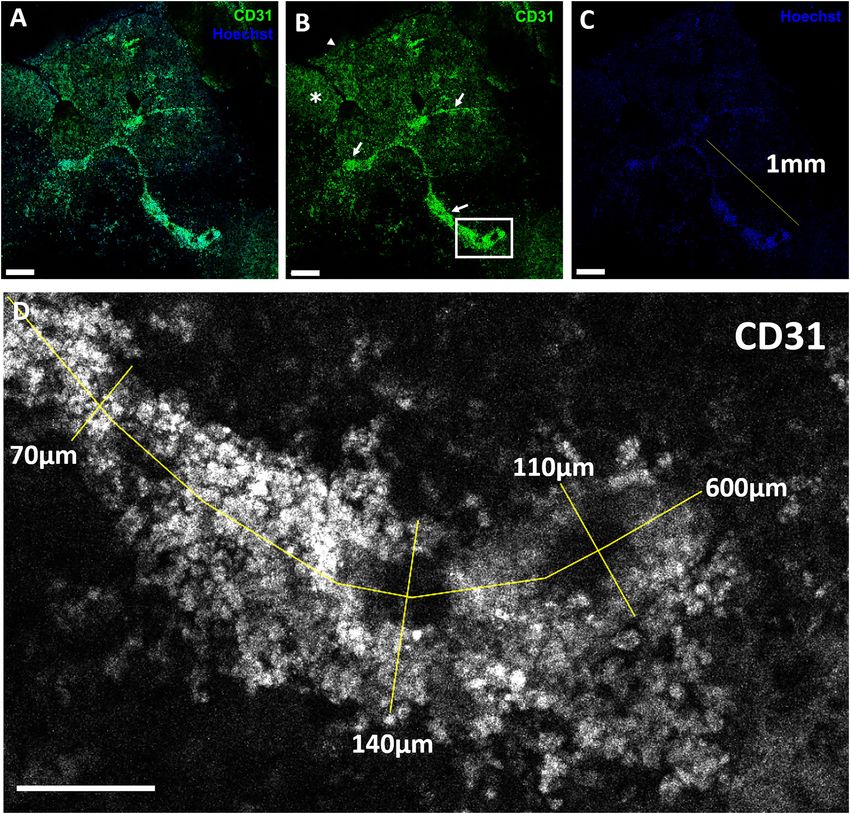
Roche et al.                                                                                                        3D Bioprinted Cardiac Endothelial Cells

(Mancha Sánchez et al., 2020), balancing printability against               neonatal heart dissociation kit according to the manufacturer’s
durability characteristics in culture, whilst also permitting               instructions. Isolated cells (cardiac cell types present in a whole
cardiomyocyte contractility and endothelial cell network                    heart, including myocytes, endothelial cells, and fibroblasts)
formation. The extent to which vascular networks are able                   were suspended in DMEM + 10% (v/v) FBS + 1% (v/v)
to self-assemble within patches is closely linked to several                pen/strep + 1% (v/v) L-glutamine. VCSs were generated by
other characteristics: 1) printability (for instance, with poor             coculturing ∼4000 mouse cardiac cells (immediately after
printability, control of patch morphology is undermined); 2)                their isolation) in 15 µl hanging drop cultures containing
durability (if patches disintegrate too quickly in culture then             DMEM + 10% (v/v) FBS + 1% (v/v) pen/strep + 1% (v/v)
there would be no patch to host a vascular network); 3) cell                L-glutamine, using Perfecta 3D 384-well hanging drop plates
                                                                                                                R

viability (since survival of cells, including endothelial and other         (3D Biomatrix, Ann Arbor, MI, United States). Spheroids were
cell types, is critical); and 4) contractility must be permitted            allowed to form for up to five days in hanging drops in a
(patches which inhibit contractility of cardiomyocytes will be less         humidified incubator at 37◦ C with 20% (v/v) O2 and 5% (v/v)
suitable for co-culture with contractile cells) (Roche et al., 2020).       CO2 . Additional complete DMEM (7 µl) was added to each
   We hypothesised that 3D bioprinted endothelial cells can self-           hanging drop on day three. VCSs were collected and the resulting
organise into structural vascular networks using our approach.              spheroid suspension was centrifuged at 300 g for 5 mins in a 50 ml
   In testing this hypothesis, we aimed to demonstrate that                 Falcon tube. The resulting VCS pellet was ready for direct mixing
even a low starting density of endothelial cells will self-organise         with hydrogel to create bioink.
into structural networks within 3D bioprinted patches. In this
study we report on printability, durability, cell viability and             Hydrogel Preparation
endothelial cell network formation for 3D bioprinted endothelial            To prepare the alginate and gelatin (AlgGel) hydrogel 4 mg
cells in AlgGel hydrogels. Our study aims at providing new                  alginate and 8 mg gelatin powder was sterilised under UV light
insights which may overcome common challenges in the field of               for 30 min, solubilised at 50◦ C in 100 ml DMEM + 10% (v/v)
bioprinting of cardiac tissues for in vitro and in vivo applications        FBS + 1% (v/v) pen/strep + 1% (v/v) L-glut. The mixture was
(Roche et al., 2020). The major finding of our study is that                then either stored at 4◦ C or used for bioink immediately after.
the bioprinted patches generated by using our approach present              AlloECM hydrogel (AlloECM , ROKIT, Seoul, South Korea) was
                                                                                                           R

endothelial cell networks, durable structure and contractile                prepared by adding AlloECM powder to AlgGel hydrogel at
function between 14 and 28 days in culture. Our findings have               5 and 30 mg/ml. The AlloECM-AlgGel was resuspended and
the potential to directly translate in vitro testing of bioprinted          mixed thoroughly in a Falcon tube at room temperature, warmed
cardiac patches for in vivo applications for cardiac regeneration           to 37◦ C in a water bath and then was used immediately for
(Roche and Gentile, 2020).                                                  bioprinting. Gelatin-methacryloyl (GelMA) 10% w/v + lithium
                                                                            phenyl-2,4,6-trimethylbenzoylphosphinate (LAP) 0.25% (w/v)
                                                                            in HEPES buffer in light-blocking pneumatic 3 ml syringes
MATERIALS AND METHODS                                                       was purchased (product no. IK305202, CELLINK Life Sciences,
                                                                            Boston, MA, United States) for use without cells as a printability
All procedures described in this experiment were approved
                                                                            and durability control hydrogel.
by the Animal Ethics Committee at the Northern Sydney
Local Health District (project number RESP17/55;                            Generation of Bioinks
20/04/2017). Full methodological details are included in the
                                                                            To create bioinks for bioprinting, hydrogels were added to pellets
Supplementary Materials.
                                                                            of either mouse cardiac cell spheroids or a mixture of HCAECs
                                                                            and HDFs (2:1), obtained as described above. 1.5 ml prewarmed
Cultures of Human Coronary Artery                                           (37◦ C) hydrogel was added to the cell pellet by pipette, of
Endothelial Cells With Fibroblasts                                          which 0.5 ml typically produced six 10 mm2 patches. Each
Human coronary artery endothelial cells (HCAECs) (Sigma-                    patch contained either ∼20,000 HCAECS and ∼10,000 HDFs,
Aldrich, MO, United States) were cultured in MesoEndo Growth                or ∼160,000 mouse cardiac cells (∼40 spheroids/patch). The
Medium (Cell Applications, San Diego, CA, United States).                   hydrogel was resuspended until the cell pellet disappeared to
Human dermal fibroblasts (HDFs) (Sigma-Aldrich, MO,                         ensure incorporation of most of the cells. All procedures were
United States) were cultured in Dulbecco’s Modified Eagle                   performed under a biological safety cabinet for the REGEMAT3D
Medium (DMEM, Sigma-Aldrich, St Louis, MO, United States)                   bioprinter. For the INVIVO and BIO X bioprinters (which have
with added 10% (v/v) FBS + 1% (v/v) pen/strep + 1%                          their own UV steriliser and hepafilter) this was performed within
(v/v) L-glutamine. Cells were used for bioprinting between                  the bioprinting chamber itself.
passage four and five.
                                                                            3D Bioprinting
Vascularised Cardiac Spheroid                                               Bioprinting was performed as fully described in the
Formation From Mouse Cardiac Cells                                          Supplementary Materials (including preparation, parameter
Mouse hearts were isolated from neonatal C57Bl/6 mice                       setting and bioprinting processes for the three bioprinters).
(1–5 days old), diced into 0.1–0.2 mm pieces and enzymatically              AlgGel was ionically crosslinked by adding CaCl2 after
digested with the Miltenyi Biotec (Bergisch Gladbach, Germany)              bioprinting of all the patches in one six-well plate. GelMA

Frontiers in Bioengineering and Biotechnology | www.frontiersin.org     3                                      February 2021 | Volume 9 | Article 636257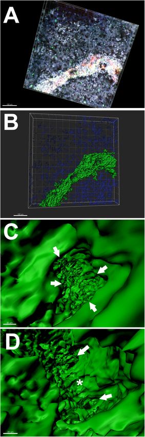
Roche et al.                                                                                                  3D Bioprinted Cardiac Endothelial Cells

was photo-crosslinked immediately after each patch layer was                Life Sciences, IL, United States) to obtain images of the
bioprinted by UV light photocuring.                                         entire patches. Quantification of stained cells was performed by
                                                                            random grid sampling and computer-based estimation using FIJI
Printability Assessments                                                    (ImageJ) software.
Printability was measured in terms of resolution, printing
accuracy, shape fidelity (after 28 days in culture) and extrudability       Patch Contractile Activity Evaluation
for grid pattern bioprinted patches. Resolution was assessed                To observe contractile activity, patches were monitored by video
as the width of deposited bioink gridlines (at day one – after              light microscopy for intrinsic oscillations. When contractile
crosslinking immediately after bioprinting); printing accuracy              oscillations were observed, phase contrast microscopy video
was measured as the number of empty squares between bioink                  recordings were obtained using an Olympus CKX53 microscope
gridlines in the grid pattern at day one (a perfect grid should have        (Olympus, Tokyo, Japan), with the beating patch contrasted with
16 empty squares between gridlines of bioink); shape fidelity was           non-contractile hydrogel in the same conditions. To count the
measured as the number of empty squares remaining with time                 rate of beating activity, videos were played in slow motion and
in culture at day 28; and extrudability outcome observations were           the average rate taken from multiple samples.
(1) whether the nozzle dripped hydrogel between the bioprinting
of patches (yes/no) and (2) the average number of times the                 3D Bioprinted Cell Staining, Imaging and
nozzle was blocked requiring the nozzle to be changed per six               Analysis
patches bioprinted (in series one after the other). To isolate              To evaluate endothelial cell organisation into structural networks,
hydrogel printability outcomes for AlgGel, it was compared                  we stained bioprinted patches with antibodies against CD31 to
to GelMA using the BIO X and bioprinting with hydrogel                      identify any structural endothelial cell network-like formation
only without cells.                                                         after 28 days in culture. After 28 days HCAEC + HDF-
                                                                            containing and mouse VCS patches were first fixed and
Durability Measurement                                                      then stained using antibody against human/mouse CD31 and
To assess survival of patches in culture, patches were monitored            Hoechst stain for endothelial cells and nuclei, respectively
daily for macroscopic disintegration for up to 28 days.                     (see Supplementary Materials for full protocol). They were
Culture medium was replaced every 3–4 days and the date of                  imaged by light microscopy and with a confocal microscope
patch disintegration was recorded and time to disintegration                (LSM 800, Zeiss, Oberkochen, Germany). Images were analysed
(durability) analysed. To evaluate the effects of layer number              with ImageJ (NIH, Bethesda, United States). For 3D rendering
on patch printability eight single-layer thickness (0.2 mm                  analysis, confocal images were processed by Imaris v7.6 (Oxford
depth) acellular AlgGels were printed as controls (without                  Instruments, Zurich, Switzerland).
cells). To evaluate the effect of minimising movement, 13
AlgGel HCAEC + HDF patches were generated and cultured                      Statistical Analysis
under minimisation of movement conditions (slow media                       Results were analysed using PRISM (GraphPad, San Diego,
replacement with 1000 µl narrow-bore non-automated pipette                  CA, United States). Hypothesis testing for categorical data
every 7–10 days with no transfer to a microscope for                        was performed using the chi-square test. Hypothesis testing
observations) and these were left in culture until the first                for continuous data was performed using the two-tailed
two patches in the set of 13 disintegrated. To evaluate the                 Mann–Whitney U test or the Kruskal–Wallis test for a
addition of exogenous AlloECM hydrogel to the AlgGel, 23                    difference between two or any of three non-parametric
AlloECM + AlgGel patches were printed at low and high                       data groups, respectively. Descriptive statistics (Kaplan–Meier
concentration of AlloECM. In addition, 13 acellular gelatin-                survival/durability data) were tested using pairwise Log-rank
methacryloyl (GelMA) patches were produced as an extended                   (Mantel Cox) tests.
durability control as GelMA is more durable in culture.

Cell Viability Assays, Imaging and                                          RESULTS
Analysis
To assess cell survival, we evaluated cell viability in bioprinted          Patch Printability
patches after 28 days in culture by staining them with                      To evaluate printability outcomes such as resolution, printing
calcein-AM, ethidium homodimer (Live/Dead Assay, Invitrogen,                accuracy, shape fidelity and extrudability, 5 × 5 line grid
Carlsbad, CA, United States) and Hoechst stain, used to identify            patches were bioprinted instead of patches completely filled with
live, dead and total cells (nuclei), respectively. Patches in               bioink, so that these printability measures could be assessed.
media with stains added were incubated at 37◦ C for 1 h.                    Patches were bioprinted with a geometry of 10 × 10 × 0.4 mm
After fresh media replacement, patches within the plate were                (length x width x depth) as shown in Figure 1. For all
moved to a microscope for automatic fluorescence imaging                    three bioprinters, printability outcomes (resolution, printing
by several automated microscopic methods (using an M7000                    accuracy, shape fidelity and extrudability) were similar. For
or EVOS Fl AUTO 1 (ThermoFisher, MA, United States),                        AlgGel hydrogel patches, resolution was similar compared to
Nikon Ti (Nikon, Tokyo, Japan), or IN Cell Analyzer (GE                     GelMA; printing accuracy was higher for AlgGel with 78/176

Frontiers in Bioengineering and Biotechnology | www.frontiersin.org     4                                   February 2021 | Volume 9 | Article 636257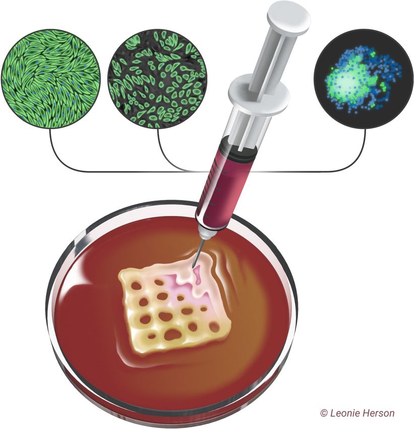
Roche et al.                                                                                                  3D Bioprinted Cardiac Endothelial Cells

(45%) of AlgGel empty internal squares in the grid pattern                day (Figures 2D,G). This was regardless of the AlloECM
preserved at day one compared to 53/208 (25%) for GelMA                   concentration in the AlgGel hydrogel (5 or 30 mg/ml), either
(p < 0.05; χ2 test; n = 384); shape fidelity after 28 days in             in the presence or absence of cellular content (Figures 2A–C).
culture was only different if it included the increased print             Overall, factors which reduced patch durability in culture were:
accuracy for AlgGel on day one, with 72/176 (41%) of squares              bioprinting patches with only one layer of depth, movement
remaining preserved at day 28 for AlgGel and 51/208 (25%)                 disturbance such as during media changes and transfer to a
for GelMA (p < 0.05; χ2 test; n = 384). However, correcting               microscopy platform and the addition of AlloECM. Factors
for differences in print accuracy on day one, the 28-day shape            which had negligible impact on durability in culture were the
fidelity rate was 72/78 (92%) for AlgGel and 51/53 (96%) for              bioprinting system used and the cellular content.
GelMA (p = 0.36; χ2 test; n = 131); for extrudability, AlgGel
dripped hydrogel from the nozzle and GelMA did not and                    Cell Viability
AlgGel had zero nozzle blockages per six patches compared to              To evaluate cell survival within patches at 28 days, our analysis of
one per six patches for GelMA (AlgGel tended toward more                  epifluorescence microscopy images at low magnification showed
flow than input software instructions and GelMA tended toward             viable cells surrounded by hydrogel components of the patches
less flow). Overall, no significant difference in printability was        which were variably autofluorescent (Figure 3). By random grid
observed between bioprinters but AlgGel had a 1.7x higher                 sampling, estimated live cell density was ∼80 cells/mm2 /layer
printing accuracy, presented some dripping from the nozzle and            (∼16,000 cells per patch) which is a viability rate of ∼53% from
had no nozzle blockages compared to GelMA hydrogel.                       the ∼30,000 cells per patch on initial bioprinting. We measured
                                                                          a live/dead ratio equal ∼2.3 at 28 days for these human cells.
Patch Durability in Culture                                               Viability in mouse mixed cardiac cell (VCS) patches (Figure 4)
To assess patch survival in culture conditions following                  could not be as reliably quantified because many patch-embedded
bioprinting, patches were cultured in media at 37◦ C for up               cells were in 3D spheroids. Using a software-based analysis of
to 28 days to evaluate durability in the post-printing, pre-              less autofluorescent confocal microscopy images we measured
transplantation phase (Figure 2). The different bioprinting               a live:dead cell ratio of 9:1 for mouse VCSs. VCS diameter was
system used had little effect on the overall durability of the            ∼150 µm at 28 days in culture as shown in Figure 4. This did not
resulting AlgGel patches (Figures 2E,F). Overall, median (and             change from day one (data not shown), confirming also the fact
IQR) for time in days to disintegration (durability) of AlgGel-           that VCSs maintained their shape in culture. Overall, our method
based patches in media in six-well plates was 10 (3–19) with              was associated with viable cells in patches, even after bioprinting
patches printed by the REGEMAT3D, 14 (13.5–18) with the                   and 28 days in culture.
INVIVO, and 14 (10–28) with the BIO X (p = 0.93; Kruskal–
Wallis test for a difference in any of the three groups). The
cellular patch content (mouse VCS, free (not in spheroid)
                                                                          Patch Contractility
                                                                          To assess whether patches were able to permit contractility
HCAECs + HDFs or acellular hydrogel alone) also had little
                                                                          of cardiomyocytes, throughout the 28 day experiments all
effect on patch durability (p = 0.17; Kruskal–Wallis test)
                                                                          mouse VCS-containing patches were evaluated under a light
(Figure 2). Kaplan–Meier survival curves suggested that the
                                                                          microscope for contractile activity (Supplementary Video 1).
addition of cells compared to hydrogel on its own might reduce
                                                                          Five patches started to display irregular contractile activity on
the number of bioprinting runs (sessions producing a set of
                                                                          day seven, two on day 10 and one on day 13. Despite six
patches) where all patches in that run survived to 28 days
                                                                          out of eight patches breaking into fragments over the course
(Figures 2A–C). However, survival analyses revealed no strong
                                                                          of the 28 days, all eight patches (or fragments) still showed
pattern for durability between different cellular contents within
                                                                          some contractile activity at the experiment end at day 28.
the AlgGel patches.
                                                                          The average rate of (non-fragmented) patch contractions was
    As the bioprinting system and cellular content had minimal
                                                                          258 beats/min (range 230–288) and the rate did not change
effect on patch durability, pooled analysis of all AlgGel
                                                                          with time in culture. Supplementary Video 1 shows VCS patch
patches was performed and median durability overall was
                                                                          contractility in real-time and the patch floating in media can
14 days (interquartile range (IQR) 10-27; n = 59; p < 0.05)
                                                                          be seen oscillating compared to adjacent static areas of non-
(Figure 2D), whereas 13/13 GelMA control patches were
                                                                          contractile hydrogel on the well floor. Altogether, the contractility
intact at the end of 28 days (p < 0.05). AlgGel acellular
                                                                          observed in our neonatal mouse VCS-AlgGel patches suggests
patches with one layer were less durable (median survival
                                                                          that there is no barrier in principle to generating contractile
3 days; IQR 2.5–4 days; n = 8; p < 0.05) than two-layer
                                                                          cardiac tissue using cardiac spheroids in AlgGel hydrogel
AlgGel acellular patches (median survival 28 days; IQR 19–
                                                                          (Supplementary Video 1).
28; n = 20; p < 0.05) (Figure 2D). The minimisation of
movement protocol allowed AlgGel patches to be cultured until
day 66 before the end point of 2/13 of these patches losing               Patch Endothelial Cell Network
their integrity (unsuitable for transplantation) was reached.             Structural Organisation
The addition of AlloECM to AlgGel reduced the durability                  To assess CD31+ endothelial cells’ ability to self-organise into
of patches: whilst AlloECM + AlgGel patches retained their                structural networks, we observed for network formation in VCS-
structure after bioprinting, 23/23 patches disintegrated in < 1           containing patches and patches with freely suspended HCAECs

Frontiers in Bioengineering and Biotechnology | www.frontiersin.org   5                                    February 2021 | Volume 9 | Article 636257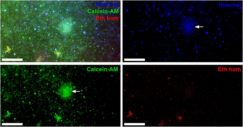
Roche et al. 3D Bioprinted Cardiac Endothelial Cells
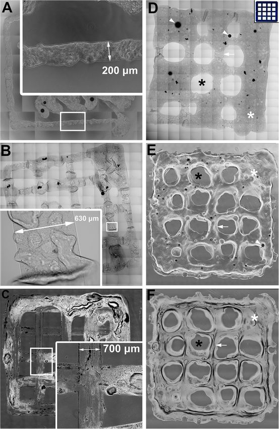
FIGURE 1 | Assessing printability for extrusion 3D bioprinting with AlgGel hydrogels and GelMA. (A–C) Representative images of 10 mm2 AlgGel patches 3D
bioprinted using three different bioprinters: a custom-made REGEMAT3D (A), the ROKIT INVIVO (B), and the CELLINK BIO X (C). The resolution (line width) of the
bioprinted grid patches was between 200 and 700 µm. Printing accuracy of AlgGel (D) and GelMA (E) was measured as the number of empty squares preserved
(Continued)
Frontiers in Bioengineering and Biotechnology | www.frontiersin.org 6 February 2021 | Volume 9 | Article 636257
Roche et al.                                                                                                                         3D Bioprinted Cardiac Endothelial Cells

  FIGURE 1 | Continued
  (black asterisks) between lines of bioink (white arrow) compared to the blueprint instructions input into the software on day one (D) inset panel). Hydrogel filled seven
  of the intended 16 empty spaces in (D), whereas in (E) two were filled (white asterisks): an example of a printing accuracy of 56 and 88% for these individually
  displayed representative patches, respectively. The day one GelMA patch in (E) is shown after 28 days in culture in (F), still with two spaces filled (a shape fidelity of
  100% for this displayed representative patch by this measure). The black dots (white arrowheads) in (D) and (E) are air bubbles. Scale bars not shown (all patches
  1 cm2 ). Overall in the complete sample (from which these displayed representative patches are taken), printing accuracy was higher for AlgGel with 78/176 (45%) of
  AlgGel empty internal squares in the grid pattern preserved at day one compared to 53/208 (25%) for GelMA (p < 0.05; χ2 test; n = 384); shape fidelity after
  28 days in culture was only different if it included the increased print accuracy for AlgGel on day one, with 72/176 (41%) of squares remaining preserved at day 28 for
  AlgGel and 51/208 (25%) for GelMA (p < 0.05; χ2 test; n = 384). However, correcting for differences in print accuracy on day one, the 28-day shape fidelity rate was
  72/78 (92%) for AlgGel and 51/53 (96%) for GelMA (p = 0.36; χ2 test; n = 131).

  FIGURE 2 | Durability assessments for 3D bioprinted patches in cell culture medium up to 28 days. (A–C) Survival analyses grouped by different patch cellular
  content, either hydrogel without cells (A), with HCAECs and HDFs (B) or mouse cardiac cell in VCSs (C). All curves are statistically significant compared to adjacent
  curves unless marked by “ns” and a black linking line connecting two or more similar (non-significant) curves; p < 0.05 Log-rank (Mantel-Cox) test of each line
  compared pairwise to each other line on the survival curve. In (A) identical curves are moved off centre to prevent complete line overlap (applies to AlloECM and
  GelMA curves). Each curve represents one print run of a series of patches bioprinted one after the other from the same batch of hydrogel (n = 5–13 per group). As
  cellular content had negligible influence on patch durability, pooled analysis of all patches is shown in (D). Overall, these results show that survival was similar
  whether patches contained AlgGel alone or with cells (A–C). They also show that AlgGel had a median survival of 14 days in culture overall (D), which was reduced
  by bioprinting single layer patches instead of standard thickness (double layer) or the addition of AlloECM (fibroblast-derived extracellular matrix hydrogel). Compared
  to GelMA (which is more durable in culture) AlgGel presented a median survival showing that it is likely to need transplanting sooner (at 14 days in culture), because
  leaving patches to culture for 28 days risks many patches fragmenting and becoming unsuitable for transplantation.

with HDFs, stained for CD31+ cells. For VCS patches, our                                     Conversely, CD31+ free (not in spheroid) HCAECs started
confocal analysis showed that some CD31+ mouse cardiac                                    to organise into structures resembling endothelial cell networks
endothelial cells in VCS remained in spheroids even after                                 (Figures 5, 6 and Supplementary Video 2). The median length of
bioprinting and 28 days in culture (Figure 4D). The median                                CD31+ linear human endothelial cell structures was 88 µm (IQR
length of CD31+ linear human endothelial cell structures was                              62–114 µm), median width was 37 µm (IQR 29–59 µm) and
149 µm (IQR 91–225 µm), median width was 46 µm (IQR                                       CD31+ endothelial cell covered area in the hydrogel was ∼2.1%.
29–80 µm) and CD31+ endothelial cell covered area in the                                  3D rendering structural analysis revealed a lumen-like space
hydrogel was ∼2.7%. For these patches (which contained VCS),                              between endothelial surfaces with endothelial cells having the
the CD31+ endothelial cells did not present extensive endothelial                         appearance of branched structures (Figure 6 and Supplementary
cell organisation into networks (Figure 4), despite some of the                           Video 2). Taken together, these findings suggest that (1) AlgGel
cells moving out of their spheroids (Figure 4D), consistent with                          hydrogels combined with this method permit the structural self-
observations in a previous study (Fleming et al., 2010). Some                             organisation of endothelial cells within bioprinted patches even
migrating CD31+ mouse cardiac endothelial cells (or clusters of                           without additional interventions such as supplementation with
these cells) were observed within the bioprinted patch outside                            angiogenic growth factors and (2) endothelial cells in spheroids
of spheroids, suggesting that at day 28 patches contained a                               with no additional angiogenic factors do not fully migrate out
mixture of cells which had migrated from their spheroids into the                         into the surrounding hydrogel for as significant a structural
hydrogel, cell clusters which had moved away from their initial                           organisation into networks to occur – instead the endothelial cells
spheroid and cells which had remained in the same spheroids                               form an irregular distribution across the patch (Figure 4) and
present at the initial bioprinting (Figure 4).                                            some remain in spheroids (Figure 4D).

Frontiers in Bioengineering and Biotechnology | www.frontiersin.org                   7                                           February 2021 | Volume 9 | Article 636257
Roche et al.                                                                                                                    3D Bioprinted Cardiac Endothelial Cells

  FIGURE 3 | Bioprinted HCAECs and HDFs in AlgGel hydrogel are viable at 28 days. HCAEC + HDFs within a patch 3D bioprinted with the REGEMAT3D shown after
  28 days in culture. This patch was stained with Hoechst (blue—nuclei), calcein-AM (green—live cell cytoplasm), ethidium homodimer (red—dead cell nuclei). Despite
  hydrogel autofluorescence (white arrow), the patch shows live cells (green), and nuclei (blue) at low magnification throughout this representative segment of patch.
  The live/dead ratio was ∼2.3 at 28 days in culture. Magnification bar = 500 µm.

DISCUSSION                                                                             patterns instead of patches completely filled with bioink to assess
                                                                                       these outcome measures. For the first time in direct comparison,
3D bioprinting is appealing for biomedical engineering as it can                       we quantified that printing accuracy was 1.7x higher for AlgGel
be used to produce uniform tissues, is scalable and automatable                        compared to GelMA with our method which was likely related
(Roche et al., 2020). It is also adaptable for use with different                      to GelMA’s less predictable extrudability (we quantified that
biomaterials and cell types, including iPSC-derived cardiac cells                      GelMA complete nozzle blockages occurred on average once
given their potential use for heart regeneration (Roche et al.,                        every six patches compared to no blockages for AlgGel). For
2020). Our study using alginate 4% (w/v)/gelatin 8% (w/v)                              durability, macroscopic disintegration (durability) was defined as
hydrogels and cardiac cells for 3D bioprinting of patches presents                     patch integrity being unacceptable for transplantation of a whole
a promising approach for cardiac bioengineering. Specifically,                         patch – for example, using our surgical patch transplantation
supporting our hypothesis, our 3D bioprinted patches showed                            method in a murine model of myocardial infarction (Roche
that it is possible for endothelial cells to self-organise into a                      and Gentile, 2020) – as this is a durability indicator of
structural network. Other studies have previously used differing                       practical relevance to the surgeon transplanting patches for
approaches to describe advances in endothelial cell network                            in vivo models. As printability and durability are critical for
assembly (Ong et al., 2017b; Cui et al., 2019; Polley et al., 2020;                    generating patches which are useable for transplantation, we
Xu et al., 2020). We have added to this a 3D rendering of                              thoroughly evaluated these characteristics. GelMA was used as
micrographic data that shows endothelial cells self-organised into                     a durable control as it is an established alternative to AlgGel
a structural network with a lumen-like space with our method                           for cardiac patch bioprinting which is highly durable in culture
(Supplementary Video 2).                                                               (Koti et al., 2019). Other studies have numerically described
   Additionally, as printability and durability are important                          detailed rheological characteristics, including (not limited to)
determinants of whether the patch survives to allow for cells                          storage modulus, viscosity and extrusion pressures for similar
to organise within it, we identified the printability/durability                       hydrogels (Mondal et al., 2019). Our study provides data on
impacts of several factors (bioprinting system, cellular patch                         highly practical printability measures (such as print accuracy) to
content, number of layers of patch depth, minimisation of                              inform hydrogel-related choices for patch culture and subsequent
movement, addition of AlloECM) which had not previously been                           transplantation.
assessed. As printability can be measured in terms of resolution                          As the effect of the bioprinting systems themselves on our
(e.g., extruded bioink line width), printing accuracy (the degree                      outcome measures was not previously compared, optimal
to which bioprinted constructs match the intended construct                            bioprinting conditions with AlgGel hydrogels were tested
set by the blueprint input into the software), shape fidelity                          with three different extrusion bioprinters: two screw-driven
(the ability of bioprinted constructs to maintain shape after                          extrusion systems (a custom made REGEMAT3D model
deposition) and extrudability (the ease of bioink extrusion/flow)                      and the commercially available Rokit INVIVO) and one
(Fisch et al., 2020; Gillispie et al., 2020), we used 5× 5 gridline                    using pneumatic extrusion (a CELLINK BIOX bioprinter)

Frontiers in Bioengineering and Biotechnology | www.frontiersin.org                8                                         February 2021 | Volume 9 | Article 636257
Roche et al. 3D Bioprinted Cardiac Endothelial Cells
FIGURE 4 | CD31-positive endothelial cells within a patch containing mouse cardiac spheroids. (A–F) Laser scanning confocal microscopy (LSCM) images of a BIO
X-bioprinted AlgGel patch containing neonatal mouse cardiac spheroids stained for cell nuclei (blue) and CD31+ endothelial cells (green). Scale bars
(A–C) = 100 µm. (A) Merged channel image showing CD31+ cells across a representative segment of patch and a spheroid still intact after bioprinting followed by
28 days in culture (inset panel). (B) and (C) show CD31 stain and Hoechst stain (nuclei), respectively. (D) Magnified image of the spheroid (white arrowheads)
highlighted in panel (A). CD31 stain and Hoechst (nuclei) are shown in (E) and (F), respectively. Scale bars (D–F) = 100 µm.
(Supplementary Figure 1). We found that 10 × 10 × 0.4 mm in culture conditions (Figure 2). AlgGel hydrogels are a suitable
patches – sized for in vivo rodent cardiac models (Roche choice for experiments that allow time for a degree of tissue
and Gentile, 2020) – can be 3D bioprinted with any system maturation in the post-printing phase before the hydrogel
with minimal difference to printability (resolution, printing disintegrates (Bociaga et al., 2019). By combining our durability
accuracy, shape fidelity and extrudability) or durability in data with our time to observation of patch contractility data
culture. No matter which system was in use, important (contractility was observed to begin between day seven and
parameters influencing the printability of a bioprinted series of 13), we are able to propose that transplantation of patches
patches would have included distance between the bioprinting for an in vivo model may be optimal just before 14 days in
nozzle tip and the six-well plate surface as well as ambient culture. Other studies have reported related durability measures,
temperature and hydrogel batch-to-batch variability (see for instance degradation rate as % weight loss of patches in
Supplementary Materials). Overall, our results suggest that culture up to 14 days (Bociaga et al., 2019). However, ours is
optimising bioprinting parameters was key to the printability and the first to report a more clinically relevant durability measure
long durability of patches as opposed to the bioprinting platform based on usefulness of the patch for its intended purpose –
used (for example, by optimisation pre-testing to determine the surgical transplantation with an established method for in vivo
ideal flow rate, nozzle speed and temperature settings which were testing (Roche and Gentile, 2020). Furthermore, we report
different for each system to work optimally with our hydrogels). comprehensive durability data with survival beyond 14 days and
It is known that bioprinting parameters and the concentration present data on three previously unevaluated determinants of
of AlgGel hydrogels are established determinants of printability durability: layer number (patch depth), minimising movement of
and durability (Mancha Sánchez et al., 2020); our study supports the patches and the addition of an exogenous factor (AlloECM).
this and also adds the finding that the bioprinting system itself AlloECM is a fibroblast-derived extracellular matrix hydrogel
was not a strong influencing factor. used to mimic properties of the extracellular matrix. We found
We also compared different hydrogel compositions for AlloECM could be added to AlgGel bioprinted patches with no
durable patches that readily retained their macrostructural shape change to printability or patch morphology on day one (when
Frontiers in Bioengineering and Biotechnology | www.frontiersin.org 9 February 2021 | Volume 9 | Article 636257Roche et al. 3D Bioprinted Cardiac Endothelial Cells FIGURE 5 | CD31-positive endothelial cell organisation within a 3D bioprinted patch containing HCAECs and HDFs. (A–C). Collapsed Z-stacks of confocal images of a 3D bioprinted AlgGel patch stained with antibodies against CD31 (green) and Hoechst stain (blue). (A) Merged channel image of CD31+ endothelial cells (green) and nuclei (blue) within a patch fragment. (B) CD31+ endothelial cells (white arrowhead shows border of hydrogel fragment and white asterisk shows hydrogel containing CD31+ cells). A small T-shaped formation of endothelial cells is starting to organise, with small 1 mm branch-like formations starting to form (white arrows). (C) The cell nuclei show the same branched organisation which maps to the CD31+ endothelial cells in (B). Magnification bars (A–C) = 200 µm. (D) Magnified black and white image taken from inset panel in (B) shows CD31+ endothelial cells with measurements (yellow lines, measurements indicated on image; scale bar 100 µm) and this structure is shown in more detail in the 3D rendering shown in Figure 6 and Supplementary Video 2. bioprinted). However, they all disintegrated by the next day. interference with the ionic crosslinking method used. Conversely, Unlike the acellular AlgGel patches which gradually broke into the addition of different cell types within a patch could also alter smaller pieces over time but left significant pieces of patch in the microenvironment and therefore affect durability but our the well (Supplementary Figure 2), the AlloECM-AlgGel patches data showed there was no major durability difference depending were completely disintegrated into pieces of residual hydrogel less on cell type when we evaluated human and mouse cells in than ∼1 mm in diameter. This is the first time durability data free and spheroid formation, respectively (Figure 2). Overall, have been reported for AlloECM mixed in with AlgGel patches we determined that bioprinting platform and cellular content and the finding may have been due to incompatibility between are not strong determinants of durability whereas layer AlloECM and AlgGel hydrogel, including overall pH changes or number, minimisation of movement in culture media and Frontiers in Bioengineering and Biotechnology | www.frontiersin.org 10 February 2021 | Volume 9 | Article 636257
Roche et al.                                                                                                                     3D Bioprinted Cardiac Endothelial Cells

                                                                                       FIGURE 6 | rendering analysis using Imaris software of a 3D bioprinted patch
                                                                                       stained with antibodies against CD31 (green) and Hoechst stain (blue). (A)
                                                                                       and (B) 3D rendering of the endothelial structure shown previously (in
                                                                                       Figure 5). Scale bars 100 µm. (C) and (D) Higher magnification images
                                                                                       depicting the inside of the structural lumen formed by endothelial cells within a
                                                                                       3D bioprinted patch. (C) Arrows indicate the inner (luminal) surface of the
                                                                                       walls (green). Scale bar 30 µm. (D) The star indicates a pillar joining two
                                                                                       opposing endothelial surfaces where the lumen branches into two smaller
                                                                                       lumens (arrows). Scale bar 20 µm. For a full view of the 3D rendering analysis
                                                                                       of the same lumen formed within the 3D bioprinted patch please see
                                                                                       Supplementary Video 2.

                                                                                      the addition of an exogenous factor (AlloECM) were strong
                                                                                      determinants of durability.
                                                                                         For the cellular component of patches, we bioprinted some
                                                                                      patches with VCS which are microtissue aggregates of mixed
                                                                                      cardiac cell types. VCS cultures can be readily adapted for other
                                                                                      cell types such as stem cell-derived cells and are generated
                                                                                      using a scaffold-free, self-sustainable approach that allows self-
                                                                                      assembly and organisation of cells in 3D (Gentile, 2016).
                                                                                      Our evaluation of cell viability at 28 days after bioprinting
                                                                                      demonstrated that over time both VCS bioink and bioink
                                                                                      with freely suspended endothelial cells and fibroblasts could
                                                                                      be cultured in bioprinted hydrogel patches (Figures 3 and
                                                                                      4). Accurate and automated quantification of viability for
                                                                                      cells embedded in autofluorescent hydrogel patches in 3D
                                                                                      is challenging (Noor et al., 2019). Nevertheless, viability of
                                                                                      freely suspended cells in bioprinted patches (Figure 3) was
                                                                                      estimated at approximately 53–61% at day 28, comparable with
                                                                                      previous reports from other studies using extrusion bioprinting
                                                                                      of 40–80% (Murphy and Atala, 2014; Fisch et al., 2020). For
                                                                                      VCS patches, live cell area/total cell area was estimated at ∼72%
                                                                                      with a favourable ratio of live to dead cells (9:1). The inability
                                                                                      to accurately quantify live cells in 3D spheroids embedded
                                                                                      in a 3D patch limits interpretation of this result without
                                                                                      further studies. Future studies would benefit from measuring
                                                                                      cell viability in 3D in a more automated way, for instance
                                                                                      with automated large specimen, serial confocal microscopy
                                                                                      techniques aimed at reducing hydrogel autofluorescence artefact.
                                                                                      Nonetheless, throughout our study, cells were viable in
                                                                                      AlgGel hydrogels.
                                                                                         All BIO X patches containing mouse VCSs presented
                                                                                      contractile activity between day seven and 13 and the contractility
                                                                                      was transmitted across the patch (Supplementary Video 1).
                                                                                      We reported that the average rate for these patches was
                                                                                      288 beats/min. Other studies have reported a beating rate
                                                                                      of approximately 180 beats/min for isolated neonatal mouse
                                                                                      cardiomyocytes in 2D culture (Ehler et al., 2013) and 500
                                                                                      beats/min for live adult mice in vivo (Mahmoud et al., 2015).
                                                                                      For these VCS patches, our confocal images of cells migrating
                                                                                      from the VCS into the hydrogel (Figure 4) suggested that at
                                                                                      day 28 patches contained both intact VCSs and clusters of cells
                                                                                      which had migrated into the hydrogel. The combined presence
  FIGURE 6 | Lumen evaluation of CD31+ endothelial cells within a
                                                                                      of cell clusters migrating away from VCSs into the hydrogel
  3D bioprinted patch generated with alginate 4%/gelatin 8% hydrogel. (A–D) 3D
                                                                   (Continued)
                                                                                      and those remaining within VCSs may have been responsible
                                                                                      for the generalised contractility observed in these patches

Frontiers in Bioengineering and Biotechnology | www.frontiersin.org              11                                           February 2021 | Volume 9 | Article 636257
Roche et al. 3D Bioprinted Cardiac Endothelial Cells
(Supplementary Video 1). This might explain why patches did (to increase their nourishing/supportive influence) but did not
not beat until between one and two weeks after bioprinting, equalise the ratio in case they interfered with endothelial cell
allowing time for some cells to migrate from spheroids and make self-assembly – as they have previously been shown to interfere
connections across the patch. Our observed contractile activity with the functioning of other cell types (cardiomyocytes) in equal
stopped and re-started with quiescent non-contractile periods ratio co-culture (Ong et al., 2017b). Future studies will be needed
of a few days between. This seemed to be related to disturbing to determine the optimal ratio of endothelial cells to fibroblasts
the patches, for example when changing culture medium, which when freely suspended in hydrogel patches.
also seemed to be a major factor in promoting macroscopic Future studies will also be needed to functionally test our
patch disintegration as well. Nonetheless, patches (or fragments self-assembled endothelial cell network. If endothelial cell self-
of patches) always resumed contractile activity (all remained assembly into networks is shown to be a successful approach,
contractile on day 28). We were unable to reliably measure it is likely to have advantages over other approaches (such as
the electrochemical discharge which causes cardiomyocyte fabricating artificial moulds and lining them with endothelial
contraction using our contractility-measuring and pacing system cells). Specifically, vascular cells which self-organise following
because the patches stopped contracting during the transport to physiological signalling in permissive hydrogel may organise
the system platform. This was probably also due to movement into a hierarchical network of different sized vessels and
disturbance from the transport itself and for this reason our present a more physiological network without exogenous scaffold
contractility analysis was limited to observance of contractions material. In summary, both the overall approach and the detailed
on video microscopy. Future workflows should minimise patch evaluations in this study pave the way for future studies aimed at
movement as much as possible, including bioprinting directly myocardial regeneration using cardiac patches.
onto a surface suitable for electrocardiographic recording and
pacing without patch transportation.
Bioengineering of heart tissues requires a vascular network CONCLUSION
for optimal cell survival and function (Fleming et al., 2010;
Roche et al., 2020; Wang et al., 2020) and our observations This study provides data of high practical relevance to inform
of stained endothelial cells showed that mouse VCSs did not bioengineering workflows focused on optimising cardiac patches
organise into endothelial network structures to the same extent prior to transplantation. Specifically, we have shown that 3D
as freely suspended HCAECs with HDFs (Figures 4–6 and bioprinted cardiac cells are viable in alginate/gelatin hydrogels
Supplementary Video 2). The endothelial network formation for at least 28 days in culture, allowing endothelial cells to self-
shown in Video 2 is structurally comparable to those reported organise into a network and for patch contractility. Bioprinted
in some other studies (Noor et al., 2019; Roche et al., 2020), cardiac patches in optimised conditions may develop/mature
which is noteworthy given our relatively low starting density of according to physiological signals in the pre-transplant phase
endothelial cells (∼2500 cells/mm3 ). One recent study (which even without being significantly coaxed or controlled by
reported on endothelial cells derived from induced pluripotent additional interventions. Taking into account patch durability, we
stem cells) reported a density of ∼15000 cells/mm3 (Noor et al., conclude that an optimal moment to transplant patches after a
2019), so even with our HCAEC density being six-fold less, a period of maturation is just before 14 days in culture.
structural network started to form. The first clinical trial for
epicardial-transplanted patch repair in humans found that a
functional benefit may be conferred to the failing heart even DATA AVAILABILITY STATEMENT
with a low starting density of stem cell derived cardiovascular
The datasets presented in this study can be found in online
progenitor cells (410/mm3 ) in large (20 cm2 ) patches (Menasché
repositories. The names of the repository/repositories and
et al., 2018). Our study supports the notion that significant
accession number(s) can be found below: Zenodo (CERN,
cell growth and self-organisation can occur in patches from
Geneva, Switzerland) repository doi: 10.5281/zenodo.4299230.
a low starting cell density. For future studies, which may use
human stem cell-derived cardiac cells or spheroids, additional
factors such as vascular endothelial growth factor (VEGF) could ETHICS STATEMENT
be added to promote endothelial cell organisation and cell
migration out of spheroids. Alternatively, a mixture of ‘free’ The animal study was reviewed and approved by the Animal
stem cell-derived endothelial cells and cardiac spheroids could Ethics Committee at the Northern Sydney Local Health District
be used. For freely suspended endothelial cells and fibroblasts, (project number RESP17/55; 20/04/2017).
we used a ratio of 2:1, respectively. Whilst optimal ratios for
various cells in spheroid co-culture have been reported (Noguchi
et al., 2016; Polonchuk et al., 2017), the optimal ratio of freely AUTHOR CONTRIBUTIONS
suspended HCAECS and HDFs in AlgGel is not known. The
physiological ratio of endothelial cells:fibroblasts in the heart is CR contributed to the conceptualisation, data generation, data
not universally agreed, but the currently accepted ratio is 4:1 curation, data analysis, data visualisation, funding acquisition,
(Pinto et al., 2016; Zhou and Pu William, 2016). In our patches, investigation, methodology, resources, validation, project
we doubled the number of fibroblasts relative to endothelial cells administration, writing (original draft), manuscript review,
Frontiers in Bioengineering and Biotechnology | www.frontiersin.org 12 February 2021 | Volume 9 | Article 636257Roche et al. 3D Bioprinted Cardiac Endothelial Cells
and editing. PS cultured and supplied the mouse cardiac cell Leonie Herson for her work in the design and generation of
spheroids used in these experiments. AA was responsible for the central image.
the supervision, writing (review), and editing. CJ and MX were
responsible for the supervision, manuscript review, and editing.
CG contributed to the conceptualisation, data generation, data SUPPLEMENTARY MATERIAL
curation, data analysis, data visualisation, funding acquisition,
methodology, project administration, supervision, manuscript The Supplementary Material for this article can be found
review, and editing. All authors contributed to the article and online at: https://www.frontiersin.org/articles/10.3389/fbioe.
approved the submitted version. 2021.636257/full#supplementary-material
Supplementary Figure 1 | 3D bioprinting platforms used: a custom-made
REGEMAT3D model (left column), the ROKIT INVIVO (middle column) and the
FUNDING CELLINK BIO X (right column). Hardware (A–C) and software (D–F) are shown for
these three extrusion-based 3D bioprinting systems. The REGEMAT3D
This work was supported by a Heart Research Australia Ph.D. customised (D) and INVIVO ‘Creator K’ (E) software were both accessed on a
Scholarship (grant number 2019-02), a Sir John Loewenthal laptop computer connected to the bioprinter by USB cable and the BIO X
software (F) was entirely inbuilt on the bioprinter touchscreen.
Scholarship 2019 (University of Sydney), the Le Gros Legacy
Fund New Zealand (grant number PhD012019), the UTS Supplementary Figure 2 | 3D bioprinted patch fragmentation. Representative
Seed Funding, a University of Sydney Kick-Start Grant, a phase images of an alginate 4%/gelatin 8% patch on day one (A) that was
fragmented in culture and became unsuitable for transplantation by day 28 (B).
University of Sydney Chancellor’s Doctoral Incentive Programme
Grant, a Catholic Archdiocese of Sydney 2019 Grant for Adult Supplementary Video 1 | Beating cardiac patches containing mouse
Stem Cell Research, and a Sydney Medical School Foundation vascularised cardiac spheroids in alginate/gelatin hydrogel. The patches shown
are oscillating in media with an average rate of 258 beats/min, consistent with
Cardiothoracic Surgery Research Grant.
mouse cardiac cells. Oscillating patches in media are compared with
non-contractile elements such as hydrogel in the well and in the video appendices
the appearances of non-contractile patches are shown with and without extrinsic
ACKNOWLEDGMENTS movement applied to the microscopy apparatus.
The authors thank Louise Cole and Chris Evenhuis and Supplementary Video 2 | Three-dimensional rendering of CD31 + endothelial
network-like structure within an alginate/gelatin patch containing HCAECs and
acknowledge the use of the equipment in the Microbial HDFs. The structure shown has a lumen space and branches. These confluent
Imaging Facility at the i3 Institute in the Faculty of Science, CD31 + endothelial cells (shown in green) self-assembled into this structure
University of Technology Sydney (UTS). The authors also thank over 28 days in culture following extrusion 3D bioprinting.
REFERENCES Fleming, P. A., Argraves, W. S., Gentile, C., Neagu, A., Forgacs, G., and Drake, C. J.
(2010). Fusion of uniluminal vascular spheroids: a model for assembly of blood
Bishop, E. S., Mostafa, S., Pakvasa, M., Luu, H. H., Lee, M. J., Wolf, J. M., et al. vessels. Dev. Dyn. 239, 398–406. doi: 10.1002/dvdy.22161
(2017). 3-D bioprinting technologies in tissue engineering and regenerative Gentile, C. (2016). Filling the gaps between the in vivo and in vitro
medicine: current and future trends. Genes Dis. 4, 185–195. doi: 10.1016/j. microenvironment: engineering of spheroids for stem cell technology. Curr.
gendis.2017.10.002 Stem. Cell Res. Ther. 11, 652–665.
Blaeser, A., Duarte Campos, D. F., Puster, U., Richtering, W., Stevens, M. M., and Gillispie, G., Prim, P., Copus, J., Fisher, J., Mikos, A. G., Yoo, J. J., et al.
Fischer, H. (2016). Controlling shear stress in 3D bioprinting is a key factor (2020). Assessment methodologies for extrusion-based bioink printability.
to balance printing resolution and stem cell integrity. Adv. Healthc Mater. 5, Biofabrication 12:022003. doi: 10.1088/1758-5090/ab6f0d
326–333. doi: 10.1002/adhm.201500677 Koti, P., Muselimyan, N., Mirdamadi, E., Asfour, H., and Sarvazyan, N. A. (2019).
Bociaga, D., Bartniak, M., Grabarczyk, J., and Przybyszewska, K. (2019). Sodium Use of GelMA for 3D printing of cardiac myocytes and fibroblasts. J. 3D Print.
alginate/gelatine hydrogels for direct bioprinting-the effect of composition Med. 3, 11–22. doi: 10.2217/3dp-2018-2017
selection and applied solvents on the bioink properties. Materials 12:2669. Mahmoud, A. I., O’Meara, C. C., Gemberling, M., Zhao, L., Bryant, D. M.,
doi: 10.3390/ma12172669 Zheng, R., et al. (2015). Nerves regulate cardiomyocyte proliferation and heart
Cattelan, G., Guerrero Gerbolés, A., Foresti, R., Pramstaller, P. P., Rossini, A., regeneration. Dev. Cell 34, 387–399. doi: 10.1016/j.devcel.2015.06.017
Miragoli, M., et al. (2020). Alginate formulations: current developments in the Maiullari, F., Costantini, M., Milan, M., Pace, V., Chirivi, M., Maiullari, S., et al.
race for hydrogel-based cardiac regeneration. Front. Bioeng. Biotechnol. 8:414. (2018). A multi-cellular 3D bioprinting approach for vascularized heart tissue
doi: 10.3389/fbioe.2020.00414 engineering based on HUVECs and iPSC-derived cardiomyocytes. Sci. Rep.
Cui, H., Zhu, W., Huang, Y., Liu, C., Yu, Z. X., Nowicki, M., et al. (2019). 8:13532. doi: 10.1038/s41598-018-31848-x
In vitro and in vivo evaluation of 3D bioprinted small-diameter vasculature Mancha Sánchez, E., Gómez-Blanco, J. C., López Nieto, E., Casado, J. G.,
with smooth muscle and endothelium. Biofabrication 12:015004. doi: 10.1088/ Macías-García, A., Díaz Díez, M. A., et al. (2020). Hydrogels for bioprinting:
1758-5090/ab402c a systematic review of hydrogels synthesis, bioprinting parameters, and
Ehler, E., Moore-Morris, T., and Lange, S. (2013). Isolation and culture of neonatal bioprinted structures behavior. Front. Bioeng. Biotechnol. 8:776. doi: 10.3389/
mouse cardiomyocytes. J. Vis. Exp. 50154. doi: 10.3791/50154 fbioe.2020.00776
Fisch, P., Holub, M., and Zenobi-Wong, M. (2020). Improved accuracy Menasché, P., Vanneaux, V., Hagege, A., Bel, A., Cholley, B., Parouchev, A., et al.
and precision of bioprinting through progressive cavity pump- (2018). Transplantation of human embryonic stem cell-derived cardiovascular
controlled extrusion. bioRxiv [preprint] doi: 10.1101/2020.01.23.9158 progenitors for severe ischemic left ventricular dysfunction. J. Am. Coll. Cardiol.
68 71, 429–438. doi: 10.1016/j.jacc.2017.11.047
Frontiers in Bioengineering and Biotechnology | www.frontiersin.org 13 February 2021 | Volume 9 | Article 636257You can also read


