Circulating long non coding RNAs HOTAIR, Linc p21, GAS5 and XIST expression profiles in diffuse large B cell lymphoma: association with R CHOP ...
←
→
Page content transcription
If your browser does not render page correctly, please read the page content below
www.nature.com/scientificreports
OPEN Circulating long non‑coding RNAs
HOTAIR, Linc‑p21, GAS5 and XIST
expression profiles in diffuse large
B‑cell lymphoma: association
with R‑CHOP responsiveness
Mahmoud A. Senousy1, Aya M. El‑Abd 2*
, Raafat R. Abdel‑Malek3 & Sherine M. Rizk1*
The reliable identification of diffuse large B-cell lymphoma (DLBCL)-specific targets owns huge
implications for its diagnosis and treatment. Long non-coding RNAs (lncRNAs) are implicated in
DLBCL pathogenesis; however, circulating DLBCL-related lncRNAs are barely investigated. We
investigated plasma lncRNAs; HOTAIR, Linc-p21, GAS5 and XIST as biomarkers for DLBCL diagnosis
and responsiveness to R-CHOP therapy. Eighty-four DLBCL patients and thirty-three healthy
controls were included. Only plasma HOTAIR, XIST and GAS5 were differentially expressed in DLBCL
patients compared to controls. Pretreatment plasma HOTAIR was higher, whereas GAS5 was lower
in non-responders than responders to R-CHOP. Plasma GAS5 demonstrated superior diagnostic
accuracy (AUC = 0.97) whereas a panel of HOTAIR + GAS5 superiorly discriminated responders from
non-responders by ROC analysis. In multivariate analysis, HOTAIR was an independent predictor of
non-response. Among patients, plasma HOTAIR, Linc-p21 and XIST were correlated. Plasma GAS5
negatively correlated with International Prognostic Index, whereas HOTAIR positively correlated
with performance status, denoting their prognostic potential. We constructed the lncRNAs-related
protein–protein interaction networks linked to drug response via bioinformatics analysis. In
conclusion, we introduce plasma HOTAIR, GAS5 and XIST as potential non-invasive diagnostic tools
for DLBCL, and pretreatment HOTAIR and GAS5 as candidates for evaluating therapy response, with
HOTAIR as a predictor of R-CHOP failure. We provide novel surrogates for future predictive studies in
personalized medicine.
Abbreviations
ATG7 Autophagy related 7
AKT1S1 AKT1 substrate 1 (proline-rich)
AKT2 AKT serine/threonine kinase 2
AUC Area under the curve
BCL2 B-cell lymphoma 2
BCL7C B-Cell CLL/Lymphoma 7 Protein Family Member C
CR Complete response
CRC Colorectal cancer
DHL Double-hit lymphoma
DLBCL Diffuse large B-cell lymphoma
ECOG Eastern Cooperative Oncology Group
eIF4E Eukaryotic translation initiation factor 4E
FDG-PET/CT Fluorodeoxyglucose-Positron Emission Tomography/Computed Tomography
1
Department of Biochemistry, Faculty of Pharmacy, Cairo University, 23 Kasr Al‑Ainy street, Cairo 11562,
Egypt. 2General Administration of Clinical Trials, Central Administration of Biological and Innovative Products and
Clinical Studies, Egyptian Drug Authority, 51 Wezaret El Zeraa Street, Agouza, Giza, Egypt. 3Department of Clinical
Oncology, Kasr Al‑Ainy Centre of Clinical Oncology & Nuclear Medicine, Faculty of Medicine, Cairo University,
Cairo, Egypt. *email: aya.elabd@gmail.com; sherine.abdelaziz@pharma.cu.edu.eg
Scientific Reports | (2021) 11:2095 | https://doi.org/10.1038/s41598-021-81715-5 1
Vol.:(0123456789)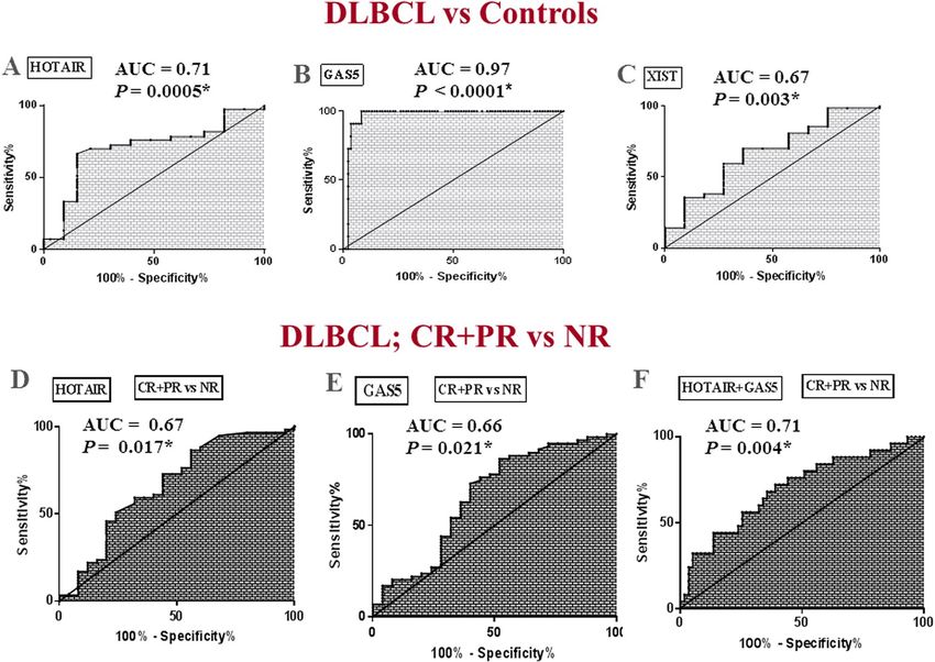
www.nature.com/scientificreports/
FDR False discovery rate
GAS5 Growth arrest-specific transcript 5
GO Gene Ontology
HOTAIR HOX transcript antisense intergenic RNA
KEGG Kyoto Encyclopedia of Genes & Genomes
lincRNAs Long intergenic RNA
IPI International Prognostic Index
LDH Lactate dehydrogenase
lncRNAs Long non-coding RNAs
mTOR Mammalian target of rapamycin
NFKBIA Nuclear Factor Kappa-B Inhibitor Alpha
NSCLC Non-small cell lung cancer
NR Non-responders
PR Partial response
PRC2 Polycomb repressive complex 2
PS Performance status
PPI Protein–protein interaction
R-CHOP Rituximab, cyclophosphamide, doxorubicin, vincristine, and prednisone
PI3K Phosphoinositide-3-kinase
PIK3R1 Phosphoinositide-3-Kinase Regulatory Subunit 1
ROC Receiver-operating-characteristic
SOX2 SRY-box 2
SETDB1 SET Domain Bifurcated 1
S1PR1 Sphingosine-1-Phosphate Receptor 1
STAT Signal transducer and activator of transcription
THL Triple-hit lymphomas
TP53 Tumor protein p53
XIST X-inactive-specific transcript
Diffuse large B-cell lymphoma (DLBCL) is the most common subtype of non-Hodgkin lymphoma (NHL), con-
stituting up to 40% of all cases globally1. It is a cancer of B-cells that have been exposed to antigens, the annual
incidence of which is rising year by y ear1. Notably, DLBCL is one of the most common NHL subtypes in North
Africa and Middle East (49.4%) compared to North America (29.3%)2. Egypt exceptionally has high incidence
of lymphoma and is claimed to have higher incidence of NHL among all hematopoietic c ancers3.
DLBCL is a fast growing tumor that occurs in lymph nodes within the neck, armpit or groin area, but may
appear elsewhere. It is diagnosed primarily by biopsy, complete blood count and computed tomography4. Largely,
DLBCL is a rapidly progressive fatal malignancy that responds badly to existing treatment, with more than one-
third of affected patients are resistant to various therapies5. The current standard initial therapy for DLBCL is
a combination of Rituximab (CD20 antibody), cyclophosphamide, doxorubicin, vincristine, and prednisone
(R-CHOP)5. Prognosis has improved significantly by adding Rituximab to the conventional t herapy6, however
approximately 30% to 50% of patients still respond badly to R-CHOP, depending on disease stage or prognostic
index7. The most commonly used prognostic tool in DLBCL is the International Prognostic Index (IPI), which
takes only into account clinical parameters such as age, clinical stage and performance status8. Recently, the
deconvolution of the complex molecular genetics of DLBCL has unraveled key oncogenic pathways that improved
the understanding of its biological d iversity5. Thus, exploring novel genetic and/or epigenetic markers may be
of clinical value for the diagnosis, prognosis, and therapy of DLBCL.
Long non-coding RNAs (lncRNAs), a class of ncRNAs longer than 200 nucleotides, are implicated in cancer
initiation, development and progression through epigenetic regulation of multiple cellular p aradigms9. Indeed,
dysregulated lncRNAs act as oncogenes or tumor suppressors in diverse cancers including haematological malig-
nancies, and have come out as interesting predictive biomarkers for diagnosis, prognosis, therapy responsiveness
and also as therapeutic targets9,10. Intriguingly, the possible application of lncRNA-based therapies in clinical
practice has attracted much attention in the last decade and many clinical trials are already started e.g., the
DTA-H19 vector in bladder, ovarian, and pancreatic c ancer11,12. Recently, the clinical application of lncRNAs
in B-cell malignancies is increasingly evident based on their involvement in normal B-cell development as well
as the pathogenesis of B-cell t umors13,14, however, the biological functions, expression pattern, and prognostic
value of many lncRNAs in DLBCL are still largely unelucidated13. In addition, data about circulating DLBCL-
related lncRNAs are scarce. Thus, profiling circulating lncRNAs may open a new avenue for non-invasive DLBCL
diagnosis, treatment and prediction of its response to therapy.
HOX transcript antisense intergenic RNA (HOTAIR) is reported as an oncogenic lncRNA that promotes cell
proliferation, tumor invasiveness and metastasis, and its overexpression is a marker of poor prognosis in various
cancer types, including lymphoma15. LincRNA-p21 (Linc-p21), a p53-dependent lncRNA, is reported to be a
tumor suppressor lncRNA in B-cell m alignancies16. Growth arrest-specific transcript 5 (GAS5) is an another
tumor suppressor lncRNA that regulates cell survival17, and was linked to B-cell lymphoma18. X-inactive-specific
transcript (XIST) is a 17 kb lncRNA that sculpts the cis-inactivation of the over one thousand X-linked genes19.
Indeed, ample evidence demonstrated aberrant XIST regulation in various cancers, including lymphoma and
male testicular germ-cell tumors, where XIST hypomethylation was o bserved20.
In this study, we selected these 4 lncRNA candidates (HOTAIR, Linc-p21, GAS5 and XIST) based on the
reported biological link with DLBCL or other lymphomatic cancers. HOTAIR and Linc-p21 have been shown
Scientific Reports | (2021) 11:2095 | https://doi.org/10.1038/s41598-021-81715-5 2
Vol:.(1234567890)
www.nature.com/scientificreports/

                                                            Patients     Control
                                   Parameter                n = 84       n = 33          P value
                                   Age > 18, mean ± SD      50 ± 13.30   47.21 ± 10.52   0.28
                                   Range, years             21–79        30–70
                                   Sex, n (%)
                                   Male                     37 (44%)     18 (54.5%)
                                                                                         0.41
                                   Female                   47 (56%)     15 (45.5%)
                                   Ann Arbor stage, n (%)
                                   I                        14 (17%)     –
                                   II                       29 (34%)
                                   III                      19 (23%)
                                   IV                       22 (26%)
                                   ECOG PS, n (%)
                                   0                        13 (15.5%)
                                   1                        39 (46%)
                                   2                        13 (15.5%)
                                   3                        16 (19%)
                                   4                        3 (4%)
                                   IPI, n (%)
                                   0                        17 (20%)     –
                                   1                        21 (25%)
                                   2                        16 (19%)
                                   3                        16 (19%)
                                   4                        14 (17%)
                                   Extranodal lymph nodes, n (%)
                                   ≥2                       37 (44%)     –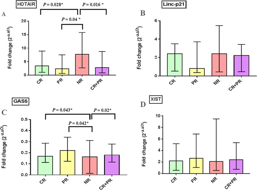
www.nature.com/scientificreports/
Figure 1. Flow chart of study design, patient classification, treatment response and analysis data sets. CR
complete response, PR partial response, NR non-responders, N number.
into complete responders (CR), n = 38; partial responders (PR), n = 21; and non-responders (NR), n = 25. Thus,
the overall responders (CR + PR) were 59/84 (70%), while the NR patients were 25/84 (30%). Comparison of the
clinicopathological data of overall responder and NR groups revealed statistically significant higher IPI scores in
NRs compared to the overall responders (P = 0.014). NRs were of higher age than overall responders (P = 0.01).
Other clinicopathological data showed no significant difference between the two groups (Table 2).
Plasma lncRNAs levels in DLBCL patients. All studied lncRNAs were expressed in control plasma
with varying levels (Supplementary Fig. S1). HOTAIR and XIST levels were significantly upregulated with a
median fold change = 3.77, P = 0.0004 and 2.265, P = 0.003, respectively, whereas GAS5 expression was signifi-
cantly downregulated with a median fold change = 0.159 (P < 0.0001) in the overall DLBCL patients compared
to the control group. On the other hand, Linc-p21 expression was not statistically significant between the two
groups (P = 0.76) (Fig. 2).
Pretreatment plasma lncRNAs levels and responsiveness to R‑CHOP therapy. Baseline plasma
lncRNAs levels in DLBCL patients were analyzed in relation to response outcome (Fig. 3). Pretreatment levels
of plasma HOTAIR were significantly higher in NR than those in CR or PR groups (P = 0.028, P = 0.04 respec-
tively). Indeed, further analysis revealed that baseline plasma HOTAIR levels were higher in NR than overall
responders (CR + PR) (P = 0.016). On the other hand, GAS5 levels were significantly higher in CR or PR than NR
groups (P = 0.043, P = 0.042, respectively). Further analysis showed that the levels of GAS5 in plasma of overall
responders were significantly higher than those in NRs (P = 0.02). Comparisons of the pretreatment HOTAIR
and GAS5 levels between CR vs PR + NR revealed no statistical difference (P > 0.05). On the other hand, pre-
treatment plasma Linc-p21 and XIST levels were not statistically different at all comparisons (P > 0.05) (Fig. 3).
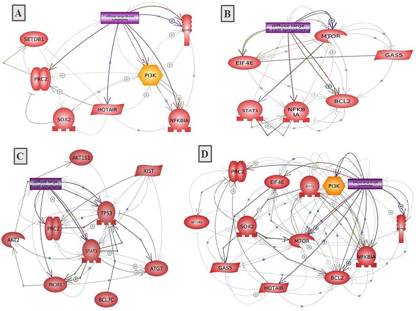
Diagnostic and prognostic potentials of studied plasma lncRNAs. Receiver-operating-character-
istic (ROC) analysis was performed to explore the clinical value of HOTAIR, GAS5 and XIST in the diagno-
Scientific Reports | (2021) 11:2095 | https://doi.org/10.1038/s41598-021-81715-5 4
Vol:.(1234567890)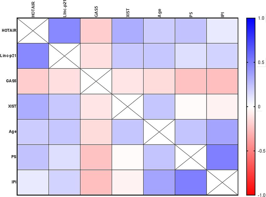
www.nature.com/scientificreports/

                                                   Overall responders   Non-responders
                                   Parameter       n = 59               n = 25           P value
                                   Age ≤ 60        50 (85%)             14 (56%)
                                                                                         0.01*
                                   > 60            9 (15%)              11 (44%)
                                   Sex, n (%)
                                   Male            29 (49%)             8 (32%)
                                                                                         0.16
                                   Female          30 (51%)             17 (68%)
                                   Ann Arbor stage, n (%)
                                   I/II            33 (56%)             10 (40%)
                                                                                         0.23
                                   III/IV          26 (44%)             15 (60%)
                                   ECOG PS
                                   0–2             48 (81%)             17 (68%)
                                                                                         0.25
                                   3–4             11 (19%)             8 (32%)
                                   IPI, n (%)
                                   0–2             43 (73%)             11 (44%)
                                                                                         0.014*
                                   3–4             16 (27%)             14 (56%)
                                   Extranodal lymph nodes, n (%)
                                    1.34 for HOTAIR, 91.67% and 100%, respectively at a cutoff fold change < 0.45 for GAS5, 70.24% and
                                  63.64%, respectively at a cutoff fold change > 1.05 for XIST. These results demonstrate the impact of these lncR-
                                  NAs as diagnostic biomarkers in DLBCL. Comparison of the ROC curve results suggested that plasma GAS5
                                  performed much better (AUC = 0.97) than HOTAIR and XIST (AUC = 0.71, 0.67, respectively, differences = 0.26,
                                  0.3, P < 0.0001, respectively).
                                      The prognostic significance of plasma HOTAIR and GAS5 which were differentially expressed in overall
                                  responders and NR groups were evaluated using a ROC curve (Fig. 4D,E). Results revealed that baseline plasma
                                  HOTAIR and GAS5 levels discriminated patients with different treatment outcome among DLBCL patients
                                  with AUC of 0.67 (95%CI = 0.5302 to 0.802, P = 0.017), and 0.66 (95%CI = 0.521–0.798, P = 0.021), respectively.
                                  The optimal sensitivity and specificity to discriminate overall responders from NR patients were 72.8% and
                                  56%, respectively at a cutoff fold change < 2.37 for HOTAIR and 67.8% and 60%, respectively at a cutoff fold
                                  change > 0.13 for GAS5. Combination analysis of HOTAIR + GAS5 (Fig. 4F) revealed that a panel of baseline
                                  plasma HOTAIR plus GAS5 levels discriminated patients with different treatment outcome among DLBCL
                                  patients with AUC = 0.71 (95%CI = 0.574 to 0.824, P = 0.004), with optimal sensitivity and specificity of 72% and
                                  61.02%, respectively. These results demonstrate the impact of these lncRNAs as biomarkers of therapy outcome
                                  in DLBCL.

                                  Correlation between lncRNAs levels and clinical data. We further examined correlations between
                                  studied plasma lncRNAs with each other and with clinical data in the overall studied DLBCL patients (Fig. 5
                                  and Supplementary Table S1). HOTAIR levels were positively correlated with Linc-p21 (r = 0.46, P < 0.0001),
                                  XIST (r = 0.32, P = 0.003) levels and PS (r = 0.24, P = 0.029). Linc-p21 levels were positively correlated with XIST

Scientific Reports |   (2021) 11:2095 |                      https://doi.org/10.1038/s41598-021-81715-5                                             5

                                                                                                                                               Vol.:(0123456789)
www.nature.com/scientificreports/
Figure 2. Plasma fold change levels of studied lncRNAs in DLBCL patients compared to healthy controls. Data
are presented by median interquartile range. DLBCL, n = 84, controls, n = 33. *Indicates statistical significance
(P < 0.05).
levels (r = 0.215, P = 0.049) and patient age (r = 0.217, P = 0.047). Plasma GAS5 levels were negatively correlated
with IPI (r = -0.251, P = 0.022) and PS (r = -0.243, P = 0.026). Finally, XIST levels were positively correlated with
patient age (r = 0.22, P = 0.044).
Prediction of DLBCL diagnosis and therapy outcome. Univariate and multivariate logistic regres-
sion analyses were performed to identify predictors of the risk of DLBCL diagnosis and its treatment outcome.
Neither studied lncRNA was able to predict the risk of being diagnosed with DLBCL (DLBCL vs healthy con-
trols) in the univariate analysis (Supplementary Table S2). Interestingly, HOTAIR was selected as significant
negative predictor of overall response to therapy in DLBCL patients (P = 0.008) in the univariate analysis. GAS5
demonstrated marginal association (P = 0.045) in the univariate analysis. In the multivariate analysis, HOTAIR
was the final independent negative predictor of overall response. In other words, HOTAIR was an independ-
ent predictor of non-response. Results were adjusted with age, sex, family history and IPI score as confounders
(Table 3). These results suggested that HOTAIR may offer potential as biomarker for R-CHOP response evalu-
ation in DLBCL patients.
Results of functional analysis in relation to therapy response. We carried out a functional analy-
sis of DLBCL-associated lncRNAs in our study in relation to drug response. Of the large number of lncRNA-
RNA interactions found in the starBase platform (http://starbase.sysu.edu.cn/) for HOTAIR, GAS5 and XIST,
we selected the protein-coding genes, which were then filtered according to their possible biological relation to
drug responsiveness using molecular annotation (MAS) system (http://bioinfo.capitalbio.com/mas3/). Finally,
PI3K, PRC2, SOX2, IkBa, SETDB1 and S1PR1 were selected for HOTAIR; eIF4E, mTOR, STAT1, NFκBIA and
BCL2 for GAS5; and TP53, STAT3, ATG7, PRC2, BCL7C, BCL79L, PIK3R1, AKT2, AKT1S1 for XIST (Table 4).
To further identify the role of these lncRNAs-related genes, we analyzed the lncRNA-related protein–protein
interactions (PPI) as well as the biological processes and KEGG pathways of the PPI network using the STRING
online software. P values and the results of Gene ontology (GO) and KEGG pathway analyses for each lncRNA-
related PPI are listed in Table 4. The lncRNA-related PPI network construction is visualized in Fig. 6.
Discussion
The pathogenesis of DLBCL involve multi-step and heterogeneous processes with different genetic and epigenetic
changes, and that high epigenomic heterogeneity correlated with a higher relapse rate and poor outcome25,26.
The lack of clear symptoms and early detection makes it difficult to diagnose at an early stage, leading to poor
prognosis. Existing molecular prognostic markers of DLBCL include MYC, P53, BCL2, and Ki-67. However,
Scientific Reports | (2021) 11:2095 | https://doi.org/10.1038/s41598-021-81715-5 6
Vol:.(1234567890)www.nature.com/scientificreports/
Figure 3. Baseline plasma fold change of studied lncRNAs in relation to treatment response. Data are presented
by median interquartile range. Complete response (CR), n = 38, partial response (PR), n = 21 and non-
responders (NR), n = 25, overall responders (CR + PR), n = 59. *Indicates statistical significance (P < 0.05).
they have several limitations. MYC and P53 mutations are found in only 10% and 15–30% of DLBCL patients,
respectively27. BCL2 is upregulated in 40–60% of patients and is associated with worse outcomes only in certain
subtypes of DLBCL, while data about Ki-67 were controversial27, necessitating the identification of new pre-
dictive markers. Recently, expectations have been raised regarding the potential role of lncRNAs as predictive
markers28–30 and as potential mediators of resistance to cancer t herapy31,32, however, these studies were carried
out on tumor tissue samples and/or cell lines.
Herein, we found that plasma HOTAIR, XIST and GAS5 were differentially expressed in DLBCL patients
indicating their involvement in the pathogenesis of DLBCL. To the best of our knowledge, we are the first to
provide evidence about XIST expression in DLBCL and its diagnostic and prognostic significance. In addition, we
demonstrated that plasma levels of HOTAIR, GAS5 and XIST showed a discriminative ability for DLBCL, sug-
gesting them as surrogate non-invasive biomarkers of DLBCL diagnosis, with GAS5 was of superior diagnostic
performance. We also recorded that baseline plasma HOTAIR and GAS5 were associated with prognosis and
therapy outcome, with HOTAIR demonstrated predictive ability for R-CHOP failure. We also constructed the
HOTAIR- and GAS5-related PPI networks to explore their role in drug response in DLBCL. Our results intro-
duce GAS5 and HOTAIR as novel candidates for future large scale predictive studies in personalized medicine.
First-line early R-CHOP failure in DLBCL still represents a dramatic situation in routine clinical p ractice33.
Among patients for whom R-CHOP therapy fails, 20% suffer from primary refractory disease (progress during or
right after treatment) whereas 30% relapse after achieving complete remission34. Herein, we found that R-CHOP
failure was 30%, that is in marginal compliance with the reported 30–50% failure7,33,34. Our findings could be due
to the short-term follow up study design aiming at early detection of NR patients for shifting them to another
treatment. Actually, a high rate of relapse usually appears during longer follow up periods34. Our results showed
higher IPI scores in NR relative to overall responders, confirming that patients with a more advanced disease
state have poorer prognosis and are more liable to R-CHOP failure.
Several mechanisms of resistance may account for refractoriness to R-CHOP in DLBCL. The majority of
DLBCL patients present a double rearrangement of MYC and BCL2 genes called double-hit lymphoma (DHL),
a chromosomal breakpoint, affecting the MYC/8q24 locus in combination with another recurrent breakpoint,
usually BCL2 [t(14;18)(q32;q21)], although BCL6/MYC-positive DHLs or BCL2/BCL6/MYC-positive triple-hit
Scientific Reports | (2021) 11:2095 | https://doi.org/10.1038/s41598-021-81715-5 7
Vol.:(0123456789)www.nature.com/scientificreports/
Figure 4. Plasma lncRNAs as biomarkers of DLBCL diagnosis and therapy outcome. (A–C) ROC curve
analysis of plasma (A) HOTAIR, (B) GAS5 and (C) XIST as diagnostic biomarkers differentiating DLBCL
patients (n = 84) from healthy controls (n = 33). (D–F) ROC analysis of plasma (D) HOTAIR and (E) GAS5, (F)
HOTAIR + GAS5 to differentiate overall responders (n = 59) from NR (n = 25) among DLBCL patients.
lymphomas (THLs) may also be observed. All studies that focused on DHLs or THLs concluded that the patients
outcomes were poor, with R-CHOP probably not being the best therapeutic o ption35,36. Furthermore, TP53,
FOXO1, MLL3, CCND3, NFKBIZ, and STAT6 were identified as top candidate genes for therapeutic resistance
in DLBCL37. In addition, lncRNAs play a crucial role in the chromosome breaks involved in typical gene rear-
rangements in hematologic m alignancies38, and indirectly affect drug resistance through regulating the expression
of some intermediate regulatory factors31,32.
Only a limited number of studies have examined circulatory levels of lncRNAs in B-cell malignancies39,40.
Herein, the observed upregulation of plasma HOTAIR in DLBCL patients agreed with the previously reported
overexpression in DLBCL tissues21,41 and cell lines21. Our recorded correlation between HOTAIR and perfor-
mance status links this lncRNA to DLBCL prognosis. Similarly, HOTAIR upregulation in DLBCL tumor tissues
was correlated with clinical stage, B symptoms, IPI scores and tumor volumes, and predicted poor prognosis
and poor survival rates in DLBCL patients21. In addition, we highlighted an association of plasma HOTAIR with
non-response to R-CHOP. Similarly, HOTAIR upregulation was associated with resistance to different chemo-
therapeutic drugs in non-small cell lung cancer (NSCLC), breast, and ovarian cancers via activating multiple
oncogenic events32. In fact, HOTAIR upregulation seems to be a common event underlying cancer progression
and resistance to therapy via a key pro-oncogenic role15, however, its role in inducing drug resistance in DLBCL
was not fully clear.
Using a bioinformatics approach, we identified a HOTAIR-related PPI network linked to drug resistance in
DLBCL, including PI3K, PRC2, SOX2, IkBa, SETDB1 and S1PR1. This PPI was enriched in cell chemotaxis and
angiogenesis process and in B-cell and T-cell receptors signaling, TNF signaling and apoptotic pathways. To put
this in context, HOTAIR promotes cell growth and inhibits apoptosis by regulating H3K27me3 and activating
the PI3K/AKT/NF-κB p athway42, which is considered a checkpoint for R-CHOP resistance in DLBCL; PI3K/
AKT inhibition was found to reverse R-CHOP resistance by destabilizing SOX2 in DLBCL43. HOTAIR also
regulates chromatin remodeling in DLBCL via recruiting of polycomb repressive complex 2 (PRC2) proteins
and inducing silencing of target genes through H3K27 t rimethylation41. HOTAIR was hypothesized to inhibit
IkBa (an inhibitor of NF-kB), and then activates c-MYC expression, which in turn induces HOTAIR expression
through SETDB1/STAT3 signaling pathway involved in cisplatin-resistant ovarian cancer44. NF-κB mutations and
Scientific Reports | (2021) 11:2095 | https://doi.org/10.1038/s41598-021-81715-5 8
Vol:.(1234567890)www.nature.com/scientificreports/
Figure 5. Correlation between plasma lncRNAs levels with each other and with clinical data. A correlation
map with a blue-red scale. The blue color corresponds to a correlation close to 1 and the red color corresponds
to a correlation close to − 1. Correlations are made by Spearman correlation. IPI International Prognostic
Index, PS performance status.
Univariate analysis Multivariate analysis
Variable B S.E P OR 95%CI B S.E Pa OR 95%CI
HOTAIR − 0.51 0.196 0.008* 0.60 0.407–0.877 − 0.44 0.206 0.032* 0.64 0.43–0.964
GAS5 7.06 3.130 0.045* 68.74 1.005–151 4.23 3.930 0.281 68.84 0.311–152.3
Table 3. Plasma lncRNAs as predictors of overall response to R-CHOP in DLBCL patients. HOTAIR and
GAS5 were included in a multivariate analysis with age, sex, family history and IPI score as covariates. − 2
Log likelihood of the best model, P < 0.0001. a Adjusted with age, sex, family history and IPI score. *Indicates
statistical significance (P < 0.05).
high S1PR1 and S1PR1/pSTAT3 expression were known pathways contributed to increase relapse in D LBCL34.
HOTAIR may also contribute to drug resistance through regulation of miR-130a that was associated with higher
risk of R-CHOP failure in D LBCL34. HOTAIR is a direct target of c-MYC through interaction with putative
c-MYC target response element in the upstream region of HOTAIR by harboring a miR-130a binding site44. Taken
together, these results conceptualize the critical role of HOTAIR in drug resistance for R-CHOP in DLBCL, and
provide HOTAIR as a therapeutic target.
Our study also demonstrated plasma GAS5 downregulation by a median 6.29 fold in DLBCL patients. Simi-
lar findings have been reported in B-cell neoplasm such as multiple m yloma39. GAS5 was also reported to be
abnormally expressed in DLBCL in an in silico analysis23. We recorded an inverse correlation of GAS5 and IPI,
suggesting that low plasma GAS5 levels are incorporated in the pathogenesis and development of DLBCL and
may correspond to the degree of prognosis. Indeed, patients with low GAS5 expression exhibited shorter overall
survival than those with higher expression and GAS5 expression was an independent indicator of colorectal can-
cer (CRC) p rognosis45. Additionally, we showed that higher baseline GAS5 was associated with good response to
R-CHOP. To further analyze the role of GAS5 in drug response, we identified a GAS5-related PPI network which
included eIF4E, mTOR, STAT1, NFKBIA and BCL2. This PPI was enriched in negative regulation of autophagy,
cell cycle and cell differentiation, immune response activation, promotion of apoptosis and cell response to
drugs via several pathways, including EGFR, NF-κB, JAK/STAT, and PI3K/AKT/mTOR signaling pathways. This
Scientific Reports | (2021) 11:2095 | https://doi.org/10.1038/s41598-021-81715-5 9
Vol.:(0123456789)www.nature.com/scientificreports/
Gene ontology for PPI network KEGG pathway analysis for PPI network
lncRNA lncRNA-related genes PPI P value Biological process Strength (FDR) Pathway Strength (FDR)
Cell chemotaxis 1.55 (0.0209) B-cell receptor signaling 1.96 (0.0105)
Angiogenesis 1.34 (0.0398) Chronic myeloid leukemia 1.93 (0.0105)
Negative regulation of cellular
PI3K, PRC2, SOX2, IkBa, 0.56 (0.0368) T-cell receptor signaling 1.82 (0.0105)
HOTAIR 0.019* process
SETDB1,S1PR1
Multicellular organism development 0.54 (0.0436) TNF signaling 1.78 (0.0105)
Signal transduction 0.54 (0.0436) Apoptosis 1.68 (0.0105)
Cellular response to stimulus 0.5 (0.0194) Pathways in cancer 1.1 (0.0371)
EGFR tyrosine kinase inhibitor
Negative regulation of autophagy 2.03 (0.0055) 2.0 (0.0012)
resistance
TNF-mediated signaling 1.99 (0.0056) NF-κB signaling 1.93 (0.0013)
Positive regulation of cell growth 1.69 (0.0103) Jak-STAT signaling 1.87 (0.00011)
Immune response-activating signal
1.37 (0.0203) Apoptosis 1.67 (0.0015)
transduction
eIF4E, mTOR, STAT1, NFKBIA,
GAS5 0.019* Response to drug 1.24 (0.0027) MicroRNAs in cancer 1.72 (0.0017)
BCL2
Negative regulation of cell dif-
1.24 (0.0082) Pathways in cancer 1.48 (0.00011)
ferentiation
Negative regulation of cell cycle 1.18 (0.0364) PI3K-AKT signaling 1.35 (0.0069)
Positive regulation of cell Popula-
1.13 (0.0113)
tion proliferation
Apoptotic process 1.11 (0.0115)
Regulation of cell cycle arrest 1.64 (0.0102) Acute myeloid leukemia 2.05 (0.00002)
Lymphocyte differentiation 1.34 (0.0227) Platinum drug resistance 2.02 (0.000018)
Regulation of gene expression,
1.29 (0.0258) Chronic myeloid leukemia 1.98 (0.00002)
epigenetic
Response to antibiotic 1.21 (0.0342) B-cell receptor signaling 1.84 (0.00076)
Cellular response to drug 1.2 (0.0347) Apoptosis 1.74 (0.00006)
TP53, STAT3, ATG7, PRC2, BCL7C, Negative regulation of cell death 1.11 (0.0011) mTOR signaling 1.7 (0.00007)
XIST 0.0233*
BCL79L, PIK3R1, AKT2, AKT1S1 Negative regulation of apoptotic
1.06 (0.0045) T-cell receptor signaling 1.69 (0.0013)
process
Immune effector process 0.9 (0.0282) Jak-STAT signaling 1.66 (0.00007)
Cell differentiation 0.55 (0.0326) TNF signaling 1.66 (0.0015)
PI3K-AKT signaling 1.32 (0.00065)
Pathways in cancer 1.28 (0.000085)
MAPK signaling 1.22 (0.0084)
Table 4. Bioinformatics analysis of the lncRNAs-related genes and protein–protein interactions linked to
drug responsiveness. The PPI and functional enrichment analysis for the PPI were conducted using SPRING
software. PPI, protein–protein interactions; FDR, false discovery rate. *Indicates statistical significance (P <
0.05).
agrees with previous reports that GAS5 is required for the inhibition of human T cell proliferation by mTOR
antagonists46. In fact, GAS5 influences cell survival rate by activating the apoptotic machinery. Indeed, over-
expression of GAS5 promoted apoptosis by decreasing the expression of the anti-apoptosis protein BCL-2 and
inhibited tumor resistance to therapy in bladder and cervical cancers32. In addition, GAS5 binds directly to eIF4E,
a key factor of translation initiation complex, then negatively affects the c-MYC protein through lncRNA–mRNA
interaction, denoting that GAS5 overexpression promotes favorable response by indirectly regulating c-MYC47.
We found an upregulation of plasma XIST level in DLBCL patients. Similarly, serum XIST was found to be
upregulated in NSCLC p atients48. Mechanistically, XIST binds PRC2 and propagate epigenetic silencing of an
individual X c hromosome49. The transcription factor, Yin Yang 1 has also been reported to interact with and
relocate XIST, to the inactivated X-chromosome in activated B-cells, thereby changing the X-linked gene regula-
tion in these cells compared to antigen naïve B-cells50.
Although XIST expression was linked to therapeutic response in CRC, NSCLC, and ovarian cancer24,51,52,
we failed to find a correlation between XIST and R-CHOP therapy responsiveness in DLBCL. This may be due
to different cancer type, different therapy, regimen and population. Indeed, previous studies were heterogenous
regarding the role of XIST in therapy responsiveness. While XIST was associated with doxorubicin resistance in
CRC cells24 and cisplatin resistance in NSCLC51, it was correlated with Taxol sensitivity in ovarian cell lines52. To
further unravel the role of XIST in drug resistance, our bioinformatics analysis included XIST. An XIST-related
PPI network included TP53, STAT3, ATG7, PRC2, BCL7C, BCL79L, PIK3R1, AKT2 and AKT1S1 and was
enriched in regulation of cell cycle arrest, lymphocyte differentiation, response to antibiotic, cellular response
to drug, immune effector process and negative regulation of apoptotic process. KEGG pathway analysis revealed
involvement in B-cell and T-cell receptors signaling, JAK/STAT, TNF, and PI3K/AKT, mTOR, MAPK signaling
Scientific Reports | (2021) 11:2095 | https://doi.org/10.1038/s41598-021-81715-5 10
Vol:.(1234567890)www.nature.com/scientificreports/
Figure 6. Construction of lncRNA-related PPI networks linked to drug responsiveness in DLBCL. (A)
HOTAIR, (B) GAS5, (C) XIST, (D) HOTAIR + GAS5. Pathway Studio online software was used.
pathways. Further studies are needed to explore the exact mechanism of XIST in R-CHOP therapy at the cel-
lular level.
Our finding that Linc-p21 expression was not changed in DLBCL patients compared to controls contrasts
Linc-p21 downregulation in DLBCL tumor t issues22 and in circulation of acute lymphoblastic leukemia patients39.
The reported low abundance of Linc-p21 may be due to the lack of functional tumor suppressor p53 protein
which is located on chromosome 17. Deletions of chromosome 17 are frequent events in B-cell malignancies25.
p53 may be also inactivated by the BCL6 gene during the genesis of lymphoma25.
We observed a positive correlation of Linc-p21 with HOTAIR and XIST levels, suggesting their concomitant
expression in DLBCL to orchestrate several pathologic events and co-regulatory networks. Intriguingly, tissue
Linc-p21 was correlated with clinicopathological data and considered an indicator of favorable clinical outcome
and survival rates in DLBCL p atients22. Linc-p21 was shown to impair tumerigenesis in DLBCL patients with
22
an R-CHOP regimen . However, we failed to find this relation. Discrepant results may be due to different type
of sample (plasma vs tissue), sample size, sample collection and processing and the normalization method.
Few lncRNAs have been reported to be dysregulated in DLBCL tissue samples and cell lines and their abnor-
mal expression levels were associated with poor p rognosis21,22,28–30,53,54, with little were correlated with response
22,53
to therapy . Our study improves over previous studies in that it introduces circulating lncRNAs as novel
complementary biomarkers in DLBCL diagnosis, prognosis and prediction of patient responsiveness to R-CHOP
therapy. Moreover, our data emphasize HOTAIR as a predictor of R-CHOP failure and GAS5 as a good indicator
for R-CHOP overall response in DLBCL and highlight some target genes relating them to drug resistance, which
need further validation. Our findings provide useful rationale for personalizing anti-cancer therapy.
Yet, there are few limitations in the current study, involving relatively small sample size and missing further
validation. Our study is also missing a survival analysis due to the one-end point study design which focused in
response to therapy. Therefore, future aspects should be assigned for validation and further clarification of the
biological function of circulating lncRNAs in DLBCL.
Scientific Reports | (2021) 11:2095 | https://doi.org/10.1038/s41598-021-81715-5 11
Vol.:(0123456789)www.nature.com/scientificreports/
Conclusion
Plasma HOTAIR, GAS5 and XIST could serve as novel non-invasive diagnostic biomarkers for DLBCL. Plasma
GAS5 demonstrated superior diagnostic accuracy and was a candidate for DLBCL prognosis. Baseline plasma
HOTAIR and GAS5 levels were associated with responsiveness of DLBCL patients to standard R-CHOP therapy,
with pretreatment HOTAIR was able to predict treatment failure. Our data could have impact in personalized
medicine where predicting positive response could save time, costs, and side effects. Our results also pave the way
for identification and development of new lncRNA-diagnostic and therapeutic targets that could be translated
into clinical practice.
Subjects and methods
Patients. Overall, 84 Egyptian patients with DLBCL and 33 age- and sex-matched healthy controls were
included in this prospective study. The demographic data of patients and controls are listed in Table 1. DLBCL
is diagnosed primarily by biopsy, complete blood count and computed tomography. Pathologically diagnosed
DLBCL patients were admitted at the outpatient’s clinic of Kasr Al-Ainy Centre of Clinical Oncology & Nuclear
Medicine (NEMROCK), Faculty of Medicine, Cairo University, Cairo, Egypt to receive standard treatment regi-
men; a combination of Rituximab (R) and conventional chemotherapy. The anthracycline-containing (R-CHOP)
regimen included Rituximab 375 mg/m2 on day 1, cyclophosphamide 750 mg/m2 on day 2, doxorubicin 50 mg/
m2 on day 2, vincristine 1.4 mg/m2 (up to a maximal dose of 2 mg) on day 2, and prednisone 40 mg/m2 for 5 days.
DLBCL patients received R-CHOP therapy for total 6 treatment cycles (1 cycle every 21 days)4. Cut off assess-
ment for treatment response was done one month after finishing the 6 cycles of treatment. Overall, the study
period including patient enrollment and follow up was from January 2017 to August 2018.
All patients were subjected to full history taking and clinical examination. The inclusion criteria included
patients with age > 18 years, gender of both sex, pathologically diagnosed as DLBCL patients and fit to receive
chemotherapy. Patients who received previous treatment with Rituximab were excluded.
Written informed consents were obtained from all participants. The study protocol and informed consent
were approved by the ethics committee of the Faculty of Pharmacy, Cairo University (No. BC1927) and complied
with the good clinical practice (GCP) and Declaration of Helsinki guidelines.
Definition of treatment response. After treatment cycles, patients were revaluated by using Fluorodeox-
yglucose-Positron Emission Tomography/Computed Tomography (FDG-PET/CT) which is the recommended
standard for post-treatment assessment in DLBCL. All recruited patients were successfully followed up till the
end of study. At the end of therapy, patients were divided into three groups; responded to treatment, partially
responded and non-responded according to the response evaluation (Fig. 1). Response was defined by com-
paring the residual uptake with the tumor uptake in baseline scan using FDG-PET/CT. Complete metabolic
response (CR) is defined when no residual uptake exists, partial metabolic response (PR) when the uptake has
decreased, and no metabolic response (NR) when it has not changed or progressive metabolic disease (PMD)
when it has i ncreased55. Overall response was defined as CR + PR.
Data collection. Clinical, laboratory and pathology data as well as imaging studies were collected by review-
ing the medical records of each participant. Clinical data included age, gender, lymphoma stage (Ann Arbor
stage), Eastern Cooperative Oncology Group (ECOG) performance status, and the presence of B cell-related
symptoms. IPI was calculated using age, clinical stage and performance s tatus4,8. Laboratory data included a
complete blood count and serum LDH level.
Samples collection and plasma preparation. Blood samples were taken at baseline before starting
therapy. After the patient had been diagnosed with DLBCL and complied with the inclusion criteria, a blood
sample was withdrawn at the morning on the day of starting treatment (day 1 of cycle 1 of R-CHOP for each
patient) according to the R-CHOP protocol. Samples were processed within 30 min to 2 h after collection. For
RNA analysis, we used platelet-poor plasma to exclude cellular nucleic acids. Cell and cell components-free
plasma was prepared from up to 5 ml whole blood collected on EDTA-coated tubes via a two-step centrifuga-
tion protocol (2000×g for 10 min at 4 °C and 12,000×g for 10 min at 4 °C) to thoroughly remove cellular nucleic
acids. After separation, plasma was transferred to nuclease-free tubes in aliquots and stored at -80 °C until RNA
extraction. Samples with hemolysis were excluded.
LncRNAs assay. Total RNA was drawn out from 200 μl plasma using the QIAzol reagent by miRNeasy Mini
Extraction kit (Qiagen, Valencia, CA, USA) according to the manufacturer’s instructions. The concentration and
purity of RNA were determined using NanoDrop 2000 Spectrophotometer (Thermo Fisher Scientific, USA), and
samples with a A260/A280 ratio between 1.8 and 2.0 were used in reverse transcription (RT). RNA samples were
stored in nuclease-free tubes and stored at − 80 °C till further analysis.
RT was carried out on 100 ng of total RNA in a final volume of 20 μl RT reactions (incubated for 10 min at
25 °C then for 30 min at 50 °C and finally for 5 min at 85 °C) using the Maxima First Strand cDNA Synthesis
kit (Thermo Fisher Scientific, USA) according to the manufacturer’s instructions. cDNA samples were stored in
nuclease-free tubes and stored at − 80 °C till further analysis.
Expression of lncRNAs were evaluated by quantitative PCR analysis conducted using customized primers
and Maxima SYBR Green qPCR Master Mix (Thermo Fisher Scientific, USA) according to the manufacturer’s
protocol. We used GAPDH as the endogenous control to normalize lncRNAs. GAPDH was reported to be stably
expressed in plasma and was previously selected as an internal control in plasma to normalize lncRNAs56,57. In
Scientific Reports | (2021) 11:2095 | https://doi.org/10.1038/s41598-021-81715-5 12
Vol:.(1234567890)www.nature.com/scientificreports/
addition, GAPDH level was not affected by age, sex and pathology in human plasma57, and was regarded as
an ideal internal control for plasma a ssays56,57. The primers sequences were as follows: 5′-GGTAGAAAAAGC
AACCACGAAGC-3′ (forward) and 5′-ACATAAA-CCTCTGTCTGTGAGTGCC-3′ (reverse) for HOTAIR;
5′-GGGT GGC TCA CTC TTC
TGG C-3′ (forward) and 5′-TGGC CTT
GCC CGG
GCT TGT C-3′ (reverse) for Linc-
p21; 5′-GTGTGGCTCTGG-ATAGCAC-3′ (forward) and 5′-ACCCAAGCAAGTCATCCATG-3′ (reverse) for
GAS5; 5′-GCATAACTCGGCT TAGGGCT-3′ (forward) and 5′- TCCTCTGCCTGACCTGCTAT-3′ (reverse)
for XIST; 5′-CCCTTCATTGACCTCAACTA-3′ (forward) and 5′-TGGAAGATGGTGATGGGATT -3′ (reverse)
for GAPDH.
For real-time PCR analysis of each lncRNA, 3 μl of RT products was mixed with 7.5 μl RNase-free water,
12.5 μl Maxima SYBR Green qPCR Master Mix and 1 μl forward primer and 1 μl reverse primer. The real-time
amplification was performed using 25 μl reaction mixtures using the Stratagene Mx3005P QPCR System (Agilent
Technologies, Germany) with the following conditions: 95 °C for 10 min, followed by 40 cycles at 95 °C for 15 s
and 60 °C for 60 s.
ΔCT was calculated by subtracting the Ct values of GAPDH from the Ct values of the target lncRNAs. lncR-
NAs expression relative to internal control was calculated by 2 −ΔCt. Fold change relative to healthy controls was
calculated using 2 −ΔΔCT method.
Functional analysis of lncRNAs‑related genes in relation to therapy response. The starBase
platform (http://starbase.sysu.edu.cn/) was used to check the candidate lncRNAs-RNA interactions. The output
was filtered by selecting protein-coding genes. Then data were analyzed using MAS system provided by Capi-
talBio company (Molecule Annotation System, http://bioinfo.capitalbio.com/mas3/) to determine the biological
roles of these lncRNA-related protein-coding genes. The genes most related to DLBCL, drug responsiveness
and cancer therapy in terms of biological process, molecular function, and KEGG pathway analysis were finally
selected. The cutoff P value was 0.05. STRING online software was used to analyze the interaction relationships
between the proteins encoded by the selected lncRNA-related genes (protein–protein interactions, PPI). Func-
tional enrichments; GO and KEGG pathway analysis were also conducted to determine the involvement of each
lncRNA-related PPI in different biological pathways using STRING online software. The lncRNA-related PPI
network was visualized using the Pathway Studio Online Software.
Statistical analysis. Values are expressed as mean ± SD, median interquartile range, or number (percent-
age) when appropriate. According to data normality, comparison of independent samples from two groups was
performed using Student’s t test or the Mann–Whitney U-test when appropriate. Because data were not normally
distributed according to Shapiro–Wilk and Kolmogorov–Smirnov normality tests, comparisons of lncRNAs lev-
els were performed by applying Mann–Whitney U-test or Kruskal–Wallis test followed by Dunn’s test for multi-
ple comparisons when appropriate, and the expression levels of lncRNAs were presented in median interquartile
range. To compare categorical data, Fischer exact test was performed. ROC analysis was performed to assess
the diagnostic and prognostic accuracy and the area under the curve (AUC) was calculated. Logistic regression
analysis was performed to identify predictors of DLBCL diagnosis and treatment outcome. Data that were sig-
nificant according to the univariate analysis were then entered into multivariate analysis to determine the best
model for identifying the final independent predictor variables, adjusted by confounders. Associations between
parameters were determined by Spearman correlation. We considered P to be significant at < 0.05 with a 95%
confidence interval (CI). All statistical analyses were performed using GraphPad Prism 7.0 and 8 (GraphPad
Software, CA, USA) and DTREG software (Tennessee, USA).
Ethics approval. Written informed consents were obtained from all participants. The study protocol and
informed consent were approved by the ethics committee of the Faculty of Pharmacy, Cairo University (No.
BC1927) and complied with the good clinical practice (GCP) and Declaration of Helsinki guidelines.
Data availability
All data generated or analyzed during this study are included in this article.
Received: 21 June 2020; Accepted: 11 January 2021
References
1. Union for International Cancer Control. Diffuse large B-cell lymphoma: 2014 review of cancer medicines on the WHO list of
essential medicines. 1–8 (2014).
2. Perry, A. M. et al. Relative frequency of non-Hodgkin lymphoma subtypes in selected centres in North Africa, the middle east and
India: A review of 971 cases. Br. J. Haematol. 172, 699–708 (2016).
3. Herzog, C. M. et al. Geographic distribution of hematopoietic cancers in the nile delta of Egypt. Ann. Oncol. 23, 2748–2755 (2012).
4. Tilly, H. et al. Diffuse large B-cell lymphoma (DLBCL): ESMO Clinical Practice Guidelines for diagnosis, treatment and follow-up.
Ann. Oncol. 26, v116–v125 (2015).
5. Sehn, L. H. & Gascoyne, R. D. Diffuse large B-cell lymphoma: Optimizing outcome in the context of clinical and biologic hetero-
geneity. Blood 125, 22–32 (2015).
6. Coiffier, B. et al. CHOP chemotherapy plus Rituximab compared with CHOP alone in elderly patients with diffuse large-B-cell
lymphoma. N. Engl. J. Med. 346, 235–242 (2002).
7. Friedberg, J. W. Relapsed/refractory diffuse large B-cell lymphoma. Am. Soc. Hematol. 2011, 294–505 (2011).
8. International Non-Hodgkin’s Lymphoma Prognostic Factors Project. A predictive model for aggressive non-Hodgkin’s lymphoma.
N. Engl. J. Med. 329, 987–994 (1993).
9. Vafadar, A. et al. Long non-coding RNAs as epigenetic regulators in cancer. Curr. Pharm. Des. 25, 3563–3577 (2019).
Scientific Reports | (2021) 11:2095 | https://doi.org/10.1038/s41598-021-81715-5 13
Vol.:(0123456789)www.nature.com/scientificreports/
10. Garitano-Trojaola, A., Agirre, X., Prósper, F. & Fortes, P. Long non-coding RNAs in haematological malignancies. Int. J. Mol. Sci.
14, 15386–15422 (2013).
11. Mc, S. & Bj, D. BC-819, a plasmid comprising the H19 gene regulatory sequences and diphtheria toxin A, for the potential targeted
therapy of cancers. Curr. Opin. Mol. Ther. 12, 606–616 (2010).
12. Sidi, A. A. et al. Phase I/II marker lesion study of intravesical BC-819 DNA plasmid in H19 over expressing superficial bladder
cancer refractory to bacillus Calmette-Guerin. J. Urol. 180, 2379–2383 (2008).
13. Dahl, M., Kristensen, L. S. & Grønbæk, K. Long non-coding RNAs guide the fine-tuning of gene regulation in B-cell development
and malignancy. Int. J. Mol. Sci. 19, 1–26 (2018).
14. Nobili, L., Ronchetti, D., Taiana, E. & Neri, A. Long non-coding RNAs in B-cell malignancies : a comprehensive overview. Onco-
target 8, 60605–60623 (2017).
15. Tang, Q. & Hann, S. S. HOTAIR: An oncogenic long non-coding RNA in human cancer. Cell. Physiol. Biochem. 47, 893–913 (2018).
16. Blume, C. J. et al. p53-dependent non-coding RNA networks in chronic lymphocytic leukemia C. Leukemia 29, 2015–2023 (2015).
17. Goustin, A. S., Thepsuwan, P., Kosir, M. A. & Lipovich, L. The growth-arrest-specific (GAS)-5 long non-coding RNA: A fascinating
lncrnawidely expressed in cancers. Non-coding RNA 5, E46. https://doi.org/10.3390/ncrna5030046 (2019).
18. Nakamura, Y., Takahashi, N., Kakegawa, E., Yoshida, K. & Ito, Y. The GAS5 ( growth arrest-specific transcript 5) gene fuses to
BCL6 as a result of t (1; 3)( q25; q27) in a patient with B-cell lymphoma. Cancer Genet. Cytogenet. 182, 144–149 (2008).
19. Brown, C. J. et al. A gene from the region of the human X inactivation centre is expressed exclusively from the inactive X chromo-
some. Nature 349, 38–44 (1991).
20. Kawakami, T., Okamoto, K., Ogawa, O. & Okada, Y. XIST unmethylated DNA fragments in male-derived plasma as a tumour
marker for testicular cancer. Lancet 363, 40–42 (2004).
21. Yan, Y. et al. Elevated RNA expression of long non-coding HOTAIR promotes cell proliferation and predicts a poor prognosis in
patients with diffuse large B cell lymphoma. Mol. Med. Rep. 13, 5125–5131 (2016).
22. Peng, W., Wu, J. & Feng, J. LincRNA-p21 predicts favorable clinical outcome and impairs tumorigenesis in diffuse large B cell
lymphoma patients treated with R-CHOP chemotherapy. Clin. Exp. Med. 17, 1–8 (2017).
23. Dousti, F. et al. Long non-coding RNAs expression levels in diffuse large B-cell lymphoma: An in silico analysis. Pathol. Res. Pract.
214, 1462–1466 (2018).
24. Liu, K. et al. Long non-coding RNAs regulate drug resistance in cancer. Mol. Cancer 19, 1–13 (2020).
25. Gouveia, G. R., Siqueira, S. A. C. & Pereira, J. Pathophysiology and molecular aspects of diffuse large B-cell lymphoma. Rev. Bras.
Hematol. Hemoter. 34, 447–451 (2012).
26. Pan, H. et al. Epigenomic evolution in diffuse large B-cell lymphomas. Nat. Commun. 6, 1 (2015).
27. Jamil, M. O. & Mehta, A. Diffuse Large B-cell lymphoma: Prognostic markers and their impact on therapy. Expert Rev. Hematol.
9, 471–477 (2016).
28. Zhu, D. et al. Predictive analysis of long non-coding RNA expression profiles in diffuse large B-cell lymphoma. Oncotarget 8,
23228–23236 (2017).
29. Zhou, M. et al. Discovery and validation of immune- associated long non-coding RNA biomarkers associated with clinically
molecular subtype and prognosis in diffuse large B cell lymphoma. Mol. Cancer 16, 1–13. https://doi.org/10.1186/s12943-017-
0580-4 (2017).
30. Sun, J. et al. A potential panel of six-long non- coding RNA signature to improve survival prediction of diffuse large- B-cell lym-
phoma. Sci. Rep. 6, 1–10. https://doi.org/10.1038/srep27842 (2016).
31. Vishnubalaji, R., Hibah, S., Elango, R. & Alajez, N. M. Noncoding RNAs as potential mediators of resistance to cancer immuno-
therapy. Semin. Cancer Biol. S1044, 579X (2019).
32. Zhao, W. et al. Recent progress in characterizing long noncoding RNAs in cancer drug resistance. J. Cancer 10, 6693–6702 (2019).
33. Camus, V. & Tilly, H. Managing early failures with R-CHOP in patients with diffuse large B-cell lymphoma. Expert Rev. Hematol.
10, 1047–1055 (2017).
34. Bertrand, C. & Sarkozy, C. Diffuse large B-cell lymphoma : R-CHOP failure—what to do ?. Hematology 1, 366–378 (2016).
35. Sarkozy, C., Traverse-Glehen, A. & Coiffier, B. Double-hit and double-protein-expression lymphomas: Aggressive and refractory
lymphomas. Lancet Oncol. 16, e555–e567 (2015).
36. Xu-Monette, Z. Y. et al. Clinical features, tumor biology, and prognosis associated with MYC rearrangement and Myc overexpres-
sion in diffuse large B-cell lymphoma patients treated with rituximab-CHOP. Mod. Pathol. 28, 1555–1573 (2015).
37. Morin, R. D. et al. Genetic landscapes of relapsed and refractory diffuse large B-cell lymphomas. Clin. Cancer Res. 22, 1 (2016).
38. Lu, Z. et al. Convergent BCL6 and lncRNA promoters demarcate the major breakpoint region for BCL6 translocations. Blood 126,
1730–1731 (2015).
39. Isin, M. et al. Investigation of circulating lncRNAs in B-cell neoplasms. Clin. Chim. Acta 431, 255–259 (2014).
40. Butova, R., Vychytilova-faltejskova, P., Souckova, A. & Sevcikova, S. Long non-coding RNAs in multiple myeloma. Non-coding
RNA 1, 1–15. https://doi.org/10.3390/ncrna5010013 (2019).
41. Oh, E. J., Kim, S. H., Yang, W. I., Ko, Y. H. & Yoon, S. O. Long non-coding RNA HOTAIR expression in diffuse large B-Cell
lymphoma: in relation to polycomb repressive complex pathway proteins and H3K27 trimethylation. J. Pathol. Transl. Med. 50,
369–376 (2016).
42. Huang, X., Qian, W. & Ye, X. Long noncoding rnas in diffuse large B-cell lymphoma: Current advances and perspectives. Onco.
Targets. Ther. 13, 4295–4303 (2020).
43. Chen, J. et al. PI3K/AKT inhibition reverses R-CHOP resistance by destabilizing SOX2 in diffuse large B cell lymphoma. Thera-
nostics 10, 3151–3163 (2020).
44. Zhou, X., Chen, J. & Tang, W. The molecular mechanism of HOTAIR in tumorigenesis, metastasis, and drug resistance. Acta
Biochim. Biophys. Sin. 46, 1011–1015 (2014).
45. Yin, D. et al. Long noncoding RNA GAS5 affects cell proliferation and predicts a poor prognosis in patients with colorectal cancer.
Med. Oncol. 31, 1–8 (2014).
46. Mourtada-Maarabouni, M., Hasan, A. M., Farzaneh, F. & Williams, G. T. Inhibition of human T-cell proliferation by mammalian
target of rapamycin (mTOR) antagonists requires noncoding RNA growth-arrest-specific transcript 5 (GAS5). Mol. Pharmacol.
78, 19–28 (2010).
47. Siyu, G. et al. Long noncoding RNA identification in lymphoma. Futur. Oncol. 13, 2479–2487 (2017).
48. Tantai, J., Hu, D., Yang, J. G. Combined identification of long non-coding RNA XIST and HIF1A-AS1 in serum as an effective
screening for non-small cell lung cancer. (2015).
49. Zhao, J., Sun, B. K., Erwin, J. A., Song, J. J. & Lee, J. T. Polycomb proteins targeted by a short repeat RNA to the mouse X chromo-
some. Science 322, 750–756 (2008).
50. Syrett, C. M. et al. Loss of Xist RNA from the inactive X during B cell development is restored in a dynamic YY1-dependent two-
step process in activated B cells. PLoS Genet. 13, 1–28 (2017).
51. Sun, W., Zu, Y., Fu, X. & Deng, Y. Knockdown of lncRNA-XIST enhances the chemosensitivity of NSCLC cells via suppression of
autophagy. Oncol. Rep. 1, 3347–3354. https://doi.org/10.3892/or.2017.6056 (2017).
52. Huang, K. C. et al. Relationship of XIST expression and responses of ovarian cancer to chemotherapy. Mol. Cancer Ther. 1, 769–776
(2002).
Scientific Reports | (2021) 11:2095 | https://doi.org/10.1038/s41598-021-81715-5 14
Vol:.(1234567890)www.nature.com/scientificreports/
53. Li, L. J., Chai, Y., Guo, X. J., Chu, S. L. & Zhang, L. S. The effects of the long non-coding RNA MALAT-1 regulated autophagy-related
signaling pathway on chemotherapy resistance in diffuse large B-cell lymphoma. Biomed. Pharmacother. 89, 939–948 (2017).
54. Peng, W., Fan, H., Wu, G., Wu, J. & Feng, J. Upregulation of long noncoding RNA PEG10 associates with poor prognosis in diffuse
large B cell lymphoma with facilitating tumorigenicity. Springer https://doi.org/10.1007/s10238-015-0350-9 (2015).
55. Cheson, B. D. et al. Revised response criteria for malignant lymphoma. J. Clin. Oncol. 25, 579–586 (2007).
56. Abd-Elmawla, M. A., Hassan, M., Elsabagh, Y. A., Alnaggar, A. R. L. R. & Senousy, M. A. Deregulation of long noncoding RNAs
ANCR, TINCR, HOTTIP and SPRY4-IT1 in plasma of systemic sclerosis patients: SPRY4-IT1 as a novel biomarker of scleroderma
and its subtypes. Cytokine 133, 155124 (2020).
57. Tong, Y. S. et al. Identification of the long non-coding RNA POU3F3 in plasma as a novel biomarker for diagnosis of esophageal
squamous cell carcinoma. Mol. Cancer 14, 1–13 (2015).
Acknowledgements
We acknowledge Dr. Heba Ramadan Ghaiad, PhD, Lecturer of Biochemistry, Faculty of Pharmacy, Cairo Uni-
versity for her help and support throughout the practical work.
Authors contributions
S.M.R. conceived, designed and supervised the study. M.A.S., R.R. and A.M.E. participated in study design.
R.R. and A.M.E. collected the samples and medical reports and made the patients follow up. A.M.E. supplied
the analysis tools and conducted the practical experiments. M.A.S. supervised the practical work. M.A.S. and
A.M.E. analyzed the results, conducted the statistical analysis and wrote the paper. All authors revised and
approved the final manuscript.
Funding
This research did not receive any specific grant from funding agencies in the public, commercial or not-for-profit
sectors.
Competing interests
The authors declare no competing interests.
Additional information
Supplementary Information The online version contains supplementary material available at https://doi.
org/10.1038/s41598-021-81715-5.
Correspondence and requests for materials should be addressed to A.M.E.-A. or S.M.R.
Reprints and permissions information is available at www.nature.com/reprints.
Publisher’s note Springer Nature remains neutral with regard to jurisdictional claims in published maps and
institutional affiliations.
Open Access This article is licensed under a Creative Commons Attribution 4.0 International
License, which permits use, sharing, adaptation, distribution and reproduction in any medium or
format, as long as you give appropriate credit to the original author(s) and the source, provide a link to the
Creative Commons licence, and indicate if changes were made. The images or other third party material in this
article are included in the article’s Creative Commons licence, unless indicated otherwise in a credit line to the
material. If material is not included in the article’s Creative Commons licence and your intended use is not
permitted by statutory regulation or exceeds the permitted use, you will need to obtain permission directly from
the copyright holder. To view a copy of this licence, visit http://creativecommons.org/licenses/by/4.0/.
© The Author(s) 2021
Scientific Reports | (2021) 11:2095 | https://doi.org/10.1038/s41598-021-81715-5 15
Vol.:(0123456789)You can also read