Dengue Virus Envelope Protein Domain III Induces Nlrp3 Inflammasome-Dependent NETosis-Mediated Inflammation in Mice - Frontiers
←
→
Page content transcription
If your browser does not render page correctly, please read the page content below
ORIGINAL RESEARCH
                                                                                                                                               published: 17 March 2021
                                                                                                                                       doi: 10.3389/fimmu.2021.618577

                                            Dengue Virus Envelope Protein
                                            Domain III Induces Nlrp3
                                            Inflammasome-Dependent
                                            NETosis-Mediated Inflammation in
                                            Mice
                                            Te-Sheng Lien 1 , Der-Shan Sun 1,2 , Shih-Che Hung 2 , Wen-Sheng Wu 3,4 and
                                            Hsin-Hou Chang 1,2*
                                            1
                                             Department of Molecular Biology and Human Genetics, Tzu-Chi University, Hualien, Taiwan, 2 Institute of Medical Sciences,
                                            Tzu-Chi University, Hualien, Taiwan, 3 Division of General Surgery, Department of Surgery, Hualien Tzu Chi Hospital, Buddhist
                                            Tzu Chi Medical Foundation, Hualien, Taiwan, 4 School of Medicine, Tzu Chi University, Hualien, Taiwan

                                            Abnormal immune responses and cytokine storm are involved in the development of
                                            severe dengue, a life-threatening disease with high mortality. Dengue virus-induced
                           Edited by:
                       Kuo-Feng Hua,        neutrophil NETosis response is associated with cytokine storm; while the role of viral
       National Ilan University, Taiwan     factors on the elicitation of excessive inflammation mains unclear. Here we found that
                         Reviewed by:       treatments of dengue virus envelope protein domain III (EIII), cellular binding moiety
                 Leticia Cedillo-Barron,
                                            of virion, is sufficient to induce neutrophil NETosis processes in vitro and in vivo.
      Instituto Politécnico Nacional de
         México (CINVESTAV), Mexico         Challenges of EIII in inflammasome Nlrp3−/− and Casp1−/− mutant mice resulted in
                           Jianguo Wu,      less inflammation and NETosis responses, as compared to the wild type controls.
               Wuhan University, China
                  Tadayoshi Karasawa,       Blockages of EIII-neutrophil interaction using cell-binding competitive inhibitor or selective
        Jichi Medical University, Japan     Nlrp3 inflammasome inhibitors OLT1177 and Z-WHED-FMK can suppress EIII-induced
                 *Correspondence:           NETosis response. These results collectively suggest that Nlrp3 inflammsome is a
                   Hsin-Hou Chang
                                            molecular target for treating dengue-elicited inflammatory pathogenesis.
            hhchang@mail.tcu.edu.tw
                                            Keywords: dengue envelope protein domain III, dengue hemorrhage fever, neutrophil, neutrophil extracellular
                   Specialty section:       traps, NEtosis, Nlrp3 inflammasome, pyroptosis
         This article was submitted to
                          Inflammation,
               a section of the journal     INTRODUCTION
              Frontiers in Immunology

         Received: 17 October 2020          Dengue is one the most important mosquito borne diseases in the tropical and subtropical areas
        Accepted: 22 February 2021          of the world (1, 2), while specific treatments and effective vaccines are currently unavailable (3–8).
          Published: 17 March 2021          Infections with dengue viruses (DENV) can lead to a wide range of clinical manifestations and
                              Citation:     disease severity. Severe dengue (also known as dengue hemorrhage fever, DHF) is characterized
        Lien T-S, Sun D-S, Hung S-C,        by plasma leakage and abnormal bleeding that can lead to shock and high mortality. Because
      Wu W-S and Chang H-H (2021)           DHF typically occurs during secondary infections with DENVs, abnormal adaptive immune
       Dengue Virus Envelope Protein
                                            responses are considered as part of the pathophysiology. For example, reports have suggested that
               Domain III Induces Nlrp3
            Inflammasome-Dependent
                                            antibody-dependent enhancement (9), original antigenic sin (10), autoantibody production (11)
    NETosis-Mediated Inflammation in        may be involved. However, detrimental innate immune responses such as excessive inflammation
    Mice. Front. Immunol. 12:618577.        and cytokine storm are likely the critical pathological changes that lead to exacerbated disease,
    doi: 10.3389/fimmu.2021.618577          tissue injuries and ultimate death in DHF (9, 10, 12–15).

Frontiers in Immunology | www.frontiersin.org                                      1                                            March 2021 | Volume 12 | Article 618577
Lien et al.                                                                                                   Dengue EIII-Induced Neutrophil NETosis

    The mechanism underlying dengue-induced unregulated                     NETosis pathways is also addressed. Relevant implications and
inflammation remains elusive (9, 10, 12, 13). In the innate                 applications are discussed.
immune system, neutrophils are first line of defense against
infection through engulfment of microbes, secretion of anti-
microbials and induction of neutrophil extracellular traps                  MATERIALS AND METHODS
(NETs)-releasing cell death process termed NETosis (16, 17).
NETs are extracellular DNA-protein complexed networks, which                DENV, Recombinant Protein, and
bind pathogens and modulate inflammation (16, 18). Pathogenic               Antibodies
roles of NETosis have been found in non-infectious diseases, such           Mosquito C6/36 cell line (ATCC CRL-1660) and DENV-
as autoimmunity, coagulation, acute injuries and cancer (19). In            2 (PL046) were maintained and amplified using standard
addition, NETosis has been reported associating with cytokine               cell culture methods (36–38). Soluble recombinant proteins
storm in various infectious diseases, including dengue (14, 20,             glutathione-S transferase (rGST), and EIII (rEIII) were obtained
21). Interleukin (IL)-1β, a potent proinflammatory cytokine                 from cultured bacteria (Escherichia coli) (39), after isopropyl
released by DENV-infected leukocytes, has been considered as a              β-D-1-thiogalactopyranoside induction, and were purified as
critical component in cytokine storm (22–24). Inflammasomes,                previously described (32, 33, 36). To reduce endotoxin
cellular sensors for pathogen associated molecular patterns                 (lipopolysaccharide; LPS) contamination to a desired level (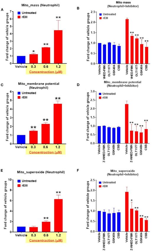
Lien et al.                                                                                                Dengue EIII-Induced Neutrophil NETosis

inhibitor chondroitin sulfate B (CSB, 10 µg/mL; Sigma-Aldrich)           anti-mouse citrulline Histone H3 antibody (1:1000) and 4’,6-
was used to suppress rEIII-induced neutrophil binding and cell           diamidino-2-phenylindole (DAPI, 5 µl/ml). A fluorescence
death. Anti-citrullinated histone H3 (CitH3; citrulline R8), anti-       microscope (Nikon Eclipse E800; Nikon Taiwan, Taipei, Taiwan)
histone H2A family member X (H2AX), and anti-gasdermin D                 (51) was used for obtaining the NETosis images. To analyze
(GSDMD) antibodies (Abcam, Cambridge, UK) were used for                  mitochondria membrane potential, superoxide, and membrane
flow cytometry NETosis analysis.                                         potential levels, mitochondria labeling reagents MitoTrackerTM
                                                                         Green FM (Thermo Fisher Scientific, Waltham, MA, USA),
Experimental Mice                                                        MitoSOXTM Red mitochondrial superoxide indicator (Thermo
Wild-type mice of ages 8–12 weeks in C57BL/6J background were            Fisher Scientific), MitoTrackerTM Red CMXRos (Thermo Fisher
purchased from the National Laboratory Animal Center (Taipei,            Scientific) were used according to the manufacturer’s instructions
Taiwan) (38, 42–46). Gene knockout mice with a C57BL/6J                  (33, 52). A flow cytometer (FACSCalibur; BD Biosciences, San
background, including Nlrp3−/− and Casp1−/− (32), were                   Jose, CA, USA) (36, 38) was used in this study to analyze RCD,
obtained from the Center National de Recherche Scientifique              cell live/death and mitochondria metabolic activities with or
(Orléans, France) (32, 33, 47). All experimental animals were            without rEIII challenges and RCD or signaling pathway (e.g.,
housed in the Animal Center of Tzu-Chi University in a                   PAD4, Nlrp3 inflammasome) inhibitor treatments (33). Levels
specific-pathogen-free, temperature-, and lighting-controlled            of cellular reactive oxygen species (ROS) were analyzed using
environment with free access to filtered water and food. All             2′ ,7′ -dichlorofluorescin diacetate (Sigma-Aldrich) staining-flow
genetic knockout strains were backcrossed with the wild-type             cytometry analysis.
C57Bl/6J mice for at least 6 generations. After challenged with
vehicle (saline), BSA (a control protein, 2 mg/kg), DENV (1.2
× 107 PFU /kg; DHF viral load) and rEIII (2 mg/kg; a dosage              Analyses of Regulated Cell Death
equivalent to 1.2 × 10⁷ PFU/kg), experimental mice were                  To analyze DENV or rEIII induced neutrophil cell death, mouse
immediately rescued with or without Nlrp3 inhibitor OLT1177              neutrophils were incubated with DENV or rEIII for 1 h and
treatments (50 mg/kg). Plasma levels of IL-1β, TNF-α, and CitH3          then subjected to flow cytometry analyses after washed with
of the experimental mice were determined through enzyme-                 PBS. Various regulated cell death (RCD) responses, including
linked immunosorbent assay (ELISA) (IL-1β, TNF-α, Biolegend;             apoptosis (CaspGLOWTM Red Active Caspase-3 Staining Kit,
CitH3, Cayman Chemical, Ann Arbor, MI, USA) 1 d after rEIII              #K193, BioVision, Milpitas, CA, USA), autophagy (Cyto-IDTM
treatments; neutrophils were isolated and analyzed (see following        Autophagy Detection Kit, Enzo Life Sciences, #ENZ51031,
“Analyses of neutrophils”).                                              Farmingdale, NY, USA), ferroptosis (C11 BODIPY 581/591,
                                                                         #27086, Cayman Chemical), necroptosis (RIP3/B-2 alexa Fluor
                                                                         488, Santa Cruz Biotechnology, #sc-374639 AF488, Santa
Ethics Statement                                                         Cruz, CA, USA), pyroptosis (Caspase-1 Assay, Green, #9146,
The animal experiments in this report were conducted in                  ImmunoChemistry Technologies, MI, USA), and live/dead
agreement with National (Taiwan Animal Protection Act, 2008)             cell labeling (Zombie NIRTM Fixable Viability Kit, #423105,
directive for protection of laboratory animals. All experimental         Biolegend), were analyzed using respective cell labeling reagents
protocols for examining the experimental animals were approved           (30 min in PBS). Treatments (1 h) of cell death inducers
by the Animal Care and Use Committee of Tzu-Chi University,              were used as positive controls for various type of regulated
Hualien, Taiwan (approval ID: 101019).                                   cell death (RCD; apoptosis: doxorubicin, 2.5 µg/mL, Nang
                                                                         Kuang Pharmaceutical, Taipei, Taiwan; autophagy: rapamycin,
Analyses of Neutrophils                                                  250 nM, #R0395, Sigma-Aldrich; ferroptosis: erastin, 10 µM,
Blood samples of mice were collected via the retro-orbital               #17754, Cayman Chemical; necroptosis, TNF-α, 2 ng/mL,
venous plexus using plain capillary tubes (Thermo Fisher                 #575202, Biolegend; pyroptosis: nigericin, 3.5 µM, #6698,
Scientific, Waltham, MA, USA), and then transferred into                 ImmunoChemistry Technologies, Minnesota, USA; NETosis,
polypropylene tubes (Eppendorf; Thermo Fisher Scientific)                TPA, #P8139, 2 nM Sigma-Aldrich) (30 min in PBS). Inhibitors
containing anticoagulant acid-citrate-dextrose solution (ACD;            were used to address the involvements of specific RCD pathways
38 mM citric acid, 75 mM sodium citrate, 100 mM dextrose)                (apoptosis: Z-DEVD-FMK, 10 µM, #FMK004, R&D Systems,
(48, 49). Following previously described methods (50), mouse             Indianapolis, IN, USA; autophagy: Chloroquine diphosphate,
neutrophils were purified from mouse blood samples using                 60 µM, #C6628, Sigma-Aldrich; ferroptosis: Ferrostatin-1
Ficoll-Paque (Ficoll-Paque Plus, 1.077 g/mL, GE Healthcare,              2.5 µM, #17729, Cayman Chemical; necroptosis: Necrostatin-1,
Chicago, IL, USA) and dextran (Sigma-Aldrich) sedimentation              50µM, #11658, Cayman Chemical; pyroptosis: Z-WHED-FMK
(3% w/v) density gradient centrifugation and red blood cell              10 µM, #FMK002, R&D Systems; pyroptosis/GSDMD: dimethyl
lysis. To obtain fluorescent NET images, mouse neutrophils (1            fumarate (DMF), 25–100 µM, #14714, Sigma–Aldrich; NETosis:
× 105 ) were treated with vehicle (the diluent, normal saline,           peptidyl arginine deiminase 4 (PAD4) inhibitor GSK484, 10 µM,
0.9 % NaCl), rEIII (50 µg/mL or an equivalent dose 0.6 µM),              #17488, Sigma–Aldrich; Nlrp3: OLT1177, 10 µM, #24671,
DENV (1 × 10⁵ PFU/mL, an equivalent dose of rEIII is 0.6 µM)             Cayman Chemical; EIII competitive blocker: Chondroitin
or 12-O-tetradecanoylphorbol-13-acetate (TPA, 2 nM, Sigma-               sulfate B, CSB, 10 µg/mL, #C3788, Sigma–Aldrich; 30 min
Aldrich) for 2 h at 37C. After fixation by 4% paraformaldehyde           pretreatments before addition of DENV, rEIII and cell-death
on coverslips, these neutrophils were then stained with rabbit           inducers according to the manufacturer’s instructions).

Frontiers in Immunology | www.frontiersin.org                        3                                    March 2021 | Volume 12 | Article 618577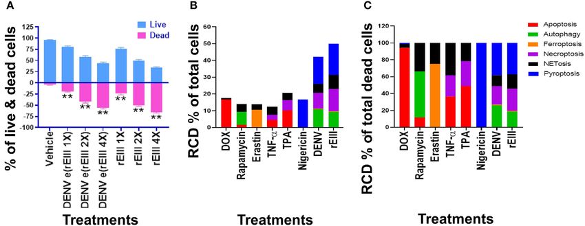
Lien et al. Dengue EIII-Induced Neutrophil NETosis
Statistical Analyses DENV and rEIII Induce Multiple Regulated
In this report, the means, standard deviations, and statistics Cell Death Pathways of Neutrophils
of the quantifiable data were calculated using the SigmaPlot NETosis is a type of regulated cell death (RCD) (58).
10 and SPSS 17 software packages. Significance of the Previous reports suggested that neutrophil RCD exacerbate
data was examined using one-way ANOVA, followed by pathogenesis in infectious diseases (59, 60). As a result, we
the post hoc Bonferroni-corrected t-test. The probability of would like to investigate whether various RCD pathways
type-1 error α = 0.05 was recognized as the threshold of are also involved in DENV- and rEIII-induced neutrophil
statistical significance. cell death.
We first found that DENV and rEIII induced neutrophil
cell death in a dose dependent manner (Figure 3A). Overall,
RESULTS various cell death inducers, including doxorubicin (apoptosis)
(61, 62), rapamycin (autophagy) (63), erastin (ferroptosis) (64),
Nlrp3 Inflammasome Is Involved in rEIII TNF-α (necroptosis) (65, 66), nigericin (pyroptosis) (67), served
Induced Neutrophil NETosis as positive control agents to induce respective RCD pathways
Flow cytometry analysis of NETosis markers citrullinated histone of the tested neutrophils (Figures 3B,C, dead cell population
H3 (CitH3) (53, 54) and histone H2A family member X (H2AX) adjusted to 100%; Supplementary Figure 2, flow cytometry
(55, 56) revealed that rEIII is sufficient to initiate NETosis gating and calculation). Notably, when compared with cell
in neutrophils; the potency of rEIII is comparable to the death agonists, DENV and rEIII treatment induced considerable
classical NETosis inducer phorbol ester (18, 57) (Figure 1A, pyroptosis, necroptosis, autophagy and NETosis responses in the
Supplementary Figure 1, flow cytometry gating; Figure 1B). In neutrophils, while only minor or no ferroptosis and apoptosis
addition, such induction of NETosis can be suppressed by levels (Figure 3B, % of total cells; Figure 3C, % of total dead
treatments of inflammasome/caspase 1 inhibitor Z-WHED- cell). In addition, the cell type specific RCD patterns/profiles
FMK (Figure 1). In agreement with this, we found that, when (CTS-RCDPs) (33) of neutrophil in the DENV-, and rEIII-treated
compared to the neutrophils from wild type mice, Nlrp3 groups were somewhat similar, with pyroptosis exhibiting the
inflammasome-deficient (Nlrp3−/− and Casp1−/− ) neutrophils highest levels in both groups among all tested RCD pathways
displayed relatively low NETosis levels after treated with rEIII (Figure 3B; ∼40%), suggesting that DENV-induced CTS-RCDP
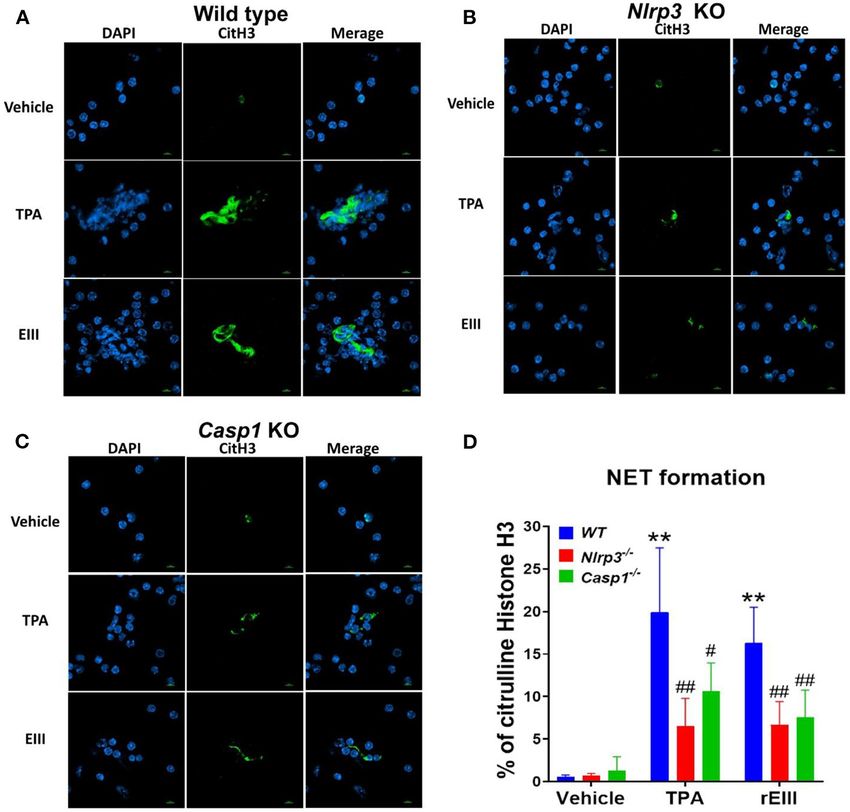
and TPA (Figures 2A–C, cell images; green channels: CitH3, in the neutrophils is likely mediated through EIII on the DENV
a NETosis marker; Figure 2D, quantified results). These data virion. In case one dead cell may display multiple RCDs, here we
suggest that Nlrp3 inflammasome plays critical role in rEIII- defined CTS-RCD as a detection ratio of RCDs in 1 cell type at a
induced NETosis. specific condition.
FIGURE 1 | Essential role of caspase 1 in DENV rEIII-induced NETosis. The gating of flow cytometry analysis of vehicle, 12-O-tetradecanoylphorbol-13-acetate (TPA,
a positive control NETosis inducer; 2 nM) and DENV rEIII (0.6 µM) challenged (1 h) wild mice neutrophils (1 × 10⁵ ) with or without caspase 1 inhibitor Z-WHED-FMK
pretreatments (30 min) (A). The quantified results of flow cytometry analysis, which reveal that TPA and rEIII treatments markedly induced NETosis formation; by
contrast, caspase 1 inhibitor Z-WHED-FMK treatments can only considerably suppress rEIII-induced NETosis, but not TPA-induced NETosis (B). n = 6, **P < 0.01,
vs. untreated controls; ## P < 0.01 vs. vehicle groups.
Frontiers in Immunology | www.frontiersin.org 4 March 2021 | Volume 12 | Article 618577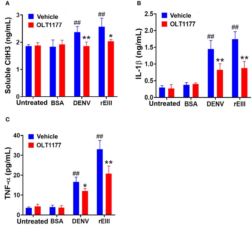
Lien et al.                                                                                                                        Dengue EIII-Induced Neutrophil NETosis

  FIGURE 2 | Essential role of Nlrp3 inflammasome in DENV rEIII-induced NETosis. After challenged (1 h) by TPA (2 nM) and DENV rEIII (0.6 µM), the images of DAPI
  (nucleus DNA staining) and citrullinated histone H3 (CitH3; NETosis marker) staining of NETosis formation of neutrophils from wild type (A), Nlrp3 null (Nlrp3−/− ) (B),
  and caspase 1 null (Casp1−/− ) (C) mouse were showed. The quantified results from flow cytometry are also indicted (D). n = 6, ** P < 0.01, vs. vehicle controls; # P
  < 0.05, ## P < 0.01 vs. wild type (WT) groups. Scale bars: 5 µm.

   An unexpected finding is that the DENV and rEIII                                      with (Figures 4I–O) or without (Figures 4B–H) normalization
induced NETosis only displayed approximately 20% of total                                of dead cell population. These results suggested that pyroptosis
RCDs (Figure 3B); and a classical NETosis inducer TPA (a                                 is the major RCD of rEIII-induced neutrophil death, and
phorbol ester) also induced NETosis about only 40% of                                    which can be rescued by selective inhibitors against Nlrp3
total RCDs (Figure 3B, TPA groups). This let us wondered                                 inflammasome. In addition, Nlrp3 likely involved in neutrophil
whether DENV and EIII-mediated induction of such a low                                   NETosis as treatments of selective inhibitor OLT1177
percentage of NETosis in total RCD, could sufficiently lead to                           rescued total neutrophil death (Figure 4A) and NETosis
neutrophil dysfunction. In addition, we would like to investigate                        (Figures 4H,O). Consistently, treatments of pyroptosis/GSDMD
whether Nlrp3 inflammasome is involved in rEIII-induced                                  inhibitor DMF (68) suppressed EIII-induced neutrophil
neutrophil death. Accordingly, Nlrp3 inhibitor OLT1177 and                               pyroptosis, NETosis, cellular ROS, surface GSDMD levels
inflammasome/caspase1 inhibitor Z-WHED-FMK were used to                                  (Supplementary Figure 4, flow cytometry analyses), and
further characterizations of whether Nlrp3 inflammasome is                               caspase-1 activation (Supplementary Figure 5, colorimetric
involved in respective RCD responses.                                                    assay). Furthermore, despite NETosis displayed only ∼20%
   We found that treatments with inflammasome inhibitors                                 cell death (Figures 4B,I), PAD4 inhibitor GSK484 suppressed
OLT1177 and Z-WHED-FMK both suppressed EIII-induced                                      considerable levels of total neutrophil death (Figure 4A)
neutrophil cell death (Figures 4A,B; Supplementary Figure 3,                             and RCDs including pyroptosis, necroptosis, autophagy,
percentage pie charts of OLT1177 and Z-WHED-FMK                                          and NETosis (Figure 4). These evidences collectively
treatments; equivalent to some data in Figures 4A,B,I).                                  suggesting that that RCDs pyroptosis, necroptosis, and
In addition, OLT1177 and Z-WHED-FMK suppressed                                           autophagy are likely associated to NETosis, and NETosis
pyroptosis     (Figures 4C,J),   necroptosis    (Figures 4D,K),                          is still a critical RCD pathway of neutrophil in response to
autophagy (Figures 4G,N), and NETosis (Figures 4H,O),                                    rEIII exposure.

Frontiers in Immunology | www.frontiersin.org                                        5                                            March 2021 | Volume 12 | Article 618577
Lien et al.                                                                                                                         Dengue EIII-Induced Neutrophil NETosis

  FIGURE 3 | DENV- and rEIII-induced regulated cell death in neutrophils. (A) Wild type mouse neutrophils treated (1 h) with vehicle and various doses of DENV and
  rEIII; the live and death cell populations were revealed by Zombie-NIR Kit labeling and flow cytometry analysis. [rEIII 1× = 0.3 µM, 2× = 0.6 µM, 4× = 1.2 µM,; DENV
  e(rEIII 1×) is a DENV level equivalent to 0.3 µM rEIII, as indicted by the methods described elsewhere (33)]. (B) Treatments (1 h) of regulated cell death (RCD) inducers,
  doxorubicin (DOX; apoptosis) (2.5 µg/mL), rapamycin (autophagy) (0.5 µM), erastin (ferroptosis) (10 µM), TNF-α (necroptosis) (2.5 ng/mL), TPA (NETosis) (2 nM), and
  nigericin (pyroptosis) (3.5 µM) induced relatively simple RCD patterns. By contrast, DENV and rEIII induced multiple RCD pathways, in which pyroptosis is the major
  RCD response, counts ∼40% of total RCD and the NETosis response displays only ∼20% total RCD. (C) If the respective RCDs are normalized by the population of
  death cells (dead cell population normalized to 100%), we can obtain a more similar RCD pattern in DENV and rEIII groups (flow cytometry gating and calculation
  methods described in Supplementary Figure 2). DENV 1 × = 4.2 × 10⁴ PFU/mL, 2 × = 8.4 × 10⁴ PFU/mL, 4 × = 1.7 × 10⁵ PFU/mL; **P < 0.01 vs. vehicle
  groups.

Nlrp3 Inflammasome Deficiency and                                                         inflammasome is a critical target for DENV and EIII to induce
Inhibitor Treatments Rescue DENV- and                                                     neutrophil dysfunction, NETosis, and inflammation.
rEIII- Exacerbated Mitochondria Metabolic
Burden and Inflammation of Neutrophils
Because inflammasome-mediated pyroptosis is a major RCD                                   DISCUSSION
involving in rEIII-induced neutrophil defect, here we would
                                                                                          Inflammasomes, cellular molecular sensors for PAMPs and
like to further investigate whether suppression of neutrophil
                                                                                          DAMPs, are multimeric protein complexes comprising of
Nlrp3 inflammasome through inhibitor treatments is sufficient
                                                                                          NLRs [nucleotide-binding domain (NBD) and leucine-
to ameliorate DENV rEIII-induced neutrophil defects. Here
                                                                                          rich-repeat-(LRR)-containing], the absent in melanoma-2
we found that treatments of rEIII-increased mitochondria
                                                                                          (AIM2)-like receptors (ALRs), an adaptor molecule ASC
mass (Figure 5A), membrane potential (Figure 5C) and
                                                                                          (apoptosis-associated speck-like protein containing a CARD),
superoxide (Figure 5E) levels in a dose dependent manner,
                                                                                          and procaspase-1 (14, 25, 69, 70). Activation of inflammasomes
while treatments of Nlrp3 inflammasome inhibitors OLT1177
                                                                                          leads to protein-cleavage processes, turning pro-caspase 1 into
and Z-WHED-FMK ameliorated such metabolic burden of
                                                                                          active form caspase 1, which converts pro-IL-1β and pro-IL-18
neutrophil mitochondria (Figures 5B,D,F). Levels of caspase-1                             into respective active forms (IL-1β and IL-18) (69). Uncontrolled
activation in the neutrophils were analyzed and confirmed in                              inflammasome activation exacerbates autoimmune and excessive
parallel using colorimetric assay (Supplementary Figure 5).                               inflammatory pathogenesis in infectious diseases, despite the
In agreement with this, mouse experiments further revealed                                adequate levels of inflammasome activation can help defending
that, treatments of Nlrp3 inflammasome inhibitors OLT1177                                 the pathogens (71). As a result, selective inhibitors against
markedly ameliorated rEIII- induced elevation of circulating                              inflammasomes have been developed for treating various
soluble CitH3 (Figure 6A), IL-1β (Figure 6B), and TNF-α                                   inflammatory and infectious diseases, and have got promising
(Figure 6C) levels in mice. Consistently, compared to wild                                advancements (70, 72). High circulating levels of IL-1β and
type mice, neutrophils from Nlrp3−/− and Casp1−/− mutant                                  IL-18 have been associated DHF (27, 73), suggesting a critical
mice displayed markedly reduced levels of total mitochondria                              role of inflammasomes in DENV-induced pathogenesis. Mouse
ROS (Figure 7A), hydrogen peroxide (Figure 7B), superoxide                                experiments further revealed that DENV induced NET formation
(Figure 7C) after in vitro treatments of rEIII. Similarly,                                and inflammasome activation was impaired in neutrophils from
rEIII treatments markedly induced circulating CitH3 in wild                               CLEC5A−/− and TLR2−/− mutants (74). DENV-induced NET
type mice, but not in Nlrp3−/− and Casp1−/− mutant mice                                   leads to endothelial cell damage and vascular leakage . Evidences
(Figure 7D). These results collective suggest that EIII is a                              have suggested that NET formation contributes significantly
virulence factor to induce neutrophil defects, and Nlrp3                                  in disease pathogenesis, and NET components were more

Frontiers in Immunology | www.frontiersin.org                                         6                                            March 2021 | Volume 12 | Article 618577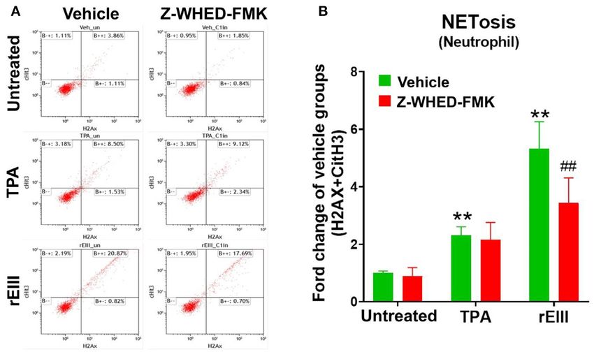
Lien et al.                                                                                                                     Dengue EIII-Induced Neutrophil NETosis

  FIGURE 4 | Protection of neutrophils from rEIII-induced pyroptosis under treatment with Nlrp3 inflammasome inhibitors. Pre-treatments (30 min) with NETosis inhibitor
  GSK484 (2 nM), Nlrp3 inhibitor OLT1177 (10 µM), and caspase 1 inhibitor Z-WHED-FMK (10 µM) on the rescue of rEIII-induced neutrophil total cell death (A).
  Pretreatments (30 min) with Nlrp3 inflammasome inhibitors OLT1177, Z-WHED-FMK, and NETosis inhibitor GSK484 rescued rEIII-induced (1 h) neutrophil pyroptosis
  (B,C), necroptosis (D), autophagy (G) and NETosis (H), but not ferroptosis and apoptosis (E,F). If the respective RCD% was normalized by the population of death
  cells [(I): dead cell population normalized to 100%], we found that CSB, OLT1177 and Z-WHED-FMK still display rescue effects on pyroptosis (J), necroptosis (K),
  autophagy (N), and NETosis (O), but not ferroptosis and apoptosis (L,M). Chondroitin sulfate B (CSB, 10 µg/mL), a competitive inhibitor against rEIII binding (33),
  serving as a positive inhibitor control to inhibit cell death. n = 6, *P < 0.05, **P < 0.01, vs. respective vehicle groups. ND, not detected.

Frontiers in Immunology | www.frontiersin.org                                      7                                           March 2021 | Volume 12 | Article 618577
Lien et al.                                                                                                                     Dengue EIII-Induced Neutrophil NETosis

  FIGURE 5 | DENV rEIII-induced mitochondria ROS production is ameliorated by Nlrp3 inflammasome and NETosis inhibitor. The rEIII (0.6 µM)-induced (1 h) elevations
  of neutrophil mitochondria mass (A) membrane potential (C) and superoxide (E) levels in a dose dependent manner. The induction of these mitochondria metabolic
  burdens in the neutrophils could be suppressed by treatments of Nlrp3 and caspase 1 inhibitors Z-WHED-FMK (10 µM) and OLT1177 (10 µM), NETosis inhibitor
  GSK484 (10 µM), and the cell-binding competitive inhibitor chondroitin sulfate B (CSB, 10 µg/mL), respectively (B,D,F). n = 6, *P < 0.05, **P < 0.01, vs. respective
  untreated vehicle groups (A,C,E); *P < 0.05, **P < 0.01, vs. respective vehicle groups (B,D,F).

Frontiers in Immunology | www.frontiersin.org                                      8                                           March 2021 | Volume 12 | Article 618577
Lien et al.                                                                                                                      Dengue EIII-Induced Neutrophil NETosis

  FIGURE 6 | Nlrp3 inhibitor OLT1177 protects mice from DENV- and rEIII-induced NETosis and IL-1β, TNF-α production in mice. After challenged with vehicle (saline),
  BSA (a control protein) 2 mg/kg), DENV (1.2 × 10⁷ PFU /kg; DHF viral load) and rEIII (2 mg/kg; a dosage equivalent to DENV 1.2 × 10⁷ PFU/kg), experimental mice
  were immediately rescued with or without Nlrp3 inhibitor OLT1177 treatments (50 mg/kg). The ELISA analyzed plasma levels of circulating soluble citrullinated histone
  H3 (CitH3; a NETosis marker) (A), circulating IL-1β levels (B), and TNF-α levels (C) after 1 d treatments were showed. n = 6, ## P < 0.01, vs. respective untreated
  groups; *P < 0.05, **P < 0.01, vs. respective vehicle groups.

predominantly displayed in serum samples of DHF patients (75).                          example, GSDMD, an effector protein of pyroptosis, plays
Despite of these findings, the mechanism underlining DENV-                              a critical role in the generation of NET (79). Consistently,
induced NETosis formation is not fully understood; particularly,                        here we found that treatments of GSDMD inhibitor DMF
the viral factor leading to NETosis remains elusive. Here we                            drastically reduced EIII-induced pyroptosis and NETosis in
found that, the DENV EIII could be a potential virulence fact                           neutrophils (Supplementary Figure 4). Accordingly, if Nlrp3
that elicits abnormal neutrophil responses and NETosis. Because                         inflammasome-activated GSDMD can further enhance NET
DENV and EIII induced similar RCD patterns of neutrophils                               formation, it is reasonable to observe an inhibition of NETosis
(Figure 3B, DENV and rEIII groups), suggesting that, during                             using inhibitors from a pyroptosis pathway. Second, the cross-
the speak viremia stage, high levels of DENV virion conduct                             talk may also explain by a model of intercellular regulations
the cytotoxicity to enhance NETosis through EIII moiety. This                           between macrophage and neutrophil. For example, the NET is
is partly consistent with the findings that NET formation is                            able to prime macrophages to produce IL-1 and IL-18 through
markedly increased in neutrophils isolated from dengue patients                         the Nlrp3 inflammasome, thus amplifying the inflammatory
during the acute phase of the infection (75). Additionally, our                         response (80). At the same time, IL-18 effectively stimulated
data suggests that such cell-type-specific RCD patterns may be                          NET release and caspase-1 activation in primed macrophages
a useful analysis method on the functional characterization of                          compared to IL-18 alone (80). This suggests a feed-forward
biologically drugs and hazardous materials at the cellular levels.                      loop that NET increase the production of IL-1β and IL-18 in
   Inflammasomes regulate and interact with various RCDs (72,                           macrophages, which in turn can stimulate NET formation in
76–78). The inhibitors of either Nlrp3 inflammasome/pyroptosis                          neutrophils (80). These evidences may explain our observations
(inhibitor OLT1177) or NETosis (inhibitor GSK484) can                                   that treatments of Nlrp3 inhibitor OLT1177 can markedly
suppress to each other (Figure 4), suggests there could exist                           suppressed rEIII-induced neutrophil pyroptosis and NETosis
a cross-talk between these 2 pathways. First, such cross-                               in vitro (Figure 4), and markedly suppressed rEIII-induced
talk could be regulated at an intracellular signaling level; for                        NETosis and inflammation in mice (Figure 6). However, as these

Frontiers in Immunology | www.frontiersin.org                                       9                                           March 2021 | Volume 12 | Article 618577
Lien et al.                                                                                                                          Dengue EIII-Induced Neutrophil NETosis

  FIGURE 7 | Nlrp3 and caspase 1 deficiencies protest rEIII-induced neutrophil ROS production and NETosis. Treatments (1 h) of rEIII (0.6 µM)-induced elevations of
  neutrophil cellular reactive oxygen species (ROS) (A) hydrogen peroxide (B), superoxide (C) in vitro. Treatments (1 d) of rEIII (2 mg/kg)-induced elevations of circulating
  CitH3 (D) levels in wild type mice. By contrast, such rEIII (0.6 µM in vitro, 2 mg/kg in mice)-induced stimulations are markedly reduced in Nlrp3−/− and Casp1−/−
  gene knockout mice in both in vitro and in vivo experiments (A–D). n = 6, # P < 0.05, ## P < 0.01, vs. respective wild type (WT) groups; *P < 0.05, **P < 0.01, vs.
  respective vehicle groups.

regulations still not been clearly demonstrated in dengue, more                            and DC-SIGN, but not CLEC2 and DC-SIGNR can block
detailed mechanism is worth of further investigations.                                     rEIII-neutrophil binding (Supplementary Figure 6). In addition,
   As a glycosaminoglycan (GAG) binding lectin (81), EIII                                  CLEC5A and DC-SIGN can reduce EIII-induced increased ROS
could have multiple neutrophil surface targets. Recent evidences                           and NETosis levels in neutrophils (Supplementary Figure 6).
have suggested that lectin DC-SIGN mediated DENV infection                                 These evidences collectively suggested that CLEC5A and DC-
in dendritic cells (82); lectins CLEC2 and CLEC5A plays                                    SIGN are cellular targets of EIII on neutrophils. As a critical
critical roles on DENV-induced inflammation (24, 74, 83,                                   inflammatory regulator, the role of CLEC5A and DC-SIGN in
84). CLEC5A and DC-SIGN are expressed by leukocyte                                         EIII- mediated pathogenesis is worthy of further investigations.
subpopulations (82, 84); while their respective isoforms CLEC2                                Cell population is heterogeneous even in one cell line. This
and DC-SIGNR preferentially expressed by the other cell                                    could be a reason that reports have revealed treatments of
types (84, 85). Here we used recombinant soluble CLEC2,                                    pathogens and cytotoxic agents leading to multiple types of
CLEC5A, DC-SIGN, DC-SIGNR, to perform EIII competition                                     RCDs simultaneously (86–92). For example, when cellular stress
experiments. Analysis results revealed that CLEC5A-, and                                   can activate both receptor-induced lysosomal-dependent, and
DC-SIGN-, but not CLEC2-, and DC-SIGNR-coated beads can                                    mitochondrial-mediated cell death pathways, which will lead
bind to rEIII (Supplementary Figure 6). Consistently, CLEC5A                               to both programmed necrosis and apoptosis (88). Similarly, as

Frontiers in Immunology | www.frontiersin.org                                         10                                            March 2021 | Volume 12 | Article 618577
Lien et al.                                                                                                              Dengue EIII-Induced Neutrophil NETosis

DENV and EIII been reported to have multiple cellular targets,                    abnormal neutrophil activation and cell death. In addition, our
it is reasonable to detect multiple RCDs after DENV and EIII                      data revealed that blockage of Nlrp3 inflammasome, through
challenges. Here, we found that DENV and EIII but not the other                   treatments of inhibitor OLT1177 or gene deficiencies in Nlrp3
RCD inducers, induced a similar RCD pattern in neutrophils                        and caspase 1 expression, protects neutrophils from rEIII-
(Figures 3B,C). Previous reports have suggested that, upon                        induced NETosis (Figures 4, 7) and proinflammatory cytokines
stimulation by PAMP, neutrophil activation results in necrotic                    TNF and IL-1 secretion in mice (Figure 6). These results
RCDs (93, 94); by contrast, neutrophil apoptosis contributes to                   collectively suggest that Nlrp3 inflammasome is a promising
the resolution of inflammation (93–96). Consistently, here we                     target for treating DENV-induced inflammation.
found that DENV and EIII (pathogen, PAMP) preferentially
induced necrotic RCDs with almost no detectable apoptosis                         DATA AVAILABILITY STATEMENT
levels in neutrophils (Figure 4). Although further investigations
are needed, our data suggested that the neutrophil CTS-RCDPs                      The original contributions presented in the study are included
are useful on the characterization of cellular tendency on the                    in the article/Supplementary Material, further inquiries can be
induction of RCDs in particular conditions.                                       directed to the corresponding author/s.
    In addition to EIII, DENV membrane protein (M) and non-
structural proteins NS2A, NS2B were also shown to induce                          ETHICS STATEMENT
inflammasome activation (14, 97, 98), and NS1 was demonstrated
to enhance inflammation through a toll-like receptor 4 (99, 100).                 The animal study was reviewed and approved by Animal Care
NS2A, NS2B have shown to serve as viroporins to increase cell-                    and Use Committee of Tzu-Chi University, Hualien, Taiwan
membrane permeability and activate inflammasome (14, 101,                         (approval ID: 101019).
102). Viroporins primarily affect virus-infected cells (103), and
clinical course of DHF occurs specifically when the viremia                       AUTHOR CONTRIBUTIONS
markedly decreased (104, 105); these evidences suggest that
NS2A, NS2B-mediated inflammasome activations may be not                           H-HC conceptualized and supervised this project and
timely associated with the clinical course of DHF. DENV virus                     wrote this manuscript. T-SL, D-SS, S-CH, and W-SW
particle-expressed EIII and soluble NS1 are detected at high                      performed experiments and analyzed the data. All
levels prior to the acute phase of DHF (104, 105), and may be                     authors contributed to the article and approved the
considered as two virulence factors for severe dengue. Here we                    submitted version.
found that both EIII and NS1 treatments can induce increased
NETosis levels in neutrophils, and EIII has a relative higher                     FUNDING
activity (Supplementary Figure 7; DENV envelop domain I,
domain II protein fragment, served as a control protein). Because                 Ministry of Science and Technology, Taiwan (101-2320-B-320-
the induction of virion-expressed EIII and soluble NS1 are                        004-MY3, 105-2923-B-320-001-MY3, and 107-2311-B-320-
induced in a similar, but not a same time course (105), the                       002-MY3), Tzu-Chi University (TCIRP95002; TCIRP98001;
respective pathogenic role of EIII and NS1 on the elicitation                     and TCIRP101001) and Tzu-Chi Medical Foundation
of DHF-related pathogenesis remains to be further studied.                        (TC-NHRI105-02; TCMMP104-06; TCMMP108-04; and
However, data obtained in the study suggested that virion-                        TCAS-108-01).
associated EIII is a candidate virulence factor that contributes to
dengue-elicited NET formation.
    In this present study, we found that treatments of both                       ACKNOWLEDGMENTS
DENV and EIII, a cell-surface-binding and cytotoxic protein, can
                                                                                  The authors want to thank Professor Yi-Ling Lin, academia
induce multiple cell death pathways in neutrophils with a similar
                                                                                  sinica, for kindly provided DENV-EIII plasmid.
cell-type-specific RCD pattern (Figure 3B). NETosis inhibitor
GSK484 treatments sufficiently suppressed EIII-induced cell
death (Figure 4A), NETosis (Figure 4G), and mitochondria                          SUPPLEMENTARY MATERIAL
metabolic burden (Figure 5) of neutrophils. This suggested
that, despite the NETosis seems to be a relatively minor                          The Supplementary Material for this article can be found
response that counts approximately 20% of total neutrophil                        online at: https://www.frontiersin.org/articles/10.3389/fimmu.
RCDs, the EIII-stimulated NETosis is sufficiently leading to an                   2021.618577/full#supplementary-material

REFERENCES                                                                         2. Gubler DJ. Dengue and dengue hemorrhagic fever. Clin Microbiol Rev.
                                                                                      (1998) 11:480–96. doi: 10.1128/CMR.11.3.480
  1. Pang T, Mak TK, Gubler DJ. Prevention and control of dengue-                  3. Izmirly AM, Alturki SO, Alturki SO, Connors J, Haddad EK. Challenges in
     the light at the end of the tunnel. Lancet Infect Dis. (2017) 17:e79–            dengue vaccines development: pre-existing infections and cross-reactivity.
     87. doi: 10.1016/S1473-3099(16)30471-6                                           Front Immunol. (2020) 11:1055. doi: 10.3389/fimmu.2020.01055

Frontiers in Immunology | www.frontiersin.org                                11                                         March 2021 | Volume 12 | Article 618577
Lien et al. Dengue EIII-Induced Neutrophil NETosis
4. Roth C, Cantaert T, Colas C, Prot M, Casademont I, Levillayer L, et al. A 23. Dinarello CA. Immunological and inflammatory functions of
modified mRNA vaccine targeting immunodominant NS epitopes protects the interleukin-1 family. Annu Rev Immunol. (2009) 27:519–
against dengue virus infection in HLA class I transgenic mice. Front 50. doi: 10.1146/annurev.immunol.021908.132612
Immunol. (2019) 10:1424. doi: 10.3389/fimmu.2019.01424 24. Wu MF, Chen ST, Yang AH, Lin WW, Lin YL, Chen NJ, et al. CLEC5A
5. Waickman AT, Friberg H, Gargulak M, Kong A, Polhemus M, Endy T, is critical for dengue virus-induced inflammasome activation in human
et al. Assessing the diversity and stability of cellular immunity generated macrophages. Blood. (2013) 121:95–106. doi: 10.1182/blood-2012-05-430090
in response to the candidate live-attenuated dengue virus vaccine TAK-003. 25. Schroder K, Tschopp J. The inflammasomes. Cell. (2010) 140:821–
Front Immunol. (2019) 10:1778. doi: 10.3389/fimmu.2019.01778 32. doi: 10.1016/j.cell.2010.01.040
6. Pinto PBA, Assis ML, Vallochi AL, Pacheco AR, Lima LM, Quaresma 26. Bergsbaken T, Fink SL, Cookson BT. Pyroptosis: host cell
KRL, et al. T cell responses Induced by DNA vaccines based on death and inflammation. Nat Rev Microbiol. (2009) 7:99–
the DENV2 E and NS1 proteins in mice: importance in protection 109. doi: 10.1038/nrmicro2070
and immunodominant epitope identification. Front Immunol. (2019) 27. Bozza FA, Cruz OG, Zagne SM, Azeredo EL, Nogueira RM, Assis EF,
10:1522. doi: 10.3389/fimmu.2019.01522 et al. Multiplex cytokine profile from dengue patients: MIP-1beta and
7. Graham N, Eisenhauer P, Diehl SA, Pierce KK, Whitehead SS, Durbin IFN-gamma as predictive factors for severity. BMC Infect Dis. (2008)
AP, et al. Rapid induction and maintenance of virus-specific CD8(+) 8:86. doi: 10.1186/1471-2334-8-86
TEMRA and CD4(+) TEM cells following protective vaccination 28. Jaiyen Y, Masrinoul P, Kalayanarooj S, Pulmanausahakul R, Ubol S.
against dengue virus challenge in humans. Front Immunol. (2020) Characteristics of dengue virus-infected peripheral blood mononuclear cell
11:479. doi: 10.3389/fimmu.2020.00479 death that correlates with the severity of illness. Microbiol Immunol. (2009)
8. Subramaniam KS, Lant S, Goodwin L, Grifoni A, Weiskopf D, Turtle 53:442–50. doi: 10.1111/j.1348-0421.2009.00148.x
L. Two is better than one: evidence for T-cell cross-protection between 29. Netea MG, Kullberg BJ, Van der Meer JW. Circulating cytokines as mediators
dengue and zika and implications on vaccine design. Front Immunol. (2020) of fever. Clin Infect Dis. (2000) 31 (Suppl. 5):S178–84. doi: 10.1086/317513
11:517. doi: 10.3389/fimmu.2020.00517 30. Dinarello CA. Infection, fever, and exogenous and endogenous
9. Kuczera D, Assolini JP, Tomiotto-Pellissier F, Pavanelli WR, Silveira pyrogens: some concepts have changed. J Endotoxin Res. (2004)
GF. Highlights for dengue immunopathogenesis: antibody-dependent 10:201–22. doi: 10.1179/096805104225006129
enhancement, cytokine storm, and beyond. J Interferon Cytokine Res. (2018) 31. Pan P, Zhang Q, Liu W, Wang W, Yu Z, Lao Z, et al. dengue virus infection
38:69–80. doi: 10.1089/jir.2017.0037 activates interleukin-1beta to induce tissue injury and vascular leakage. Front
10. Rothman AL. Immunity to dengue virus: a tale of original antigenic Microbiol. (2019) 10:2637. doi: 10.3389/fmicb.2019.02637
sin and tropical cytokine storms. Nat Rev Immunol. (2011) 11:532– 32. Lien TS, Sun DS, Chang CM, Wu CY, Dai MS, Chan H, et al. Dengue
43. doi: 10.1038/nri3014 virus and antiplatelet autoantibodies synergistically induce haemorrhage
11. Chuang YC, Wang SY, Lin YS, Chen HR, Yeh TM. Re-evaluation of through Nlrp3-inflammasome and FcgammaRIII. Thromb Haemost. (2015)
the pathogenic roles of nonstructural protein 1 and its antibodies during 113:1060–70. doi: 10.1160/TH14-07-0637
dengue virus infection. J Biomed Sci. (2013) 20:42. doi: 10.1186/1423-0127- 33. Lien TS, Sun DS, Wu CY, Chang HH. Exposure to dengue envelope
20-42 protein domain III induces Nlrp3 inflammasome-dependent endothelial
12. Srikiatkhachorn A, Mathew A, Rothman AL. Immune-mediated cytokine dysfunction and hemorrhage in mice. Front Immunol. (2021)
storm and its role in severe dengue. Semin Immunopathol. (2017) 39:563– 12:7251. doi: 10.3389/fimmu.2021.617251
74. doi: 10.1007/s00281-017-0625-1 34. Nguyen NM, Duong BT, Azam M, Phuong TT, Park H, Thuy PTB, et al.
13. Mahmud-Al-Rafat A, Majumder A, Taufiqur Rahman KM, Mahedi Hasan Diagnostic performance of dengue virus envelope domain III in acute
AM, Didarul Islam KM, Taylor-Robinson AW, et al. Decoding the enigma dengue infection. Int J Mol Sci. (2019) 20:3464. doi: 10.3390/ijms20143464
of antiviral crisis: Does one target molecule regulate all? Cytokine. (2019) 35. Khan RA, Afroz S, Minhas G, Battu S, Khan N. Dengue virus
115:13–23. doi: 10.1016/j.cyto.2018.12.008 envelope protein domain III induces pro-inflammatory signature
14. Shrivastava G, Valenzuela Leon PC, Calvo E. Inflammasome and triggers activation of inflammasome. Cytokine. (2019)
fuels dengue severity. Front Cell Infect Microbiol. (2020) 123:154780. doi: 10.1016/j.cyto.2019.154780
10:489. doi: 10.3389/fcimb.2020.00489 36. Lin GL, Chang HH, Lien TS, Chen PK, Chan H, Su MT, et al. Suppressive
15. Cloherty APM, Olmstead AD, Ribeiro CMS, Jean F. Hijacking of effect of dengue virus envelope protein domain III on megakaryopoiesis.
lipid droplets by hepatitis C, dengue and zika viruses-from viral Virulence. (2017) 8:1719–31. doi: 10.1080/21505594.2017.13
protein moonlighting to extracellular release. Int J Mol Sci. (2020) 43769
21:7901. doi: 10.3390/ijms21217901 37. Chang HH, Shyu HF, Wang YM, Sun DS, Shyu RH, Tang SS, et al. Facilitation
16. Papayannopoulos V. Neutrophil extracellular traps in immunity and disease. of cell adhesion by immobilized dengue viral nonstructural protein 1 (NS1):
Nat Rev Immunol. (2018) 18:134–47. doi: 10.1038/nri.2017.105 arginine-glycine-aspartic acid structural mimicry within the dengue viral
17. Fuchs TA, Abed U, Goosmann C, Hurwitz R, Schulze I, Wahn V, et al. Novel NS1 antigen. J Infect Dis. (2002) 186:743–51. doi: 10.1086/342600
cell death program leads to neutrophil extracellular traps. J Cell Biol. (2007) 38. Tsai CL, Sun DS, Su MT, Lien TS, Chen YH, Lin CY, et al. Suppressed
176:231–41. doi: 10.1083/jcb.200606027 humoral immunity is associated with dengue nonstructural protein NS1-
18. Yipp BG, Kubes P. NETosis: how vital is it? Blood. (2013) 122:2784– elicited anti-death receptor antibody fractions in mice. Sci Rep. (2020)
94. doi: 10.1182/blood-2013-04-457671 10:6294. doi: 10.1038/s41598-020-62958-0
19. Jorch SK, Kubes P. An emerging role for neutrophil extracellular traps in 39. Wong MS, Sun MT, Sun DS, Chang HH. Visible-Light-Responsive
noninfectious disease. Nat Med. (2017) 23:279–87. doi: 10.1038/nm.4294 antibacterial property of boron-doped titania films. Catalysts. (2020)
20. Wang J, Jiang M, Chen X, Montaner LJ. Cytokine storm and 10:1349. doi: 10.3390/catal10111349
leukocyte changes in mild versus severe SARS-CoV-2 infection: 40. Sun DS, Kau JH, Huang HH, Tseng YH, Wu WS, Chang HH. Antibacterial
review of 3939 COVID-19 patients in China and emerging properties of visible-light-responsive carbon-containing titanium dioxide
pathogenesis and therapy concepts. J Leukoc Biol. (2020) photocatalytic nanoparticles against anthrax. Nanomaterials. (2016)
108:17–41. doi: 10.1002/JLB.3COVR0520-272R 6:237. doi: 10.3390/nano6120237
21. Mozzini C, Girelli D. The role of neutrophil extracellular traps 41. Sun DS, King CC, Huang HS, Shih YL, Lee CC, Tsai WJ, et al. Antiplatelet
in covid-19: only an hypothesis or a potential new field of autoantibodies elicited by dengue virus non-structural protein 1 cause
research? Thromb Res. (2020) 191:26–7. doi: 10.1016/j.thromres.2020. thrombocytopenia and mortality in mice. J Thromb Haemost. (2007) 5:2291–
04.031 9. doi: 10.1111/j.1538-7836.2007.02754.x
22. Chang DM, Shaio MF. Production of interleukin-1 (IL-1) and IL-1 inhibitor 42. Ho YY, Sun DS, Chang HH. Silver nanoparticles protect skin
by human monocytes exposed to dengue virus. J Infect Dis. (1994) 170:811– from ultraviolet B-induced damage in mice. Int J Mol Sci. (2020)
7. doi: 10.1093/infdis/170.4.811 21:7082. doi: 10.3390/ijms21197082
Frontiers in Immunology | www.frontiersin.org 12 March 2021 | Volume 12 | Article 618577Lien et al. Dengue EIII-Induced Neutrophil NETosis
43. Lin YY, Hu CT, Sun DS, Lien TS, Chang HH. Thioacetamide- modulating ATF expression and via the ERK and Akt pathway. PLoS ONE.
induced liver damage and thrombocytopenia is associated with (2014) 9:e106812. doi: 10.1371/journal.pone.0106812
induction of antiplatelet autoantibody in mice. Sci Rep. (2019) 62. Kotamraju S, Konorev EA, Joseph J, Kalyanaraman B. Doxorubicin-induced
9:17497. doi: 10.1038/s41598-019-53977-7 apoptosis in endothelial cells and cardiomyocytes is ameliorated by nitrone
44. Perevedentseva E, Krivokharchenko A, Karmenyan AV, Chang HH, spin traps and ebselen. Role of reactive oxygen and nitrogen species. J Biol
Cheng CL. Raman spectroscopy on live mouse early embryo while Chem. (2000) 275:33585–92. doi: 10.1074/jbc.M003890200
it continues to develop into blastocyst in vitro. Sci Rep. (2019) 63. Chen HR, Chuang YC, Chao CH, Yeh TM. Macrophage migration inhibitory
9:6636. doi: 10.1038/s41598-019-42958-5 factor induces vascular leakage via autophagy. Biol Open. (2015) 4:244–
45. Chan H, Huang HS, Sun DS, Lee CJ, Lien TS, Chang HH. TRPM8 and RAAS- 52. doi: 10.1242/bio.201410322
mediated hypertension is critical for cold-induced immunosuppression in 64. Xiao FJ, Zhang D, Wu Y, Jia QH, Zhang L, Li YX, et al. miRNA-17-92
mice. Oncotarget. (2018) 9:12781–95. doi: 10.18632/oncotarget.24356 protects endothelial cells from erastin-induced ferroptosis through targeting
46. Huang CY, Yu WS, Liu GC, Hung SC, Chang JH, Chang the A20-ACSL4 axis. Biochem Biophys Res Commun. (2019) 515:448–
JC, et al. Opportunistic gill infection is associated with TiO2 54. doi: 10.1016/j.bbrc.2019.05.147
nanoparticle-induced mortality in zebrafish. PLoS ONE. (2021) 65. Sawai H. Characterization of TNF-induced caspase-independent
16:e0247859. doi: 10.1371/journal.pone.0245356 necroptosis. Leuk Res. (2014) 38:706–13. doi: 10.1016/j.leukres.2014.02.002
47. Chang YS, Ko BH, Ju JC, Chang HH, Huang SH, Lin CW. SARS 66. Choi ME, Price DR, Ryter SW, Choi AMK. Necroptosis: a
unique domain (SUD) of severe acute respiratory syndrome coronavirus crucial pathogenic mediator of human disease. JCI Insight. (2019)
induces NLRP3 inflammasome-dependent CXCL10-mediated pulmonary 4:e128834. doi: 10.1172/jci.insight.128834
inflammation. Int J Mol Sci. (2020) 21:3179. doi: 10.3390/ijms21093179 67. Xi H, Zhang Y, Xu Y, Yang WY, Jiang X, Sha X, et al. Caspase-1 inflammasome
48. Sun DS, Ho PH, Chang HH. Soluble P-selectin rescues viper activation mediates homocysteine-induced pyrop-apoptosis in endothelial
venom-induced mortality through anti-inflammatory properties and cells. Circ Res. (2016) 118:1525–39. doi: 10.1161/CIRCRESAHA.116.308501
PSGL-1 pathway-mediated correction of hemostasis. Sci Rep. (2016) 68. Humphries F, Shmuel-Galia L, Ketelut-Carneiro N, Li S, Wang B, Nemmara
6:35868. doi: 10.1038/srep35868 VV, et al. Succination inactivates gasdermin D and blocks pyroptosis. Science.
49. Sun DS, Chang YW, Kau JH, Huang HH, Ho PH, Tzeng YJ, et al. (2020) 369:1633–7. doi: 10.1126/science.abb9818
Soluble P-selectin rescues mice from anthrax lethal toxin-induced mortality 69. Martinon F, Burns K, Tschopp J. The inflammasome: a molecular platform
through PSGL-1 pathway-mediated correction of hemostasis. Virulence. triggering activation of inflammatory caspases and processing of proIL-beta.
(2017) 8:1216–28. doi: 10.1080/21505594.2017.1282027 Mol Cell. (2002) 10:417–26. doi: 10.1016/S1097-2765(02)00599-3
50. Quach A, Ferrante A. The application of dextran sedimentation 70. Swanson KV, Deng M, Ting JP. The NLRP3 inflammasome: molecular
as an initial step in neutrophil purification promotes their activation and regulation to therapeutics. Nat Rev Immunol. (2019) 19:477–
stimulation, due to the presence of monocytes. J Immunol Res. (2017) 89. doi: 10.1038/s41577-019-0165-0
2017:1254792. doi: 10.1155/2017/1254792 71. Lamkanfi M, Dixit VM. Mechanisms and functions of inflammasomes. Cell.
51. Tseng PH, Sie ZL, Liu MC, Lin HS, Yang WY, Lin TY, et al. Identification of (2014) 157:1013–22. doi: 10.1016/j.cell.2014.04.007
two novel small compounds that inhibit liver cancer formation in zebrafish 72. Yang Y, Wang H, Kouadir M, Song H, Shi F. Recent advances in the
and analysis of their conjugation to nanodiamonds to further reduce toxicity. mechanisms of NLRP3 inflammasome activation and its inhibitors. Cell
Adv Ther. (2019) 2:1900105. doi: 10.1002/adtp.201900105 Death Dis. (2019) 10:128. doi: 10.1038/s41419-019-1413-8
52. Mandal JP, Shiue CN, Chen YC, Lee MC, Yang HH, Chang HH, et al. 73. Mustafa AS, Elbishbishi EA, Agarwal R, Chaturvedi UC. Elevated
PKCdelta mediates mitochondrial ROS generation and oxidation of HSP60 levels of interleukin-13 and IL-18 in patients with dengue
to relieve RKIP inhibition on MAPK pathway for HCC progression. Free hemorrhagic fever. FEMS Immunol Med Microbiol. (2001)
Radic Biol Med. (2020) 163:69–87. doi: 10.1016/j.freeradbiomed.2020.12.003 30:229–33. doi: 10.1111/j.1574-695X.2001.tb01575.x
53. Masuda S, Nakazawa D, Shida H, Miyoshi A, Kusunoki Y, Tomaru U, et al. 74. Sung PS, Huang TF, Hsieh SL. Extracellular vesicles from CLEC2-activated
NETosis markers: Quest for specific, objective, and quantitative markers. platelets enhance dengue virus-induced lethality via CLEC5A/TLR2. Nat
Clin Chim Acta. (2016) 459:89–93. doi: 10.1016/j.cca.2016.05.029 Commun. (2019) 10:2402. doi: 10.1038/s41467-019-10360-4
54. Kuczia P, Zuk J, Iwaniec T, Soja J, Dropinski J, Malesa-Wlodzik M, et al. 75. Opasawatchai A, Amornsupawat P, Jiravejchakul N, Chan-In W, Spoerk
Citrullinated histone H3, a marker of extracellular trap formation, is NJ, Manopwisedjaroen K, et al. Neutrophil activation and early features of
increased in blood of stable asthma patients. Clin Transl Allergy. (2020) NET formation are associated with dengue virus infection in human. Front
10:31. doi: 10.1186/s13601-020-00337-8 Immunol. (2018) 9:3007. doi: 10.3389/fimmu.2018.03007
55. Cools-Lartigue J, Spicer J, McDonald B, Gowing S, Chow S, Giannias B, et al. 76. Biasizzo M, Kopitar-Jerala N. Interplay Between NLRP3
Neutrophil extracellular traps sequester circulating tumor cells and promote Inflammasome and Autophagy. Front Immunol. (2020)
metastasis. J Clin Invest. (2013) 123:3446–58. doi: 10.1172/JCI67484 11:591803. doi: 10.3389/fimmu.2020.591803
56. Tohme S, Yazdani HO, Al-Khafaji AB, Chidi AP, Loughran P, Mowen 77. Zhen Y, Zhang H. NLRP3 inflammasome and inflammatory bowel disease.
K, et al. Neutrophil extracellular traps promote the development and Front Immunol. (2019) 10:276. doi: 10.3389/fimmu.2019.00276
progression of liver metastases after surgical stress. Cancer Res. (2016) 78. Yuk JM, Silwal P, Jo EK. Inflammasome and mitophagy connection in health
76:1367–80. doi: 10.1158/0008-5472.CAN-15-1591 and disease. Int J Mol Sci. (2020) 21:4714. doi: 10.3390/ijms21134714
57. Takei H, Araki A, Watanabe H, Ichinose A, Sendo F. Rapid killing of 79. Sollberger G, Choidas A, Burn GL, Habenberger P, Di Lucrezia
human neutrophils by the potent activator phorbol 12-myristate 13-acetate R, Kordes S, et al. Gasdermin D plays a vital role in the
(PMA) accompanied by changes different from typical apoptosis or necrosis. generation of neutrophil extracellular traps. Sci Immunol. (2018)
J Leukoc Biol. (1996) 59:229–40. doi: 10.1002/jlb.59.2.229 3:eaar6689. doi: 10.1126/sciimmunol.aar6689
58. Tang D, Kang R, Berghe TV, Vandenabeele P, Kroemer G. The 80. Bonaventura A, Vecchie A, Abbate A, Montecucco F. Neutrophil
molecular machinery of regulated cell death. Cell Res. (2019) 29:347– extracellular traps and cardiovascular diseases: an update. Cells. (2020)
64. doi: 10.1038/s41422-019-0164-5 9:231. doi: 10.3390/cells9010231
59. Smith H, Rogers SL, Smith HV, Gillis D, Siskind V, Smith JA. Virus-associated 81. Watterson D, Kobe B, Young PR. Residues in domain III of the dengue
apoptosis of blood neutrophils as a risk factor for invasive meningococcal virus envelope glycoprotein involved in cell-surface glycosaminoglycan
disease. J Clin Pathol. (2013) 66:976–81. doi: 10.1136/jclinpath-2013-201579 binding. J Gen Virol. (2012) 93:72–82. doi: 10.1099/vir.0.03
60. Colamussi ML, White MR, Crouch E, Hartshorn KL. Influenza A virus 7317-0
accelerates neutrophil apoptosis and markedly potentiates apoptotic effects 82. Tassaneetrithep B, Burgess TH, Granelli-Piperno A, Trumpfheller C, Finke
of bacteria. Blood. (1999) 93:2395–403. doi: 10.1182/blood.V93.7.2395 J, Sun W, et al. DC-SIGN (CD209) mediates dengue virus infection of
61. Chen YL, Tsai YT, Lee CY, Lee CH, Chen CY, Liu CM, et al. Urotensin II human dendritic cells. J Exp Med. (2003) 197:823–9. doi: 10.1084/jem.20
inhibits doxorubicin-induced human umbilical vein endothelial cell death by 021840
Frontiers in Immunology | www.frontiersin.org 13 March 2021 | Volume 12 | Article 618577Lien et al. Dengue EIII-Induced Neutrophil NETosis
83. Chen ST, Lin YL, Huang MT, Wu MF, Cheng SC, Lei HY, et al. CLEC5A 97. Pan P, Zhang Q, Liu W, Wang W, Lao Z, Zhang W, et al. Dengue virus
is critical for dengue-virus-induced lethal disease. Nature. (2008) 453:672– m protein promotes NLRP3 inflammasome activation to induce vascular
6. doi: 10.1038/nature07013 leakage in mice. J Virol. (2019) 93:e00996–19. doi: 10.1128/JVI.00996-19
84. Sung PS, Hsieh SL. CLEC2 and CLEC5A: pathogenic host 98. Shrivastava G, Visoso-Carvajal G, Garcia-Cordero J, Leon-Juarez M, Chavez-
factors in acute viral infections. Front Immunol. (2019) Munguia B, Lopez T, et al. Dengue virus serotype 2 and its non-structural
10:2867. doi: 10.3389/fimmu.2019.02867 proteins 2A and 2B activate NLRP3 inflammasome. Front Immunol. (2020)
85. Soilleux EJ. DC-SIGN (dendritic cell-specific ICAM-grabbing non-integrin) 11:352. doi: 10.3389/fimmu.2020.00352
and DC-SIGN-related (DC-SIGNR): friend or foe? Clin Sci. (2003) 104:437– 99. Beatty PR, Puerta-Guardo H, Killingbeck SS, Glasner DR, Hopkins K,
46. doi: 10.1042/cs1040437 Harris E. Dengue virus NS1 triggers endothelial permeability and vascular
86. Chien H, Dix RD. Evidence for multiple cell death pathways during leak that is prevented by NS1 vaccination. Sci Transl Med. (2015)
development of experimental cytomegalovirus retinitis in mice with 7:304ra141. doi: 10.1126/scitranslmed.aaa3787
retrovirus-induced immunosuppression: apoptosis, necroptosis, and 100. Modhiran N, Watterson D, Muller DA, Panetta AK, Sester DP, Liu L,
pyroptosis. J Virol. (2012) 86:10961–78. doi: 10.1128/JVI.01275-12 et al. Dengue virus NS1 protein activates cells via Toll-like receptor 4
87. Panzarini E, Inguscio V, Dini L. Timing the multiple cell death pathways and disrupts endothelial cell monolayer integrity. Sci Transl Med. (2015)
initiated by rose Bengal acetate photodynamic therapy. Cell Death Dis. (2011) 7:304ra142. doi: 10.1126/scitranslmed.aaa3863
2:e169. doi: 10.1038/cddis.2011.51 101. Shrivastava G, Garcia-Cordero J, Leon-Juarez M, Oza G, Tapia-Ramirez
88. Moeckel GW. Hypertonic stress and cell death. Focus on “Multiple J, Villegas-Sepulveda N, et al. NS2A comprises a putative viroporin of
cell death pathways are independently activated by lethal hypertonicity dengue virus 2. Virulence. (2017) 8:1450–6. doi: 10.1080/21505594.2017.13
in renal epithelial cells”. Am J Physiol Cell Physiol. (2013) 305:C1009– 56540
10. doi: 10.1152/ajpcell.00263.2013 102. Leon-Juarez M, Martinez-Castillo M, Shrivastava G, Garcia-Cordero J,
89. Choi SY, Lee-Kwon W, Lee HH, Lee JH, Sanada S, Kwon HM. Multiple Villegas-Sepulveda N, Mondragon-Castelan M, et al. Recombinant Dengue
cell death pathways are independently activated by lethal hypertonicity virus protein NS2B alters membrane permeability in different membrane
in renal epithelial cells. Am J Physiol Cell Physiol. (2013) 305:C1011– models. Virol J. (2016) 13:1. doi: 10.1186/s12985-015-0456-4
20. doi: 10.1152/ajpcell.00384.2012 103. Nieva JL, Madan V, Carrasco L. Viroporins: structure and biological
90. Chen Y, Hua Y, Li X, Arslan IM, Zhang W, Meng G. Distinct types of cell functions. Nat Rev Microbiol. (2012) 10:563–74. doi: 10.1038/nrmicro2820
death and the implication in diabetic cardiomyopathy. Front Pharmacol. 104. Halstead SB. Dengue. Lancet. (2007) 370:1644–
(2020) 11:42. doi: 10.3389/fphar.2020.00042 52. doi: 10.1016/S0140-6736(07)61687-0
91. Korsnes MS. Yessotoxin as a tool to study induction of multiple cell death 105. Muller DA, Depelsenaire AC, Young PR. Clinical and laboratory
pathways. Toxins. (2012) 4:568–79. doi: 10.3390/toxins4070568 diagnosis of dengue virus infection. J Infect Dis. (2017) 215:S89–
92. Miller C, Kennington L, Cooney R, Kohjimoto Y, Cao LC, Honeyman 95. doi: 10.1093/infdis/jiw649
T, et al. Oxalate toxicity in renal epithelial cells: characteristics
of apoptosis and necrosis. Toxicol Appl Pharmacol. (2000) Conflict of Interest: The authors declare that the research was conducted in the
162:132–41. doi: 10.1006/taap.1999.8835 absence of any commercial or financial relationships that could be construed as a
93. Lawrence SM, Corriden R, Nizet V. How neutrophils meet their end. Trends potential conflict of interest.
Immunol. (2020) 41:531–44. doi: 10.1016/j.it.2020.03.008
94. Brostjan C, Oehler R. The role of neutrophil death in chronic inflammation Copyright © 2021 Lien, Sun, Hung, Wu and Chang. This is an open-access article
and cancer. Cell Death Discov. (2020) 6:26. doi: 10.1038/s41420-020-0255-6 distributed under the terms of the Creative Commons Attribution License (CC BY).
95. Serhan CN, Savill J. Resolution of inflammation: the beginning programs the The use, distribution or reproduction in other forums is permitted, provided the
end. Nat Immunol. (2005) 6:1191–7. doi: 10.1038/ni1276 original author(s) and the copyright owner(s) are credited and that the original
96. Iba T, Hashiguchi N, Nagaoka I, Tabe Y, Murai M. Neutrophil cell death in publication in this journal is cited, in accordance with accepted academic practice.
response to infection and its relation to coagulation. J Intensive Care. (2013) No use, distribution or reproduction is permitted which does not comply with these
1:13. doi: 10.1186/2052-0492-1-13 terms.
Frontiers in Immunology | www.frontiersin.org 14 March 2021 | Volume 12 | Article 618577You can also read