Short-term radiofrequency exposure from new generation mobile phones reduces EEG alpha power with no effects on cognitive performance - bioRxiv
←
→
Page content transcription
If your browser does not render page correctly, please read the page content below

bioRxiv preprint first posted online Jul. 6, 2018; doi: http://dx.doi.org/10.1101/363929. The copyright holder for this preprint
(which was not peer-reviewed) is the author/funder, who has granted bioRxiv a license to display the preprint in perpetuity.
                             It is made available under a CC-BY-NC-ND 4.0 International license.

    Short-term radiofrequency exposure from new generation
    mobile phones reduces EEG alpha power with no effects on
    cognitive performance

    Zsuzsanna Vecsei1,2, Balázs Knakker2,3, Péter Juhász1, György Thuróczy1, Attila Trunk2,3, István
    Hernádi2,3,4, CA
    1
     Department of Non-ionizing Radiation, National Public Health Institute, Budapest, Hungary
    2
     Department of Experimental Zoology and Neurobiology, Faculty of Sciences, University of Pécs, Hungary
    3
     Szentágothai Research Center, University of Pécs, Hungary and Centre for Neuroscience, University of Pécs,
    Hungary
    4
     Institute of Physiology, Medical School, University of Pécs, Hungary

    Corresponding author’s (CA) address:
    University of Pécs, Department of Experimental Zoology and Neurobiology
    Ifjúság útja 6, H-7624, Pécs, Hungary
    Phone: +36 72 503607, Fax: +36 72 501517
    E-mail: hernadi.istvan@pte.hu

          1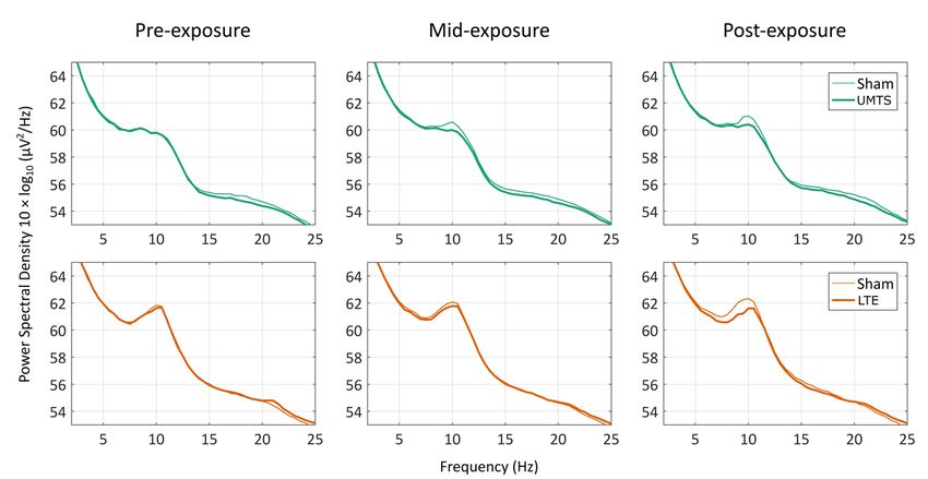
bioRxiv preprint first posted online Jul. 6, 2018; doi: http://dx.doi.org/10.1101/363929. The copyright holder for this preprint
(which was not peer-reviewed) is the author/funder, who has granted bioRxiv a license to display the preprint in perpetuity.
                             It is made available under a CC-BY-NC-ND 4.0 International license.

          Abstract
          Although mobile phone (MP) use has been steadily increasing in the last decades and similar
    positive trends are expected for the near future, systematic investigations on neurophysiological and
    cognitive effects caused by recently developed technological standards for MPs are scarcely
    available. Here, we investigated the effects of radiofrequency (RF) fields emitted by new-generation
    mobile technologies, specifically, Universal Mobile Telecommunications System (UMTS) and Long-
    Term Evolution (LTE), on intrinsic scalp EEG activity in the alpha band (8–12 Hz) and cognitive
    performance in the Stroop test. The study involved 60 healthy, young-adult university students (34
    for UMTS and 26 for LTE) with double-blind administration of Real and Sham exposure in separate
    sessions. EEG was recorded before, during and after RF exposure, and Stroop performance was
    assessed before and after EEG recording. Both RF exposure types caused a notable decrease in the
    alpha power over the whole scalp that persisted even after the cessation of the exposure, whereas
    no effects were found on any aspects of performance in the Stroop test. The results imply that the
    brain networks underlying global alpha oscillations might require minor reconfiguration to adapt to
    the local biophysical changes caused by focal RF exposure mimicking MP use.

          Introduction
          The worldwide use of mobile phones (MPs) is still rapidly growing. By 2020, almost three-
    quarters of the world’s population—or 5.7 billion people—will subscribe to mobile services.1 When
    MPs are used in close proximity to the head, it absorbs a considerable portion of the energy of
    radiofrequency (RF) output power.2 To date, research does not offer any consistent evidence
    concerning the adverse health effects from short-term exposure to RF fields at levels below those
    that cause tissue heating.3,4 However, given the large number of MP users, it is important to
    investigate, understand and monitor any potential public health impact5 they may pose. Owing to
    this, there has been an increasing concern about the effects of RF exposure emitted by MPs on
    psychophysiological and cognitive indices of brain function.
          Third-generation (3G) MPs use Wideband Code Division Multiple Access (WCDMA) standard RF
    signals, also called Universal Mobile Telecommunication System (UMTS), which operate in the
    frequency range of 1920 to 2170 MHz. The WCDMA modulation is a non-periodic mixture of signals
    with 5-MHz bandwidth.6 Fourth-generation (4G) mobile services are based on the Long-Term
    Evolution (LTE) technology, which is the recent generation global mobile communication standard.
    The LTE service may utilise all of the frequency bands currently in use by legacy communication

          2
bioRxiv preprint first posted online Jul. 6, 2018; doi: http://dx.doi.org/10.1101/363929. The copyright holder for this preprint
(which was not peer-reviewed) is the author/funder, who has granted bioRxiv a license to display the preprint in perpetuity.
It is made available under a CC-BY-NC-ND 4.0 International license.
technologies (e.g. 1710–1785 MHz (uplink) and 1805–1880 MHz (downlink)), as well as a few new
bands (e.g. 2500–2570 MHz (uplink) and 2620–2690 MHz (downlink)). LTE is a fully digital, Internet
Protocol-based technology. In 2016, new-generation connections (3G and 4G technologies)
accounted for 55% of total mobile broadband connections, and the proportion of 4G connections
alone is forecasted to reach 41% by the end of the decade.1
Previous papers focused mainly on health effects of exposure to RF emitted by the classic
second-generation (2G) MPs using the Global System of Mobile (GSM) technology.7,8 In contrast, only
a few studies examined the possible effects of 3G mobile radiation on cognition,9-15 whereas for the
LTE system used by 4G MPs, no indices of human cognitive performance have been investigated thus
far. Moreover, findings regarding the possible cognitive effects of MP use are rather inconsistent; for
example, some suggest adverse11,16,17 or facilitating effects,18-21 whereas others found no
effects.12,14,22-26 Previous reviews do not support the short-term impact of high frequency
electromagnetic fields (EMF) emitted by several types of MPs on human cognitive performance (for
7,8,22,27,28
an overview, see the following reviews: ). It is argued that the heterogeneity of the results
may be due to differences in methodology, statistical power and interpretation criteria.27
Nevertheless, almost all cited studies examined the effects of the classic GSM system, with only a
limited range of cognitive tasks applied.28 Most studies applied simple reaction time (RT)
measurements or go/no-go tests.27 Based on a recent review study, GSM-like low-intensity EMF
exposure does not seem to cause any measurable cognitive and/or psychomotor effects measured in
simple reaction tasks.29 However, despite the fact that evidence suggests that basic sensorimotor
mechanisms probed in simple tasks are robust to EMF exposure, subtle disturbances may well be
caused by RF EMF in more complex and thus more vulnerable cognitive processes, which should be
investigated using suitable experimental paradigms. Given that the Stroop test measures selective
attention, cognitive flexibility, processing speed and executive functions within the same task, it
enables the testing of various higher-order cognitive functions well beyond the capacity of the
usually applied simple RT tasks.30,31 Certain mental illnesses, including dementias,32,33
schizophrenia34,35 and attention-deficit hyperactivity disorder (ADHD)36,37 have been successfully
screened by measuring the size of the so-called Stroop-effect (see below) within the task. Thus, by
using the Stroop test, complex and finely tuned cognitive networks can be investigated.
The Stroop-effect reflects various cognitive phenomena, especially (semantic) interference,
facilitation and the automatic nature of reading.30 Stroop interference (IF) occurs when the written
word on the screen is incongruent with the colour of the font used to write that word (e.g. the word
‘green’ written in blue). Longer response times can be recorded in the incongruent condition relative
to the neutral condition (in which the conflicting information is absent from the stimulus), as the
subject has to suppress automatic reading to determine colour. Stroop facilitation (FAC) occurs in the
3bioRxiv preprint first posted online Jul. 6, 2018; doi: http://dx.doi.org/10.1101/363929. The copyright holder for this preprint
(which was not peer-reviewed) is the author/funder, who has granted bioRxiv a license to display the preprint in perpetuity.
It is made available under a CC-BY-NC-ND 4.0 International license.
congruent condition (e.g. the word ‘green’ written in green), as reading the matching word speeds up
colour-naming, which is manifested in shortened response time. Both IF and FAC appear only in the
colour-naming (CN) task condition and not in the word-naming (WN) task condition.38
The most prominent methods for non-invasive measurements of human brain function are
magnetoencephalography (MEG), electroencephalography (EEG) and functional magnetic resonance
imaging (fMRI). Most previous research on the effects of RF on human brain function utilise EEG,
presumably because it has excellent temporal resolution, it is widely available (relatively to MEG) and
the measurement itself does not involve the use of RF pulses (unlike fMRI). Although scalp-measured
EEG is the result of spatiotemporal summation of a plethora of cellular level processes, decades of
research suggest that it can still provide rich and valuable information of the neurophysiological
processes underlying healthy and pathological brain function.39-41 A recent review article about the
effects of EMF on resting EEG42 covered 36 experiments (minimum criteria for inclusion were
randomised, single-blind studies with crossover procedure, and mostly involved GSM MPs) where
MP-EMF effects were tested on resting wakeful EEG in healthy humans, 72% of which confirmed the
existence of the EMF-EEG relationship, most prominently in the alpha (8–12 Hz) frequency band.
Most of these studies involved second-generation (2G) MP technology, the methodology was very
heterogeneous and most reviews concluded that the findings varied even in the direction of effects,
and thus are hard to reconcile. In addition, a systematic investigation of UMTS and LTE technologies
with identical EEG methodology has not been performed thus far, and the possible cognitive effects
of exposure to LTE RF have remained unexplored.
In an attempt to contribute to the systematic investigation of RF exposure effects, we studied
the neurophysiological and cognitive effects of UMTS and LTE technologies within the same
experimental framework. The question we sought to answer was whether short-term (20 minutes)
RF exposure with the two most widely used new-generation technologies (3G, 4G) would induce any
measurable effects on brain function as indexed by intrinsic alpha band EEG activity and cognitive
performance assessed by the Stroop test.
Materials and Methods
Participants
Two identical investigations were performed, only differing in the type of RF exposure used
(UMTS and LTE), which are hereinafter referred to as UMTS experiment and LTE experiment. Thirty-
four healthy participants (20 females, aged 20 ± 3years) participated in the UMTS experiment. In the
LTE experiment, 26 healthy participants were involved (13 females, aged 21 ± 3 years).
4bioRxiv preprint first posted online Jul. 6, 2018; doi: http://dx.doi.org/10.1101/363929. The copyright holder for this preprint
(which was not peer-reviewed) is the author/funder, who has granted bioRxiv a license to display the preprint in perpetuity.
It is made available under a CC-BY-NC-ND 4.0 International license.
Participants were right-handed and medication-free according to self-report. They were full-
time students of the University of Pécs, Hungary. All participants had normal or corrected-to-normal
vision. Participants reported that they use their right hand for phone calls. They were instructed to
abstain from smoking, alcohol and caffeine consumption at least six hours prior to the investigation
and to moderate phone use before investigation (less than 30 minutes on investigation day). They
were compensated by practical course credit for the time spent in the experiment. All participants
signed a written informed consent after the experiment was fully explained. The study was
conducted in accordance with the declaration of Helsinki and the protocol was approved by the
Regional and Institutional Research Ethics Committee of the University of Pécs (file number 5348).
Recordings were carried out in the Psychophysiology Laboratory of the Department of Experimental
Neurobiology, University of Pécs.
Experimental design
Two separate experimental sessions were conducted with at least a one-week interval between
the sessions. The timelines of the two sessions are shown in Fig. 1. Both sessions were carried out at
an identical time of the day (in the morning or in the afternoon) between 8 am and 6 pm, balanced
across participants to avoid possible interference with circadian regulation effects.12 Participants
were seated in a comfortable armchair in front of the screen in a dimly lit room. Both the subject and
the experimenter were present in the recording room, but no visual contact was possible during the
Stroop tests and EEG recordings. To sustain the alertness of subjects, a muted nature film was played
during the EEG recording; the film was started exactly at the beginning and stopped at the end of the
resting EEG period (Fig. 1). This controlled visual stimulus contributed to diverting the possible
imagery of the investigation or of the RF itself. The order of the two documentary clips, the order of
the RF exposure types (Real, Sham) across the two sessions, and the order of the Stroop tasks (WN,
CN) of one individual Stroop test within one session and between sessions were randomly assigned
and counterbalanced across participants. Exposure to RF energy was administered in a double-blind
fashion.
5bioRxiv preprint first posted online Jul. 6, 2018; doi: http://dx.doi.org/10.1101/363929. The copyright holder for this preprint
(which was not peer-reviewed) is the author/funder, who has granted bioRxiv a license to display the preprint in perpetuity.
It is made available under a CC-BY-NC-ND 4.0 International license.
Figure 1. Schematic illustration of experimental design. First, the Stroop test was performed.
After that, a 30-min long rEEG was registered continuously. After five minutes of EEG recording (Pre),
RF exposure (either Real or Sham) was turned on and lasted for 20 minutes (Mid). Five additional
minutes rEEG (Post) was registered continuously after ending of exposure. After that rEEG
registration (and the documentary clip) was stopped, the RF exposure headset was quickly removed
and participants did the second Stroop test of the session. The data of the two studies were collected
by the same person, and later processed identically at the same time. The random elements of the
counterbalancing are separated by horizontal line.
Stroop test
A computerised version of the Stroop colour word test was designed to assess interference and
facilitation effects between two different types of information: colour and word. The test was
programmed in the freeware experiment programming language PEBL,43 a free psychology software
package that allows users to create or develop experiments freely without licence or charge
(http://pebl.sourceforge.net). Test stimuli (Fig. 2) were coloured words sequentially projected on the
screen and the participants had to respond with appropriate key presses according to the task rules
on a keyboard from which task-irrelevant buttons were removed. RTs were recorded in ms accuracy
on a PC microcomputer for later processing. Stimuli were projected on a screen at a distance of
approx. 1.4 m in front of the participant, subtending vertical visual angles of approx. 0.3°. The
monitor refresh rate was 120 Hz and the response time was 2 ms. Every Stroop test consisted of
three blocks: one training block and two task blocks: CN (in which one had to react to the colour of
the word—response by shade) and WN (in which one had to establish the meaning of the word—
response by meaning). Each block consisted of only one kind of task. The type of the initial task (WN,
CN) was randomly selected for each participant and then alternated within and between sessions
6bioRxiv preprint first posted online Jul. 6, 2018; doi: http://dx.doi.org/10.1101/363929. The copyright holder for this preprint
(which was not peer-reviewed) is the author/funder, who has granted bioRxiv a license to display the preprint in perpetuity.
It is made available under a CC-BY-NC-ND 4.0 International license.
(see Fig. 1, mixed elements are separated by horizontal lines). The number of trials per block was 20
in the training block and 60 in the task block, with an equal number of trials from each condition.
Stimuli were coloured words "red", "green", "blue", "yellow" (in Hungarian). Three stimulus
conditions were used: congruent (=, colour name and colour match), incongruent (≠, colour name
does not match colour) and neutral (×, either the colour is black or colour name is 'xxxxx') as control
stimuli for comparison purposes.
Figure 2. Arrangement of the Stroop test. Each individual Stroop test consisted of 140 stimuli:
20 training stimuli and 120 test stimuli in equal amounts per task types and per conditions.
Abbreviations: [TR] training, [TASK] task – [CN] colour-naming: to pay attention to the colour of the
font (ignoring the word itself), word-naming [WN]: to pay attention to the word itself (ignoring the
colour of the word); [COND] condition—congruent [=]: colour and name of the word is the same;
incongruent [≠]: colour and name differ; and neutral [×]: neither the colour nor the name is among
the targets. (A) Number of stimuli per one Stroop test. (B) Stimulus types. (C) Schematic depictions of
stimulus layout. The words here are in English, but in the study, we used words in the participants’
mother tongue (in Hungarian).
Stroop test data analysis
To test differences in performance in the two separate sessions (Real/Sham exposure), RTs were
recorded and median RT values of different task conditions were computed. Considering the median
of RTs instead of mean stems from a concern that RT distributions are positively skewed—similar to
the ex-Gaussian distribution44,45—and the median value is less affected by outliers and less sensitive
to deviation from normality.46 Based on the distribution of all RTs from all participants in the Sham
condition, responses with RTs more than two standard deviations above the mean were considered
slow. Participants with more than 10% erroneous responses or more than 10% slow responses to
non-training targets were excluded from data analysis, assuming that they were not sufficiently
motivated or focused on the task.47 Only the RTs of correct responses of remaining participants were
analysed. In the UMTS experiment, 7 of the 34 participants were excluded from data analysis as
7bioRxiv preprint first posted online Jul. 6, 2018; doi: http://dx.doi.org/10.1101/363929. The copyright holder for this preprint
(which was not peer-reviewed) is the author/funder, who has granted bioRxiv a license to display the preprint in perpetuity.
It is made available under a CC-BY-NC-ND 4.0 International license.
exclusion criteria were met: 5 participants were excluded due to errors and 2 due to extreme
slowness. In the LTE experiment, 3 of the 26 participants were excluded: 2 participants due to errors
and 1 due to extreme slowness.
RT data were analysed with mixed ANOVA to assess possible RF exposure effects on cognitive
performance. As IF and FAC are surely detectable only in the CN task,38 RTs of the WN task were not
the focus of statistical analysis. Interference was probed by (2 × 2 × 2) × 2 ANOVA (within-subject
factors: exposure (two levels: Real, Sham exposure) × time (two levels: Pre, Post), IF (two levels: CN
incongruent, CN neutral)), between-subject factor: RF type (UMTS, LTE). Likewise, FAC was tested
with ANOVA (within-subject factors: exposure (two levels: Real, Sham exposure) × time (two levels:
Pre, Post), FAC (two levels: CN congruent, CN neutral)), between-subject factor: RF type (UMTS, LTE).
In those significant interactions where condition IF or FAC were concerned, post-hoc tests were
further investigated. Alpha level was set to 0.05 and Tukey correction was used to control for
multiple testing. Assumption checks of normality and of sphericity (Mauchly's Test of Sphericity)
were conducted.
Responses are fastest in the congruent condition because of facilitation and slowest in the
incongruent condition due to interference, so the difference in RT between these conditions
(congruent minus incongruent) yields a cumulative index of the two Stroop-effects (CUM), which is
potentially more sensitive to concordant changes in the IF and FAC effects. CUM was studied with a
design similar to that of the previous ones: (2 × 2 × 2) × 2 ANOVA (within-subject factors: exposure
(two levels: Real, Sham exposure) × time (two levels: Pre, Post), CUM (two levels: CN incongruent, CN
congruent)), between-subject factor: RF type (UMTS, LTE).
Subjective ratings
At the end of the second session in the UMTS experiment, participants were asked the following
question (in Hungarian): “What do you think, in which of the two sessions did you get exposed to the
actual, Real irradiation? At the first (A) session? /At the second (B) session? /No idea?” Fourteen of
them correctly identified the session of Real exposure, 14 participants gave a wrong answer and 6
could not tell the answer. When A and B responses in which the subjects expressed their uncertainty
(saying e.g. “…but it is only a guess”) were counted as “No idea”, the proportion of right and wrong
answers was still similar: 11 right, 10 wrong, 13 no idea. Thus, we can conclude that participants in
the UMTS experiment really could not establish when the Real UMTS exposure was applied. In the
previously conducted LTE experiment, no subjective ratings query was done.
8bioRxiv preprint first posted online Jul. 6, 2018; doi: http://dx.doi.org/10.1101/363929. The copyright holder for this preprint
(which was not peer-reviewed) is the author/funder, who has granted bioRxiv a license to display the preprint in perpetuity.
It is made available under a CC-BY-NC-ND 4.0 International license.
RF exposure
UMTS experiment
The UMTS MP exposure system was developed in our laboratories and was successfully used in
previous studies.14,48-51 The UMTS RF source was a standard Nokia 6650 (Nokia, Espoo, Finland) MP
operated by Phoenix Nokia Service Software (v. 2005/44_4_120; Nokia, Espoo, Finland). The MP was
connected to an RF amplifier (Bonn Hungary Electronics Ltd., Hungary) via the external antenna
output of the MP. A patch antenna (Reinheimer Elektronik, Wettenberg, Germany; model no:
M30EXO-0250-XX) was connected to the output of the RF amplifier. The patch antenna was mounted
in a position mimicking the normal use of an MP: the centre of the patch antenna was near the exit
of the ear canal, above the tragus, at a distance of 7 mm. A two-position switch located on the front
panel of the RF amplifier enabled double-blind experimental conditions: one position was associated
with Real exposure, and the other with Sham exposure. Neither the investigator nor the participants
were aware of the actual exposure condition. The system was set to WCDMA mode operating at the
1947 MHz carrier frequency (which corresponds to the operating frequency of UMTS MPs in Europe)
with a wideband 5 MHz modulation of the RF carrier signal.6 SAR measurements were performed
using the same exposure setup as in this study in Trunk et al. (2013).49 The averaged SAR was below 2
W/kg in any position within the phantom, meeting the limit of public exposure to RF radiation in the
European Union (EU) Recommendation [1999/519/EC Recommendation, Brussels, Belgium]. The
duration of exposure was 20 minutes. For a detailed description of exposure, see Supplementary
Material 1 (Exposure Systems and RF Dosimetry).
LTE experiment
The LTE RF exposure system consisted of a programmable signal generator, a power amplifier,
the antenna holding fixture and the same patch antenna as in the UMTS study. The LTE “signal
cocktail” was generated by an Anritsu MG3700A (Anritsu Co., Japan) programmable signal generator.
The signal generator was remotely controlled by a computer in a way that was not visible to the
investigator or the participants, either enabling or disabling signal output (corresponding to Real or
Sham exposure, respectively), thus enforcing double-blind experimental conditions. For this
experiment, we chose a carrier frequency of 1750 MHz (corresponding to one of the operating
frequencies of LTE systems in Europe), and the LTE signal used 20 MHz of bandwidth (the allowed
maximum). The signal generator was connected to the RF power amplifier BPAM14 (Bonn Hungary
Electronics Ltd., Hungary). In our experiment, we set the maximum peak SAR to 1.8 W/kg and the
ear-antenna distance to 7 mm. Figure 3 shows a block diagram of both exposure setups. For a
detailed description of exposure, see Supplementary Material 1 (Exposure Systems and RF
Dosimetry).
9bioRxiv preprint first posted online Jul. 6, 2018; doi: http://dx.doi.org/10.1101/363929. The copyright holder for this preprint
(which was not peer-reviewed) is the author/funder, who has granted bioRxiv a license to display the preprint in perpetuity.
It is made available under a CC-BY-NC-ND 4.0 International license.
Figure 3. Scheme of UMTS (A) and LTE (B) exposure system.
EEG data acquisition
Participants were fitted with an elastic EEG cap (Easycap, Munich, Germany) mounted with 32
Ag/AgCl electrodes positioned according to the international 10/20 System, referenced to the nose
and grounded on the forehead. Data were recorded with a BrainAmp amplifier (Brain Products
GmbH, München, Germany) at sampling rate of 1 kHz, band-pass filtered online with 0.1 Hz low and
450 Hz high cut-off and 45–50 Hz notch. Electrode impedances were below 5 kOhm at the beginning
of each session. Electrooculogram (EOG) was recorded from the outer canthus of the right eye for
off-line artefact rejection. During the recording, to minimise muscle-derived artefacts, participants
were asked to avoid head and eye movements as much as possible. In each session, participants
watched a documentary clip with the sound turned off during EEG recording. They were informed
that no film-related tasks would be given during the experiment.
EEG data processing
Pre-processing
EEG data were analysed off-line on a personal computer using EEGLab 14.0.0b52 in the MATLAB
software environment (Mathworks Inc., Natick, MA, USA). Continuous EEG data were cut into three
consecutive segments: block Pre (before exposure–5 min), block Mid (during exposure–20 min) and
block Post (after exposure–5 min). EEG data from each block was filtered with zero-phase Hamming-
windowed sinc filters (eegfiltnew.m in EEGLAB): a high-pass filter with a passband edge at 0.5 Hz (-6
10bioRxiv preprint first posted online Jul. 6, 2018; doi: http://dx.doi.org/10.1101/363929. The copyright holder for this preprint
(which was not peer-reviewed) is the author/funder, who has granted bioRxiv a license to display the preprint in perpetuity.
It is made available under a CC-BY-NC-ND 4.0 International license.
dB cut-off at 0.25 Hz, filter order: 6601), a low-pass filter with a passband edge at 80 Hz (-6 dB cut-off
at 95.625 Hz, filter order: 157) and a notch filter with passband edges at 45–55 Hz (-6 dB cut-offs at
46–54 Hz, filter order: 1651). Independent component analysis (ICA) was computed to decompose
the EEG signals. ICA was only used to aid artefact rejection (see below), no components were
removed from the data, and all the analyses were conducted in channel space. Data were segmented
into 2 s epochs, yielding 150 Pre, 150 Post and 600 Mid segments. Bad epochs were rejected using a
semi-automatic method based on ICA component time series. Bad epochs were first marked based
on the joint probability distribution of all trials, kurtosis and temporal trends. The thresholds were as
follows: 6 standard deviations for single-component kurtosis and probability, 5 standard deviations
for all-component kurtosis and probability, a maximum slope (across the 2 s epochs) of 10 and an R2
limit of 0.8. After this, all segments were visually inspected to discard segments potentially missed by
the automatic method. Less than 10% of epochs were rejected. Data were re-referenced to the
average of all channels, and a fast Fourier transformation (FFT) was applied to artefact-free, epoched
data with 1 Hz resolution to obtain spectral power values. Log-transformed power values were
calculated for all electrodes within the alpha (8–12 Hz) frequency band for each block for statistical
analysis.
Statistical analysis
First, alpha power averaged across all EEG channels (the EOG channel was excluded) was
analysed with three-way (2 × 3 × 2) mixed ANOVA: within-subject factors: exposure (two levels: Real,
Sham exposure) × time (three levels: Pre, Mid, Post), between-subject factor: RF type (UMTS, LTE). To
further characterise exposure effects across time, a follow-up analysis was conducted where pre-
exposure alpha power was subtracted from mid- and post-exposure values (hence, the time factor
has only two levels here, Mid and Post). Further ANOVAs were performed to examine the effects of
the two different RF types separately.
After this, we examined the topographic distribution of the effects using mass univariate
analyses that were analogous with the ANOVA analyses we conducted. First, we tested for the
exposure effect pooling data across RF types, followed by separate analyses for UMTS and LTE
exposure. Finally, we explicitly tested whether exposure effects differed between UMTS and LTE
exposure types. Each analysis was run on all three time windows and all the channels, yielding 90
tests per analysis (3 time windows × 30 electrodes); the within-analysis familywise error rate (α =
0.05, two-sided) was controlled by means of the cluster-based permutation method as implemented
in the FieldTrip toolbox.53,54 The tests were based on parametric statistics, which were paired-sample
t-tests for exposure effects and independent samples t-test on individual Real minus Sham difference
scores calculated for each subject for the exposure × RF type interaction. Spatiotemporal clusters
11bioRxiv preprint first posted online Jul. 6, 2018; doi: http://dx.doi.org/10.1101/363929. The copyright holder for this preprint
(which was not peer-reviewed) is the author/funder, who has granted bioRxiv a license to display the preprint in perpetuity.
It is made available under a CC-BY-NC-ND 4.0 International license.
were formed using the conventional two-sided αCluster = 0.05 threshold of the local t-tests. During the
clustering, channels were considered neighbours if they were closer than 5 cm from each other,
except for Fp1, Fp2, O1 and O2, for which this threshold was 7 cm, because they were more distant
from the rest of the electrodes. No minimal spatial cluster extent (in terms of minimum number of
neighbouring channels) criterion was set. The number of permutations was 9999 and the maximum
sum cluster statistic was used. The resulting p-values, reported as pCluster, were multiplied by 2 to
account for the fact that positive and negative effects were tested against separate null distributions
(cfg.correcttail = ‘prob’ option in FieldTrip).
Although factors were carefully balanced, additional ANOVAs were conducted as control
analyses involving factors that may have affected our results, the order of exposure sessions and the
order of movies played in the two sessions. The two ANOVAs were designed as follows: within-
subject factors: exposure (two levels: Real, Sham exposure) × time (three levels: Pre, Mid, Post), and
one of them with between-subject factor session order (Real exposure on first week, Sham exposure
on first week) and the other with the between-subject factor film order (film A on first week, film B
on first week).
Results
The effect of exposure on Stroop-effects
During Stroop CN task performance, participants gave slower responses in the incongruent
condition (IF: F1,48 = 132.155; p < 0.001) and faster responses in the congruent condition (FAC: F1,48 =
34.116; p < 0.001) as compared to the neutral condition (Fig. 4). This was also reflected in a
significant cumulative Stroop-effect (CUM: F1,48 = 215.247; p < 0.001). Participants got faster with
practice, resulting in RTs being lower during post-exposure than pre-exposure testing (main effect of
time, IF: F1,48 = 6.958; p = 0.011; FAC: F1,48 = 40.710; p < 0.001; CUM: F1,48 = 10.318; p = 0.002). We did
not find main effects of exposure in any of the conditions (IF: F1,48 = 1.038; p = 0.313; FAC: F1,48 =
9.72e-6; p = 0.998; CUM: F1,48 = 0.936; p = 0.338). We also failed to find any veridical exposure effect
in terms of interactions throughout all of our analyses. Unfortunately, in the incongruent condition,
Real RTs differed from Sham RTs before exposure, in the baseline period, which resulted in several
spurious interactions. In Supplementary Material 2 (Stroop Test Quality Control), we provide a
detailed analysis of the Stroop test results (also involving the WN task) from the Sham condition.
12bioRxiv preprint first posted online Jul. 6, 2018; doi: http://dx.doi.org/10.1101/363929. The copyright holder for this preprint
(which was not peer-reviewed) is the author/funder, who has granted bioRxiv a license to display the preprint in perpetuity.
It is made available under a CC-BY-NC-ND 4.0 International license.
Figure 4. Comparison of RTs in the CN task pooled across RF types in the case of Facilitation and
Interference before (Pre) and after (Post) RF exposure. Both FAC and IF appear both before and
after exposure, but RF has no effect on either of the measures. Legends: [=] congruent, [×] neutral,
[≠] incongruent. Error bars: Standard error of mean.
EEG results
Alpha power significantly decreased in the RF exposure conditions relative to Sham control
(main effect of exposure: F1,53 = 6.338; p = 0.015). Importantly, this effect was the result of differing
temporal trends across exposure periods, as evidenced by a main effect of time (F1.7,87.4 = 7.309; p =
0.002) and a time × exposure interaction (F1.8,95.3 = 4.459; p = 0.017). A follow-up analysis on mid-
exposure and post-exposure data with pre-exposure baseline values subtracted showed that the
time × exposure interaction was driven by the exposure effect being stronger in the mid- and post-
exposure periods compared to the pre-period (exposure in mid and post vs. baseline: F1,53 = 6.475; p
= 0.014). However, the effect did not appear to differ between the mid-exposure and post-exposure
periods (exposure × time mid vs. post: F1,53 = 2.202; p = 0.144).
We also performed the latter analysis (on baseline corrected data) on UMTS and LTE exposure
separately. The results were analogous: alpha power significantly decreased as a result of LTE
exposure (LTE exposure in mid and post vs. baseline: F1,20 = 5.095; p = 0.035), a similar (but marginally
significant) effect was present for UMTS (UMTS exposure in Mid and Post vs. baseline: F1,33 = 3.772; p
= 0.061), and the mid-post temporal interactions were not significant (exposure × time mid vs. post,
UMTS: F1,33 = 0.037; p = 0.849; LTE: F1,20 = 2.932; p = 0.102). Also in line with this, analysis on the
pooled data with RF type (UMTS vs. LTE) as a between-subject factor found no significant main effect
13bioRxiv preprint first posted online Jul. 6, 2018; doi: http://dx.doi.org/10.1101/363929. The copyright holder for this preprint
(which was not peer-reviewed) is the author/funder, who has granted bioRxiv a license to display the preprint in perpetuity.
It is made available under a CC-BY-NC-ND 4.0 International license.
of (RF type F1,53 = 0.724, p = 0.399) or interaction with RF type (time × RF type F1,53 = 2.927; p = 0.093;
exposure × RF type F1,53 = 0.061; p = 0.806; time × exposure × RF type F1,53 = 1.562; p = 0.217).
The mass univariate analysis also yielded concordant results (Fig. 5). In the data pooled across
RF types, a significant exposure effect was found (pCluster = 0.005). The topographic distribution of the
difference covered the whole scalp in the mid- and post-exposure period (with only very subtle
apparent differences between them), but with no difference in the pre-exposure period (top row of
Fig. 5A). UMTS and LTE data analysed separately resulted in marginally significant differences with
this method as well (UMTS: pCluster = 0.067, LTE: pCluster = 0.055; see Fig. 5A, second row, left and right
topoplots for each time period), whereas testing for an RF type × exposure interaction yielded no
suprathreshold clusters (see Fig. 5A, second row, smaller topoplots in the middle for each time
period). Note that there are several apparent subtle differences in topographic patterns depending
on exposure type and between the mid-exposure and post-exposure time periods, but as detailed
above, these differences did not result in any significant interaction.
Our analyses focused on the alpha band that was a priori selected based on previous results. We
do not present statistics from any other frequency range, but it is nevertheless important to verify
that the spectral pattern of the results matches our expectations.55 In Fig. 6, it is evident that the
effect is indeed confined to modulations of the peak in the alpha frequency band.
With regard to our results of control analyses, neither the order of the Real and Sham exposure
sessions (session order: F1,53 = 2.42; p = 0.13, time × session order: F2,106 = 1.143; p = 0.67, exposure ×
session order: F1,53 = 0.0877; p = 0.77, time × exposure × session order: F2,106 = 1.74; p = 0.18) nor the
film order factors (film order: F1,53 = 0.135; p = 0.71, time × film order: F2,106 = 0.096; p = 0.91,
exposure × film order: F1,53 = 0.43; p = 0.51, time × exposure × film order: F2,106 = 1.92; p = 0.15)
displayed effects or interactions with any of the examined variables. Thus, we can exclude that these
factors could have influenced our RF exposure related results.
14bioRxiv preprint first posted online Jul. 6, 2018; doi: http://dx.doi.org/10.1101/363929. The copyright holder for this preprint
(which was not peer-reviewed) is the author/funder, who has granted bioRxiv a license to display the preprint in perpetuity.
It is made available under a CC-BY-NC-ND 4.0 International license.
Figure 5. Real-Sham alpha band (8–12 Hz) power (dB) changes over the three exposure periods
(pre-exposure, mid-exposure, post-exposure). (A) Topographic distribution of Real-Sham alpha band
power difference. The colour scale bar represents t-values (left), white markers show suprathreshold
clusters (cluster-corrected p-values for each cluster are shown in the legend on the left side). (B)
Distributions of Real minus Sham alpha band power values. The layout matches that of panel A,
boxplots and individual difference plots correspond to the large topographic plots (UMTS, UMTS and
LTE together, LTE). On individual difference plots, one triangle marks the Real minus Sham difference
in whole-scalp average alpha power of one subject in the corresponding time interval. Legends of
boxes: line within the box: median (Q2); upper box edge: third quartile (Q3); lower box edge: first
quartile (Q1); error bar upwards: Q3 + 1.5 × interquartile range (IQR) or maximum value, whichever is
lower (IQR = Q3 – Q1); error bar downwards: Q1 - 1.5 × IQR or minimum value, whichever is greater;
points: individual data points outside the range defined by the error bar (if any).
15bioRxiv preprint first posted online Jul. 6, 2018; doi: http://dx.doi.org/10.1101/363929. The copyright holder for this preprint
(which was not peer-reviewed) is the author/funder, who has granted bioRxiv a license to display the preprint in perpetuity.
It is made available under a CC-BY-NC-ND 4.0 International license.
Figure 6. Pre-exposure, mid-exposure and post-exposure power spectral density from Real and
Sham sessions (thick and thin lines, respectively) averaged over all EEG electrodes and across
subjects in the UMTS and the LTE exposure groups.
Discussion
We examined the effect of 3G and 4G RF EMF exposure on cognitive functions manifested in
Stroop test performance and resting state spontaneous EEG in a double-blind, randomised,
counterbalanced, crossover study.
UMTS and LTE exposure caused no measurable effects on the observed parameters of
attentional processing in the Stroop test—neither on IF nor on FAC. Both IF and FAC were reliably
present during the Real RF exposure in both experiments. Moreover, we did not find any change in
the effect of RF even when we considered a more robust variable—the difference between RTs in
congruent and incongruent situations. In contrary to the present results obtained in the acute
exposure conditions, some epidemiological studies showed shorter19 or longer16,17 IF RTs in groups
with higher frequency of MP use. It is possible that the change in performance found in the
epidemiological studies was not directly caused by RF exposure itself but by other (confounding)
factors. Evaluating long-term epidemiological studies (involving data about cognitive abilities,
personality and user habits) together with well-controlled laboratory studies could help us to
differentiate the general effects of MP using habits from the effects of the RF energy exposure itself.
Analysing the effects RF exposure on resting EEG with data pooled across the UMTS and LTE RF
types revealed that, relative to the same time periods in Sham exposure sessions, alpha band power
decreased both during and after irradiation. There was no difference between Sham and Real
16bioRxiv preprint first posted online Jul. 6, 2018; doi: http://dx.doi.org/10.1101/363929. The copyright holder for this preprint
(which was not peer-reviewed) is the author/funder, who has granted bioRxiv a license to display the preprint in perpetuity.
It is made available under a CC-BY-NC-ND 4.0 International license.
sessions in the pre-exposure baseline, and the exposure effects (decrease of alpha power) in the mid-
exposure and post-exposure periods were also significant when assessed relative to the baseline
period, but did not differ from each other. The effect was not at all focal to the site of irradiation, but
rather appeared to be global, suggesting a brain-wide modulation of alpha oscillations by the RF
exposure. UMTS and LTE exposure had similar effects, which imply that either the biophysical
mechanism that mediates the neural effect should be to some extent shared between the two RF
types or that the differences are too subtle to detect with the current data and methodology.
It is consistent that the EMF can affect the alpha band of resting EEG,56 although the reason why
alpha power reacts differently (increases/decreases) to the RF exposure between studies remains
unclear. In terms of waking EEG, the effect of RF (considering GSM systems) has been predominantly
shown to increase alpha band power,57-61 although some reductions of alpha band power have also
been reported.56,62-64 In a single-blind study by D’Costa et al.,62 during a single 25-min Real GSM
exposure, statistically significant decreases were found in EEG power at 8 Hz and 9 Hz in the occipital
region, and at 7 Hz and 9 Hz in both the occipital end central recording sites compared to Sham
exposure. Perentos et al.64 observed a suppression of the global alpha band (8–12.75 Hz) activity
under a 20-min low-level Real GSM-like extremely low frequency (ranging from direct current to at
least 40 kHz) exposure compared to Sham in the exposed hemisphere only. In their follow-up study,63
it was found that this decrease in alpha band power was present also both under pulse modulated
and continuous GSM RF exposure. Another article56 reported a statistically significant decrease of the
alpha band spectral power in the post-exposure session after GSM RF exposure. The most relevant
factors that may explain the bidirectional alpha power changes across the different studies are the
use of diverse methods, intensities or frequencies and different experimental protocols as discussed
by Loughran et al.65 Individual variability is also one of the factors that can cause discrepancies
between the different results concerning the alpha power.65,66
A recent investigation of LTE exposure on resting EEG67 modulation in the alpha power was also
detected. The double-blind, counterbalanced experiment involving 25 male adult subjects used a
standard formulation for LTE signals and found reduced alpha band power during and after LTE
exposure as compared to the pre-exposure period. In another recent LTE study,68 the functional
connectivity reflected in the neural synchronisation of EEG signals was assessed, and LTE exposure
was found to modulate the synchronisation patterns of brain activation also in areas remote to the
exposure—even in the contralateral hemisphere. This phenomenon was similar to a previous finding
of the same group based on fMRI data69 where a 30-min LTE RF exposure modulated spontaneous
low frequency fluctuations in temporal and frontomedial brain regions, a finding which is also in line
with other studies.57,70
17bioRxiv preprint first posted online Jul. 6, 2018; doi: http://dx.doi.org/10.1101/363929. The copyright holder for this preprint
(which was not peer-reviewed) is the author/funder, who has granted bioRxiv a license to display the preprint in perpetuity.
It is made available under a CC-BY-NC-ND 4.0 International license.
The strength of alpha oscillations measurable on the scalp is associated with local modulations
of excitability related to sensory or congnitive processes within an area: lower alpha power is often
related to more intensive processing and higher cortical excitability.71-73 From this perspective, our
results are compatible with the findings of recent transcranial magnetic stimulation (TMS)
experiments showing incresased motor cortical excitability following acute exposure to GSM-EMF.74
Alpha oscillations are also thought to form important communication channels providing the basic
scaffold of brain-wide visual attentional and memory networks.75-77 The fact that the exposure was
localised to the right temporal cortex in our experiment and the alpha power decrease appeared as a
global modulation over the whole scalp implies that local biophysical effects of the exposure in the
temporal cortex may have led to global network-level changes in the brain. For example, it could be
speculated that a change in the local signal-to-noise ratio in a few nodes of a network might require
the whole network to adapt by re-tuning the sensitivity of oscillatory communication channels
represented by alpha oscillations. The notion that, in the present experiment, the alpha modulation
persisted even after the cessation of the irradiation could be due to the local changes also persisting,
but also possibly from the temporal dynamics of the underlying network-level adaptation
mechanism. Using the Stroop test, we could not show that such network-level alteration would be
followed by or manifested in any observable changes in cognitive function. It is possible, again, that a
different task might be more sensitive to network-level changes that might be reflected by the alpha
modulation, but the presence of an EEG effect and the absence of a cognitive effect together suggest
that the effects on neural processing might be subtle and stay well within the adaptation range of the
functional brain networks involved in successful performance in the Stroop test.
Conclusions
We can conclude that 20 min 3G (UMTS) or 4G (LTE) MP emitted RF exposure below the limit
proposed by the International Commission of Non-Ionizing Radiation Protection (ICNIRP) does not
interfere with executive function measures, processing speed and selective attention as assessed by
the Stroop test. In addition, both RF exposure types caused a notable decrease in EEG alpha power
over the whole scalp that persisted even after cessation of the exposure. These results imply that the
brain networks underlying global alpha oscillations might require minor reconfiguration to adapt to
the acute local biophysical changes in the temporal cortex caused by focal RF exposure mimicking MP
use.
18bioRxiv preprint first posted online Jul. 6, 2018; doi: http://dx.doi.org/10.1101/363929. The copyright holder for this preprint
(which was not peer-reviewed) is the author/funder, who has granted bioRxiv a license to display the preprint in perpetuity.
It is made available under a CC-BY-NC-ND 4.0 International license.
Author Contributions
ZV, AT, PJ and IH designed research; ZV performed experiments; GT and PJ designed the
exposure system; ZV, AT and BK analysed data; ZV, BK, IH and GT wrote the paper. All authors read
and approved the final manuscript.
Acknowledgements
We thank Viktória Báló and Norbert Zentai for their valuable assistance during data collection
and Norbert Zentai for his kind IT support. The project was supported by the Hungarian National
Program in Brain Sciences (NPIBS, KTIA NAP 13-1-2013-0001) and EFOP-3.6.2-16-2017-00008 „The
role of neuro-inflammation in neurodegeneration: from molecules to clinics” at the University of
Pécs. We thank all members of WG #2 at EU-COST Action BM1309 (EMF-MED) for valuable
discussions on the experimental design and data interpretation.
Additional Information
Competing interests: The authors declare no competing interests.
19bioRxiv preprint first posted online Jul. 6, 2018; doi: http://dx.doi.org/10.1101/363929. The copyright holder for this preprint
(which was not peer-reviewed) is the author/funder, who has granted bioRxiv a license to display the preprint in perpetuity.
It is made available under a CC-BY-NC-ND 4.0 International license.
References
1 Kenechi Okeleke, M. R., Xavier Pedros. The Mobile Economy 2017. (© GSMA Intelligence
2017, 2017).
2 Gandhi, O. P. Electromagnetic fields: human safety issues. Annual Review of Biomedical
Engineering 4, 211-234, doi:10.1146/annurev.bioeng.4.020702.153447 (2002).
3 Hyland, G. J. Physics and biology of mobile telephony. Lancet 356, 1833-1836 (2000).
4 Foster, K. R. & Glaser, R. Thermal mechanisms of interaction of radiofrequency energy with
biological systems with relevance to exposure guidelines. Health Physics 92, 609-620,
doi:10.1097/01.HP.0000262572.64418.38 (2007).
5 No.193., W. F. S. WHO Fact Sheet No.193. Electromagnetic fields and public health: mobile
phones. (2014).
6 Ndoumbe Mbonjo Mbonjo, H. et al. Generic UMTS test signal for RF bioelectromagnetic
studies. Bioelectromagnetics 25, 415-425, doi:10.1002/bem.20007 (2004).
7 Kwon, M. S. & Hamalainen, H. Effects of mobile phone electromagnetic fields: critical
evaluation of behavioral and neurophysiological studies. Bioelectromagnetics 32, 253-272,
doi:10.1002/bem.20635 (2011).
8 Zhang, J., Sumich, A. & Wang, G. Y. Acute effects of radiofrequency electromagnetic field
emitted by mobile phone on brain function. Bioelectromagnetics 38, 329-338,
doi:10.1002/bem.22052 (2017).
9 Kleinlogel, H. et al. Effects of weak mobile phone - electromagnetic fields (GSM, UMTS) on
event related potentials and cognitive functions. Bioelectromagnetics 29, 488-497,
doi:10.1002/bem.20418 (2008).
10 Kleinlogel, H. et al. Effects of weak mobile phone - electromagnetic fields (GSM, UMTS) on
well-being and resting EEG. Bioelectromagnetics 29, 479-487, doi:10.1002/bem.20419
(2008).
11 Leung, S. et al. Effects of 2G and 3G mobile phones on performance and electrophysiology
in adolescents, young adults and older adults. Clinical Neurophysiology 122, 2203-2216,
doi:10.1016/j.clinph.2011.04.006 (2011).
12 Sauter, C. et al. Effects of Exposure to Electromagnetic Fields Emitted by GSM 900 and
WCDMA Mobile Phones on Cognitive Function in Young Male Subjects.
Bioelectromagnetics 32, 179-190, doi:10.1002/bem.20623 (2011).
13 Unterlechner, M., Sauter, C., Schmid, G. & Zeitlhofer, J. No effect of an UMTS mobile
phone-like electromagnetic field of 1.97 GHz on human attention and reaction time.
Bioelectromagnetics 29, 145-153, doi:10.1002/bem.20374 (2008).
14 Trunk, A. et al. Effects of concurrent caffeine and mobile phone exposure on local target
probability processing in the human brain. Scientific Reports 5, 14434,
doi:10.1038/srep14434 (2015).
15 Schmid, G., Sauter, C., Stepansky, R., Lobentanz, I. S. & Zeitlhofer, J. No influence on
selected parameters of human visual perception of 1970 MHz UMTS-like exposure.
Bioelectromagnetics 26, 243-250, doi:10.1002/bem.20076 (2005).
16 Abramson, M. J. et al. Mobile telephone use is associated with changes in cognitive
function in young adolescents. Bioelectromagnetics 30, 678-686, doi:10.1002/bem.20534
(2009).
20bioRxiv preprint first posted online Jul. 6, 2018; doi: http://dx.doi.org/10.1101/363929. The copyright holder for this preprint
(which was not peer-reviewed) is the author/funder, who has granted bioRxiv a license to display the preprint in perpetuity.
It is made available under a CC-BY-NC-ND 4.0 International license.
17 Redmayne, M. et al. Use of mobile and cordless phones and cognition in Australian primary
school children: a prospective cohort study. Environmental Health 15, 26,
doi:10.1186/s12940-016-0116-1 (2016).
18 Mortazavi, S. A., Tavakkoli-Golpayegani, A., Haghani, M. & Mortazavi, S. M. Looking at the
other side of the coin: the search for possible biopositive cognitive effects of the exposure
to 900 MHz GSM mobile phone radiofrequency radiation. Journal of Environmental Health
Science and Engineering 12, 75, doi:10.1186/2052-336X-12-75 (2014).
19 Arns, M., Van Luijtelaar, G., Sumich, A., Hamilton, R. & Gordon, E. Electroencephalographic,
personality, and executive function measures associated with frequent mobile phone use.
The International Journal of Neuroscience 117, 1341-1360,
doi:10.1080/00207450600936882 (2007).
20 Edelstyn, N. & Oldershaw, A. The acute effects of exposure to the electromagnetic field
emitted by mobile phones on human attention. Neuroreport 13, 119-121 (2002).
21 Koivisto, M. et al. Effects of 902 MHz electromagnetic field emitted by cellular telephones
on response times in humans. Neuroreport 11, 413-415 (2000).
22 Curcio, G. Exposure to Mobile Phone-Emitted Electromagnetic Fields and Human Attention:
No Evidence of a Causal Relationship. Frontiers in Public Health 6, 42,
doi:10.3389/fpubh.2018.00042 (2018).
23 Loughran, S. P. et al. No increased sensitivity in brain activity of adolescents exposed to
mobile phone-like emissions. Clinical Neurophysiology, doi:10.1016/j.clinph.2013.01.010
(2013).
24 Haarala, C. et al. 902 MHz mobile phone does not affect short term memory in humans.
Bioelectromagnetics 25, 452-456, doi:10.1002/bem.20014 (2004).
25 Hamblin, D. L., Croft, R. J., Wood, A. W., Stough, C. & Spong, J. The sensitivity of human
event-related potentials and reaction time to mobile phone emitted electromagnetic
fields. Bioelectromagnetics 27, 265-273, doi:10.1002/bem.20209 (2006).
26 Trunk, A. et al. Lack of interaction between concurrent caffeine and mobile phone exposure
on visual target detection: an ERP study. Pharmacology, Biochemistry, and Behavior 124,
412-420, doi:10.1016/j.pbb.2014.07.011 (2014).
27 Valentini, E., Ferrara, M., Presaghi, F., De Gennaro, L. & Curcio, G. Systematic review and
meta-analysis of psychomotor effects of mobile phone electromagnetic fields.
Occupational and Environmental Medicine 67, 708-716, doi:10.1136/oem.2009.047027
(2010).
28 Barth, A., Ponocny, I., Gnambs, T. & Winker, R. No effects of short-term exposure to mobile
phone electromagnetic fields on human cognitive performance: a meta-analysis.
Bioelectromagnetics 33, 159-165, doi:10.1002/bem.20697 (2012).
29 Valentini, E., Ferrara, M., Presaghi, F., De Gennaro, L. & Curcio, G. Republished review:
systematic review and meta-analysis of psychomotor effects of mobile phone
electromagnetic fields. Postgraduate Medical Journal 87, 643-651,
doi:10.1136/pgmj.2009.047027rep (2011).
30 Stroop. Studies of interference in serial verbal reactions. Journal of Experimental
Psychology, 18, 643-662. (1935).
31 Treisman, A. & Fearnley, S. The Stroop test: selective attention to colours and words.
Nature 222, 437-439 (1969).
32 Hutchison, K. A., Balota, D. A. & Duchek, J. M. The utility of Stroop task switching as a
marker for early-stage Alzheimer's disease. Psychology and Aging 25, 545-559,
doi:10.1037/a0018498 (2010).
21bioRxiv preprint first posted online Jul. 6, 2018; doi: http://dx.doi.org/10.1101/363929. The copyright holder for this preprint
(which was not peer-reviewed) is the author/funder, who has granted bioRxiv a license to display the preprint in perpetuity.
It is made available under a CC-BY-NC-ND 4.0 International license.
33 Koss, E., Ober, B. A., Delis, D. C. & Friedland, R. P. The Stroop color-word test: indicator of
dementia severity. The International Journal of Neuroscience 24, 53-61 (1984).
34 Henik, A. & Salo, R. Schizophrenia and the stroop effect. Behavioral and Cognitive
Neuroscience Reviews 3, 42-59, doi:10.1177/1534582304263252 (2004).
35 McGrath, J., Scheldt, S., Welham, J. & Clair, A. Performance on tests sensitive to impaired
executive ability in schizophrenia, mania and well controls: acute and subacute phases.
Schizophrenia Research 26, 127-137 (1997).
36 Assef, E. C., Capovilla, A. G. & Capovilla, F. C. Computerized stroop test to assess selective
attention in children with attention deficit hyperactivity disorder. The Spanish Journal of
Psychology 10, 33-40 (2007).
37 Lopez-Villalobos, J. A. et al. [Usefulness of the Stroop test in attention deficit hyperactivity
disorder]. Revista de Neurologia 50, 333-340 (2010).
38 MacLeod, C. M. Half a century of research on the Stroop effect: an integrative review.
Psychological Bulletin 109, 163-203 (1991).
39 Nunez, P. L. & Srinivasan, R. Electric fields of the brain: the neurophysics of EEG. (Oxford
University Press, 2006).
40 Buzsaki, G. Rhythms of the Brain. (Oxford University Press, 2006).
41 Cohen, M. X. Where Does EEG Come From and What Does It Mean? Trends in
Neurosciences 40, 208-218, doi:10.1016/j.tins.2017.02.004 (2017).
42 Gjoneska, B., Markovska-Simoska, S., Hinrikus, H., Pop-Jordanova, N. & Pop-Jordanov, J.
Brain Topography of EMF-Induced EEG-Changes in Restful Wakefulness: Tracing Current
Effects, Targeting Future Prospects. Prilozi 36, 103-112, doi:10.1515/prilozi-2015-0085
(2015).
43 Mueller, S. T. & Piper, B. J. The Psychology Experiment Building Language (PEBL) and PEBL
Test Battery. Journal of Neuroscience Methods 222, 250-259,
doi:10.1016/j.jneumeth.2013.10.024 (2014).
44 Luce, R. D. Response times: Their Role in Inferring Elementary Mental Organization. (Oxford
University Press, 1986).
45 Heathcote, A., Popiel, S. J. & Mewhort, D. J. K. Analysis of Response-Time Distributions - an
Example Using the Stroop Task. Psychological Bulletin 109, 340-347, doi:10.1037/0033-
2909.109.2.340 (1991).
46 Whelan, R. Effective Analysis of Reaction Time Data. The Psychological Record 58, 475-482
(2008).
47 Ratcliff, R. Methods for dealing with reaction time outliers. Psychological Bulletin 114, 510-
532 (1993).
48 Vecsei, Z., Csatho, A., Thuroczy, G. & Hernadi, I. Effect of a single 30 min UMTS mobile
phone-like exposure on the thermal pain threshold of young healthy volunteers.
Bioelectromagnetics 34, 530-541, doi:10.1002/bem.21801 (2013).
49 Trunk, A. et al. No effects of a single 3G UMTS mobile phone exposure on spontaneous EEG
activity, ERP correlates, and automatic deviance detection. Bioelectromagnetics 34, 31-42,
doi:10.1002/bem.21740 (2013).
50 Parazzini, M. et al. Effects of UMTS cellular phones on human hearing: results of the
European project EMFnEAR. Radiation Research 172, 244-251, doi:10.1667/RR1679.1
(2009).
51 Stefanics, G., Thuroczy, G., Kellenyi, L. & Hernadi, I. Effects of twenty-minute 3G mobile
phone irradiation on event related potential components and early gamma
synchronization in auditory oddball paradigm. Neuroscience 157, 453-462,
doi:10.1016/j.neuroscience.2008.08.066 (2008).
22You can also read