Substantial near-infrared radiation-driven photosynthesis of chlorophyll f-containing cyanobacteria in a natural habitat
←
→
Page content transcription
If your browser does not render page correctly, please read the page content below
SHORT REPORT

                                    Substantial near-infrared radiation-driven
                                    photosynthesis of chlorophyll f-containing
                                    cyanobacteria in a natural habitat
                                    Michael Kühl1,2*, Erik Trampe1, Maria Mosshammer1, Michael Johnson3,
                                    Anthony WD Larkum2, Niels-Ulrik Frigaard1, Klaus Koren1,4
                                    1
                                     Marine Biological Section, Department of Biology, University of Copenhagen,
                                    Copenhagen, Denmark; 2Climate Change Cluster, University of Technology Sydney,
                                    Sydney, Australia; 3iThree Institute, University of Technology Sydney, Sydney,
                                    Australia; 4Centre for Water Technology, Section for Microbiology, Department of
                                    Bioscience, University of Aarhus, Aarhus, Denmark

                                    Abstract Far-red absorbing chlorophylls are constitutively present as chlorophyll (Chl) d in the
                                    cyanobacterium Acaryochloris marina, or dynamically expressed by synthesis of Chl f, red-shifted
                                    phycobiliproteins and minor amounts of Chl d via far-red light photoacclimation in a range of
                                    cyanobacteria, which enables them to use near-infrared-radiation (NIR) for oxygenic
                                    photosynthesis. While the biochemistry and molecular physiology of Chl f-containing cyanobacteria
                                    has been unraveled in culture studies, their ecological significance remains unexplored and no data
                                    on their in situ activity exist. With a novel combination of hyperspectral imaging, confocal laser
                                    scanning microscopy, and nanoparticle-based O2 imaging, we demonstrate substantial NIR-driven
                                    oxygenic photosynthesis by endolithic, Chl f-containing cyanobacteria within natural beachrock
                                    biofilms that are widespread on (sub)tropical coastlines. This indicates an important role of NIR-
                                    driven oxygenic photosynthesis in primary production of endolithic and other shaded habitats.

*For correspondence:
mkuhl@bio.ku.dk                     Introduction
                                    The persisting textbook notion that oxygenic photosynthesis is mainly driven by visible wavelengths
Competing interests: The
                                    of light (400–700 nm) and chlorophyll (Chl) a as the major photopigment is challenged; recent find-
authors declare that no
                                    ings indicate that cyanobacteria with red-shifted chlorophylls and phycobiliproteins capable of har-
competing interests exist.
                                    vesting near-infrared-radiation (NIR) at wavelengths > 700–760 nm and exhibiting a pronounced
Funding: See page 12                plasticity in their photoacclimatory responses (Gan et al., 2014; Gan and Bryant, 2015) are wide-
Received: 05 August 2019            spread in natural habitats (Gan et al., 2015; Zhang et al., 2019; Behrendt et al., 2019). Besides the
Accepted: 05 January 2020           Chl d-containing cyanobacterium Acaryochloris marina, which was originally isolated from tropical
Published: 21 January 2020          ascidians (Miyashita, 2014) but has now been found in many other habitats (Behrendt et al., 2011;
                                    Zhang et al., 2019), the discovery of Chl f (Chen et al., 2010) and its occurrence in many different
Reviewing editor: Dianne K
Newman, California Institute of
                                    cyanobacteria (Gan et al., 2015) has triggered a substantial amount of research on the biochemistry
Technology, United States           and molecular physiology of Chl f-containing cyanobacterial strains (Airs et al., 2014;
                                    Allakhverdiev et al., 2016; Chen, 2014; Ho et al., 2016; Nürnberg et al., 2018). In comparison,
   Copyright Kühl et al. This
                                    the in situ distribution and activity of Chl f-containing cyanobacteria and their role in primary produc-
article is distributed under the
                                    tivity remain largely unexplored. Here, we used a novel combination of hyperspectral imaging, con-
terms of the Creative Commons
Attribution License, which          focal laser scanning microscopy, and chemical imaging of O2 for high-resolution mapping of the
permits unrestricted use and        distribution of Chl f–containing cyanobacteria and their NIR-driven oxygenic photosynthesis in an
redistribution provided that the    intertidal beachrock habitat. Our study gives novel insight into the ecological niche and importance
original author and source are      of endolithic Chl f-containing cyanobacteria, and indicates that high rates of NIR-driven oxygenic
credited.                           photosynthesis can contribute to primary production in natural biofilm habitats.

Kühl et al. eLife 2020;9:e50871. DOI: https://doi.org/10.7554/eLife.50871                                                            1 of 15
Short report                                                                                                          Ecology

                                        With an in vivo absorption range of 700–760 nm, Chl f is the most red-shifted chlorophyll, which
                                    was first found in the filamentous cyanobacterium Halomicronema hongdechloris isolated from stro-
                                    matolites in Western Australia (Chen et al., 2010; Chen et al., 2012), and in an unicellular cyanobac-
                                    terium (strain KC1 related to Aphanocapsa spp.) isolated from Lake Biwa, Japan (Akutsu et al.,
                                    2011). However, Bryant and coworkers discovered that the ability to synthesize Chl f, far-red shifted
                                    phycobiliproteins, and small amounts of Chl d can be induced in many different cyanobacteria,
                                    including representatives from all five major subdivisons, when grown under far-red light-enriched
                                    conditions (Gan et al., 2014; Gan et al., 2015). Such far-red light photoacclimation (FaRLiP) involves
                                    remodeling of the photosynthetic apparatus via synthesis and modification of pigments and pig-
                                    ment-protein complexes. This remodeling is primarily regulated at the transcriptional level via upre-
                                    gulation of paralogous photosynthesis-related genes in a 21-gene cluster, which seems largely
                                    conserved in cyanobacteria exhibiting FaRLiP (Gan and Bryant, 2015) and contains genes for a red
                                    phytochrome-triggered control cascade of FaRLiP (Zhao et al., 2015). Recently, it was shown that
                                    FaRLiP also involves the modification and inclusion of Chl f in both PSI and PSII (Itoh et al., 2015;
                                    Nürnberg et al., 2018).
                                        Besides their strong relevance for exploring the biophysical and biochemical limits and controls
                                    of oxygenic photosynthesis, cyanobacteria with Chl d and Chl f have been employed in speculations
                                    on more efficient light harvesting of solar energy, as organisms with red-shifted photopigments can
                                    in principle exploit ~20% more photons in the solar spectrum for their photosynthesis (Chen and
                                    Blankenship, 2011). However, FaRLiP involves remodeling of the photosynthetic apparatus for bet-
                                    ter performance under 700–760 nm light and is apparently not induced in absence of NIR
                                    (Gan et al., 2014), and Acaryochloris marina has largely exchanged its Chl a with Chl d (Miya-
                                    shita, 2014). Hence, the selective benefit of employing Chl d, Chl f, and red-shifted phycobilipro-
                                    teins in oxygenic photosynthesis may be more related to the capability of exploring special
                                    ecological niches in the shadow of other oxygenic phototrophs (Ohkubo and Miyashita, 2017;
                                    Kühl et al., 2005). But we know nothing about the quantitative role of cyanobacteria with far red-
                                    shifted photopigments for primary production in natural habitats.
                                        There is a growing database of cyanobacteria and habitats wherein Chl f has been detected
                                    (Supplementary file 1). However, only three studies have reported on the actual distribution and
                                    niche of Chl f-containing cyanobacteria in their natural habitats (Trampe and Kühl, 2016;
                                    Ohkubo and Miyashita, 2017; Behrendt et al., 2019), and hitherto NIR-driven oxygenic photosyn-
                                    thesis by Chl f-containing cyanobacteria has not been demonstrated in situ. This is experimentally
                                    challenging, as cyanobacteria exhibiting FaRLiP respond to the local light microenvironment and typ-
                                    ically occur in dense proximity with other oxygenic phototrophs harboring a plethora of photopig-
                                    ments fueling Chl a-based oxygenic photosynthesis by visible light (400–700 nm) (Ohkubo and
                                    Miyashita, 2017; Behrendt et al., 2015). Consequently, photosynthetic activity of Chl f-containing
                                    cyanobacteria has only been measured on strains (e.g. Gan et al., 2014; Nürnberg et al., 2018) or
                                    enrichments isolated from their natural habitat (e.g. Behrendt et al., 2015). Detailed microscopic
                                    investigation of pigmentation in cyanobacteria with Chl f has also largely been limited to culture
                                    material (Majumder et al., 2017; Zhang et al., 2019).
                                        Beachrock is a widespread sedimentary rock formation on (sub)tropical, intertidal shorelines,
                                    where a mixture of biogeochemical processes cement carbonate sands together into a porous solid
                                    matrix (Vousdoukas et al., 2007). The upper surface of light-exposed beachrock is colonized by
                                    dense microbial biofilms dominated by cyanobacteria (Cribb, 1966; Dı́ez et al., 2007), which are
                                    embedded in a dense exopolymeric matrix covering the surface and endolithic pore space of the
                                    beachrock (Petrou et al., 2014). We previously demonstrated the presence of Chl f-containing cya-
                                    nobacteria in an endolithic niche below the surface biofilm along with the ability of beachrock sam-
                                    ples to upregulate their Chl f-content upon incubation under NIR, indicative of FaRLiP (Trampe and
                                    Kühl, 2016). Here, we correlate the spatial organization of Chl f-containing cyanobacteria with direct
                                    in vivo measurements of their NIR-driven O2 production in a natural beachrock habitat.

                                    Results and discussion
                                    Hyperspectral reflectance imaging on vertical cross-sections of beachrock submerged in seawater
                                    (23˚C and salinity = 35) revealed the presence of a dense ~1 mm thick surface biofilm with high
                                    amounts of Chl a, while a more patchy zone containing Chl f, and less Chl a was found below the

Kühl et al. eLife 2020;9:e50871. DOI: https://doi.org/10.7554/eLife.50871                                                          2 of 15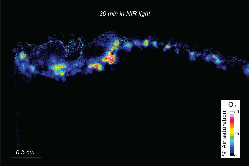
Short report                                                                                                                     Ecology

                                    Figure 1. Spatial distribution of photopigments and near-infrared radiation-driven oxygenic photosynthesis in
                                    beachrock as mapped with hyperspectral reflectance imaging and chemical imaging of O2. (A) RGB image
                                    composite, constructed from the hyperspectral image stack (R = 650 nm, G = 550 nm, B = 450 nm), showing ‘true’
                                    colors of beachrock material and the biofilm community in a cross-section of the top layer. (B) False color coded
                                    image of the same hyperspectral image stack as in panel A mapping pixels with Chl a absorption (670–680 nm) in
                                    green, and Chl f absorption (718–722 nm) in red. Representative reflectance spectra of the two regions are given
                                    in Figure 1—figure supplement 1. (C) Overlay of beachrock structure obtained in panel A and the Chl a signature
                                    from panel B with map of the relative abundance of Chl f obtained from the amplitude of Chl f absorption (color
                                    coded between 0 and 1), as acquired from hyperspectral image analysis. (D) Distribution of O2 concentration
                                    (color coded in units of % air saturation) in the beachrock under illumination of 740 nm light (half-bandwidth = 25
                                    nm; photon irradiance = 28 mmol photons m 2 s 1) when immersed in anoxic seawater, as imaged with the
                                    beachrock section covered with a thin paint of agarose containing O2–sensitive nanoparticles. The O2
                                    concentration image was superimposed onto the structural image of the beachrock cross section. The insert is a
                                    digital zoom corresponding to the insert in panel C. Additional data on two other beachrock sections are available
                                    in the Suppl. Materials (Figure 1—figure supplements 2 and 6).
                                    Figure 1 continued on next page

Kühl et al. eLife 2020;9:e50871. DOI: https://doi.org/10.7554/eLife.50871                                                                     3 of 15
Short report Ecology
Figure 1 continued
The online version of this article includes the following figure supplement(s) for figure 1:
Figure supplement 1. Reflectance spectra of beachrock.
Figure supplement 2. Macroscopic distribution of Chl f in beachrock.
Figure supplement 3. Pigment analysis of black beachrock.
Figure supplement 4. Confocal laser scanning microscopy (CLSM) of a beachrock cross-sectional area.
Figure supplement 5. Higher resolution CLSM of beachrock.
Figure supplement 6. Application of agarose paint with nanoparticles.
Figure supplement 7. NIR-driven O2 production by endolithic cyanobacteria in beachrock.
Figure supplement 8. Calibration curve of the nanoparticle-based O2 sensor paint.
surface biofilm of the beachrock (Figure 1A,B), exhibiting localized hot spots of Chl f concentration
(Figure 1C). Representative reflectance spectra from these regions carrying spectral signatures of
maximal Chl a and Chl f absorption at 670–680 nm and 715–725 nm, respectively, are presented in
the Supplementary Materials (Figure 1—figure supplement 1) along with additional examples of
hyperspectral imaging of beachrock cross-sections (Figure 1—figure supplement 2). The presence
of Chl f (and absence of microalgal Chl b and c) in our samples was confirmed by HPLC analysis of
beachrock pigment extracts (see Materials and methods), both in a ~ 0–2 mm thick black beachrock
sample, and in a more distinct green endolithic layer (~2–5 mm below the beachrock surface) (Fig-
ure 1—figure supplement 3). The amount of Chl f relative to Chl a in these samples ranged from
3.5% in the mixed layer to 6% in the green layer, while Trampe and Kühl (2016) reported Chl f
amounts ranging from 0.5% to 5% of Chl a in beachrock.
To further describe the microscale distribution of cells with different photo-pigmentation, we
employed hyperspectral fluorescence imaging with confocal laser scanning microscopy (CLSM; 488
nm excitation) on beachrock cross-sections (Figure 1—figure supplements 4 and 5). The CLSM
data confirmed the occurrence of patches of filamentous and unicellular Chl f-containing cyanobac-
teria with a characteristic fluorescence peak around 740–750 nm (cf. Majumder et al., 2017) in
deeper endolithic zones (Figure 1—figure supplements 4C and 5A–D). Besides filamentous mor-
photypes, brightfield microscopy of Chl f hot spots revealed the presence of larger round cell aggre-
gates (Figure 1—figure supplement 5E,F) typical of pleurocapsalean cyanobacteria
(Waterbury and Stanier, 1978).
In order to confirm the apparent dominance of cyanobacteria over microalgal oxygenic photo-
trophs, we employed 16S rRNA gene amplicon sequencing on black beachrock samples taken from
the same area as the samples used for hyperspectral and O2 imaging. Among ~39,000 cyanobacte-
rial-like sequences obtained from the black beachrock, none were classified as chloroplasts. In con-
trast, among ~17,000 cyanobacterial-like sequences obtained from two samples of seawater, 18%
were classified as chloroplasts (Supplementary file 2). Microalgae are thus likely to be completely
absent from the black beachrock, where the oxygenic phototrophic community consists exclusively
of cyanobacteria.
Analysis of a distinct green layer below the black beachrock surface biofilm showed that most
cyanobacterial OTU’s were clustering with Halomicronema (harboring the Chl f-containing species
Halomicronema hongdechloris; Chen et al., 2012) as well as coccoid Pseudocapsa and Chroococci-
diopsis (Supplementary file 2). The surface layer of the black beachrock harbored a higher cyano-
bacterial diversity with most OTU’s clustering with Rivularia, Calothrix, and Halomicronema, as well
as numerous smaller populations clustering with Chroococcidiopsis and other coccoid cyanobacteria.
These data confirm earlier findings of i) Chl f (and only minor amounts of Chl d) in beachrock
(Trampe and Kühl, 2016), and ii) a predominance of cyanobacteria as the major oxygenic photo-
trophs in beachrock (Cribb, 1966; Dı́ez et al., 2007). We note that a comprehensive description of
the cyanobacterial diversity associated with beachrock was beyond the scope of the present study,
and a detailed study of the microbial diversity in beachrock (based on 16S rRNA amplicon and meta-
genomic sequencing) will be presented elsewhere. Here, we focus on the photosynthetic activity of
Chl f-containing cyanobacteria in beachrock.
By coating beachrock cross-sections with a thin (Short report Ecology
section when illuminated with weak NIR levels (740 nm, 25 nm half bandwidth; 28 mmol photons m 2
s 1). We observed hot spots of NIR-driven photosynthesis driving local O2 levels from 0% to >40–
50% air saturation within 15–20 min (Figure 1D, Figure 1—figure supplement 7), which overlapped
with regions of high Chl f absorption (Figure 1C, Figure 1—figure supplement 3). The build-up of
O2 in the hotspots harboring Chl f occurred rapidly after onset of NIR illumination and dissipated
rapidly back to anoxia within a few minutes after darkening (see Video 1). Based on O2 concentra-
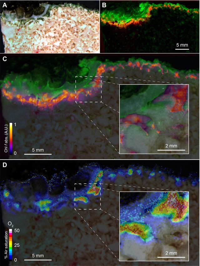
tion images recorded at 5 min intervals after experimental light-dark shifts, we calculated images of
apparent dark respiration and NIR-driven net and gross photosynthesis that could be mapped onto
the beachrock structure (Figure 2A–D) showing that hotspots of activity aligned with the presence
of Chl f (see Figure 1B,C). We extracted estimates of maximum O2 conversion rates in particular
regions of interest (ROI) showing high rates of NIR-driven gross photosynthesis of ~5–15 mmol O2
L 1 min 1 in the beachrock under the given actinic irradiance of 28 mmol photons m 2 s 1; a similar
range was found for two other beachrock cross-sections (data not shown). These volume-specific
rates fall among the upper range of maximal gross photosynthesis rates in aquatic phototrophs, and
are comparable with photosynthetic rates found in benthic microalgae (Krause-Jensen and Sand-
Jensen, 1998).
The Chl f-driven oxygenic photosynthesis by endolithic cyanobacteria thus seems very efficient
given the low actinic NIR level applied. High photosynthetic efficiency of Chl f-containing cyanobac-
teria under NIR has been shown in previous ex situ studies on cultivated strains and enrichments.
Gan et al. (2014) thus found that Leptolyngbya strain JSC-1 cells showed a 40% higher O2 produc-
tion rate with NIR after undergoing FaRLiP relative to cells grown under red light (Gan et al., 2014),
and a similar high photosynthetic efficiency was found in a Chroococcidiopsis strain
(Nürnberg et al., 2018). Using similar NIR levels as used in the present study, Behrendt et al.
(2015) showed rapid saturation of NIR-driven oxygenic photosynthesis already at 25–30 mmol pho-
tons m 2 s 1 (740 nm) in a cell enrichment with Chl f-containing, Aphanocapsa-like cyanobacteria
from a cavernous biofilm.
Our structural and chemical imaging of beachrock showed that Chl f and NIR-driven O2 produc-
tion was confined to a relative narrow zone 1–2 mm below the beachrock surface in the investigated
samples (Figure 1, Figure 2; Figure 1—figure supplements 2, 4 and 5). Assuming a NIR-driven
oxygenic photosynthesis rate of 10 mmol O2 L 1 min 1 (=nmol O2 cm 3 beachrock min 1) in a 1 mm
thick layer (Figure 2), and a conservative esti-
mate of beachrock porosity of ~0.4 in the upper-
most 1–2 mm (Vousdoukas et al., 2007), we can
estimate the areal NIR-driven gross photosynthe-
sis rate to (10 mmol L 1 min 1 0.40.1 cm x
10 6 x 10000 x 60 =) ~0.24 mmol O2 m 2 beach-
rock h 1. Total beachrock primary productivity
remains to be quantified in detail, but it is well
known that beachrock habitats sustain high graz-
ing rates of epifauna (McLean, 1974;
McLean, 2011) and herbivorous reef fish
(Stephenson and Searles, 1960).
To our knowledge, beachrock primary pro-
duction has only been reported by Krumbein,
Video 1. Animation of NIR-driven O2 dynamics over a who studied a Red Sea beachrock habitat
beachrock cross-section (see Figures 1 and 2) coated (Krumbein, 1979). Based on his data on O2
with a thin (Short report Ecology
Figure 2. Oxygen consumption and NIR-driven oxygenic photosynthesis in beachrock. Cross-sectional images of
initial O2 consumption after onset of darkness (A) and maximum net photosynthetic O2 production (B) after onset
of actinic NIR illumination (740 nm, 28 mmol photons m 2 s 1) of the beachrock cross-section shown in Figure 1.
(C) The NIR-driven gross photosynthesis was estimated by summing the absolute rates of net photosynthesis
under NIR and O2 consumption in the dark. (D) Overlay of gross photosynthesis distribution over a structural
image of the beachrock cross-section. (E) Data for O2 consumption, and NIR-driven net and gross photosynthesis
were extracted for 11 regions of interest (ROI) in panel C and are presented as means ± standard deviation within
the ROI.
in a thin agar layer adjacent to the actual phototrophic cells, which will underestimate the true
dynamics (see Materials and methods), and our measurements were conducted at low NIR levels
that may not represent saturated photosynthesis rates. Obviously, the realized total daily primary
productivity will also be strongly modulated by the actual biomass distribution, porosity and diel
light exposure within the beachrock, as well as the pronounced diel cycles of environmental
Kühl et al. eLife 2020;9:e50871. DOI: https://doi.org/10.7554/eLife.50871 6 of 15Short report Ecology
conditions on the beachrock platform (Petrou et al., 2014; Schreiber et al., 2002). Nevertheless,
we argue that our experimental data point to a significant role of NIR-driven oxygenic photosynthe-
sis in beachrock and potentially other endolithic habitats.
Based on the origin of isolates shown to employ FaRLiP (Supplementary file 1), it has been spec-
ulated that shaded soils, caves, plant canopies and thermal springs may be prime terrestrial habitats
for cyanobacteria with Chl f (Gan et al., 2015; Zhang et al., 2019). Beachrock is widespread in inter-
tidal zones on a global scale (Vousdoukas et al., 2007) and our study shows that these habitats may
present a major ecological niche for cyanobacteria with FaRLiP and NIR-driven oxygenic photosyn-
thesis in marine intertidal habitats. While we present the hitherto most detailed insight into the dis-
tribution and in situ activity of Chl f-containing cyanobacteria in intact samples from a natural
habitat, there is now a need for more precise characterization of the microenvironment and meta-
bolic activities of microorganisms in beachrock to assess the quantitative importance of NIR-driven
oxygenic photosynthesis for system productivity.
The present study and the few other studies of natural habitats (Supplementary file 1) demon-
strate that a key trait of such cyanobacteria is the formation of biofilms in strongly shaded environ-
ments below other algae, cyanobacteria or terrestrial plants, or in the twilight zone of caves
(Behrendt et al., 2019). It remains to be explored to what extent the presence of cyanobacteria
with FaRLiP-capability or constitutive high Chl d levels play a role for overall photosynthetic produc-
tivity in such habitats. That is, does the complementary photopigmentation of cyanobacteria with
far-red shifted photopigments in the ‘understory’ of other oxygenic phototrophs lead to higher pho-
tosynthetic efficiency and productivity at the community/system level, for example in analogy to
plant canopies? Such studies are complicated due to the compacted and often stratified structure of
the natural communities, but the novel combination of structural and chemical imaging presented
here seems a promising toolset for unraveling the ecological importance of FaRLiP and cyanobacte-
ria with far red-shifted photopigments.
Materials and methods
Key resources table
Reagent type Additional
(species) or resource Designation Source Identifiers information
Chemical Agarose Thermo Fisher 16520100
compound, drug
Chemical PtTFPP Frontier Scientific PtT975 O2-sensitive dye in the
compound, drug www.frontiersci.com/ O2 sensor nanoparticles
(Koren et al., 2015)
Chemical Macrolex Kremer Pigments Reference dye in the
compound, drug fluorescent yellow www.kremer-pigmente.com/en/ O2 sensor nanoparticles
(Koren et al., 2015)
Chemical PSMA (XIRAN) Polyscope www.polyscope.eu/ Polymer used in the
compound, drug O2 sensor nanoparticles
(Koren et al., 2015)
Software, Image J http://rsb.info.nih.gov/ij/ RRID:SCR_003070 Used for calculations of O2
algorithm concentration images and
visualization of
hyperspectral images,
structure and O2
concentration
Software, Ratio Plus http://rsb.info.nih.gov/ij/ Used for calculations
algorithm Image J plugin plugins/ratio-plus.html of image ratios
Software, look@RGB http://imaging.fish-n-chips.de Used for camera and
algorithm LED control during
image acquisition
(Larsen et al., 2011)
Software, Hyperspectral PhiLumina, LLC, Used for hyperspectral
algorithm Imager V. 4.2 Gulfport, MS, USA image stack acquisition
www.philumina.com/
Continued on next page
Kühl et al. eLife 2020;9:e50871. DOI: https://doi.org/10.7554/eLife.50871 7 of 15Short report Ecology
Continued
Reagent type Additional
(species) or resource Designation Source Identifiers information
Software, ENVI L3 Harris Geospatial, Used for hyperspectral
algorithm Brromfield, CO, USA image stack conversion
www.harrisgeospatial.com/
Software, Look@MOSI www.microsenwiki.net/ Used for hyperspectral
algorithm doku.php/hsimaging: image analysis
hs_iman_howto
Chemical Acetone Sigma Aldrich 650501 Solvents used in
compound, drug Methanol 34860 HPLC analysis
Ammonium acetate 543834
Acetonitrile 114291
Ethylacetate 103649
Software, OpenLAB CDS Agilent Technologies Used for HPLC analysis
algorithm ChemStation Edition
Other Supor-200 PALL 63025 Filters for
polyethersulfone seawater filtration
membrane disc
filters (47 mm diameter,
0.2 mm pore size)
Commercial DNeasy PowerLyzer QIAGEN 12855 Extraction of
assay or kit PowerSoil kit DNA from beachrock
Commercial DNeasy Power QIAGEN 14900 Extraction of
assay or kit Water kit DNA from seawater
Commercial PCR reaction PCRBIO PB10.41–02 Amplification of
assay or kit 16S rRNA gene
Sequence- V3 Eurofins 5’-CCTAYGGGRBGCASCAG-3’ PCR primer for
based reagent 16S rRNA gene
Sequence- V4 Eurofins 5’-GGACTACHVGGGTWTCTAAT-3’ PCR primer for
based reagent 16S rRNA gene
Software, BLAST NCBI RRID:
algorithm http://blast.ncbi.nlm. SCR_004870
nih.gov/Blast.cgi
Field site and beachrock sampling
Beachrock fragments (5–10 cm) from the upper black-brown zone of the intertidal beachrock plat-
form on Heron Island (Great Barrier Reef, Queensland, Australia; 23˚26.5540S, 151˚54.7420E); the
field site is described in detail elsewhere (Cribb, 1966; Dı́ez et al., 2007; Trampe and Kühl, 2016).
Beachrock was sampled at low tide and cut into smaller subsamples with smooth vertical cross sec-
tions (ca. 2 2 1 cm3) using a seawater-flushed stone saw under dim light. These samples were
subsequently stored and transported dry and dark prior to the imaging experiments at University of
Technology Sydney, which commenced within a few days after sampling. Other black beachrock
samples were either processed at the Heron Island Research Station for DNA extraction, or were fro-
zen in liquid N2 on site and shipped on dry ice to Denmark for subsequent pigment extraction and
HPLC analysis.
Hyperspectral imaging
Imaging setup
Hyperspectral reflectance imaging was done on vertical cross-sections of beachrock as previously
described Trampe and Kühl (2016). The samples were submerged in seawater (23˚C and salin-
ity = 35) with the cross-section facing the objective of a dissection zoom microscope (Leica, Ger-
many) with a hyperspectral camera system (100T-VNIR; Themis Vision Systems, St. Louis, MO)
(Kühl and Polerecky, 2008) connected via the C-mount video adapter. A fiber-optic halogen lamp
with an annular ring-light (KL-2500 and Annular Ring-light; Schott AG, Mainz, Germany) mounted on
the objective of the dissection microscope was used as a light source for the hyperspectral image
acquisition. Additional hyperspectral measurements of reflected light were performed (at similar
Kühl et al. eLife 2020;9:e50871. DOI: https://doi.org/10.7554/eLife.50871 8 of 15Short report Ecology
distance and microscope settings as used for the beachrock samples) on a calibrated 20% reflec-
tance standard (Spectralon SRM-20; LabSphere Inc, North Sutton, NH, USA).
Hyperspectral image analysis
Using the manufacturers software, (PhiLumina Hyperspectral Imager V. 4.2; PhiLumina, LLC, Gulf-
port, MS), dark-corrected, hyperspectral image stacks of beachrock reflection were converted to
hyperspectral reflectance images (in units of % reflectance) by normalization to dark-corrected
reflection stacks recorded using the reflectance standard. Subsequent file format conversion, and
image cropping were performed in ENVI (Exelis Visual Information Solutions, Boulder, CO), before
further image processing with the software Look@MOSI (freeware available at www.microsen-wiki.
net/doku.php/hsimaging:hs_iman_howto).
RGB images were constructed from the reflectance measurements at 650 nm (Red), 550 nm
(Green), and 450 nm (Blue), as extracted from the calibrated hyperspectral image stacks. The fourth
derivative of the hyperspectral reflectance stacks was calculated using the Look@MOSI software,
yielding the relative extent of light attenuation at wavelengths representative for Chl a (670–680 nm)
and Chl f (718–722 nm) absorption. The resulting grayscale images were then used for construction
of false-color-coded images, showing the spatial coverage of the two defined spectral signatures.
The extraction of spectral information from areas of interest (covering hotspots of Chl f) was per-
formed as previously described (Polerecky et al., 2009). The resulting images were first cropped to
be of computational sizes for the Look@MOSI software, and were stitched back together in Photo-
shop CC 2015.1.2 (Adobe Systems Incorporated, San Jose, CA) after computation. The relative
absorbance/abundance of Chl f was quantified by scaling of the relative extent of light attenuation
as calculated above for Chl f. Pixel intensity values from the earlier calculated gray scale images
were assigned a scale ranging from 0 to 1, and were finally displayed using a color scaled lookup
table in ImageJ (http://rsbweb.nih.gov/ij/). This yielded false-color coded images with values
between 0 and 1, displaying a relative measure of Chl f abundance according to the amount of light
attenuation obtained from Chl f absorption.
Pigment analysis
Two vertically separated layers from the black beachrock zone were analyzed for composition of
photopigments, that is in the top layer (0–2 mm) and in a more distinct green deeper layer (2–5
mm). Samples from the two layers were kept at 80˚C until further analysis by high-pressure liquid
chromatography (HPLC) as described in detail elsewhere (Trampe and Kühl, 2016). Homogenized
beachrock samples were transferred to 1.5 mL Eppendorf tubes for pigment extraction. The pig-
ments were extracted by adding 0.8 mL acetone:methanol (7:2 vol:vol) to each tube, which was
briefly vortexed, and then kept on ice for a 2 min extraction time in darkness. Subsequently, samples
were sonicated in an ice-cooled high-power ultrasonicating bath (Misonix 4000; Qsonica LLC., New-
town, CT) in darkness for 10 s consisting of 10 pulses of 1 s ON and 1 s OFF (with an amplitude set-
ting of 100%), and were then centrifuged at ~12,000 g for 1 min in a mini centrifuge (MiniSpin,
Eppendorf AG, Hamburg, Germany). The supernatant was filtered through a 0.45 mm pore size
syringe filter (Satorius Minisart SRP 4 filter; Sartorius AG, Goettingen, Germany), and 200 mL filtrate
was then mixed with 15 mL ammonium acetate (1 M) in 0.3 mL HPLC vials. 100 mL of the extract was
then immediately injected into the HPLC. Pigment extracts were separated and analyzed in the
HPLC by a diode array detector (HPLC-DAD and Agilent 1260 Infinity; Agilent Technologies, Santa
Clara, CA) fitted with Ascentis C18 column (25 cm 4.6 mm, Sigma-Aldrich cat. no. 581325 U),
detecting specific absorption wavelengths of compounds. The extracts were run at a constant col-
umn temperature of 30˚C for 69 min., and a flow-rate of 1.0 mL min 1 in a changing gradient of sol-
vent A (methanol:acetonitrile:water, 42:33:25, vol/vol/vol), and solvent B (methanol:acetonitrile:
ethylacetate, 50:20:30, vol/vol/vol), where the mobile phase changed linearly from 30% solvent B at
the time of injection to 100% at 52 min, staying at 100% for15 min before returning to 30% within 2
min. Spectral comparisons from Chen and Blankenship (2011), and Trampe and Kühl (2016) were
used for identification of Chl a, Chl d, and Chl f from the HPLC chromatograms. Elution profiles from
the absorbance detector signal at 430 nm were used to calculate pigment ratios from the derived
integrated peak areas for each of the identified pigments of interest, using the manufacturer’s soft-
ware (OpenLAB CDS ChemStation Edition; Agilent Technologies).
Kühl et al. eLife 2020;9:e50871. DOI: https://doi.org/10.7554/eLife.50871 9 of 15Short report Ecology
Amplicon sequencing
A survey of the microbial communities in the beachrock was performed based on high-throughput
sequencing of the 16S rRNA community gene pool, of which details will be published elsewhere. In
brief, genomic DNA was isolated from portions (0.1–0.4 g) of freshly sampled black beachrock using
the DNeasy PowerLyzer PowerSoil kit (QIAGEN, Germany). Costal seawater was sampled by filtra-
tion of 2.3 L seawater onto Supor-200 polyethersulfone membrane disc filters (47 mm diameter, 0.2
mm pore size; PALL cat. no. 63025) and genomic DNA was isolated using the DNeasy PowerWater
kit (QIAGEN, Germany). This genomic DNA was used as template for PCR amplification with primers
targeting the V3 and V4 regions of the bacterial/chloroplast 16S rRNA gene. The PCR products
were sequenced with the Illumina MiSeq platform and the sequence data analyzed with the Qiime2
pipeline at the Section for Microbiology, Department of Biology, University of Copenhagen.
Experimental setup for O2 imaging
The experiments were performed in a small glass aquarium (15 10 15 cm3) filled with filtered
seawater (salinity 35). A lid with two inlets was placed on the aquarium. One inlet was used as a gas
inlet, the second inlet was used to insert a robust fiber-optic O2 sensor (OXR430 connected to Fire-
Sting GO2 meter; PyroScience GmbH, Aachen, Germany) for monitoring the O2 concentration in the
bulk seawater. The beachrock sample was mounted with a smooth vertical cross-section parallel to
the aquarium glass wall. The camera used for O2 imaging and the excitation LED were placed per-
pendicular to the sample cross-section. Actinic NIR illumination was provided by a 740 nm LED (LZ4-
40R300; LED Engin, Inc, San Jose, CA; HBW 25 nm) providing a NIR photon irradiance (integrated
over 715–765 nm) of 28 mmol photons m 2 s 1, as measured with a calibrated spectroradiometer
(MSC15, GigaHertz-Optik GmbH, Germany).
A beachrock sample painted on one side with the O2 sensor nanoparticle paint (see below and
Figure 1—figure supplement 6) was placed into the aquarium, and the surrounding seawater was
flushed with pure N2 for at least 30 min to completely remove O2, as confirmed by the fiber-optic
O2 sensor. After anoxic conditions were reached, measurements of the change in O2 concentration
over the beachrock cross section in darkness and under NIR illumination were performed under stag-
nant anoxic conditions in the surrounding seawater (Figure 1C, Figure 1—figure supplement 7).
Images were recorded at 5 min intervals relative to onset of actinic light or darkness. This enabled
both recording of steady-state O2 images in light and darkness (=homogeneous anoxia), as well as
the dynamic change in O2 distribution upon NIR irradiation. Proxies for the NIR-driven net photosyn-
thesis, PN, and apparent dark respiration, RD, were calculated by subtracting images taken with a 5
min interval just before and after onset or eclipse of the actinic NIR light, respectively. Gross photo-
synthesis was estimated as PG = PN+|RD|. To avoid any interference from surrounding light reaching
the sample, the entire setup was covered thoroughly with black fabric. All measurements were per-
formed at room temperature (23 ± 1˚C).
It should be noted that the O2 measurements were done over minute intervals in the thin agar
layer with O2-sensitive nanoparticles coating the beachrock with the photosynthetic cells, where the
observed O2 levels are affected both by cell activity and diffusional exchange with the surroundings.
This leads to some diffusive smearing, and the observed dynamics in O2 and thus the derived reac-
tion rates likely represent an underestimate of ‘true’ reaction rates (Santner et al., 2015).
Chemical imaging of O2
Imaging system and application of O2 sensor nanoparticles
We used recently published protocols for imaging O2 concentration over complex bioactive surfaces
by coating beachrock cross-sections with O2-sensitive luminescent sensor nanoparticles
(Koren et al., 2015; Koren et al., 2016) followed by ratiometric luminescence imaging of the
coated surface with a DSLR camera (without NIR filter and with a 530 nm long pass filter mounted
on the camera objective) during brief excitation with blue LED light (445 nm). Details of the imaging
system and image acquisition software (Larsen et al., 2011), nanoparticle fabrication (Koren et al.,
2015; Moßhammer et al., 2019) and biocompatibility (Trampe et al., 2018) are given elsewhere.
Kühl et al. eLife 2020;9:e50871. DOI: https://doi.org/10.7554/eLife.50871 10 of 15Short report Ecology
Optical O2 sensor nanoparticle paint
First, we attempted to spray-paint beachrock sections with sensor nanoparticles using a paintbrush
according to Koren et al. (2016), but the porous structure of the beachrock prevented saturation
and a homogenous coating of cross-sections. Instead, we coated beachrock cross-sections (as well
as glass slides used for calibration) with a thin (680 nm) excited by the excitation light (445 nm) during
O2 imaging can potentially overlap with the red channel. Furthermore, such Chl a-based photosyn-
thesis may also generate a small amount of O2 during the brief excitation pulse. To account for such
potential artefacts, and as we aimed to visualize changes in O2 concentration (DO2) attributed to Chl
f-based photosynthesis, all ratio images were subtracted from ratio images recorded after 45 min in
the dark in anoxic water, Rdark, (DR = Rdark R). Further, to avoid any cross-talk of NIR into the red
channel, O2 images were always recorded during a brief (Short report Ecology
Confocal laser scanning microscopy
After the O2-imaging, the nanoparticle paint was peeled off the beachrock sections, and a smaller
subsection of the flat beachrock cross section (previously used for hyperspectral or chemical imag-
ing) was imaged at 200x and 400x magnification on an inverted confocal laser scanning microscope
(Nikon A1R, Japan) with the beachrock cross section facing downwards in a 35 mm coverslip glass
bottom petri dish (World Precision Instruments). Care was taken to keep the beachrock cross sec-
tions intact and oriented to enable identification and alignment of CLSM data to areas of interest
exhibiting NIR-driven O2 production.
The microscope was equipped with a motorized xyz sample holder and was able to acquire
hyperspectral fluorescence image stacks over a large sample area by sequential scanning, with sub-
sequent automatic stitching of the acquired images in the microscope software (NIS elements AR,
Nikon, Japan). The sample was excited by 488 nm laser light (laser power 1.2 mW, 0.5 frames per
seconds) and spectral fluorescence (500–750 nm) was acquired at 10 nm resolution by the spectral
PMT-array detector on the CLSM microscope. First, the beachrock cross-section was imaged at 200x
magnification obtaining hyperspectral image stacks of 5 confocal layers (step size 5 mm) for each
field of view. Scanned images were stitched to make a 4 mm x 11 mm large image and were then
de-convolved (numerical aperture of 0.75, pinhole size 177.52, refractive index 1 for 26 emission
channels 500–750 nm) using the NIS elements AR software (Version 4.60, Nikon, Japan). Subse-
quently, a more detailed scan of the same cross sectional area was done at 400x magnification (laser
power 1.2 mW, 0.063 frames per seconds), and spectral fluorescence (500–750 nm) was acquired at
10 nm resolution by the spectral PMT-array detector on the CLSM microscope obtaining hyperspec-
tral fluorescence images in one z- plane.
We note that the beachrock cross-section was not perfectly smooth and single plane recording at
400x led to less signal in parts of the cross-section, where the surface was out of focus. Nevertheless,
we could still identify most of the hot spots recorded at 200x magnification and resolve the shape of
individual cells and cell clusters. Scanned images were stitched to make a 3.5 mm x 5.2 mm large
image and were then de-convolved (numerical aperture of 1.00, pinhole size 177.52, refractive index
1.515 for 26 emission channels 500–750 nm). Regions of interest (ROI) were selected from the
obtained cross-sectional scans and spectral fluorescence features were captured and false-color
coded for particular regions of interest. CLSM scans were false-color coded to highlight the fluores-
cence of Chl a (690–700 nm), phycobiliproteins (650–660 nm), and Chl f (740–750 nm; cf.
Majumder et al., 2017).
Acknowledgements
We acknowledge the excellent technical assistance in the field and during laboratory measurements
by Sofie L Jakobsen, Veronica M Petersen, and the staff at Heron Island Research Station. Lorrie
Maccario is thanked for assistance with the MiSeq amplicon sequencing. Work at Heron Island was
conducted under permit no. G16/38423.1 from the Great Barrier Reef Marine Parks authority
Additional information
Funding
Funder Grant reference number Author
Det Frie Forskningsråd DFF-8021-00308B Michael Kühl
Det Frie Forskningsråd DFF-8022-00301B Michael Kühl
Det Frie Forskningsråd DFF-4184-00515B Michael Kühl
Villum Fonden 00023073 Michael Kühl
Poul Due Jensen Fonden Klaus Koren
The funders had no role in study design, data collection and interpretation, or the
decision to submit the work for publication.
Kühl et al. eLife 2020;9:e50871. DOI: https://doi.org/10.7554/eLife.50871 12 of 15Short report Ecology
Author contributions
Michael Kühl, Conceptualization, Resources, Data curation, Formal analysis, Supervision, Funding
acquisition, Validation, Investigation, Visualization, Methodology, Project administration, Writing,
Original draft, Review and editing; Erik Trampe, Conceptualization, Data curation, Formal analysis,
Validation, Investigation, Visualization, Methodology, Review and editing; Maria Mosshammer, Inves-
tigation, Methodology, Review and editing; Michael Johnson, Resources, Software, Formal analysis,
Investigation, Visualization, Methodology, Review and editing; Anthony WD Larkum, Investigation,
Visualization, Methodology, Review and editing; Niels-Ulrik Frigaard, Conceptualisation, Resources,
Data curation, Formal analysis, Validation, Investigation, Methodology, Review and editing; Klaus
Koren, Conceptualization, Data curation, Software, Formal analysis, Validation, Investigation, Visuali-
zation, Methodology, Review and editing
Author ORCIDs
Michael Kühl https://orcid.org/0000-0002-1792-4790
Erik Trampe https://orcid.org/0000-0003-3249-0297
Maria Mosshammer https://orcid.org/0000-0002-7296-8673
Niels-Ulrik Frigaard https://orcid.org/0000-0002-9389-8109
Klaus Koren https://orcid.org/0000-0002-7537-3114
Decision letter and Author response
Decision letter https://doi.org/10.7554/eLife.50871.sa1
Author response https://doi.org/10.7554/eLife.50871.sa2
Additional files
Supplementary files
. Supplementary file 1. Overview of cyanobacterial strains and enrichments reported to contain Chl
f.
. Supplementary file 2. List of most abundant OTUs related to oxygenic phototrophs in black beach-
rock and seawater.
. Transparent reporting form
Data availability
All data generated or analysed during this study are included in the manuscript and supporting files.
References
Airs RL, Temperton B, Sambles C, Farnham G, Skill SC, Llewellyn CA. 2014. Chlorophyll f and chlorophyll d are
produced in the Cyanobacterium chlorogloeopsis fritschii when cultured under natural light and near-infrared
radiation. FEBS Letters 588:3770–3777. DOI: https://doi.org/10.1016/j.febslet.2014.08.026, PMID: 25176411
Akutsu S, Fujinuma D, Furukawa H, Watanabe T, Ohnishi-Kameyama M. 2011. Pigment analysis of a chlorophyll f-
containing cyanobacterium strain KC1 isolated from Lake Biwa. Photomedicine and Photobiology 33:35–40.
Allakhverdiev SI, Kreslavski VD, Zharmukhamedov SK, Voloshin RA, Korol’kova DV, Tomo T, Shen JR. 2016.
Chlorophylls d and f and their role in primary photosynthetic processes of cyanobacteria. Biochemistry.
Biokhimiia 81:201–212. DOI: https://doi.org/10.1134/S0006297916030020, PMID: 27262189
Behrendt L, Larkum AW, Norman A, Qvortrup K, Chen M, Ralph P, Sørensen SJ, Trampe E, Kühl M. 2011.
Endolithic chlorophyll d-containing phototrophs. The ISME Journal 5:1072–1076. DOI: https://doi.org/10.1038/
ismej.2010.195, PMID: 21160540
Behrendt L, Brejnrod A, Schliep M, Sørensen SJ, Larkum AW, Kühl M. 2015. Chlorophyll f-driven photosynthesis
in a cavernous cyanobacterium. The ISME Journal 9:2108–2111. DOI: https://doi.org/10.1038/ismej.2015.14,
PMID: 25668158
Behrendt L, Trampe EL, Nord NB, Nguyen J, Kühl M, Lonco D, Nyarko A, Dhinojwala A, Hershey OS, Barton H.
2019. Life in the dark: far-red absorbing cyanobacteria extend photic zones deep into terrestrial caves.
Environmental Microbiology 81. DOI: https://doi.org/10.1111/1462-2920.14774
Chen M, Schliep M, Willows RD, Cai ZL, Neilan BA, Scheer H. 2010. A red-shifted chlorophyll. Science 329:1318–
1319. DOI: https://doi.org/10.1126/science.1191127, PMID: 20724585
Kühl et al. eLife 2020;9:e50871. DOI: https://doi.org/10.7554/eLife.50871 13 of 15Short report Ecology
Chen M, Li Y, Birch D, Willows RD. 2012. A Cyanobacterium that contains chlorophyll f–a red-absorbing
photopigment. FEBS Letters 586:3249–3254. DOI: https://doi.org/10.1016/j.febslet.2012.06.045, PMID: 227
96191
Chen M. 2014. Chlorophyll modifications and their spectral extension in oxygenic photosynthesis. Annual Review
of Biochemistry 83:317–340. DOI: https://doi.org/10.1146/annurev-biochem-072711-162943
Chen M, Blankenship RE. 2011. Expanding the solar spectrum used by photosynthesis. Trends in Plant Science
16:427–431. DOI: https://doi.org/10.1016/j.tplants.2011.03.011, PMID: 21493120
Cribb AB. 1966. The Algae of Heron Island, Great Barrier Reef, Australia: Part I, A General Account. St. Lucia,
Brisbane, Australia: University of Queensland Press.
Dı́ez B, Bauer K, Bergman B. 2007. Epilithic cyanobacterial communities of a marine tropical beach rock (Heron
Island, great barrier reef): diversity and diazotrophy. Applied and Environmental Microbiology 73:3656–3668.
DOI: https://doi.org/10.1128/AEM.02067-06, PMID: 17416688
Gan F, Zhang S, Rockwell NC, Martin SS, Lagarias JC, Bryant DA. 2014. Extensive remodeling of a cyanobacterial
photosynthetic apparatus in far-red light. Science 345:1312–1317. DOI: https://doi.org/10.1126/science.
1256963
Gan F, Shen G, Bryant D. 2015. Occurrence of Far-Red light photoacclimation (FaRLiP) in diverse cyanobacteria.
Life 5:4–24. DOI: https://doi.org/10.3390/life5010004
Gan F, Bryant DA. 2015. Adaptive and acclimative responses of cyanobacteria to far-red light. Environmental
Microbiology 17:3450–3465. DOI: https://doi.org/10.1111/1462-2920.12992
Ho MY, Shen G, Canniffe DP, Zhao C, Bryant DA. 2016. Light-dependent chlorophyll f synthase is a highly
divergent paralog of PsbA of photosystem II. Science 353:aaf9178. DOI: https://doi.org/10.1126/science.
aaf9178, PMID: 27386923
Itoh S, Ohno T, Noji T, Yamakawa H, Komatsu H, Wada K, Kobayashi M, Miyashita H. 2015. Harvesting Far-Red
light by chlorophyll f in photosystems I and II of unicellular Cyanobacterium strain KC1. Plant and Cell
Physiology 56:2024–2034. DOI: https://doi.org/10.1093/pcp/pcv122, PMID: 26320210
Koren K, Brodersen KE, Jakobsen SL, Kühl M. 2015. Optical sensor nanoparticles in artificial sediments–a new
tool to visualize O2 dynamics around the rhizome and roots of seagrasses. Environmental Science & Technology
49:2286–2292. DOI: https://doi.org/10.1021/es505734b, PMID: 25610948
Koren K, Jakobsen SL, Kühl M. 2016. In-vivo imaging of O2 dynamics on coral surfaces spray-painted with sensor
nanoparticles. Sensors and Actuators B: Chemical 237:1095–1101. DOI: https://doi.org/10.1016/j.snb.2016.05.
147
Koren K, Kühl M. 2018. Optical O2 sensing in aquatic systems and organisms. In: Papkovsky D. B, Dmitriev R
(Eds). Quenched-Phosphorescence Detection of Molecular Oxygen: Applications in Life Sciences. 11 Royal
Society of Chemistry, Detection Science Series. p. 145–174.
Krause-Jensen D, Sand-Jensen K. 1998. Light attenuation and photosynthesis of aquatic plant communities.
Limnology and Oceanography 43:396–407. DOI: https://doi.org/10.4319/lo.1998.43.3.0396
Krumbein WE. 1979. Photolithotropic and chemoorganotrophic activity of bacteria and algae as related to
beachrock formation and degradation (gulf of Aqaba, Sinai). Geomicrobiology Journal 1:139–203. DOI: https://
doi.org/10.1080/01490457909377729
Kühl M, Chen M, Ralph PJ, Schreiber U, Larkum AWD. 2005. A niche for cyanobacteria containing chlorophyll d.
Nature 433:820. DOI: https://doi.org/10.1038/433820a
Kühl M, Polerecky L. 2008. Functional and structural imaging of phototrophic microbial communities and
symbioses. Aquatic Microbial Ecology 53:99–118. DOI: https://doi.org/10.3354/ame01224
Larsen M, Borisov SM, Grunwald B, Klimant I, Glud RN. 2011. A simple and inexpensive high resolution color
ratiometric planar optode imaging approach: application to oxygen and pH sensing. Limnology and
Oceanography: Methods 9:348–360. DOI: https://doi.org/10.4319/lom.2011.9.348
Majumder EL, Wolf BM, Liu H, Berg RH, Timlin JA, Chen M, Blankenship RE. 2017. Subcellular pigment
distribution is altered under far-red light acclimation in cyanobacteria that contain chlorophyll f. Photosynthesis
Research 134:183–192. DOI: https://doi.org/10.1007/s11120-017-0428-1, PMID: 28895022
McLean RF. 1974. Geologic significance of bioerosion of beachrock. Proceedings of the Second International
Coral Reef Symposium 2:401–408.
McLean RF. 2011. “Beach Rock” in Encyclopedia of Modern Coral Reefs. In: Hopley D (Ed). Encyclopedia of
Earth Sciences Series. Springer.
Miyashita H. 2014. Discovery of chlorophyll d in Acaryochloris marina and chlorophyll f in a unicellular
Cyanobacterium, strain KC1, isolated from lake biwa. Journal of Physical Chemistry & Biophysics 4:1–9.
DOI: https://doi.org/10.4172/2161-0398.1000149
Moßhammer M, Brodersen KE, Kühl M, Koren K. 2019. Nanoparticle- and microparticle-based luminescence
imaging of chemical species and temperature in aquatic systems: a review. Microchimica Acta 186:126.
DOI: https://doi.org/10.1007/s00604-018-3202-y, PMID: 30680465
Nürnberg DJ, Morton J, Santabarbara S, Telfer A, Joliot P, Antonaru LA, Ruban AV, Cardona T, Krausz E,
Boussac A, Fantuzzi A, Rutherford AW. 2018. Photochemistry beyond the red limit in chlorophyll f-containing
photosystems. Science 360:1210–1213. DOI: https://doi.org/10.1126/science.aar8313, PMID: 29903971
Ohkubo S, Miyashita H. 2017. A niche for cyanobacteria producing chlorophyll f within a microbial mat. The ISME
Journal 11:2368–2378. DOI: https://doi.org/10.1038/ismej.2017.98, PMID: 28622287
Petrou K, Trimborn S, Kühl M, Ralph PJ. 2014. Desiccation stress in two intertidal beachrock biofilms. Marine
Biology 161:1765–1773. DOI: https://doi.org/10.1007/s00227-014-2458-y
Kühl et al. eLife 2020;9:e50871. DOI: https://doi.org/10.7554/eLife.50871 14 of 15Short report Ecology
Polerecky L, Bissett A, Al-Najjar M, Faerber P, Osmers H, Suci PA, Stoodley P, de Beer D. 2009. Modular
spectral imaging system for discrimination of pigments in cells and microbial communities. Applied and
Environmental Microbiology 75:758–771. DOI: https://doi.org/10.1128/AEM.00819-08, PMID: 19074609
Santner J, Larsen M, Kreuzeder A, Glud RN. 2015. Two decades of chemical imaging of solutes in sediments and
soils–a review. Analytica Chimica Acta 878:9–42. DOI: https://doi.org/10.1016/j.aca.2015.02.006,
PMID: 26002324
Schreiber U, Gademann R, Bird P, Ralph PJ, Larkum AWD, Kuhl M. 2002. Apparent light requirement for
activation of photosynthesis upon rehydration of desiccated beachrock microbial mats1. Journal of Phycology
38:125–134. DOI: https://doi.org/10.1046/j.1529-8817.2002.01103.x
Stephenson W, Searles RB. 1960. Experimental studies on the ecology of intertidal environments at Heron
Island. I. exclusion of fish from beach rock. Marine and Freshwater Research 11:241–268. DOI: https://doi.org/
10.1071/MF9600241
Trampe E, Koren K, Akkineni AR, Senwitz C, Krujatz F, Lode A, Gelinsky M, Kühl M. 2018. Functionalized Bioink
with Optical Sensor Nanoparticles for O 2 Imaging in 3D-Bioprinted Constructs . Advanced Functional Materials
28:1804411. DOI: https://doi.org/10.1002/adfm.201804411
Trampe E, Kühl M. 2016. Chlorophyll f distribution and dynamics in cyanobacterial beachrock biofilms. Journal of
Phycology 52:990–996. DOI: https://doi.org/10.1111/jpy.12450, PMID: 27439961
Vousdoukas MI, Velegrakis AF, Plomaritis TA. 2007. Beachrock occurrence, characteristics, formation
mechanisms and impacts. Earth-Science Reviews 85:23–46. DOI: https://doi.org/10.1016/j.earscirev.2007.07.
002
Waterbury JB, Stanier RY. 1978. Patterns of growth and development in pleurocapsalean cyanobacteria.
Microbiological Reviews 42:2–44. DOI: https://doi.org/10.1128/MMBR.42.1.2-44.1978, PMID: 111023
Zhang ZC, Li ZK, Yin YC, Li Y, Jia Y, Chen M, Qiu BS. 2019. Widespread occurrence and unexpected diversity of
red-shifted chlorophyll producing cyanobacteria in humid subtropical forest ecosystems. Environmental
Microbiology 21:1497–1510. DOI: https://doi.org/10.1111/1462-2920.14582, PMID: 30838735
Zhao C, Gan F, Shen G, Bryant DA. 2015. RfpA, RfpB, and RfpC are the master control elements of Far-Red light
photoacclimation (FaRLiP). Frontiers in Microbiology 6:1–13. DOI: https://doi.org/10.3389/fmicb.2015.01303
Kühl et al. eLife 2020;9:e50871. DOI: https://doi.org/10.7554/eLife.50871 15 of 15You can also read