The Golgi as a "Proton Sink" in Cancer - Frontiers
←
→
Page content transcription
If your browser does not render page correctly, please read the page content below
HYPOTHESIS AND THEORY
                                                                                                                                          published: 13 May 2021
                                                                                                                                  doi: 10.3389/fcell.2021.664295

                                            The Golgi as a “Proton Sink” in
                                            Cancer
                                            Koen M. O. Galenkamp* and Cosimo Commisso*
                                            Cell and Molecular Biology of Cancer Program, NCI-Designated Cancer Center, Sanford Burnham Prebys Medical Discovery
                                            Institute, La Jolla, CA, United States

                                            Cancer cells exhibit increased glycolytic flux and adenosine triphosphate (ATP)
                                            hydrolysis. These processes increase the acidic burden on the cells through the
                                            production of lactate and protons. Nonetheless, cancer cells can maintain an alkaline
                                            intracellular pH (pHi) relative to untransformed cells, which sets the stage for optimal
                                            functioning of glycolytic enzymes, evasion of cell death, and increased proliferation and
                                            motility. Upregulation of plasma membrane transporters allows for H+ and lactate efflux;
                                            however, recent evidence suggests that the acidification of organelles can contribute
                                            to maintenance of an alkaline cytosol in cancer cells by siphoning off protons, thereby
                                            supporting tumor growth. The Golgi is such an acidic organelle, with resting pH ranging
                                            from 6.0 to 6.7. Here, we posit that the Golgi represents a “proton sink” in cancer
                            Edited by:
                         Daniel Ungar,      and delineate the proton channels involved in Golgi acidification and the ion channels
   University of York, United Kingdom       that influence this process. Furthermore, we discuss ion channel regulators that can
                        Reviewed by:        affect Golgi pH and Golgi-dependent processes that may contribute to pHi homeostasis
                   Sakari Kellokumpu,
            University of Oulu, Finland
                                            in cancer.
                   Francois Foulquier,
                                            Keywords: Golgi pH, proton sink, pH homeostasis, ion transport, intracellular pH, cancer
   UMR 8576 Unité de Glycobiologie
  Structurale et Fonctionnelle (UGSF),
                                France
                                           INTRODUCTION
                 *Correspondence:
              Koen M. O. Galenkamp
                                           Cancer cells require large amounts of energy in the form of adenosine triphosphate (ATP)
       kgalenkamp@sbpdiscovery.org
                  Cosimo Commisso
                                           to drive rapid proliferation and support cellular processes including the activation of cell
       ccommisso@sbpdiscovery.org          signaling pathways, membrane transport, and DNA and protein synthesis (Alberts, 2015; Zhu and
                                           Thompson, 2019). Interestingly, cancer cells frequently switch to a less efficient glucose metabolism
                    Specialty section:     pathway for ATP production, compared to untransformed cells (Heiden et al., 2009; Liberti and
         This article was submitted to     Locasale, 2016). This cancer hallmark phenomenon is known as the Warburg effect. Untransformed
                     Membrane Traffic,     cells mostly rely on mitochondrial oxidative phosphorylation (OXPHOS) for energy production,
                a section of the journal   which is a highly efficient process where oxygen and glucose-derived pyruvate fuel the TCA cycle
  Frontiers in Cell and Developmental
                                           and drive the electron transport chain to produce 36 ATPs per glucose molecule (Heiden et al.,
                                Biology
                                           2009; Liberti and Locasale, 2016). Although cancer cells are generally equipped with a functional
        Received: 04 February 2021
                                           OXPHOS pathway, glucose metabolism is frequently switched to aerobic glycolysis where pyruvate
           Accepted: 21 April 2021
                                           is fermented to lactate, even when oxygen is available. This switch in metabolism is thought
           Published: 13 May 2021
                                           to provide essential building blocks for the rapid production of biomass and to facilitate rapid
                             Citation:
                                           proliferation. However, the pathway leads to the production of only two ATPs per glucose molecule
               Galenkamp KMO and
    Commisso C (2021) The Golgi as
                                           and thus requires the cells to boost glucose consumption to meet energy demands. A byproduct
           a “Proton Sink” in Cancer.      of this metabolic rewiring is the increased production of lactate and a surge in H+ . Further
     Front. Cell Dev. Biol. 9:664295.      contributing to the surge in H+ is the hydrolysis of ATP that supports cellular processes and
     doi: 10.3389/fcell.2021.664295        accelerated proliferation. High lactate and H+ levels can have devastating effects on cellular fitness

Frontiers in Cell and Developmental Biology | www.frontiersin.org               1                                           May 2021 | Volume 9 | Article 664295
Galenkamp and Commisso Golgi as a “Proton Sink”
by lowering the intracellular pH (pHi). Surprisingly though, than those found in the cytosol of cancer cells, which has pH
the pHi of cancer cells is not acidic, but is even more alkaline values of 7.4 and higher (Webb et al., 2011, 2021; Corbet and
relative to untransformed cells (Webb et al., 2011; Corbet Feron, 2017; Zheng et al., 2020). Lysosomes, endosomes, and
and Feron, 2017; Zheng et al., 2020). The maintenance of this the Golgi each occupy 3% or less of the total volume of a cell
alkaline pHi provides cancer cells with an optimal environment (Alberts, 2015; Valm et al., 2017). But due to these high luminal
for glycolytic enzyme activity and cellular advantages to proton concentrations, these organelles may function as proton
proliferate, migrate, and withstand cell death cues. To avoid repositories that contribute to maintaining an alkaline pHi in
the accumulation of cytosolic H+ , cancer cells express multiple cancer. Even small changes in the pH of these organelles can
families of transporters on the plasma membrane, including translate to high molar quantities of H+ ions at the cytosolic
vacuolar H+ -ATPases, sodium-hydrogen exchangers, and level. For instance, in cervical cancer, with lysosomes showing a
monocarboxylate transporters, all of which can extrude protons resting pH of 4.6, an increase of ∼0.7 pH units in the lysosome
(Figure 1). However, increasing evidence shows that the pH was able to bring about a ∼0.4 pH unit decrease in the cytosol
regulation of pH homeostasis in cancer is not as straightforward (Liu et al., 2018). Moreover, in pancreatic cancer, a ∼0.5 pH
as extrusion of protons at the plasma membrane level, as unit increase in trans-Golgi network pH resulted in a ∼0.5 pH
acidification of organelles, such as lysosomes and the Golgi, unit decrease in pHi and ablation of the Golgi through Brefeldin
contributes to the maintenance of an alkaline cytosol (Liu A administration produced similar effects on pHi homeostasis
et al., 2018; Funato et al., 2020; Galenkamp et al., 2020). These (Galenkamp et al., 2020). In these cases of induced pH alteration
organelles therefore function as repositories for H+ storage of the lysosome and Golgi, the increase in organelle pH and the
or means to extrude protons through an alternative pathway. concomitant decrease in cytosolic pH reduced the viability of the
Hence, because of their role in siphoning off cytosolic protons, cells, marking the importance of these organelles in maintaining
these organelles can be considered the “proton sinks” of the cell. an alkaline pHi and cell fitness in cancer cells.
In addition to storing protons, the Golgi and lysosomes can
potentially target protons to the extracellular space through
PROTON SINKS IN CANCER exocytosis, which adds an extra layer by which these organelles
can regulate pHi homeostasis (Figure 1; Jaiswal et al., 2009;
The presence of acidic organelles and proton pumping into Rivera-Molina and Toomre, 2013; Deng et al., 2016; Nugues
their lumen can conceptually contribute to the upkeep of an et al., 2018; Funato et al., 2020). Interestingly, exocytosis may
alkaline cytosol by sequestering protons or targeting them for be further stimulated in cancer cells by the acidification of
the extracellular space through exocytosis. While it is conceivable the extracellular surroundings and alkalinization of the cytosol.
that both normal and cancer cells might exploit organelle- During tumor acidosis, the extracellular fluid can reach pH 6.5
mediated sequestering of protons for pHi maintenance, the through increased proton and lactate extrusion (Webb et al.,
available literature specifically points to this process as being 2011; Corbet and Feron, 2017; Zheng et al., 2020). As an
selective to cancer cells (Liu et al., 2018; Funato et al., 2020; adaptation to this acidic environment, lysosomes are targeted
Galenkamp et al., 2020). This idea is consistent with the for fusion with the plasma membrane to protect the cells from
notion that cancer cells produce more acidic moieties relative to acidity, but thereby also contribute to maintaining an alkaline
untransformed cells due to their altered metabolism. cytosol (Steffan et al., 2009; Damaghi et al., 2015; Funato et al.,
Acidic organelles in mammalian cells belong to the endocytic 2020). Moreover, cytosolic acidification was discovered to inhibit
and secretory pathway and each of these organelles has a distinct Golgi to plasma membrane trafficking (Cosson et al., 1989), while
resting pH (Paroutis et al., 2004; Casey et al., 2010). Notably, an alkaline pHi promotes exocytosis (Pernas-Sueiras et al., 2005;
the Golgi pH gradually descends through the sub-compartments; Huck et al., 2007). An alkaline cytosol was recently shown to
starting at pH 6.7 at the cis-Golgi, reaching pH 6.0 at the change the protonation status of PI4P, a phosphatidylinositol that
trans-Golgi network and ultimately ending in the formation predominantly localizes to the Golgi and is required for secretory
of secretory vesicles and granules which can reach pH values vesicle formation (Dippold et al., 2009; Rahajeng et al., 2019;
as low as 5.2. Lysosomes are the cell’s most acidic organelle Waugh, 2019; Shin et al., 2020). Using a yeast model system,
with a resting pH that ranges between pH 4.7 and 5.5, and it was demonstrated that reduced PI4P protonation in response
endosomes acidify as they mature; early endosomes have a resting to cytosolic alkalinization increased PI4P protein binding (Shin
pH of 6.3–6.5 that decreases to pH 5.5 in late endosomes. The et al., 2020). It remains to be determined whether this protonation
luminal pH of these organelles is required for proper organelle status promotes secretory vesicle formation. Nonetheless, PI4P
function and regulation of the organelle pH is important is an important docking station for GOLPH3, a Golgi-localizing
for maintaining cellular fitness. For instance, in the Golgi, oncoprotein frequently upregulated in cancer, which is described
luminal pH levels regulate the activity of glycosyltransferases and to increase secretory vesicle formation and Golgi-to-plasma
vesicular trafficking (Axelsson et al., 2001; Kornak et al., 2008; membrane trafficking (Halberg et al., 2016; Waugh, 2019; Sechi
Maeda et al., 2008; Hucthagowder et al., 2009; Rivinoja et al., et al., 2020). Multiple cancer types have been found to display
2009), while in the lysosomes the pH activates hydrolases and increased and malignant Golgi-dependent secretion that drives
mediates cargo degradation (Chen et al., 2020). metastasis, a process enhanced by extracellular acidification
The pH scale is a negative logarithm and thus these organelles (Webb et al., 2011; Halberg et al., 2016; Corbet and Feron,
contain proton concentrations that are 10–1,000 times higher 2017; Capaci et al., 2020; Gupta et al., 2020; Tan et al., 2020;
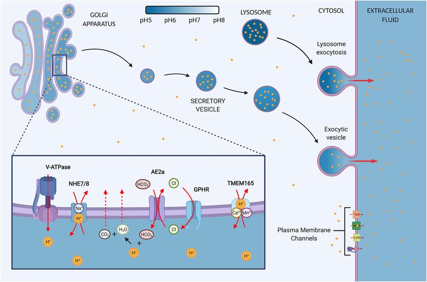
Frontiers in Cell and Developmental Biology | www.frontiersin.org 2 May 2021 | Volume 9 | Article 664295Galenkamp and Commisso Golgi as a “Proton Sink” FIGURE 1 | The Golgi contains proton channels and inherent properties that may convert the organelle into a proton sink in cancer. (A) The Golgi is an acidic organelle that shows a decreasing pH gradient along the sub-compartments, eventually leading to the formation of acidic secretory vesicles and granules. The luminal proton concentration is 10–100 times higher as the cytosol and thus the organelle may function as a proton repository that contributes to the upkeep of an alkaline intracellular pH (pHi) in cancer cells. The resting pH of the Golgi and vesicles is thought to be mediated by proton loading and counter ion conductance. Additionally, a proton leak pathway allows for reducing luminal proton content, but the pathway is suggested to be absent in secretory vesicles. (B) Ion channels at the Golgi regulate the luminal H+ content. V-ATPase: Vacuolar H+ -ATPases load the lumen constitutively with protons in an ATP-dependent manner. NHE7/8: Sodium-hydrogen exchanger 7 (NHE7) has been implicated in proton loading by exchanging protons for luminal Na+ . However, the directionality of NHEs at the Golgi is debated since other studies propose that NHE7 and NHE8 function as a proton leak pathway. AE2a: Anion exchanger 2a is a Golgi-residing AE2 isoform that buffers the Golgi through HCO3 − loading in exchange for Cl− . The buffering presents a sought-after proton leak pathway by providing means to neutralize luminal protons through the production of water and carbon dioxide, which can exit the lumen through diffusion. The directionality of AE2 is reversible and therefore bicarbonate influx is gradient-dependent. GPHR: Golgi pH regulator loads the lumen with Cl− in a voltage-gated manner. Hence, it provides the chloride ions required for counterion conductance to sustain the constitutive activity of V-ATPases. In addition, GHPR is thought to provide the chloride ions to allow for AE2a-mediated HCO3 − buffering. TMEM165: Transmembrane protein 165 ion selectivity and directionality is still under investigation but data points toward Ca2+ /Mn2+ transport in exchange for H+ . (C) Secretory vesicles are targeted for exocytosis and thus present a pathway by which the Golgi may target protons to the extracellular space and convert the Golgi into a proton sink by siphoning off cytosolic H+ . Secretion is upregulated in cancer, but the role in regulating pHi homeostasis remains to be determined. (D) In response to an acidic extracellular environment, lysosomes have been shown to be targeted for exocytosis, thereby maintaining an alkaline pHi and protecting the cells from extracellular acid. Zheng et al., 2020). Whether the malignant secretion presents normal kidney or mammary cells with mutant KRAS or mutant a pathway to boost proton extrusion through the Golgi and HRAS, respectively, results in a lysosome acidification similar to whether that contributes to the metastatic potential of cancer cancer cells. Interestingly, mutant RAS transformation-mediated cells and pHi homeostasis is a concept that requires further reduction in lysosome pH coincides with cytosol alkalinization, investigation (Figure 1). but a direct link between these observations remains to be Organelle pH levels are tightly regulated to support cell established. Nonetheless, a direct link between alkaline pHi and function; however, changes in organelle pH are detected in lysosome pH was found for cervical cancer cells, where loss of cancer. Multiple cancer types display a decreased lysosome lysosome acidification coincides with a reduction in cytosolic pH, pH compared to tissue-matched untransformed cells (Webb which points to lysosomes functioning as a proton sink (Liu et al., et al., 2021). Lysosomes of triple negative breast cancer cells 2018). The lysosome as a proton sink in cancer is extensively are significantly more acidic than lysosomes of untransformed reviewed by Chen et al. (2020). The medial/trans-Golgi is mammary epithelial or benign breast cancer cells. Similarly, proposed to become more alkaline in cancer, as determined by the lysosomes of pancreatic ductal adenocarcinoma (PDAC) comparing various human cancer cell lines to canine kidney cells with mutant KRAS show a >0.5 pH unit decrease epithelial cells and human and monkey fibroblasts (Rivinoja et al., compared to normal human pancreatic duct epithelial cells 2006; Kokkonen et al., 2019; Khosrowabadi et al., 2021). However, or PDAC cells lacking oncogenic KRAS. Transformation of no differences in luminal pH were observed when comparing Frontiers in Cell and Developmental Biology | www.frontiersin.org 3 May 2021 | Volume 9 | Article 664295
Galenkamp and Commisso                                                                                                     Golgi as a “Proton Sink”

the trans-Golgi network of Chinese hamster ovary (CHO) and                  Golgi dysfunction observed in cutis laxa patients, which harbor a
cervical cancer cells (Demaurex et al., 1998). If there is indeed an        mutation in ATP6V0A2 (Kornak et al., 2008; Hucthagowder et al.,
increase in Golgi pH in cancer cells, this might not necessarily            2009). In cancer, V0a2 subunit expression is mostly unaffected
be linked to reduced proton loading since organelle pH also                 at the transcriptional level (ATP6V0A2,1 ) (Tang et al., 2019),
relies on buffering capacity. Such a buffering effect could result          but the subunit may show differential localization to the plasma
in increased luminal pH, while still allowing for the capture               membrane, as observed in ovarian cancer (Kulshrestha et al.,
of protons from the cytosol. The alkalinization in the Golgi                2015). Here, knockdown of the subunit results in cytosolic
is proposed to occur through exchanger-mediated bicarbonate                 acidification, indicating that V-ATPase activity is important for
buffering that reduces the luminal proton content by converting             pHi homeostasis, but additional studies are required to determine
HCO3 − and H+ into H2 O and CO2 , which can readily occur                   if the Golgi is involved (Kulshrestha et al., 2016).
at the resting pH of the Golgi since the pKa of HCO3 − is 6.4                   The V-ATPase proton pump has been demonstrated to
(Khosrowabadi et al., 2021). With this increased pH buffering               transport protons constitutively into the Golgi lumen, where the
capacity, the Golgi in cancer cells is possibly better equipped             resting pH is dictated by a balance between proton pumping
to tolerate proton sequestration and allow for additional proton            and proton leakage (Schapiro and Grinstein, 2000; Wu et al.,
loading relative to normal cells, which may contribute to clearing          2001). Inhibition of V-ATPases causes luminal alkalinization
cytosolic protons. However, additional studies are required to              of the Golgi and secretory vesicles, highlighting the role for
confirm this potential.                                                     V-ATPase in acidifying these organelles. However, artificial
                                                                            luminal acidification in combination with V-ATPase inhibition
                                                                            only results in gradual alkalinization of the Golgi and is not
GOLGI pH REGULATION                                                         detected in secretory vesicles (Wu et al., 2001). These results
                                                                            indicate the possible presence of a proton leak pathway at the
The Golgi pH is thought to be regulated at multiple levels                  Golgi, which is non-existent or minimal in secretory vesicles.
through proton pumping, proton leakage, buffering, and counter              Interestingly, at the lysosome, V-ATPase is shown to be regulated
ion transport. Significant advances have been made in the                   by the oncoprotein STAT3, which increases proton pump activity
understanding of these processes, but a comprehensive model                 in response to sensing cytosolic acidification, thereby restoring
for luminal Golgi pH regulation in cancer largely remains                   the alkaline pHi in cancer (Liu et al., 2018). The role of STAT3
unresolved. Regulators of Golgi pH have mainly been identified              in regulating Golgi acidification seems to be absent, since the
in non-cancerous models and the variety of cancer types and their           study did not find evidence for STAT3 localization at the Golgi.
genomic differences may contribute to alternative regulation.               Nevertheless, a large number of proteins have been identified to
Moreover, cancer frequently displays changes in ion channel                 interact with the V-ATPase proton pump, which could possibly
expression levels and localization and is therefore proposed as             affect its Golgi acidification capacity (Merkulova et al., 2015).
a channelopathy, which, given the multitude of ion channels
that can set off pH changes, beclouds the direct translation of             Na+ /H+ Exchangers
non-cancerous findings to cancer models (Litan and Langhans,                Sodium-hydrogen exchangers (NHEs) are a family of
2015; Prevarskaya et al., 2018). Notably, it should be considered           electroneutral membrane ion transporters that transfer protons
that changes in expression levels do not necessarily translate              across membranes in a 1:1 exchange for Na+ , and in some
to changes in pH levels since ion channel activity can also                 cases Li+ and K+ (Pedersen and Counillon, 2019). The family
be driven by membrane potential and ion gradients. Here, we                 consists of nine isoforms that share a conserved architecture
will outline the regulators of Golgi pH homeostasis thus far                and can be classified into two groups: the plasma membrane
identified (Figure 1).                                                      NHEs (NHE1-5) and the endomembrane NHEs (NHE6-9),
                                                                            although NHE8 can be considered a separate group as it exerts
Vacuolar H+ -ATPases                                                        its function at both the plasma membrane and endomembranes.
The Vacuolar H+ -ATPase (V-ATPases) is the main acidifier                   Both NHE7 and NHE8 have been identified to localize to
of organelles along the endocytic and secretory pathway and                 the Golgi and have been implicated in the regulation of the
its activity is required for both endocytosis and exocytosis                Golgi resting pH (Numata and Orlowski, 2001; Nakamura
(Vasanthakumar and Rubinstein, 2020). The importance of the                 et al., 2005). NHE7 expression is frequently increased in
proton pump in Golgi acidification is well illustrated by studies           cancer (SLC9A7,1 (Tang et al., 2019) and overexpression of
from Llopis et al. (1998) who showed that V-ATPase inhibition               NHE7 in a breast cancer cell line enhances cell adhesion,
with bafilomycin A1 led to Golgi alkalinization, reaching pH                invasion, and anchorage-independent growth (Onishi et al.,
values close to the cytosolic pH levels. The proton pump is a               2012). No significant differences are observed for NHE8
multi-subunit protein complex that transports H+ ions across                expression in tumors relative to tissue-matched controls
the membrane using ATP as an energy source. Expression levels               and no data is available for its role in cancer (SLC9A8,1 )
of the different subunits are tissue-, cell-, and organelle-specific,       (Tang et al., 2019).
and, in cancer, the subunits are frequently differentially expressed            The NHE7 and NHE8 exchangers were originally
(Couto-Vieira et al., 2020). The localization of V-ATPase to the            proposed to function as a Golgi leak pathway for protons
Golgi and endosomes is thought to occur through the V0a2
subunit, and its role in proper Golgi function is illustrated by the        1
                                                                                http://gepia2.cancer-pku.cn

Frontiers in Cell and Developmental Biology | www.frontiersin.org       4                                      May 2021 | Volume 9 | Article 664295
Galenkamp and Commisso Golgi as a “Proton Sink”
(Numata and Orlowski, 2001; Nakamura et al., 2005), allowing Celay et al., 2018; Tang et al., 2019; Khosrowabadi et al.,
H+ to flow out of the lumen through the ion gradient formed 2021). The AE2a isoform is reported to increase the Golgi
by the elevated H+ levels relative to the cytosol. The notion resting pH and therefore represents a likely candidate for
that sodium-hydrogen exchangers act as a proton leak pathway the proton leak observed at the Golgi (Wu et al., 2001;
was supported by the original findings that exogenous NHE8 Khosrowabadi et al., 2021).
expression in monkey fibroblasts increased the pH of the Golgi
and NHE7 expression in CHO cells increased the Na+ influx Golgi pH Regulator
into endomembrane structures (Numata and Orlowski, 2001; The Golgi pH regulator (GPHR) is a voltage-gated chloride
Nakamura et al., 2005). However, CHO cells lacking plasma channel that regulates Golgi pH through counter ion
membrane NHE1 did not confirm the NHE7 leak pathway conductance (Maeda et al., 2008). The influx of chloride ions into
since the Golgi resting pH was unaltered after exogenous the Golgi allows for H+ pumping by reducing the membrane
NHE7 expression (Khayat et al., 2019). Nevertheless, exogenous potential, which increases through continuous H+ pumping by
expression of an NHE7 mutant linked to intellectual disability the V-ATPases (Llopis et al., 1998; Maeda et al., 2008). Moreover,
did cause alkalinization of the Golgi. According to the proton- GPHR can conceivably provide the chloride ions required for
leak hypothesis, the authors proposed a model in which the anion exchanger-mediated bicarbonate transport at the Golgi
mutation transformed the exchanger into a hyperactive proton (Becker and Deitmer, 2020). GPHR is mainly localized to the
leak responsible for the luminal alkalization (Khayat et al., Golgi and the introduction of an inactivating mutation or
2019). Subsequent studies on NHE8 have pointed toward a role downregulation increases the Golgi resting pH, causes disruption
in regulating the function and morphology of multivesicular of Golgi integrity, impairs glycosylation, and Golgi to plasma
bodies, with no observed changes in luminal pH (Lawrence membrane trafficking (Maeda et al., 2008; Vavassori et al., 2013;
et al., 2010). The proton-leak hypothesis warrants further Sou et al., 2019). Little is known about the role of GPHR in
scrutiny, especially since an increase of cytosolic Na+ to 103 and cancer and its expression is largely unaffected by transformation
140 mM, concentrations similar to physiological extracellular (GPR89A/B,1 ) (Tang et al., 2019).
concentrations known to drive NHE1-mediated proton
extrusion, had minimal effects on the Golgi pH, suggesting that
the Na+ /H+ leak activity at the Golgi is insignificant (Demaurex
Transmembrane Protein 165
The ion specificity of transmembrane protein 165 (TMEM165)
et al., 1998; Schapiro and Grinstein, 2000).
is still debated, but research findings point to the function of
An opposing hypothesis for the function of NHE7 at the Golgi
a Ca2+ /Mn2+ transporter in exchange for H+ (Lebredonchel
is in a role as a proton loader. NHE7 expression is frequently
et al., 2019; Stribny et al., 2020; Wang et al., 2020). TMEM165
increased in cancer, as is the case for PDAC (Galenkamp et al.,
localizes to the Golgi and knockdown of TMEM165 in normal
2020). Here, endogenous NHE7 localization was confirmed at
liver cells results in Golgi acidification, suggesting TMEM165 is
the trans-Golgi network and knockdown of NHE7 resulted
a proton leak pathway (Foulquier et al., 2012; Wang et al., 2020).
in alkalization of the organelle and a concomitant increase
However, in cervical cancer cells knockdown of TMEM165 was
in cytosolic pH. A thorough assessment of NHE7 features by
shown to cause acidification of lysosomes (Demaegd et al., 2013).
Milosavljevic et al. (2014) revealed that the exchanger functions
Nonetheless, the role of TMEM165 as a possible regulator of
as an acid loader. Importantly, NHE7 was shown to display
Golgi pH is supported by glycosylation abnormalities found in
non-reversible proton transport from the cytosol to the lumen,
patients harboring a TMEM165 mutation, which is tightly linked
arguing against NHE7 being able to function as a proton
to Golgi pH homeostasis (Foulquier et al., 2012). TMEM165 is
leak pathway at the Golgi. Moreover, the study indicated that
upregulated in a few cancer types and is linked to promoting
NHE7-mediated proton loading is only effectuated by high Na+
migration and invasion (TMEM165,1 ) (Lee et al., 2018; Tang et al.,
concentrations, not by K+ , and is constitutively activated by
2019; Murali et al., 2020).
cytosolic H+ .
Anion Exchangers Cystic Fibrosis Transmembrane
The anion exchangers (AEs) family contains membrane Conductance Regulator
transporters that electroneutrally and reversibly exchange Cl− The role of cystic fibrosis transmembrane conductance regulator
for HCO3 − (Romero et al., 2013). The exchangers mediate (CFTR) in regulating Golgi pH is controversial since studies have
bicarbonate buffering that contributes to pH homeostasis by described seemingly contradictory findings. Moreover, CFTR is
sequestering H+ at acidic pH levels and which leads to the not a Golgi-resident protein per se but traffics through the Golgi
conversion of protons and bicarbonate into water and CO2. Both to reach the cell surface. Nonetheless, this counterion channel
products can readily escape the lumen of organelles through has been shown to change Golgi pH in a cystic fibrosis model
diffusion and, possibly, aquaporin water channels (Nozaki et al., (Poschet et al., 2001). CFTR is a cAMP-activated Cl− /HCO3 −
2008; Alberts, 2015). Of special interest is the Golgi-localized AE2 channel best known for causing the life-limiting cystic fibrosis
isoform AE2a (Holappa et al., 2001). AE2 gene transcription is disease through the F508del mutation, but mutations and
upregulated in multiple cancer types and is linked to promoting differential expression are also linked to cancer predisposition
cell viability, proliferation, migration and invasion of cancer (Amaral et al., 2020). Gene expression is increased or reduced
cells (SLC4A2,1 ) (Hwang et al., 2009, 2019; Zhang et al., 2017; depending on the cancer type (CFTR,1 ) (Tang et al., 2019). The
Frontiers in Cell and Developmental Biology | www.frontiersin.org 5 May 2021 | Volume 9 | Article 664295Galenkamp and Commisso Golgi as a “Proton Sink”
F508del mutation causes CFTR misfolding, ER retention, and in response to amplified aerobic glycolysis and ATP hydrolysis
degradation (Cheng et al., 1990; Denning et al., 1992). In (Webb et al., 2011; Corbet and Feron, 2017; Zheng et al., 2020).
a PDAC cell line derived from a cystic fibrosis patient, the Alternatively, acidic organelles in cancer may exhibit increased
F508del mutation was shown to cause Golgi dispersion which or altered activity that exacerbates the effect on cytosolic pH
was reverted by wild-type CFTR expression (Hollande et al., when perturbed. Indeed, cancer cells show increased Golgi-
2005). Cells harboring the mutation show hyperacidification mediated secretion which would increase the number of protons
of the Golgi, which is counteracted by restoring 1508-CFTR secreted through this pathway (Halberg et al., 2016; Capaci et al.,
folding or reintroduction of wild-type CFTR (Chandy et al., 2020; Gupta et al., 2020; Tan et al., 2020). Given this enhanced
2001; Poschet et al., 2001). However, reduced Cl− influx by secretory flux, perturbation of the secretory pathway might result
decreased expression of CFTR at the Golgi cannot explain the in greater accumulation of protons in the cytosol, relative to
increased acidity, since Cl− is a H+ counterion that reduces untransformed cells. Additionally, cancer cells display altered
the membrane potential. This led to the proposal of a model luminal ion levels, and differential transporter levels or activity,
in which Na+ efflux from the organelle is increased in the which could potentially bring about changes in proton loading
absence of CFTR, allowing for additional H+ pumping (Poschet (Litan and Langhans, 2015; Prevarskaya et al., 2018).
et al., 2002). The CFTR chloride channel represses sodium efflux The contribution of additional Golgi proton loaders or
by inhibiting the epithelial sodium channel, ENaC, resulting leak pathways and their physiological relevance alongside
in Na+ build up due to the action of Na+ /K+ -ATPases. This V-ATPase, which is the main acidifier of secretory and endocytic
leads to reduced proton pumping in response to the increase organelles, in cancer and normal cells is limitedly studied.
in membrane potential caused by Na+ ions. Nonetheless, the In cancer, the Golgi is proposed to display elevated buffering
role of CFTR in regulating Golgi pH remains controversial capacity via increased AE2a expression and bicarbonate loading
as administration of cAMP had little effect on Golgi pH of (Khosrowabadi et al., 2021). A conceivable source of bicarbonate
CFTR mutant and wild-type cells (Llopis et al., 1998; Chandy for transport into the Golgi lumen are the carbonic anhydrases
et al., 2001). This in contrast to previous studies where cAMP present at the cell surface, which are upregulated in cancer
was found to alkalinize the Golgi, but where overexpression and convert carbon dioxide and water into H+ and HCO3 −
of CFTR did not significantly change the pH of the organelle (Mboge et al., 2018). In turn, bicarbonate is imported by the cell
(Seksek et al., 1995, 1996). through Na+ /HCO3 − cotransporters that utilize the existing Na+
gradient between the cytosol and extracellular fluid (Becker and
Deitmer, 2020). Additionally, the cytosolic carbonic anhydrase
DISCUSSION CAII may provide HCO3 − as it has been determined to localize
to the Golgi (Alvarez et al., 2001). Importantly, CAII and AE2
The concept of the Golgi as a proton sink remains to be fully are able to form a transport metabolon, a complex between the
explored and requires more extensive studies that specifically anion exchanger and carbonic anhydrase, which is proposed
address the contribution of the Golgi to maintenance of cytosolic to be required for full bicarbonate transport activity by AE2
pH. Moreover, plasma membrane transporters are historically (Vince and Reithmeier, 2000; Sterling et al., 2001; Gonzalez-
thought to predominately regulate pHi and the role of organelles Begne et al., 2007; Becker and Deitmer, 2020). Luminal chloride
in this homeostatic process needs additional scrutinization ions required for the counter ion transport in the HCO3 −
(Paroutis et al., 2004; Casey et al., 2010; Webb et al., 2011; exchange are most likely transported to the lumen through
Corbet and Feron, 2017; Zheng et al., 2020). It would be chloride channels, such as GPHR, present at the Golgi (Maeda
interesting to examine the level of contribution that alternative et al., 2008). Although AE2a does not directly mediate proton
pathways have on H+ efflux, such as exocytosis and proton leakage, but provides a means for proton neutralization, it
loading of lysosomes and the Golgi, and whether these pathways’ presents a plausible option for the proton leak pathway that had
contributions are distinctive for cancer vs. untransformed cells. previously been identified, but for which thus far a compelling
In cancer, both the perturbation of Golgi and lysosome pH candidate is lacking (Wu et al., 2001). Given that the pKa of
has been shown to affect the pH of the cytosol, but equivalent bicarbonate is 6.4, in the Golgi lumen, bicarbonate and H+
analyses of normal cells is lacking (Liu et al., 2018; Funato can convert to water and carbon dioxide without requiring
et al., 2020; Galenkamp et al., 2020). As an acidic organelle, carbonic anhydrases (Mboge et al., 2018). This alkalinization
the Golgi subtracts protons from the cytosol and, as a part of of the Golgi by increased bicarbonate buffering may provide
the secretory pathway, might target H+ for the extracellular means for additional subtraction of protons from the cytosol.
space. Conceptually, it may be possible that the Golgi can The contribution of the pathway to cytosolic pH alkalinization
contribute to pHi homeostasis in both cancer and normal cells. in cancer ultimately depends on the fate of the carbon dioxide
However, one observation that this might not be the case is and whether it remains within the cell and converts back to
that the depletion of NHE7 in normal pancreatic cells did not bicarbonate in a carbonic anhydrase-dependent or independent
affect the cytosolic pH, while loss of NHE7 in PDAC cells manner, or whether it leaves the cell and thereby leads to the
resulted in alkalinization of the Golgi that caused a decrease in neutralization of intracellular protons.
cytosolic pH (Galenkamp et al., 2020). A likely explanation is The sodium-hydrogen exchanger NHE7 had previously been
that organelle acidification plays a greater role in cancer cells proposed as a proton leak pathway at the Golgi, but more
due to the acidic burden that these cells have to withstand recent data has pointed toward NHE7 functioning as a proton
Frontiers in Cell and Developmental Biology | www.frontiersin.org 6 May 2021 | Volume 9 | Article 664295Galenkamp and Commisso Golgi as a “Proton Sink”
loader (Numata and Orlowski, 2001; Milosavljevic et al., 2014; of these transporters to function remains to be determined
Galenkamp et al., 2020). Endogenous NHE7 localizes to the on a contextual basis and could be differentially regulated in
trans-Golgi network; however, the transporter can be present untransformed cells vs. cancer.
on post-Golgi vesicles, endosomes, and traffic to the plasma
membrane when overexpressed or when endocytosis is inhibited
(Numata and Orlowski, 2001; Lin et al., 2005, 2007; Nakamura CONCLUSION
et al., 2005; Fukura et al., 2010; Onishi et al., 2012; Khayat
et al., 2019; Galenkamp et al., 2020; Lopez-Hernandez et al., Cancer is a pathological state in which cells are exposed
2020). Whether NHE7 is required for the formation of post-Golgi to chronic stresses that can alter cellular processes in
vesicles and endosomes, or whether it is a passenger, and if it order to provide growth benefits despite the harsh tumor
regulates post-Golgi vesicle acidification has not been carefully microenvironment (Webb et al., 2011; Corbet and Feron, 2017;
assessed. When forced to express at the plasma membrane, Zheng et al., 2020). One such stress is the increased production
NHE7 is shown to function as a proton loader of endosomes, of H+ ions, which can lead to cellular acidification and tumor
with no leak activity (Milosavljevic et al., 2014). However, cells acidosis. Despite these conditions, cancer cells maintain an
with and without NHE7 expression displayed similar steady- alkaline pHi that allows proliferation to thrive. The Golgi is
state endosomal pH levels. Inhibition of NHE7 with the pan- shown to contribute to the upkeep of this alkaline cytosolic pH
NHE inhibitor EIPA reduced acidification of endosomes to a in cancer by functioning as a proton sink (Galenkamp et al.,
similar extent as bafilomycin A1, whereas the effect of EIPA was 2020). Whether the Golgi also plays a role in the maintenance
absent in cells without NHE7 expression. These data indicate that of cytosolic pH in normal cells remains to be determined. In
NHE7 can function alongside V-ATPases in mediating luminal addition to the proton storage capacity, the Golgi contains
acidification and that the steady-state pH might involve both inherent properties, the secretory pathway, to target protons
transporters. NHE7 knockdown in PDAC cells was shown to for the extracellular space. This pathway becomes malignant in
alkalinize the Golgi, but a direct evaluation of Na+ -mediated cancer and may promote proton extrusion and provide means
proton loading of the Golgi lumen remains to be carried out to drive metastasis (Halberg et al., 2016; Capaci et al., 2020;
(Galenkamp et al., 2020). Nonetheless, the involvement of NHEs Gupta et al., 2020; Tan et al., 2020). The limited understanding
in Golgi acidification in PDAC was confirmed by treatment of the players involved in Golgi pH homeostasis in cancer
with EIPA. Altogether, the data fits a model in which NHE7 impedes the targeting of this pathway as a therapeutic strategy.
acidifies the trans-Golgi network, possibly alongside V-ATPases. Further investigation is warranted to fully comprehend the
It cannot be completely ruled out that the downregulation or contribution of the Golgi ion channels to Golgi pH and pHi
inhibition of NHE7 affects V-ATPase activity or that proton homeostasis in cancer. It would be beneficial to scrutinize
extrusion at the plasma membrane partially contributes to the how ion channels in the Golgi are regulated, as they may
observed effects. NHE7 constitutively binds protons (Km = 2.5 be affected by changes in expression levels or subcellular
10−7 M), but has a low affinity for Na+ (Km = 240 mM) and localization, or through activation cascades, such as observed
thus needs high Na+ concentrations to drive proton transport in the WNK signaling pathway, and by secondary messengers,
against the gradient between the Golgi lumen and the cytosol such a cAMP and PDGF (Seksek et al., 1995; Alessi et al.,
(Milosavljevic et al., 2014). These findings suggest that, if NHE7 2014). Such examinations may provide valuable insights into
functions as an acid loader at the Golgi, a source of luminal ways to therapeutically disrupt pH homeostasis in cancer and
Na+ should be present. Thus far the concentrations of Na+ at may open new avenues for pharmacological intervention,
the Golgi have not been determined and might be differentially which are eagerly needed to improve clinical outcomes for
regulated in cancer. Na+ /K+ -ATPases present at the Golgi are cancer patients.
likely not the source of Na+ , since their inhibition did not
affect Golgi pH in cervical cancer cells (Llopis et al., 1998)
and reduced Golgi resting pH in cystic fibrosis control cells AUTHOR CONTRIBUTIONS
(Poschet et al., 2001). However, luminal Na+ concentration
Both authors conceived, organized, and wrote the manuscript.
could be driven by retrograde transport from endosomes
to the trans-Golgi network, which delivers extracellular Na+
obtained through endocytosis (Tu et al., 2020). This process is FUNDING
thought to contribute to the high Na+ concentrations observed
in lysosomes, where Na+ is the predominant cation at a This work was supported by a Department of Defense
concentration of ∼150 mM (Wang et al., 2012; Xu and Ren, Career Developmental Award (W81XWH-17-10316 to CC).
2015). A role for this pathway in Na+ delivery to the Golgi has KG is the recipient of a TRDRP Postdoctoral Fellowship
yet to be assessed. Award (T30FT0952).
Altogether, the available data indicates that additional
transporters besides V-ATPase can co-regulate the luminal pH
of the Golgi and contribute to the extraction of protons from ACKNOWLEDGMENTS
the cytosol. The role of each of these transporters and whether
the correct physiological conditions are met to allow for each The figure was created with BioRender.com.
Frontiers in Cell and Developmental Biology | www.frontiersin.org 7 May 2021 | Volume 9 | Article 664295Galenkamp and Commisso Golgi as a “Proton Sink”
REFERENCES Dippold, H. C., Ng, M. M., Farber-Katz, S. E., Lee, S. K., Kerr, M. L., Peterman,
M. C., et al. (2009). GOLPH3 Bridges Phosphatidylinositol-4-Phosphate and
Alberts, B. (2015). Molecular biology of the cell. New York, NY: Garland Science, Actomyosin to Stretch and Shape the Golgi to Promote Budding. Cell 139,
Taylor and Francis Group. 337–351. doi: 10.1016/j.cell.2009.07.052
Alessi, D. R., Zhang, J. W., Khanna, A., Hochdorfer, T., Shang, Y. Z., and Kahle, Foulquier, F., Amyere, M., Jaeken, J., Zeevaert, R., Schollen, E., Race, V., et al.
K. T. (2014). The WNK-SPAK/OSR1 pathway: Master regulator of cation- (2012). TMEM165 Deficiency Causes a Congenital Disorder of Glycosylation.
chloride cotransporters. Sci. Signal. 7:334. doi: 10.1126/scisignal.2005365 Am. J. Hum. Genet. 91, 15–26. doi: 10.1016/j.ajhg.2012.05.002
Alvarez, L., Fanjul, M., Carter, N., and Hollande, E. (2001). Carbonic anhydrase Fukura, N., Ohgaki, R., Matsushita, M., Nakamura, N., Mitsui, K., and Kanazawa,
II associated with plasma membrane in a human pancreatic duct cell H. (2010). A Membrane-Proximal Region in the C-Terminal Tail of NHE7 Is
line (CAPAN-1). J. Histochem. Cytochem. 49, 1045–1053. doi: 10.1177/ Required for Its Distribution in the Trans-Golgi Network, Distinct from NHE6
002215540104900812 Localization at Endosomes. J. Membr. Biol. 234, 149–158. doi: 10.1007/s00232-
Amaral, M. D., Quaresma, M. C., and Pankonien, I. (2020). What Role Does CFTR 010-9242-9
Play in Development, Differentiation, Regeneration and Cancer? Int. J. Mole. Funato, Y., Yoshida, A., Hirata, Y., Hashizume, O., Yamazaki, D., and Miki,
Sci. 21:21093133. doi: 10.3390/ijms21093133 H. (2020). The Oncogenic PRL Protein Causes Acid Addiction of Cells by
Axelsson, M. A. B., Karlsson, N. G., Steel, D. M., Ouwendijk, J., Nilsson, T., and Stimulating Lysosomal Exocytosis. Dev. Cell 55, 387. doi: 10.1016/j.devcel.2020.
Hansson, G. C. (2001). Neutralization of pH in the Golgi apparatus causes 08.009
redistribution of glycosyltransferases and changes in the O-glycosylation of Galenkamp, K. M. O., Sosicka, P., Jung, M., Recouvreux, M. V., Zhang, Y. J.,
mucins. Glycobiology 11, 633–644. doi: 10.1093/glycob/11.8.633 Moldenhauer, M. R., et al. (2020). Golgi Acidification by NHE7 Regulates
Becker, H. M., and Deitmer, J. W. (2020). Transport Metabolons and Acid/Base Cytosolic pH Homeostasis in Pancreatic Cancer Cells. Cancer Discov. 10,
Balance in Tumor Cells. Cancers 12:12040899. doi: 10.3390/cancers12040899 822–835. doi: 10.1158/2159-8290.Cd-19-1007
Capaci, V., Bascetta, L., Fantuz, M., Beznoussenko, G. V., Sommaggio, R., Cancila, Gonzalez-Begne, M., Nakamoto, T., Nguyen, H. V., Stewart, A. K., Alper, S. L., and
V., et al. (2020). Mutant p53 induces Golgi tubulo-vesiculation driving a Melvin, J. E. (2007). Enhanced formation of a HCO3- transport metabolon in
prometastatic secretome. Nat. Comm. 11:5. doi: 10.1038/s41467-020-17596-5 exocrine cells of Nhe1(-/-) mice. J. Biol. Chem. 282, 35125–35132. doi: 10.1074/
Casey, J. R., Grinstein, S., and Orlowski, J. (2010). Sensors and regulators of jbc.M707266200
intracellular pH. Nat. Rev. Mole. Cell Biol. 11, 50–61. doi: 10.1038/nrm2820 Gupta, R., Malvi, P., Parajuli, K. R., Janostiak, R., Bugide, S., Cai, G. P., et al. (2020).
Celay, J., Lozano, T., Concepcion, A. R., Beltran, E., Rudilla, F., Garcia-Barchino, KLF7 promotes pancreatic cancer growth and metastasis by up-regulating ISG
M. J., et al. (2018). Targeting the anion exchanger 2 with specific peptides as expression and maintaining Golgi complex integrity. Proc. Natl. Acad. Sci. U S
a new therapeutic approach in B lymphoid neoplasms. Haematologica 103, A 117, 12341–12351. doi: 10.1073/pnas.2005156117
1065–1072. doi: 10.3324/haematol.2017.175687 Halberg, N., Sengelaub, C. A., Navrazhina, K., Molina, H., Uryu, K., and Tavazoie,
Chandy, G., Grabe, M., Moore, H. P. H., and Machen, T. E. (2001). Proton leak and S. F. (2016). PITPNC1 Recruits RAB1B to the Golgi Network to Drive
CFTR in regulation of Golgi pH in respiratory epithelial cells. Am. J. Physiol. Malignant Secretion. Can. Cell 29, 339–353. doi: 10.1016/j.ccell.2016.02.013
Cell Physiol. 281, C908–C921. Heiden, M. G. V., Cantley, L. C., and Thompson, C. B. (2009). Understanding the
Chen, R., Jaattela, M., and Liu, B. (2020). Lysosome as a Central Hub for Rewiring Warburg Effect: The Metabolic Requirements of Cell Proliferation. Science 324,
PH Homeostasis in Tumors. Cancers 12, doi: 10.3390/cancers12092437 1029–1033. doi: 10.1126/science.1160809
Cheng, S. H., Gregory, R. J., Marshall, J., Paul, S., Souza, D. W., White, G. A., Holappa, K., Suokas, M., Soininen, P., and Kellokumpu, S. (2001). Identification
et al. (1990). Defective intracellular transport and processing of CFTR is the of the full-length AE2 (AE2a) isoform as the Golgi-associated anion
molecular basis of most cystic fibrosis. Cell 63, 827–834. doi: 10.1016/0092- exchanger in fibroblasts. J. Histochem. Cytochem. 49, 259–269. doi: 10.1177/
8674(90)90148-8 002215540104900213
Corbet, C., and Feron, O. (2017). Tumour acidosis: from the passenger to the Hollande, E., Salvador-Cartier, C., Alvarez, L., and Fanjul, M. (2005). Expression of
driver’s seat. Nat. Rev. Cancer 17, 577–593. doi: 10.1038/nrc.2017.77 a wild-type CFTR maintains the integrity of the biosynthetic/secretory pathway
Cosson, P., de Curtis, I., Pouyssegur, J., Griffiths, G., and Davoust, J. (1989). in human cystic fibrosis pancreatic duct cells. J. Histochem. Cytochem. 53,
Low cytoplasmic pH inhibits endocytosis and transport from the trans-Golgi 1539–1552. doi: 10.1369/jhc.4A6587.2005
network to the cell surface. J. Cell Biol. 108, 377–387. doi: 10.1083/jcb.108.2.377 Huck, V., Niemeyer, A., Goerge, T., Schnaeker, E. M., Ossig, R., Rogge, P.,
Couto-Vieira, J., Nicolau-Neto, P., Costa, E. P., Figueira, F. F., Simao, T. D., et al. (2007). Delay of acute intracellular pH recovery after acidosis decreases
Okorokova-Facanha, A. L., et al. (2020). Multi-cancer V-ATPase molecular endothelial cell activation. J. Cell. Physiol. 211, 399–409. doi: 10.1002/jcp.20947
signatures: A distinctive balance of subunit C isoforms in esophageal Hucthagowder, V., Morava, E., Kornak, U., Lefeber, D. J., Fischer, B., Dimopoulou,
carcinoma. Ebiomedicine 51:42. doi: 10.1016/j.ebiom.2019.11.042 A., et al. (2009). Loss-of-function mutations in ATP6V0A2 impair vesicular
Damaghi, M., Tafreshi, N. K., Lloyd, M. C., Sprung, R., Estrella, V., Wojtkowiak, trafficking, tropoelastin secretion and cell survival. Hum. Mole. Genet. 18,
J. W., et al. (2015). Chronic acidosis in the tumour microenvironment selects 2149–2165. doi: 10.1093/hmg/ddp148
for overexpression of LAMP2 in the plasma membrane. Nat. Comm. 6:9752. Hwang, J. M., Kao, S. H., Hsieh, Y. H., Li, K. L., Wang, P. H., Hsu, L. S., et al.
doi: 10.1038/ncomms9752 (2009). Reduction of anion exchanger 2 expression induces apoptosis of human
Demaegd, D., Foulquier, F., Colinet, A. S., Gremillon, L., Legrand, D., Mariot, P., hepatocellular carcinoma cells. Mole. Cell. Biochem. 327, 135–144. doi: 10.1007/
et al. (2013). Newly characterized Golgi- localized family of proteins is involved s11010-009-0051-3
in calcium and pH homeostasis in yeast and human cells. Proc. Natl. Acad. Sci. Hwang, S., Shin, D. M., and Hong, J. H. (2019). Drug Repurposing as an Antitumor
U S A 110, 6859–6864. doi: 10.1073/pnas.1219871110 Agent: Disulfiram-Mediated Carbonic Anhydrase 12 and Anion Exchanger 2
Demaurex, N., Furuya, W., D’Souza, S., Bonifacino, J. S., and Grinstein, S. Modulation to Inhibit Cancer Cell Migration. Molecules 24:18. doi: 10.3390/
(1998). Mechanism of acidification of the trans-Golgi network (TGN) - In situ molecules24183409
measurements of pH using retrieval of TGN38 and furin from the cell surface. Jaiswal, J. K., Rivera, V. M., and Simon, S. M. (2009). Exocytosis of Post-Golgi
J. Biol. Chem. 273, 2044–2051. doi: 10.1074/jbc.273.4.2044 Vesicles Is Regulated by Components of the Endocytic Machinery. Cell 137,
Deng, Y. Q., Rivera-Molina, F. E., Toomre, D. K., and Burd, C. G. (2016). 1308–1319. doi: 10.1016/j.cell.2009.04.064
Sphingomyelin is sorted at the trans Golgi network into a distinct class of Khayat, W., Hackett, A., Shaw, M., Ilie, A., Dudding-Byth, T., Kalscheuer, V. M.,
secretory vesicle. Proc. Natl. Acad. Sci. U S A 113, 6677–6682. doi: 10.1073/pnas. et al. (2019). A recurrent missense variant in SLC9A7 causes nonsyndromic
1602875113 X-linked intellectual disability with alteration of Golgi acidification and
Denning, G. M., Anderson, M. P., Amara, J. F., Marshall, J., Smith, A. E., and aberrant glycosylation. Hum. Mole. Genet. 28, 598–614. doi: 10.1093/hmg/
Welsh, M. J. (1992). Processing Of Mutant Cystic-Fibrosis Transmembrane ddy371
Conductance Regulator Is Temperature-Sensitive. Nature 358, 761–764. doi: Khosrowabadi, E., Rivinoja, A., Risteli, M., Tuomisto, A., Salo, T., Mäkinen,
10.1038/358761a0 M. J., et al. (2021). PREPRINT: SLC4A2 Anion Exchanger Promotes Tumor
Frontiers in Cell and Developmental Biology | www.frontiersin.org 8 May 2021 | Volume 9 | Article 664295Galenkamp and Commisso Golgi as a “Proton Sink” Cell Malignancy via Enhancing H+ Leak across Golgi Membranes. bioRxiv migration and invasion for breast carcinoma. Oncotarget 11, 2747–2762. doi: 2021:428406. doi: 10.1101/2021.02.09.428406 10.18632/oncotarget.27668 Kokkonen, N., Khosrowabadi, E., Hassinen, A., Harrus, D., Glumoff, T., Nakamura, N., Tanaka, S., Teko, Y., Mitsui, K., and Kanazawa, H. (2005). Kietzmann, T., et al. (2019). Abnormal Golgi pH Homeostasis in Cancer Four Na+/H+ exchanger isoforms are distributed to Golgi and post-Golgi Cells Impairs Apical Targeting of Carcinoembryonic Antigen by Inhibiting Its compartments and are involved in organelle pH regulation. J. Biol. Chem. 280, Glycosyl-Phosphatidylinositol Anchor-Mediated Association with Lipid Rafts. 1561–1572. doi: 10.1074/jbc.M410041200 Antiox. Redox Signal. 30, 5–21. doi: 10.1089/ars.2017.7389 Nozaki, K., Ishii, D., and Ishibashi, K. (2008). Intracellular aquaporins: clues for Kornak, U., Reynders, E., Dimopoulou, A., van Reeuwijk, J., Fischer, B., Rajab, intracellular water transport? Pflugers Archiv-Eur. J. Physiol. 456, 701–707. doi: A., et al. (2008). Impaired glycosylation and cutis laxa caused by mutations 10.1007/s00424-007-0373-5 in the vesicular H+-ATPase subunit ATP6V0A2. Nat. Genet. 40, 32–34. doi: Nugues, C., Helassa, N., Rajamanoharan, D., Burgoyne, R. D., and Haynes, L. P. 10.1038/ng.2007.45 (2018). PREPRINT: Lysosome exocytosis is required for mitosis. bioRxiv 2018, Kulshrestha, A., Katara, G. K., Ginter, J., Pamarthy, S., Ibrahim, S. A., Jaiswal, M. K., 375816. doi: 10.1101/375816 et al. (2016). Selective inhibition of tumor cell associated Vacuolar-ATPase ’a2’ Numata, M., and Orlowski, J. (2001). Molecular cloning and characterization of isoform overcomes cisplatin resistance in ovarian cancer cells. Mole. Oncol. 10, a novel (Na+,K+)/H+ exchanger localized to the trans-Golgi network. J. Biol. 789–805. doi: 10.1016/j.molonc.2016.01.003 Chem. 276, 17387–17394. doi: 10.1074/jbc.M101319200 Kulshrestha, A., Katara, G. K., Ibrahim, S., Pamarthy, S., Jaiswal, M. K., Sachs, Onishi, I., Lin, P. J. C., Numata, Y., Austin, P., Cipollone, J., Roberge, M., et al. A. G., et al. (2015). Vacuolar ATPase ’a2’ isoform exhibits distinct cell surface (2012). Organellar (Na+, K+)/H+ exchanger NHE7 regulates cell adhesion, accumulation and modulates matrix metalloproteinase activity in ovarian invasion and anchorage-independent growth of breast cancer MDA-MB-231 cancer. Oncotarget 6, 3797–3810. doi: 10.18632/oncotarget.2902 cells. Oncol. Rep. 27, 311–317. doi: 10.3892/or.2011.1542 Lawrence, S. P., Bright, N. A., Luzio, J. P., and Bowers, K. (2010). The Paroutis, P., Touret, N., and Grinstein, S. (2004). The pH of the secretory pathway: Sodium/Proton Exchanger NHE8 Regulates Late Endosomal Morphology and Measurement, determinants, and regulation. Physiology 19, 207–215. doi: 10. Function. Mole. Biol. Cell 21, 3540–3551. doi: 10.1091/mbc.E09-12-1053 1152/physiol.00005.2004 Lebredonchel, E., Houdou, M., Potelle, S., de Bettignies, G., Schulz, C., Recchi, Pedersen, S. F., and Counillon, L. (2019). The Slc9a-C Mammalian Na+/H+ M. A. K., et al. (2019). Dissection of TMEM165 function in Golgi glycosylation Exchanger Family: Molecules. Mechanisms, and physiology. Physiol. Rev. 99, and its Mn2+ sensitivity. Biochimie 165, 123–130. doi: 10.1016/j.biochi.2019. 2015–2113. doi: 10.1152/physrev.00028.2018 07.016 Pernas-Sueiras, O., Alfonso, A., Vieytes, M. R., and Botana, L. M. (2005). Mast Lee, J. S., Kim, M. Y., Park, E. R., Shen, Y. N., Jeon, J. Y., Cho, E. H., et al. cell exocytosis can be triggered by ammonium chloride with just a cytosolic (2018). TMEM165, a Golgi transmembrane protein, is a novel marker for alkalinization and no calcium increase. J. Cell. Physiol. 204, 775–784. doi: 10. hepatocellular carcinoma and its depletion impairs invasion activity. Oncol. 1002/jcp.20334 Rep. 40, 1297–1306. doi: 10.3892/or.2018.6565 Poschet, J., Perkett, E., and Deretic, V. (2002). Hyperacidification in cystic fibrosis: Liberti, M. V., and Locasale, J. W. (2016). The Warburg Effect: How Does it Benefit links with lung disease and new prospects for treatment. Trends Mole. Med. 8, Cancer Cells? Trends Biochem. Sci. 41, 211–218. doi: 10.1016/j.tibs.2015.12.001 512–519. doi: 10.1016/s1471-4914(02)02414-0 Lin, P. J. C., Williams, W. P., Kobiljski, J., and Numata, M. (2007). Caveolins bind Poschet, J. F., Boucher, J. C., Tatterson, L., Skidmore, J., Van Dyke, R. W., and to (Na+, K+)/H+ exchanger NHE7 by a novel binding module. Cell. Signal. 19, Deretic, V. (2001). Molecular basis for defective glycosylation and Pseudomonas 978–988. doi: 10.1016/j.cellsig.2006.11.006 pathogenesis in cystic fibrosis lung. Proc. Natl. Acad. Sci. U S A 98, 13972–13977. Lin, P. J. C., Williams, W. P., Luu, Y., Molday, R. S., Orlowski, J., and Numata, M. doi: 10.1073/pnas.241182598 (2005). Secretory carrier membrane proteins interact and regulate trafficking Prevarskaya, N., Skryma, R., and Shuba, Y. (2018). Ion channels in cancer: are of the organellar (Na+,K+)/H+ exchanger NHE7. J. Cell Sci. 118, 1885–1897. cancer hallmarks oncochannelopathies? Physiol. Rev. 98, 559–621. doi: 10.1152/ doi: 10.1242/jcs.02315 physrev.00044.2016 Litan, A., and Langhans, S. A. (2015). Cancer as a channelopathy: ion channels and Rahajeng, J., Kuna, R. S., Makowski, S. L., Tran, T. T. T., Buschman, M. D., Li, pumps in tumor development and progression. Front. Cell. Neurosci. 9:00086. S., et al. (2019). Efficient Golgi Forward Trafficking Requires GOLPH3-Driven, doi: 10.3389/fncel.2015.00086 PI4P-Dependent Membrane Curvature. Dev. Cell 50:573. doi: 10.1016/j.devcel. Liu, B., Palmfeldt, J., Lin, L., Colaco, A., Clemmensen, K. K. B., Huang, J., et al. 2019.05.038 (2018). STAT3 associates with vacuolar H+-ATPase and regulates cytosolic and Rivera-Molina, F., and Toomre, D. (2013). Live-cell imaging of exocyst links its lysosomal pH. Cell Res. 28, 996–1012. doi: 10.1038/s41422-018-0080-0 spatiotemporal dynamics to various stages of vesicle fusion. J. Cell Biol. 201, Llopis, J., McCaffery, J. M., Miyawaki, A., Farquhar, M. G., and Tsien, R. Y. (1998). 673–680. doi: 10.1083/jcb.201212103 Measurement of cytosolic, mitochondrial, and Golgi pH in single living cells Rivinoja, A., Hassinen, A., Kokkonen, N., Kauppila, A., and Kellokumpu, S. with green fluorescent proteins. Proc. Natl. Acad. Sci. U S A 95, 6803–6808. (2009). Elevated Golgi pH Impairs Terminal N-Glycosylation by Inducing doi: 10.1073/pnas.95.12.6803 Mislocalization of Golgi Glycosyltransferases. J. Cell. Physiol. 220, 144–154. Lopez-Hernandez, T., Puchkov, D., Krause, E., Maritzen, T., and Haucke, V. (2020). doi: 10.1002/jcp.21744 Endocytic regulation of cellular ion homeostasis controls lysosome biogenesis. Rivinoja, A., Kokkonen, N., Kellokumpu, I., and Kellokumpu, S. (2006). Elevated Nat. Cell Biol. 22:815. doi: 10.1038/s41556-020-0535-7 Golgi pH in breast and colorectal cancer cells correlates with the expression of Maeda, Y., Ide, T., Koike, M., Uchiyama, Y., and Kinoshita, T. (2008). GPHR oncofetal carbohydrate T-antigen. J. Cell. Physiol. 208, 167–174. doi: 10.1002/ is a novel anion channel critical for acidification and functions of the Golgi jcp.20653 apparatus. Nat. Cell Biol. 10, 1135–1145. doi: 10.1038/ncb1773 Romero, M. F., Chen, A. P., Parker, M. D., and Boron, W. F. (2013). The SLC4 Mboge, M. Y., Mahon, B. P., McKenna, R., and Frost, S. C. (2018). Carbonic family of bicarbonate (HCO3-) transporters. Mole. Aspects Med. 34, 159–182. Anhydrases: Role in pH Control and Cancer. Metabolites 8:8010019. doi: 10. doi: 10.1016/j.mam.2012.10.008 3390/metabo8010019 Schapiro, F. B., and Grinstein, S. (2000). Determinants of the pH of the Golgi Merkulova, M., Paunescu, T. G., Azroyan, A., Marshansky, V., Breton, S., and complex. J. Biol. Chem. 275, 21025–21032. doi: 10.1074/jbc.M002386200 Brown, D. (2015). Mapping the H+ (V)-ATPase interactome: identification of Sechi, S., Frappaolo, A., Karimpour-Ghahnavieh, A., Piergentili, R., and Giansanti, proteins involved in trafficking, folding, assembly and phosphorylation. Sci. M. G. (2020). Oncogenic Roles of GOLPH3 in the Physiopathology of Cancer. Rep. 5:14827. doi: 10.1038/srep14827 Int. J. Mole Sci. 21:3. doi: 10.3390/ijms21030933 Milosavljevic, N., Monet, M., Lena, I., Brau, F., Lacas-Gervais, S., Feliciangeli, Seksek, O., Biwersi, J., and Verkman, A. S. (1995). Direct Measurement Of Trans- S., et al. (2014). The Intracellular Na+/H+ Exchanger NHE7 Effects a Na+- Golgi Ph In Living Cells And Regulation By 2nd Messengers. J. Biol. Chem. 270, Coupled, but Not K+-Coupled Proton-Loading Mechanism in Endocytosis. Cell 4967–4970. doi: 10.1074/jbc.270.10.4967 Rep. 7, 689–696. doi: 10.1016/j.celrep.2014.03.054 Seksek, O., Biwersi, J., and Verkman, A. S. (1996). Evidence against defective Murali, P., Johnson, B. P., Lu, Z., Climer, L., Scott, D. A., Foulquier, F., et al. trans-Golgi acidification in cystic fibrosis. J. Biol. Chem. 271, 15542–15548. (2020). Novel role for the Golgi membrane protein TMEM165 in control of doi: 10.1074/jbc.271.26.15542 Frontiers in Cell and Developmental Biology | www.frontiersin.org 9 May 2021 | Volume 9 | Article 664295
You can also read