Mechanical forces induce an asthma gene signature in healthy airway epithelial cells
←
→
Page content transcription
If your browser does not render page correctly, please read the page content below

www.nature.com/scientificreports

            OPEN           Mechanical forces induce an
                           asthma gene signature in healthy
                           airway epithelial cells
                           Ayşe Kılıç1,10, Asher Ameli1,2,10, Jin-Ah Park3,10, Alvin T. Kho4, Kelan Tantisira1, Marc Santolini            1,5
                                                                                                                                                ,
                           Feixiong Cheng6,7,8, Jennifer A. Mitchel3, Maureen McGill3, Michael J. O’Sullivan3,
                           Margherita De Marzio1,3, Amitabh Sharma1, Scott H. Randell9, Jeffrey M. Drazen3,
                           Jeffrey J. Fredberg3 & Scott T. Weiss1,3*
                           Bronchospasm compresses the bronchial epithelium, and this compressive stress has been implicated
                           in asthma pathogenesis. However, the molecular mechanisms by which this compressive stress alters
                           pathways relevant to disease are not well understood. Using air-liquid interface cultures of primary
                           human bronchial epithelial cells derived from non-asthmatic donors and asthmatic donors, we applied
                           a compressive stress and then used a network approach to map resulting changes in the molecular
                           interactome. In cells from non-asthmatic donors, compression by itself was sufficient to induce
                           inflammatory, late repair, and fibrotic pathways. Remarkably, this molecular profile of non-asthmatic
                           cells after compression recapitulated the profile of asthmatic cells before compression. Together, these
                           results show that even in the absence of any inflammatory stimulus, mechanical compression alone is
                           sufficient to induce an asthma-like molecular signature.

                           Bronchial epithelial cells (BECs) form a physical barrier that protects pulmonary airways from inhaled irritants
                           and invading pathogens1,2. Moreover, environmental stimuli such as allergens, pollutants and viruses can induce
                           constriction of the airways3 and thereby expose the bronchial epithelium to compressive mechanical stress. In
                           BECs, this compressive stress induces structural, biophysical, as well as molecular changes4,5, that interact with
                           nearby mesenchyme6 to cause epithelial layer unjamming1, shedding of soluble factors, production of matrix
                           proteins, and activation matrix modifying enzymes, which then act to coordinate inflammatory and remodeling
                           processes4,7–10.
                               Growing evidence supports the notion that mechanical stress in the airway induces not only developmental,
                           homeostatic and reparative responses in the healthy lung but also pathophysiologic processes in the asthmatic
                           lung2,11–13. For example, mechanical stimuli induce early inflammatory and remodeling factors6,7,14,15, microRNAs
                           (miRs)16,17, and cell proliferation18. Using protein-protein interaction networks, here we compare responses to
                           compressive stress in human BECs (HBECs) obtained from non-asthmatic versus asthmatic donors. Our hypoth-
                           esis was, that using a systems biology approach to examine the effect of mechanical forces acting on structural
                           cells in the airway, will elucidate the complex transcriptional programs that contribute to disease development
                           and pathophysiology.

                           1
                            Channing Division of Network Medicine, Department of Medicine, Brigham and Women’s Hospital, Boston, MA,
                           USA. 2Department of Physics, Northeastern University, Boston, MA, USA. 3Program in Molecular Integrative
                           Phyisological Sciences, Department of Environmental Health, Harvard TH Chan School of Public Health, Boston,
                           MA, USA. 4Computational Health Informatics Program, Boston Children’s Hospital, Boston, MA, USA. 5Centre
                           for Research and Interdisciplinarity (CRI), Paris, F-75014, France. 6Genomic Medicine Institute, Lerner Research
                           Institute, Cleveland Clinic, Cleveland, OH, 44195, USA. 7Department of Molecular Medicine, Cleveland Clinic Lerner
                           College of Medicine, Case Western Reserve University, Cleveland, OH, 44195, USA. 8Case Comprehensive Cancer
                           Center, Case Western Reserve University School of Medicine, Cleveland, Ohio, 44106, USA. 9Marsico Lung Institute/
                           Cystic Fibrosis Center, University of North Carolina, Chapel Hill, NC, USA. 10These authors contributed equally: Ayşe
                           Kılıç, Asher Ameli and Jin-Ah Park. *email: scott.weiss@channing.harvard.edu

Scientific Reports |   (2020) 10:966 | https://doi.org/10.1038/s41598-020-57755-8                                                                   1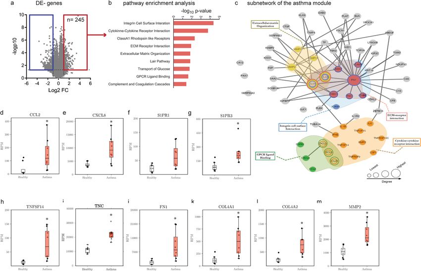
www.nature.com/scientificreports/                                                              www.nature.com/scientificreports

                           Figure 1. Workflow for the analysis of compression induced pathological signatures in HBECs from non-
                           asthmatic donors. HBECs from non-asthmatic donors were compressed and the mRNA expression was
                           detected by RNA-sequencing. Using the human PPI, the compression modules were defined. Pathway
                           enrichment analysis was performed in both, compressed cells and asthmatic HBECs, both normalized to
                           healthy non-asthmatic HBECs.

                           Results
                           Uncompressed asthmatic HBECs express a distinct gene signature.                        There is an ongoing debate
                           whether the development of asthma arises from early disruption of the airway epithelium or imbalance of
                           immune responses19. To investigate the former possibility, we used both non-asthmatic and asthmatic HBECs
                           grown in air-liquid interface (ALI) culture in the absence of immune cells or stimuli. We combined RNA-
                           sequencing and the human protein-protein interaction network to identify relevant genes and pathways (Fig. 1).
                               Initial analysis indicated that 774 genes were differentially expressed (DE) between non-asthmatic and
                           asthmatic HBECs, with 254 being upregulated and 520 downregulated (Fig. 2a). A network approach, using
                           gene expression data levels20 then identified a neighborhood of DE genes between non-asthmatic and asth-
                           matic HBECs, that formed a significantly connected module in the PPI network and defined a disease signa-
                           ture, which we termed the “asthma module” (see methods section for details). Genes mapping to this asthma
                           module were validated with available literature, ascertaining the significance of the identified gene list. Pathway
                           analysis revealed the increased expression of genes belonging to G-protein coupled receptor (GPCR) ligand
                           binding (p = 2.24E-02)21–26, integrin cell surface interaction (p = 4.25E-05), cytokine - cytokine receptor inter-
                           action (p = 1.70E-04), extracellular matrix (ECM) receptor interaction (p = 1.18E-02) and ECM organization
                           (p = 1.49E-02) were enriched (Fig. 2b and Table 1). Since a disease module usually comprises several hundred
                           genes, we visualized the upregulated genes and the pathways that they mapped to in a subnetwork entailing
                           inflammatory and remodeling mechanisms (Fig. 2c). The GPCR pathway included the chemokines CCL221,
                           CCL2022, CXCL323, CXCL524, CXCL824 (Interleukin (IL)-8) and sphingosine 1-phosphate receptors S1PR1 and
                           S1PR325 (Fig. 2d–g). The cytokine - cytokine receptor interaction pathway comprised chemokines as well as sev-
                           eral cytokines and growth factors, including IL-626 and IL-1β27. Genes coding for matrix proteins as well as matrix
                           modifying enzymes mapped to the pathways associated with matrix interaction and remodeling. The expression
                           of the most central gene fibronectin 1 (FN1) and IL-8 as a peripheral gene were validated by real-time RT-PCR in
                           independent samples (Supplementary Fig. S2). In asthma, these pathways regulate recruitment of inflammatory
                           cells as well as remodeling processes (Fig. 2h–m). To further validate and confirm above listed results, the overlap
                           with previously described and published asthma data sets (see methods section) was assessed. A highly significant
                           overlap was found with ALI-grown healthy epithelial cells exposed to interleukin (IL) -13 (GSE37693; p = 1.61E-
                           36) and freshly isolated epithelial cells from severe asthmatic patients (GSE63142; p = 1.24E-08) (Fig. 2m).

                           Compressive stress induces pronounced molecular changes in bronchial epithelial cells. The
                           asthma module described above was determined in the absence of compression and then compared to gene
                           expression profiles occurring in non-asthmatic cells exposed to compression. Specifically, we analysed gene
                           expression induced soon after compression (the 3 hr time point) and later (the 24 hr time point). Control
                           (non-asthmatic) treated cells for each donor and time point were used to normalize gene expression (Fig. 3a).
                           Differential gene expression for each time point was assessed and the respective “compression modules” were
                           defined. The same fold change (FC) cut-offs used for compression module description (FC 3 hr: 1.87 and FC
                           24 hr: 1.67) were applied to characterize alterations in gene expression. Within 3 hours of compressive stress,
                           expression of 343 genes was altered (upregulated: 200; downregulated: 143; Fig. 3b). Pathway analysis performed
                           by mapping all DE genes revealed alteration of the developmental Hedgehog pathway (Hedgehog signalling,
                           adjusted p-value = 3.55e-07), the focal adhesion pathway (p-value = 4.32e-06), the G-protein coupled receptor
                           signalling pathway (GPCR ligand binding, p-value = 1.12e-05), and the MAPK signalling pathway (p = 2.46e-05)
                           (Fig. 3c–f and Table 2). The group of down regulated genes is associated with growth and development, including
                           wingless proteins (WNT)28 and bone morphogenic protein (BMP) 729,30 (Supplementary Fig. S3a–d). HBECs
                           responded with the strong induction of genes associated with wound healing, matrix remodeling and epithelial
                           repair, including adrenomedullin (ADM)31, osteopontin (SPP1)32, zyxin (ZYX)33 and tenascin (TNC)34. Subjecting

Scientific Reports |   (2020) 10:966 | https://doi.org/10.1038/s41598-020-57755-8                                                            2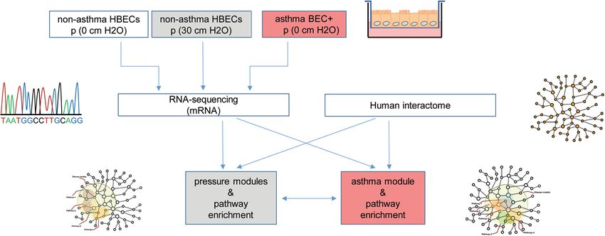
www.nature.com/scientificreports/                                                           www.nature.com/scientificreports

                           Figure 2. Asthma signature in HBECs. (a) Volcano plot representation of gene expression changes between
                           healthy non-asthmatic and asthmatic HBECs. (b) Pathway enrichment analysis for genes upregulated in the
                           asthmatic HBECs compared to non-asthmatic HBECs. (c) A subnetwork of the asthma disease module is
                           shown. Activated pathways are highlighted in colours. RNA expression of genes promoting inflammation,
                           including the chemokines (d) Ccl2, (e) Cxcl8, sphingosine-1-phosphate receptors (f) S1PR1, (g) S1PR3
                           as well as the secreted protein (h) TNFSF14. Asthmatic HBECs express elevated remodeling associated
                           factors, including (i) tenascin (TNC), (j) fibronectin 1(FN1), (k) collagen 4 chain Col4a1 and (l) matrix
                           metalloproteinase MMP2. (m) Venn diagram summarizes the gene overlap between the asthma module and
                           publicly available epithelial derived datasets deposited under GSE63142 and GSE37693. Values summarize the
                           expression levels for n = 8 independent samples per group. The box and whisker plots represent the minimum,
                           25th percentile, median, 75th percentiles and the maximum. *p < 0.05, to control was considered significant.

                           the upregulated genes to gene ontology analyses for cellular compartments revealed an enrichment in focal adhe-
                           sions (GO:0005925) and the cytoskeleton (GO:0005856) as summarized in Table 3.
                               This prompt reaction of the epithelium subsequently transitioned into a strong repair/fibrotic response at
                           24 hr post-compression. Among the 512 DE genes (upregulated: 294; downregulated: 220) (Fig. 3d,e), were those
                           coding for the pro-fibrotic factors TGF-β and PDGF-β (Fig. 4a,b), various collagen chains and matrix metallo-
                           proteinases (MMPs) (Fig. 4c–f). We validated single molecules in independent samples using real-time RT-PCR
                           (Supplementary Fig. S4). These molecules and pathways have been described in chronic disease conditions and
                           had previously been thought to emerge as a result of a chronic inflammatory response (Fig. 3g and Table 4).
                               In addition to extracellular matrix proteins, compressive stress induced the expression of soluble immune
                           modulatory factors, including 4-1BB (TNFSF9; FC: 1.75) and LIGHT (TNFSF14; FC: 2.12) in the later phase
                           (Supplementary Fig. S5a,b). These data provide evidence for the ability of compressive stress in the absence of
                           inflammation to enforce pronounced gene alterations in non-asthmatic HBECs.

                           Compressive stress induces a disease signature resembling asthma.                   To address the question of
                           whether compressive stresses might initiate an immune response as observed in asthma, we assessed the expres-
                           sion level of epithelium derived Th2-promoting factors in non-asthmatic HBECs exposed to compression.
                           Epithelial alarmins, including IL-33 (Fig. 5a), thymic stromal lymphopoietin (TSLP; Fig. 5b) as well as CXCL8
                           (Fig. 5c) were strongly induced in HBECs from non-asthmatic donors immediately after mechanical stimulus
                           and receded at the 24 hr time point. Induction of IL-33 and IL-8 were validated in independent samples using
                           real-time RT-PCR (Supplementary Fig. S6a,b).
                               In non-asthmatic HBECs, compressive stress promptly (3 hr time point) induced a set of genes mapping to
                           ligands and receptors coupled to G-proteins in the asthma module. These included S1PR1 and prostaglandin
                           E receptor (PTGER) 4 (Fig. 5d,e). Real-time RT-PCR confirmed the induction of PTGER4 expression in com-
                           pressed epithelial cells (Supplementary Fig. S6c). Genes induced at 24 hr post-compression cover the area of the

Scientific Reports |   (2020) 10:966 | https://doi.org/10.1038/s41598-020-57755-8                                                         3
www.nature.com/scientificreports/ www.nature.com/scientificreports
adj. p-valuea
Pathway (Bonferroni) genes in pathway
COL4A2, ICAM1, COL4A1, LAMA1, ITGB6, THBS1, TNC,
1 Integrin cell surface interaction 4.25E-05
VCAM1, FN1
CXCL8, IL1B, CCL2, TNFRSF9, CXCL3, CXCL5, IL6, IFNE,
2 Cytokine- cytokine- receptor interaction 1.70E-04
TNFSF14, TNFSF15, CSF1, BMP2, TNFRSF10C, CCL20
CCL2, CXCL3, CXCL8, HTR7, S1PR1, AGT, EDNRA, MTNR1A,
3 Class A1 Rhodopsin receptor interaction 4.74E-03
GPR68, CXCL5, S1PR3, NPBWR1, CCL20
4 Platelet amyloid precursor protein pathway 6.39E-03 SERPINE1, COL4A2, PLAT, COL4A1
5 ECM receptor interaction 1.18E-02 COL4A2, FN1, COL4A1, LAMA1, ITGB6, THBS1, TNC
6 Extracellular matrix organization 1.49E-02 COL4A2, COL4A1, MMP2, TLL2, MMP7, MMP17, COL22A1
7 local acute inflammatory response pathway 1.49E-02 VCAM1, CXCL8, ICAM1, IL6
Transport of glucose and other sugars, bile, SLC22A3, SLC13A5, SLC39A8, SLC2A3, RHCG, SLC39A2,
8 1.73E-02
salts, acids, metal ion, and amine compounds SLC6A12
CCL2, CXCL3, CXCL8, HTR7, S1PR1, AGT, ENDRA, FZD7,
9 GPCR ligand binding 2.24E-02
MTNR1A, GPR68, CXCL5, S1PR3, NPBWR1, CCL20
10 Cartilage oligomeric matrix protein pathway 3.99E-02 C1S, C1R, CFB, C4A
Table 1. Active pathways in asthmatic HBECs compared to healthy HBEC at baseline. aAdjusted p-value:
p-value was calculated using the Fisher exact test and adjustment was done using Bonferroni correction.
asthma module associated with matrix remodeling and map to extracellular –matrix organization, ECM-receptor
interaction and Integrin-cell surface interaction (yellow, blue and red highlighted pathways (Fig. 3g).
These findings support the hypothesis that mechanical stimuli can induce disease-relevant gene expression in
non-asthmatic HBECs and do so in the absence of prior inflammatory stimuli.
Discussion
We report here that application of mechanical compression —akin to that which occurs during bronchospasm—
is sufficient to evoke far-reaching molecular changes in human bronchial epithelial cells. We show, further, that
these changes merge into an asthma-like molecular phenotype. Over time, initial transcriptional differences
between non-asthmatic and asthmatic HBECs gradually faded and aligned to a similar molecular signature, com-
prising the induction of remodeling associated genes. Compression induced a prompt expression of inflamma-
tory mediators promoting a type 2 inflammatory response. In non-asthmatic HBECs, early alterations in the
compression response translated into a long-term repair/fibrotic response.
The airway epithelial lining forms a continuous tight barrier to protect the host from inhaled irritants and
invading pathogens. Upon insult, epithelial cells become activated, secrete alarmins and pro-inflammatory mol-
ecules to recruit accessory immune cells and to induce proliferation and polarization programs aiming to restore
epithelial integrity35. The asthmatic HBECs used in this study display a stable activated gene expression profile
at baseline, which suggests a stable transcriptional program in these cells. It is unclear if a genetic or epigenetic
signature determines the activated profile of asthmatic cells. Future studies might be directed towards the identi-
fication of molecular mechanisms to determine inherent differences between non-asthmatic and asthmatic air-
way epithelial cells and whether mechanical stimuli regulate any of these events. However, this work represents
only the compression and asthmatic response of well-differentiated HBECs in vitro, so we cannot rule out that
additional factors come into play in vivo. Furthermore, extending these approaches by capturing the dynamics of
the cellular responses initiated by compressive stress and how these are propagated into an asthmatic phenotype
will shed light on mechanisms which remain hidden by analysing only static networks. Analysing further biolog-
ical information from other omics such as miRNA, proteomics and metabolomics, and integrating these, with
the existing gene expression data will likely enhance the knowledge of how asthma can manifest at the airway
epithelium.
In previous studies analysing the compression mediated response in HBECs only single genes have been
described, while there has not been a network-based approach integrating the early events occurring upon appli-
cation of acute compression to an asthma phenotype. Here we used HBECs from non-asthmatic and asthmatic
donors to assess the functional consequences of compression on normal airway epithelial cells. Despite the small
sample size, with which marginal differences due to high variability in gene expression were expected, we were
able to detect marked differences in gene signature between HBECs from non-asthmatic and asthmatic donors
at baseline as depicted in the asthma module, that overlapped with previously reported asthma-related genes and
pathways in both human and mouse2,36–38. To further address the issue of relatively few asthmatic and mostly
female donors in our experiment, we compared our retrieved disease signature, DE gene expression between
asthmatic and non-asthmatic HBECs at baseline, with published asthma GEO datasets and calculated the overlap
in DE-gene signature. The datasets were chosen to reflect the experimental set up of ALI-cultures (GSE37693)
as well as the in vivo condition by analysing bronchial brushings isolated from severe asthmatics (GSE63142). In
latter data set, both male and female donors were included. In GSE37693, stimulation of non-asthmatic HBECs
with IL-13, mimicking the impact of Th2-inflammation on HBECs, induced a gene signature, that significantly
overlapped (60.1%) with our asthma module. Even in epithelial cells isolated from severe asthmatic patients, a
significant overlap (37%) in gene expression was observed (Fig. 2m).
The goal of this network approach was to map out the connectivity structure between genes that are affected
by disease or perturbation (compressive pressure), instead of aiming for the identification of single differential
Scientific Reports | (2020) 10:966 | https://doi.org/10.1038/s41598-020-57755-8 4www.nature.com/scientificreports/ www.nature.com/scientificreports
Figure 3. Compression induced molecular changes in HBECs. (a) Workflow of the methodology used to
describe the overlapping and aligning expression signature between healthy HBECs exposed to compression
and asthmatic HBECs at baseline. Expression data are collected and then, differentially expressed genes and
miRs are mapped to the human interactome, resulting in an early and a late compression disease module.
These were compared with the asthma disease module generated in Fig. 1. (b) Volcano plot visualizing DE
genes at 3 hr post pressure application and pathways enriched in the early compression module. (c) Volcano
plot visualizing DE genes at 24 hr post pressure application (d) and pathways enriched in the late compression
module. (e) Visualization of the early (f) and late (g) pressure subnetworks. Activated pathways, overlapping
with the asthma subnetwork are highlighted in colour.
expressed genes. For this, DE- gene information was used to generate the disease and pressure modules. In
order to not apriori exclude genes and thus allow a more holistic view, a minimum p-value of 0.05 was used as a
cut-off20. We validate this approach by retrieving many known genes based on the literature and our GEO confir-
mation analysis. To capture the molecular response, two time points were chosen to reflect initial inflammation
and later remodeling. Besides the previously described autocrine and paracrine acting growth factors, including
TGF-β39–41 and HB-EGF42, we describe multiple pathways initially affected by mechanical stimuli (Tables 2 and
3). A strong suppression of developmental genes, including Wnt and BMP proteins (Supplementary Fig. S3),
was detectable promptly after compression and was accompanied by a marked induction of epithelial alarmins,
including IL-33 and TSLP. While a supportive activity of these factors has been documented for a variety of
accessory immune cells43,44, IL-33 and TSLP are central for example to the initiation and enhancement of type
2 response typically seen in asthmatic inflammation45–48. Following compression (3 hr), an increased release of
these factors from airway epithelial cells could activate type 2 innate lymphoid cells (ILC2) and augment type 2
inflammation in the lung43,48,49.
Increased expression of PTGER4 and S1PR1 further support the pro-inflammatory and immune-modulatory
outcome. Signalling via PTGER4 stimulates the activation of PKA via cAMP and induces transcription of
CREB-dependent genes 50. As a downstream target, we could detect increased IL-11 expression at 24 hrs
post-compression (Supplementary Fig. S7)51. IL-11 is essential for allergic sensitization, inflammation and airway
remodeling in mice52 and reported to drive fibrotic responses, including cardiac and renal fibroblasts in humans53.
Sphingolipid levels are elevated in the lungs of patients with allergic asthma. Specific blockade of sphingosine
Scientific Reports | (2020) 10:966 | https://doi.org/10.1038/s41598-020-57755-8 5www.nature.com/scientificreports/ www.nature.com/scientificreports
adj. p-valuea
Pathway (Bonferroni) genes in pathway
1 Hedgehog signalling 3.55E-07 RAB23, GLI3, WNT3A, GAS1, BMP7, WNT4, BMP2, WNT10A
2 Focal adhesion 4.32E-06 CAV1, ACTG1, ITGB3, ITGA9, PDGFA, TNC, THBS4, MYL9, VCL, ZYX, JUN, SPP1
P2RY1, GPR39, PTGER4, WNT3A, ACKR3, ADM2, ADM, FZD8, WNT4, EDNRA,
3 GPCR ligand binding 1.12E-05
EDN1, EDN2, WNT10A, FZD2, S1PR5, NPBWR1, S1PR1
DUSP6, DUSP8, DDIT3, CACNA2D2, DUSP5, PDGFA, FOS, SRF, RASA2, JUN,
4 MAPK signalling pathway 2.46E-05
HSPA6, FGF1, DUSP9
Table 2. Pathways enriched immediately after compressive stress. (3 hr). aAdjusted p-value: p-value was
calculated using the Fisher exact test and adjustment was done using Bonferroni correction.
adj.
Term p-valuea Genes
DST, TPM4, FBLIM1, ITGB3, TGFB1I1, FHL1, SPRY4, FHL2,
Focal Adhesion (GO:0005925) 9.15E-10 PLAUR, TNC, MSN, RHOB, CSRP1, PALLD, DLC1, MYH9, LCP1,
FERMT2, PDLIM7, VCL
DST, TPM4, MSN, NUAK1, KRT17, PALLD, CDC42EP2, MYH9,
Cytoskeleton (GO:0005856) 2.23E-04
SPRY2, STK38L, LCP1, PDLIM5, NES, ULBP1, PDLIM7, VCL
TPM4, PALLD, DLC1, MYADM, MYH9, STK38L, LCP1, PDLIM5,
Actin Cytoskeleton (GO:0015629) 4.40 E-3
ULBP1, PDLIM7
Contractile Actin Filament bundle (GO:0097517) 6.15 E-03 TPM4, FBLIM1, MYH9, LCP1
Stress Fibre (GO:0001725) 6.15 E-03 TPM4, FBLIM1, MYH9, LCP1
Actomyosin (GO:0042641) 1.02 E-02 TPM4, FBLIM1, MYH9, LCP1
Table 3. Gene ontology analysis of genes immediately induced after compression. aAdjusted p-value: p-value
was calculated using the Fisher exact test and adjustment was done using Bonferroni correction.
kinase 1 attenuates airway inflammation and hyper reactivity in mice54 further supporting the pro-inflammatory
role of S1P signalling in bronchial epithelial cells.
In this acute compressive stress response, an induced expression of FOS and JUN, which together form the
AP-1 transcription factor implicate a longer lasting change in protein expression post the analysis time points55.
The later response is marked by an increased expression of several collagen chains as well as matrix metallopro-
teinases, accompanied by elevated pro-fibrotic factors TGF-β and PDGF-β. Taken together we observed a gradual
alignment of the compression response with the asthma module, linking both phenomena with each other.
This analysis represents the first attempt in any biological system to understand the impact of mechani-
cal forces on disease pathways using a network approach. The distinct activated profile of asthmatic epithelial
cells suggests an “imprinted” signature in the absence of inflammation. It remains unclear, however, if a single
compression may suffice to prime HBE cells for future reactions or if a repetitive stimulus would be needed to
imprint an inflammatory signature as observed in asthmatic HBECs at baseline. The secretory activity of asth-
matic HBECs at baseline could provide survival signals for resident innate and adaptive immune cells in the lung
post-inflammation. Thus far it is postulated that the presence of antigenic molecules at the site of inflamma-
tion could activate survival programs in tissue resident memory immune cells and retain these in the previously
inflamed organ56,57. Data provided here support the notion of a fundamental role of the airway epithelium in the
control of local immune responses (Supplementary Fig. S8).
In the previous studies we have shown that compressive stress applied to HBECs induces a transition of the
epithelial layer from a solid-like, immobile, jammed phase to a fluid-like mobile, unjammed phase, which is
termed an unjamming transition5,58,59. This unjamming transition is reflected in the shape and mobility of the
cells5,58,59. While the molecular mechanisms underlying this unjamming transition are not understood, the gene
ontology analysis performed in this study provides new insights into the potential molecular mechanisms. For
example, when mechanical compression caused cell layer unjamming the genes that were upregulated included
those associated with focal adhesions and cytoskeleton (Table 3), which are responsible for internal force gener-
ation and maintaining cell shape. It remains unclear, however, as to whether the identified genes are the cause of
the unjamming transition or the effect.
Material and Methods
Primary human bronchial epithelial cells. Primary HBECs were cultured in air-liquid interface (ALI)
conditions as previously described5,7,60–62. We used HBECs from 4 donors with no pre-existing chronic lung dis-
ease (from here on referred to as non-asthmatic) and 4 asthmatic donors (Supplementary Table 1). HBECs were
obtained at passage 0 or passage 1 from the Marisco Lung Institute at the University of North Carolina, Chapel
Hill. Passage 2 cells were plated on transwell inserts coated with type I collagen and grown under submerged
conditions for 5-6 days until the cells reached confluence. For each donor, two samples were introduced into the
polarization process. Since every polarization runs independently for each well, these samples were considered as
independent samples. To initiate ALI culture, apical media were removed and only basal media were subsequently
fed every 2 days for additional 15 days, where the cells were well-differentiated with the appearance of basal, gob-
let, and ciliated cells (Supplementary Fig. S1)61.
Scientific Reports | (2020) 10:966 | https://doi.org/10.1038/s41598-020-57755-8 6www.nature.com/scientificreports/ www.nature.com/scientificreports
Figure 4. Compression induced remodeling associated genes. Genes, related to fibrotic responses, are elevated
at 24 hr post compression. This list includes the soluble factors (a) platelet-derived growth factor β (PDGFB)
and (b) transforming growth factor-β2 (TGFB2). As downstream targets of the fibrotic response we highlight
(c) Col1a1, (d) Col4a1, (e) matrix metalloproteinases MMP-2 and (f) MMP-10. Values summarize the
expression levels for n = 8 independent samples per group. The box and whisker plots represent the minimum,
25th percentile, median, 75th percentiles and the maximum. *p < 0.05 compared to control was considered
significant.
Human lungs unsuitable for transplantation, including two cases of fatal asthma and two with asthma in the
medical social history, were obtained from either Carolina Donor Services (Durham, NC), the National Disease
Research Interchange (Philadelphia, PA), or the International Institue for Advancement of Medicine (Edison, NJ)
under protocol #03-1396 approved by the University of North Carolina at Chapel Hill Biomedical Institutional
Review Board. Informed consent was obtained from authorized representatives of all organ donors.
Mechanical compression of HBECs. During asthma exacerbations, the excessive smooth muscle contrac-
tion causes the airway wall to buckle63. In the original development of mechanical compression model, Drazen
and colleagues first used finite element analysis to compute the degree of compressive stresses imposed on the
buckled airway epithelium by bronchospasm64. The estimated mechanical forces during maximal bronchos-
pasm, is approximately 30 cm H2O. Using those computations as a guide, this team then went on to test the
effect of mechanical compression with 30 cm H2O empirically in ALI culture of bronchial epithelial cells so as to
optimize epithelial mechano-transduction responses9. Subsequently, this same apico-to-basal mechanical com-
pression, was found to induce events that occur in the remodeled asthmatic airway, including increased matrix
deposition62, goblet cell hyperplasia, and airway smooth muscle hyperplasia and hypercontraction, as well as
production of asthma-associated mediators, including maspin, YKL-40, and tissue-factor positive extracellular
vesicles7,9,13,61,65–67.
Well-differentiated HBECs cultured from non-asthmatic or asthmatic donors were mechanically compressed
on ALI day 145,7,61,62. At 20 hours prior to initiation of compression, cells were starved of bovine pituitary extract
and epidermal growth factor. Cells were then exposed to 0 or 30 cm H2O of apical-to-basal transcellular pressure
for 3 hours, thus mimicking the mechanical compression that occurs during bronchospasm3. Both control and
compressed HBECs were harvested either immediately at 3 hr post compression, immediately after releasing
pressure or at 24hr-post compression initiation, including an incubation period of 21 hours post pressure release.
Harvested cells were used for isolation of RNA.
RNA-Isolation, Library preparation and RNA sequencing. Total RNA was isolated organically using
QIAzol lysis reagent and the Qiagen miRNeasy Kit (Qiagen). Quality was assessed using the Nanodrop 8000
®
spectrophotometer. Sequencing libraries were constructed with the TruSeq Stranded Total RNA Library Prep
Globin Kit (Illumina). Sequencing was performed using a HiSeq. 2500 instrument (Illumina). Trimmed reads
Scientific Reports | (2020) 10:966 | https://doi.org/10.1038/s41598-020-57755-8 7www.nature.com/scientificreports/ www.nature.com/scientificreports
adj.
Pathway p-valuea genes in pathway
COL4A3, COL4A2, COL16A1, MMP3, COL4A1, MMP11, MMP13, COL4A4,
1 Extracellular matrix organization 6.03E-11
COL1A1, MMP2, MMP10, COL5A2, COL8A2, COL7A1
COL4A2, CAV1, COMP, COL4A1, ITGB3, ITGB6, ITGA9, PDGFB, THBS1,
2 Focal adhesion 2.30E-09
TNC, COL1A1, COL4A4, PDGFD, MYL9, JUN, COL5A2, ITGA4, ACTN1
COL4A2, COL4A4, COMP, COL4A1, ITGB3, ITGA9, ITGB6, THBS1, TNC,
3 ECM receptor interaction 5.84E-09
COL1A1, COL5A2, ITGA4,
COL4A3, COL4A2, COL4A4, COL4A1, ITGB3, ITGA9, ITGB6, THBS1,
4 Integrin cell surface interaction 3.53E-07
TNC, COL1A1
SMAD7, LTBP1, COMP, THBS1, INHBA, BMP6, BMP7, TGFB2, CDKN2B,
5 TGFbeta pathway 7.88E-07
FST
CXCL8, CXCR2, KIT, TNFSF13B, IL11, INHBA, TNFSF9, PDGFB, IL6R,
6 Cytokine- cytokine- receptor interaction 9.01E-07
TNFSF14, CXCR4, CXCL14, BMP7, LIF, TGFB2, NGFR, GHR
COL4A3, COL4A2, COL4A4, COL4A1, PDGFB, THBS1, COL1A1, PDGFD,
7 PDGF pathway 1.88E-05
COL5A2, CAMK4,
8 Kinesins 2.72E-05 KIF4A, KIF15, KIF2C, KIF11, KIFC1
Table 4. Pathways enriched after compressive stress. (24 h). aAdjusted p-value: p-value was calculated using the
Fisher exact test and adjustment was done using Bonferroni correction.
were mapped to The GRCh38 reference genome using STAR68. Read counts were computed with htseq.69. Data
were normalized and analysed with DESeq. 270 with an FDR < 0.05.
Gene ontology analysis. To describe the immediate changes induced by compression in BECs, we per-
formed functional annotation analysis for upregulated genes. We used the Enrich71 method to obtain enriched
GOCC terms.
Gene expression data for ALI-grown bronchial epithelial cells stimulated with IL-13 (GSE37693; n = 6 for
each condition)72 and bronchial brushings from severe asthmatics (n = 56) compared to healthy controls (n = 27)
(GSE63142)73 were retrieved from the GSE database.
Protein-protein interaction network. To build a comprehensive human protein-protein interactome
(PPI), we combined 15 databases with various kinds of experimental evidence that are currently available. The
current updated human interactome includes 246,995 interactions connecting 16,706 unique proteins, which is
more than 40% larger in number in comparison with our previously used human interactome74. Specifically, we
focused on the high-quality PPIs with four types of data:
(1) binary PPIs tested by high-throughput yeast-two-hybrid (Y2H) systems: we combined binary PPIs tested
from two public available high-quality Y2H datasets75,76 and one unpublished dataset. This resource is
available online at (https://ccsb.dana-farber.org/interactome-data.html).
(2) kinase-substrate interactions by literature-derived low-throughput and high-throughput experiments
from KinomeNetworkX77, Human Protein Resource Database (HPRD)78, PhosphoNetworks79,80, Phos-
phositePlus81, DbPTM 3.082, and Phospho. ELM83.
(3) carefully literature-curated PPIs identified by affinity purification followed by mass spectrometry (AP-MS),
Y2H and by literature-derived low-throughput experiments, and protein three-dimensional structures
from BioGRID84, PINA85, Instruct86, HPRD78, MINT87, IntAct19, and InnateDB88.
(4) signaling network by literature-derived low-throughput experiments as annotated in SignaLink2.089.
All data were downloaded in December 2015. The genes were mapped to their Entrez ID based on the NCBI
database90 as well as their official gene symbols based on GeneCards (http://www.genecards.org/). Duplicated
pairs were removed. Data from inferred data, such as evolutionary analysis, gene expression data, and metabolic
associations were excluded.
Quantitative real-time RT-PCR. Two μg of total RNA was used to synthesize cDNA using MultiScribe
reverse transcriptase (Thermo Fisher Scientific), as described previously65. Quantitative real-time PCR was per-
formed using iTaq Universal SYBR Green Supermix (BioRad). Following an initial denaturation step at 95 °C
for 10 minutes, 40 cycles of PCR at 95 °C for 15 seconds followed by at 60 °C for 60 seconds were performed in
a Bio-rad CFX96 real-time PCR detection system. Primers specific for COL1A1 (5′-CAC ACG TCT CGG TCA
TGG TA -3′/5′-AAG AGG AAG GCC AAG TCG AG-3′); FN1 (5′-CCC CAT TCC AGG ACA CTT CT-3′/5′-
TGC CTC CAC TAT GAC GTT GT-3′); IL-8 (5′-CAC CGG AAG GAA CCA TCT CA-3′/5′-AGA GCC ACG GCC AGC
TT-3′); IL-33 (5′- TGC ATG CCA ACA ACA AGG AA-3′/5′-AAG GAC AAA GAA GGC CTG GT-3′); (5′- ACT CTT
TTG ATG GCC CAG GA-3′/5′- GAG TGG CCA AGT TCA TGA GC-3′); MMP-10 (5′- ACT CTT TTG ATG GCC
CAG GA-3′/5′- GAG TGG CCA AGT TCA TGA GC-3′); PTGER4 (5′-TAC TCA TTG CCA CCT CCC
TG-3′/5′-ATT CGG ATG GCC TGC AAA TC-3′) and GAPDH (5′- TGG GCT ACA CTG AGC ACC AG -3′/5′-
GGG TGT CGC TGT TGA AGT CA-3′) were used to test the mRNA expression. PCR quantification was done
with the 2−ΔΔCT method, with normalization to Gapdh as the housekeeping gene. All standard procedures were
performed according to the manufacturer′s instructions.
Scientific Reports | (2020) 10:966 | https://doi.org/10.1038/s41598-020-57755-8 8www.nature.com/scientificreports/ www.nature.com/scientificreports
Figure 5. Compression induced alarmins and Th2-promoting mediators in healthy non-asthmatic HBECs.
Compression on HBECs immediately (3 hr) induced the expression of (a) IL33, (b) Th2-promoting thymic
stromal lymphopoietic protein (TSLP) as well as Cxcl8 (c). This response was accompanied by immediate
increase in lipid-mediator receptors (d) S1PR1 and (e) Prostaglandin E Receptor 4 (PTGER4). Values
summarize the expression levels for n = 8 independent samples per group. The box and whisker plots represent
the minimum, 25th percentile, median, 75th percentiles and the maximum. *p < 0.05 compared to control was
considered significant.
Statistical analysis. Results summarize the RNA sequencing results of four individuals per group. Each
individual was assessed in replicate and the average values were used for further analysis. All computations
were performed with Python 2.7. Log2 transformed values were used to compare experimental groups with
Mann-Whitney U test. Where appropriate, a Wilcoxon matched-pairs signed rank test was done. p < 0.05 was
considered significant. We calculated the significance of the overlap of the here described “disease signature” and
the DE-genes from publicly available data sets GSE37693 and GSE63142 using a gene set enrichment analysis.
Data availability
The RNA-sequencing data that support the findings of this study are available upon request from the senior
author of this article.
Received: 23 July 2019; Accepted: 23 December 2019;
Published: xx xx xxxx
References
1. Jackson, D. J. et al. Evidence for a causal relationship between allergic sensitization and rhinovirus wheezing in early life. American
journal of respiratory and critical care medicine 185, 281–285, https://doi.org/10.1164/rccm.201104-0660OC (2012).
2. Holgate, S. T. The sentinel role of the airway epithelium in asthma pathogenesis. Immunological reviews 242, 205–219, https://doi.
org/10.1111/j.1600-065X.2011.01030.x (2011).
3. Jartti, T. & Gern, J. E. Role of viral infections in the development and exacerbation of asthma in children. The Journal of allergy and
clinical immunology 140, 895–906, https://doi.org/10.1016/j.jaci.2017.08.003 (2017).
4. Park, J.-A. et al. Compressive Stress Causes an Unjamming Transition and an Epithelial-Mesenchymal Transition in the Airway
Epithelium in Asthma. Annals of the American Thoracic Society 13(Suppl 1), S102, https://doi.org/10.1513/AnnalsATS.201506-
382MG (2016).
5. Park, J.-A. et al. Unjamming and cell shape in the asthmatic airway epithelium. Nature materials 14, 1040–1048, https://doi.
org/10.1038/nmat4357 (2015).
6. Tschumperlin, D. J. & Drazen, J. M. Chronic effects of mechanical force on airways. Annual review of physiology 68, 563–583, https://
doi.org/10.1146/annurev.physiol.68.072304.113102 (2006).
Scientific Reports | (2020) 10:966 | https://doi.org/10.1038/s41598-020-57755-8 9www.nature.com/scientificreports/ www.nature.com/scientificreports
7. Park, J.-A. et al. Tissue factor-bearing exosome secretion from human mechanically stimulated bronchial epithelial cells in vitro and
in vivo. The Journal of allergy and clinical immunology 130, 1375–1383, https://doi.org/10.1016/j.jaci.2012.05.031 (2012).
8. Shiomi, T. et al. TNF-α-converting enzyme/a disintegrin and metalloprotease-17 mediates mechanotransduction in murine tracheal
epithelial cells. American journal of respiratory cell and molecular biology 45, 376–385, https://doi.org/10.1165/rcmb.2010-0234OC
(2011).
9. Tschumperlin, D. J. et al. Mechanotransduction through growth-factor shedding into the extracellular space. Nature 429, 83–86,
https://doi.org/10.1038/nature02543 (2004).
10. Mitchel, J. A. et al. IL-13 Augments Compressive Stress-Induced Tissue Factor Expression in Human Airway Epithelial Cells.
American journal of respiratory cell and molecular biology 54, 524–531, https://doi.org/10.1165/rcmb.2015-0252OC (2016).
11. Grainge, C. L. et al. Effect of bronchoconstriction on airway remodeling in asthma. The New England journal of medicine 364,
2006–2015, https://doi.org/10.1056/NEJMoa1014350 (2011).
12. Loxham, M. & Davies, D. E. Phenotypic and genetic aspects of epithelial barrier function in asthmatic patients. The Journal of allergy
and clinical immunology 139, 1736–1751, https://doi.org/10.1016/j.jaci.2017.04.005 (2017).
13. Tschumperlin, D. J., Shively, J. D., Kikuchi, T. & Drazen, J. M. Mechanical stress triggers selective release of fibrotic mediators from
bronchial epithelium. American journal of respiratory cell and molecular biology 28, 142–149, https://doi.org/10.1165/rcmb.2002-
0121OC (2003).
14. Ressler, B., Lee, R. T., Randell, S. H., Drazen, J. M. & Kamm, R. D. Molecular responses of rat tracheal epithelial cells to
transmembrane pressure. American journal of physiology. Lung cellular and molecular physiology 278, L1264–72, https://doi.
org/10.1152/ajplung.2000.278.6.L1264 (2000).
15. Park, J.-A., Fredberg, J. J. & Drazen, J. M. Putting the Squeeze on Airway Epithelia. Physiology (Bethesda, Md.) 30, 293–303, https://
doi.org/10.1152/physiol.00004.2015 (2015).
16. Huang, Y., Crawford, M., Higuita-Castro, N., Nana-Sinkam, P. & Ghadiali, S. N. miR-146a regulates mechanotransduction and
pressure-induced inflammation in small airway epithelium. FASEB journal: official publication of the Federation of American Societies
for Experimental Biology 26, 3351–3364, https://doi.org/10.1096/fj.11-199240 (2012).
17. Neth, P., Nazari-Jahantigh, M., Schober, A. & Weber, C. MicroRNAs in flow-dependent vascular remodelling. Cardiovascular
research 99, 294–303, https://doi.org/10.1093/cvr/cvt096 (2013).
18. Iwawaki, Y. et al. MiR-494-3p induced by compressive force inhibits cell proliferation in MC3T3-E1 cells. Journal of bioscience and
bioengineering 120, 456–462, https://doi.org/10.1016/j.jbiosc.2015.02.006 (2015).
19. Orchard, S. et al. The MIntAct project–IntAct as a common curation platform for 11 molecular interaction databases. Nucleic acids
research 42, D358–63, https://doi.org/10.1093/nar/gkt1115 (2014).
20. Kılıç, A. et al. A systems immunology approach identifies the collective impact of 5 miRs in Th2 inflammation. JCI Insight 3; https://
doi.org/10.1172/jci.insight.97503 (2018).
21. Lee, Y. G. et al. Recruited alveolar macrophages, in response to airway epithelial-derived monocyte chemoattractant protein 1/CCl2,
regulate airway inflammation and remodeling in allergic asthma. American journal of respiratory cell and molecular biology 52,
772–784, https://doi.org/10.1165/rcmb.2014-0255OC (2015).
22. Post, S. et al. ADAM10 mediates the house dust mite-induced release of chemokine ligand CCL20 by airway epithelium. Allergy 70,
1545–1552, https://doi.org/10.1111/all.12730 (2015).
23. Al-Alwan, L. A. et al. Differential roles of CXCL2 and CXCL3 and their receptors in regulating normal and asthmatic airway smooth
muscle cell migration. Journal of immunology (Baltimore, Md.: 1950) 191, 2731–2741, https://doi.org/10.4049/jimmunol.1203421
(2013).
24. Rohde, G. et al. CXC chemokines and antimicrobial peptides in rhinovirus-induced experimental asthma exacerbations. Clinical
and experimental allergy: journal of the British Society for Allergy and Clinical Immunology 44, 930–939, https://doi.org/10.1111/
cea.12313 (2014).
25. Rivera, J., Proia, R. L. & Olivera, A. The alliance of sphingosine-1-phosphate and its receptors in immunity. Nature reviews.
Immunology 8, 753–763, https://doi.org/10.1038/nri2400 (2008).
26. Peters, M. C. et al. Plasma interleukin-6 concentrations, metabolic dysfunction, and asthma severity: a cross-sectional analysis of
two cohorts. The Lancet. Respiratory medicine 4, 574–584, https://doi.org/10.1016/S2213-2600(16)30048-0 (2016).
27. Kim, R. Y. et al. Role for NLRP3 Inflammasome-mediated, IL-1β-Dependent Responses in Severe, Steroid-Resistant Asthma.
American journal of respiratory and critical care medicine 196, 283–297, https://doi.org/10.1164/rccm.201609-1830OC (2017).
28. Lehmann, M., Baarsma, H. A. & Königshoff, M. WNT Signaling in Lung Aging and Disease. Annals of the American Thoracic Society
13, S411–S416, https://doi.org/10.1513/AnnalsATS.201608-586AW (2016).
29. Hines, E. A. & Sun, X. Tissue crosstalk in lung development. Journal of cellular biochemistry 115, 1469–1477, https://doi.org/10.1002/
jcb.24811 (2014).
30. Tadokoro, T., Gao, X., Hong, C. C., Hotten, D. & Hogan, B. L. M. BMP signaling and cellular dynamics during regeneration of airway
epithelium from basal progenitors. Development (Cambridge, England) 143, 764–773, https://doi.org/10.1242/dev.126656 (2016).
31. Idrovo, J.-P. et al. Combination of adrenomedullin with its binding protein accelerates cutaneous wound healing. PloS one 10,
e0120225, https://doi.org/10.1371/journal.pone.0120225 (2015).
32. Kohan, M., Breuer, R. & Berkman, N. Osteopontin induces airway remodeling and lung fibroblast activation in a murine model of
asthma. American journal of respiratory cell and molecular biology 41, 290–296, https://doi.org/10.1165/rcmb.2008-0307OC (2009).
33. Sperry, R. B. et al. Zyxin controls migration in epithelial-mesenchymal transition by mediating actin-membrane linkages at cell-cell
junctions. Journal of cellular physiology 222, 612–624, https://doi.org/10.1002/jcp.21977 (2010).
34. Snyder, J. C., Zemke, A. C. & Stripp, B. R. Reparative capacity of airway epithelium impacts deposition and remodeling of
extracellular matrix. American journal of respiratory cell and molecular biology 40, 633–642, https://doi.org/10.1165/rcmb.2008-
0334OC (2009).
35. Ritchie, A. I., Jackson, D. J., Edwards, M. R. & Johnston, S. L. Airway Epithelial Orchestration of Innate Immune Function in
Response to Virus Infection. A Focus on Asthma. Annals of the American Thoracic Society 13(Suppl 1), S55–63, https://doi.
org/10.1513/AnnalsATS.201507-421MG (2016).
36. Alrifai, M. et al. Compartmental and temporal dynamics of chronic inflammation and airway remodelling in a chronic asthma
mouse model. PloS one 9, e85839, https://doi.org/10.1371/journal.pone.0085839 (2014).
37. Cohen, L. et al. Epithelial cell proliferation contributes to airway remodeling in severe asthma. American journal of respiratory and
critical care medicine 176, 138–145, https://doi.org/10.1164/rccm.200607-1062OC (2007).
38. Fahy, J. V. Type 2 inflammation in asthma–present in most, absent in many. Nature reviews. Immunology 15, 57–65, https://doi.
org/10.1038/nri3786 (2015).
39. Ojiaku, C. A., Yoo, E. J. & Panettieri, R. A. Transforming Growth Factor β1 Function in Airway Remodeling and
Hyperresponsiveness. The Missing Link? American journal of respiratory cell and molecular biology 56, 432–442, https://doi.
org/10.1165/rcmb.2016-0307TR (2017).
40. Akdis, M. et al. Interleukins (from IL-1 to IL-38), interferons, transforming growth factor β, and TNF-α: Receptors, functions, and
roles in diseases. The Journal of allergy and clinical immunology 138, 984–1010, https://doi.org/10.1016/j.jaci.2016.06.033 (2016).
41. Spanjer, A. I. R. et al. TGF-β-induced profibrotic signaling is regulated in part by the WNT receptor Frizzled-8. FASEB journal:
official publication of the Federation of American Societies for Experimental Biology 30, 1823–1835, https://doi.org/10.1096/
fj.201500129 (2016).
Scientific Reports | (2020) 10:966 | https://doi.org/10.1038/s41598-020-57755-8 10www.nature.com/scientificreports/ www.nature.com/scientificreports
42. Hirota, N. et al. Histamine may induce airway remodeling through release of epidermal growth factor receptor ligands from
bronchial epithelial cells. FASEB journal: official publication of the Federation of American Societies for Experimental Biology 26,
1704–1716, https://doi.org/10.1096/fj.11-197061 (2012).
43. Lund, S. J. et al. Leukotriene C4 Potentiates IL-33-Induced Group 2 Innate Lymphoid Cell Activation and Lung Inflammation.
Journal of immunology (Baltimore, Md.: 1950) 199, 1096–1104, https://doi.org/10.4049/jimmunol.1601569 (2017).
44. Salter, B. M. A. et al. Human Bronchial Epithelial Cell-derived Factors from Severe Asthmatic Subjects Stimulate Eosinophil
Differentiation. American journal of respiratory cell and molecular biology 58, 99–106, https://doi.org/10.1165/rcmb.2016-0262OC
(2018).
45. Han, H., Roan, F. & Ziegler, S. F. The atopic march: current insights into skin barrier dysfunction and epithelial cell-derived
cytokines. Immunological reviews 278, 116–130, https://doi.org/10.1111/imr.12546 (2017).
46. Liu, T. et al. Type 2 Cysteinyl Leukotriene Receptors Drive IL-33-Dependent Type 2 Immunopathology and Aspirin Sensitivity.
Journal of immunology (Baltimore, Md.: 1950) 200, 915–927, https://doi.org/10.4049/jimmunol.1700603 (2018).
47. Li, Y. et al. Elevated Expression of IL-33 and TSLP in the Airways of Human Asthmatics In Vivo: A Potential Biomarker of Severe
Refractory Disease. Journal of immunology (Baltimore, Md.: 1950) 200, 2253–2262, https://doi.org/10.4049/jimmunol.1701455
(2018).
48. Nechama, M. et al. The IL-33-PIN1-IRAK-M axis is critical for type 2 immunity in IL-33-induced allergic airway inflammation.
Nature communications 9, 1603, https://doi.org/10.1038/s41467-018-03886-6 (2018).
49. Stier, M. T. et al. IL-33 promotes the egress of group 2 innate lymphoid cells from the bone marrow. The Journal of experimental
medicine 215, 263–281, https://doi.org/10.1084/jem.20170449 (2018).
50. Holgate, S. T., Peters-Golden, M., Panettieri, R. A. & Henderson, W. R. Roles of cysteinyl leukotrienes in airway inflammation,
smooth muscle function, and remodeling. The Journal of allergy and clinical immunology 111, S18–34; discussion S34–6 (2003).
51. Kawaguchi, M. et al. IL-17F-induced IL-11 release in bronchial epithelial cells via MSK1-CREB pathway. American journal of
physiology. Lung cellular and molecular physiology 296, L804–10, https://doi.org/10.1152/ajplung.90607.2008 (2009).
52. Lee, C. G. et al. Endogenous IL-11 signaling is essential in Th2- and IL-13-induced inflammation and mucus production. American
journal of respiratory cell and molecular biology 39, 739–746, https://doi.org/10.1165/rcmb.2008-0053OC (2008).
53. Schafer, S. et al. IL-11 is a crucial determinant of cardiovascular fibrosis. Nature 552, 110–115, https://doi.org/10.1038/nature24676
(2017).
54. Price, M. M. et al. A specific sphingosine kinase 1 inhibitor attenuates airway hyperresponsiveness and inflammation in a mast cell-
dependent murine model of allergic asthma. The Journal of allergy and clinical immunology 131, 501–11.e1, https://doi.org/10.1016/j.
jaci.2012.07.014 (2013).
55. Hermann-Kleiter, N. & Baier, G. NFAT pulls the strings during CD4+ T helper cell effector functions. Blood 115, 2989–2997,
https://doi.org/10.1182/blood-2009-10-233585 (2010).
56. Khan, T. N., Mooster, J. L., Kilgore, A. M., Osborn, J. F. & Nolz, J. C. Local antigen in nonlymphoid tissue promotes resident memory
CD8+ T cell formation during viral infection. The Journal of experimental medicine 213, 951–966, https://doi.org/10.1084/
jem.20151855 (2016).
57. McMaster, S. R. et al. Pulmonary antigen encounter regulates the establishment of tissue-resident CD8 memory T cells in the lung
airways and parenchyma. Mucosal immunology; https://doi.org/10.1038/s41385-018-0003-x (2018).
58. Park, J.-A., Atia, L., Mitchel, J. A., Fredberg, J. J. & Butler, J. P. Collective migration and cell jamming in asthma, cancer and
development. Journal of cell science 129, 3375–3383, https://doi.org/10.1242/jcs.187922 (2016).
59. Atia, L. et al. Geometric constraints during epithelial jamming. Nature Phys 14, 613–620, https://doi.org/10.1038/s41567-018-0089-
9 (2018).
60. Park, J.-A. et al. Human neutrophil elastase induces hypersecretion of mucin from well-differentiated human bronchial epithelial
cells in vitro via a protein kinase C{delta}-mediated mechanism. The American journal of pathology 167, 651–661 (2005).
61. Park, J.-A. & Tschumperlin, D. J. Chronic intermittent mechanical stress increases MUC5AC protein expression. American journal
of respiratory cell and molecular biology 41, 459–466, https://doi.org/10.1165/rcmb.2008-0195OC (2009).
62. Swartz, M. A., Tschumperlin, D. J., Kamm, R. D. & Drazen, J. M. Mechanical stress is communicated between different cell types to
elicit matrix remodeling. Proceedings of the National Academy of Sciences of the United States of America 98, 6180–6185, https://doi.
org/10.1073/pnas.111133298 (2001).
63. Yager, D. et al. Amplification of airway constriction due to liquid filling of airway interstices. Journal of applied physiology (Bethesda,
Md.: 1985) 66, 2873–2884, https://doi.org/10.1152/jappl.1989.66.6.2873 (1989).
64. Wiggs, B. R., Hrousis, C. A., Drazen, J. M. & Kamm, R. D. On the mechanism of mucosal folding in normal and asthmatic airways.
Journal of applied physiology (Bethesda, Md.: 1985) 83, 1814–1821, https://doi.org/10.1152/jappl.1997.83.6.1814 (1997).
65. Park, J.-A., Drazen, J. M. & Tschumperlin, D. J. The chitinase-like protein YKL-40 is secreted by airway epithelial cells at base line
and in response to compressive mechanical stress. The Journal of biological chemistry 285, 29817–29825, https://doi.org/10.1074/jbc.
M110.103416 (2010).
66. Kim, S.-H. et al. Increased extracellular maspin levels after mechanical compression in vitro or allergen challenge in vivo. The Journal
of allergy and clinical immunology. https://doi.org/10.1016/j.jaci.2019.06.006 (2019).
67. Tschumperlin, D. J. et al. Bronchial epithelial compression regulates MAP kinase signaling and HB-EGF-like growth factor
expression. American journal of physiology. Lung cellular and molecular physiology 282, L904–11, https://doi.org/10.1152/
ajplung.00270.2001 (2002).
68. Dobin, A. & Gingeras, T. R. Mapping RNA-seq Reads with STAR. Current protocols in bioinformatics 51, 11.14.1–19, https://doi.
org/10.1002/0471250953.bi1114s51 (2015).
69. Anders, S., Pyl, P. T. & Huber, W. HTSeq–a Python framework to work with high-throughput sequencing data. Bioinformatics
(Oxford, England) 31, 166–169, https://doi.org/10.1093/bioinformatics/btu638 (2015).
70. Love, M. I., Huber, W. & Anders, S. Moderated estimation of fold change and dispersion for RNA-seq data with DESeq. 2. Genome
biology 15, 550, https://doi.org/10.1186/s13059-014-0550-8 (2014).
71. Chen, E. Y. et al. Enrichr: interactive and collaborative HTML5 gene list enrichment analysis tool. BMC bioinformatics 14, 128,
https://doi.org/10.1186/1471-2105-14-128 (2013).
72. Alevy, Y. G. et al. IL-13-induced airway mucus production is attenuated by MAPK13 inhibition. The Journal of clinical investigation
122, 4555–4568, https://doi.org/10.1172/JCI64896 (2012).
73. Modena, B. D. et al. Gene expression in relation to exhaled nitric oxide identifies novel asthma phenotypes with unique biomolecular
pathways. American journal of respiratory and critical care medicine 190, 1363–1372, https://doi.org/10.1164/rccm.201406-1099OC
(2014).
74. Menche, J. et al. Disease networks. Uncovering disease-disease relationships through the incomplete interactome. Science (New York,
N.Y.) 347, 1257601, https://doi.org/10.1126/science.1257601 (2015).
75. Rolland, T. et al. A proteome-scale map of the human interactome network. Cell 159, 1212–1226, https://doi.org/10.1016/j.
cell.2014.10.050 (2014).
76. Rual, J.-F. et al. Towards a proteome-scale map of the human protein-protein interaction network. Nature 437, 1173–1178, https://
doi.org/10.1038/nature04209 (2005).
Scientific Reports | (2020) 10:966 | https://doi.org/10.1038/s41598-020-57755-8 11www.nature.com/scientificreports/ www.nature.com/scientificreports
77. Cheng, F., Jia, P., Wang, Q. & Zhao, Z. Quantitative network mapping of the human kinome interactome reveals new clues for
rational kinase inhibitor discovery and individualized cancer therapy. Oncotarget 5, 3697–3710, https://doi.org/10.18632/
oncotarget.1984 (2014).
78. Peri, S. et al. Human protein reference database as a discovery resource for proteomics. Nucleic acids research 32, D497–501, https://
doi.org/10.1093/nar/gkh070 (2004).
79. Newman, R. H. et al. Construction of human activity-based phosphorylation networks. Molecular systems biology 9, 655, https://doi.
org/10.1038/msb.2013.12 (2013).
80. Hu, J. et al. PhosphoNetworks: a database for human phosphorylation networks. Bioinformatics (Oxford, England) 30, 141–142,
https://doi.org/10.1093/bioinformatics/btt627 (2014).
81. Hornbeck, P. V. et al. PhosphoSitePlus, 2014: mutations, PTMs and recalibrations. Nucleic acids research 43, D512–20, https://doi.
org/10.1093/nar/gku1267 (2015).
82. Lu, C.-T. et al. DbPTM 3.0: an informative resource for investigating substrate site specificity and functional association of protein
post-translational modifications. Nucleic acids research 41, D295–305, https://doi.org/10.1093/nar/gks1229 (2013).
83. Dinkel, H. et al. Phospho.ELM: a database of phosphorylation sites–update 2011. Nucleic acids research 39, D261–7, https://doi.
org/10.1093/nar/gkq1104 (2011).
84. Chatr-Aryamontri, A. et al. The BioGRID interaction database: 2015 update. Nucleic acids research 43, D470–8, https://doi.
org/10.1093/nar/gku1204 (2015).
85. Cowley, M. J. et al. PINA v2.0: mining interactome modules. Nucleic acids research 40, D862–5, https://doi.org/10.1093/nar/gkr967
(2012).
86. Meyer, M. J., Das, J., Wang, X. & Yu, H. INstruct: a database of high-quality 3D structurally resolved protein interactome networks.
Bioinformatics (Oxford, England) 29, 1577–1579, https://doi.org/10.1093/bioinformatics/btt181 (2013).
87. Licata, L. et al. MINT, the molecular interaction database: 2012 update. Nucleic acids research 40, D857–61, https://doi.org/10.1093/
nar/gkr930 (2012).
88. Breuer, K. et al. InnateDB: systems biology of innate immunity and beyond–recent updates and continuing curation. Nucleic acids
research 41, D1228–33, https://doi.org/10.1093/nar/gks1147 (2013).
89. Fazekas, D. et al. SignaLink 2 - a signaling pathway resource with multi-layered regulatory networks. BMC systems biology 7, 7,
https://doi.org/10.1186/1752-0509-7-7 (2013).
90. Database resources of the National Center for Biotechnology Information. Nucleic acids research 46, D8–D13, https://doi.
org/10.1093/nar/gkx1095 (2018).
Acknowledgements
We thank Avron Spira MD, Professor of Medicine, Boston University School of Medicine for excellent advice and
help with sequencing protocols. This work was supported by the German National Science Foundation, Deutsche
Forschungsgemeinschaft to AK, P01 HL132825 from the Lung Division, National Heart, Lung and Blood Institute
to JF, NIH; R01 HL127332 to KT and NIH grant DK065988 to SHR.
Author contributions
A.K. and A.A. analysed data, wrote the manuscript. J.A.P., J.A.M., M.M., M.J.O., J.M.D., J.J.F. and S.T.W. designed
the experiment, generated the experimental data, and wrote the manuscript. A.T.K., K.G.T., M.S., M.D.M. and
A.S. performed preliminary analysis and wrote the manuscript. F.C. provided the most up-to-date Protein-
Protein interaction network. S.H.R. provided cells and supporting information. All authors critically evaluated
the manuscript.
Competing interests
The authors declare no competing interests.
Additional information
Supplementary information is available for this paper at https://doi.org/10.1038/s41598-020-57755-8.
Correspondence and requests for materials should be addressed to S.T.W.
Reprints and permissions information is available at www.nature.com/reprints.
Publisher’s note Springer Nature remains neutral with regard to jurisdictional claims in published maps and
institutional affiliations.
Open Access This article is licensed under a Creative Commons Attribution 4.0 International
License, which permits use, sharing, adaptation, distribution and reproduction in any medium or
format, as long as you give appropriate credit to the original author(s) and the source, provide a link to the Cre-
ative Commons license, and indicate if changes were made. The images or other third party material in this
article are included in the article’s Creative Commons license, unless indicated otherwise in a credit line to the
material. If material is not included in the article’s Creative Commons license and your intended use is not per-
mitted by statutory regulation or exceeds the permitted use, you will need to obtain permission directly from the
copyright holder. To view a copy of this license, visit http://creativecommons.org/licenses/by/4.0/.
© The Author(s) 2020
Scientific Reports | (2020) 10:966 | https://doi.org/10.1038/s41598-020-57755-8 12You can also read