Outer Membrane Vesicles of Gram-Negative Bacteria: An Outlook on Biogenesis
←
→
Page content transcription
If your browser does not render page correctly, please read the page content below
REVIEW
                                                                                                                                                published: 04 March 2021
                                                                                                                                        doi: 10.3389/fmicb.2021.557902

                                              Outer Membrane Vesicles of
                                              Gram-Negative Bacteria: An
                                              Outlook on Biogenesis
                                              Eric Daniel Avila-Calderón 1,2*, María del Socorro Ruiz-Palma 1,3, Ma. Guadalupe
                                              Aguilera-Arreola 1, Norma Velázquez-Guadarrama 4, Enrico A. Ruiz 5,
                                              Zulema Gomez-Lunar 1, Sharon Witonsky 6,7 and Araceli Contreras-Rodríguez 1*
                                              1
                                               Departamento de Microbiología, Escuela Nacional de Ciencias Biológicas, Instituto Politécnico Nacional, México City,
                                              Mexico, 2 Departamento de Biología Celular, Centro de Investigación y de Estudios Avanzados, Instituto Politécnico Nacional,
                                              CINVESTAV-IPN, México City, Mexico, 3 División Químico Biológicas, Universidad Tecnológica de Tecámac, Tecámac,
                                              Mexico, 4 Unidad de Investigación en enfermedades infecciosas, Hospital Infantil de México Federico Gómez, Ciudad de
                                              México, Mexico, 5 Departamento de Zoología, Escuela Nacional de Ciencias Biológicas, Instituto Politécnico Nacional,
                           Edited by:         México City, Mexico, 6 Center for One Health Research, Virginia-Maryland College of Veterinary Medicine, Virginia Tech,
                   Daniela De Biase,          Blacksburg, VA, United States, 7 Large Animal Clinical Sciences, Virginia-Maryland College of Veterinary Medicine,
    Sapienza University of Rome, Italy        Virginia Tech, Blacksburg, VA, United States

                         Reviewed by:
                     Paola Sperandeo,         Outer membrane vesicles (OMVs) from Gram-negative bacteria were first described more
               University of Milan, Italy
                        Meta J. Kuehn,
                                              than 50 years ago. However, the molecular mechanisms involved in biogenesis began to
       Duke University, United States         be studied only in the last few decades. Presently, the biogenesis and molecular
                      Jonathan Lynch,         mechanisms for their release are not completely known. This review covers the most
 University of California, Los Angeles,
                          United States       recent information on cellular components involved in OMV biogenesis, such as lipoproteins
                  *Correspondence:            and outer membrane proteins, lipopolysaccharide, phospholipids, quorum-sensing
         Araceli Contreras-Rodríguez          molecules, and flagella.
      aracelicontreras21@gmail.com;
                  acontrerasr@ipn.mx          Keywords: outer membrane vesicles, bacterial vesicles, extracellular vesicles, OMVs biogenesis, phospholipids,
           Eric Daniel Avila-Calderón         LPS, PQS, flagellin
              mosi6286@hotmail.com

                   Specialty section:
         This article was submitted to
                                              INTRODUCTION
Microbial Physiology and Metabolism,
               a section of the journal       Outer membrane vesicles (OMVs) are nanostructures released by pathogenic and non-pathogenic
             Frontiers in Microbiology        Gram-negative bacteria in vivo and in vitro. OMVs range in size from 20 to 300 nm and are
           Received: 30 April 2020
                                              released during bacterial growth (Jan, 2017). These vesicles are formed from the bacterial
        Accepted: 04 February 2021            outer membrane; thus, they contain phospholipids, lipopolysaccharide (LPS), outer membrane
         Published: 04 March 2021             proteins (OMPs), and periplasmic proteins trapped during membrane budding (Jan, 2017).
                             Citation:        Because of their composition, these vesicles have been linked to many physiological processes,
Avila-Calderón ED, Ruiz-Palma MdS,            such as protein transport, nutrient acquisition, cell-intercellular communication, antibacterial
                 Aguilera-Arreola MG,         activity, toxin delivery, and host-immune response modulation (Avila-Calderón et al., 2015).
 Velázquez-Guadarrama N, Ruiz EA,                Outer membrane vesicle generation begins with outer membrane bulging and ends with
     Gomez-Lunar Z, Witonsky S and            the release of vesicles into the external surroundings. The molecular mechanism for OMV
Contreras-Rodríguez A (2021) Outer
                                              production is still unclear. However, genetic and biochemical evidence has revealed some clues
                Membrane Vesicles of
Gram-Negative Bacteria: An Outlook
                                              towards a better understanding of this complex process (Pathirana and Kaparakis-Liaskos, 2016).
                       on Biogenesis.            The present review discusses new findings concerning OMV biogenesis as well as some
         Front. Microbiol. 12:557902.         important molecular determinants involved in vesicle formation, including lipoproteins, LPS,
    doi: 10.3389/fmicb.2021.557902            phospholipids, quorum-sensing molecules, and flagella.

Frontiers in Microbiology | www.frontiersin.org                                      1                                             March 2021 | Volume 12 | Article 557902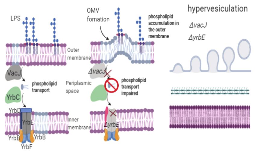
Avila-Calderón et al.                                                                                                             OMVs Biogenesis

Models for OMV Biogenesis                                                 to favor the release of OMVs because of the repulsion caused
The first observation of bacterial vesicles dates back to the             by negative charges in the outer membrane (Kadurugamuwa
1960s, but only in recent decades has there been a significant            and Beveridge, 1995; Sabra et al., 2003).
increase in reports on the biogenesis, physiological roles, and              These models highlighted the importance of lipoproteins,
applications of OMVs. Some of the first reports were performed            LPS, and peptidoglycan during OMV formation. However, it
using Escherichia coli strains, and researchers observed that             remains unknown whether these mechanisms act in tandem.
the lysine-requiring mutant E. coli 12,408 produced “globules”            Some of the most important and widely accepted mechanisms
surrounded by membranes with no evidence of cell lysis (Knox              for OMV formation are outlined in the following sections.
et al., 1966). Moreover, E. coli JC411 released vesicles containing
lipids and proteins (Hoekstra et al., 1976). Other experiments            Some Lipoproteins and the Outer
using E. coli W3110 showed that membrane vesicles were                    Membrane Protein OmpA Are Involved in
released from the cell surface after heating the cells at 55°C.           OMV Biogenesis
These vesicles contained lipid, LPS, and protein compositions             The envelope of Gram-negative bacteria is made up of the
similar to those of the outer membrane from whole cells (Katsui           inner and outer membranes, which are separated by the
et al., 1982). After these findings were reported, the number             periplasmic space. Peptidoglycan is bound to proteins in the
of published studies concerning OMVs has increased (Gamazo                outer and inner membranes through covalent and noncovalent
and Moriyón, 1987; Grenier and Bélanger, 1991; Kadurugamuwa               bonds (Silhavy et al., 2010). Peptidoglycan is composed of
and Beveridge, 1995).                                                     linear glycans (g β-1,4-connected N-acetylglucosamine and
    Three models propose a role for lipoproteins, LPS, and                N-acetylmuramic acid) that are cross-linked by short peptides
peptidoglycan in the biogenesis of OMVs. Recently, new reports            (Pazos and Peters, 2019). Braun’s lipoprotein, commonly referred
contributed to the knowledge of OMV production, some of                   to as Lpp, is the major lipoprotein in E. coli and is the only
which agree with the early models proposed, whereas others                lipoprotein covalently that is linked to the peptidoglycan layer
shed light on new molecules involved in vesiculation. In this             and plays a unique role in the envelope architecture. The
section, the three early models are described initially; in the           N-terminal domain of Lpp is acylated and inserted into to
subsequent section, new insights into the molecular determinants          the outer membrane, while the C-terminal domain is covalently
involved in OMVs are described.                                           linked to the peptidoglycan layer (Lee and Inouye, 1974).
    The first model explains the formation of OMVs due to                 OmpA in E. coli is a β-barrel, and its C-terminal domain
the low number of lipoproteins attached to the peptidoglycan              interacts with peptidoglycan through a 20-aa residue linker
layer. The low number of lipoprotein linkages leads to outer              region (Smith et al., 2007). In 2017, Samsudin et al. have
membrane bulging, affecting vesicle production. In 1976,                  shown that interactions among OmpA, peptidoglycan, and Lpp
Hoekstra et al. determined the number of lipoproteins found               are essential in maintaining the integrity of the cellular envelopes.
in the outer membrane of E. coli as well as those contained               The research team showed that Lpp aids in the interaction of
in the vesicles. These authors reported fewer lipoproteins in             monomeric OmpA with the peptidoglycan layer. In the absence
the vesicles than in the outer membrane. Based on these                   of Lpp, the C-terminal domain of OmpA binds to the outer
results, the authors proposed that vesicles are released from             membrane. Additionally, an OmpA homodimer can easily
outer membrane sites with few lipoprotein linkages (Hoekstra              interact with the peptidoglycan layer in the absence of Lpp
et al., 1976; Mashburn-Warren and Whiteley, 2006). The second             (Samsudin et al., 2017). These lipoproteins and outer membrane
model was based on the presence of peptidoglycan residues                 proteins are associated with outer membrane stability.
with autolysins in the OMVs. This model proposes that during                 Initially, the production of “blebs” (OMVs) was considered
synthesis of the peptidoglycan layer, sites exist where the               an alteration or instability of the outer membrane rather than
concentration of peptidoglycan is higher, causing protrusions             a physiological phenomenon. For example, in 1969, Koike et al.
in the outer membrane and indicating the beginning of vesicle             observed bleb formation on the surface of E. coli after treatment
formation. An important finding that supported this model                 with polymyxin B. However, the same authors observed that
was the presence of muramic acid, a known peptidoglycan                   polymyxin B induced outer membrane projections rather than
layer precursor, in OMVs purified from Porphyromonas gingivalis           blebs in P. aeruginosa. These projections diminished as the
(Zhou et al., 1998). Furthermore, mutation of an autolysin                concentration of the antibiotic was decreased. Thus, blebs and
involved in peptidoglycan replacement increased the synthesis             outer membrane projections were considered to be affected
of OMVs (Hayashi et al., 2002). These findings suggest that               by the action of the antibiotic on the outer membrane (Koike
the accumulation of peptidoglycan residues increases outer                et al., 1969). Subsequently, the release of OMVs was observed
membrane bulging, triggering the release of OMVs (Zhou                    in more bacterial species, and vesicles were found not to be the
et al., 1998; Hayashi et al., 2002). The third and last model             product of cell lysis (Zhou et al., 1998). The exclusion of some
involves the electric charge of LPS in OMV formation.                     periplasmic and outer membrane components and enrichment
Pseudomonas aeruginosa produces two types of LPS: negatively              of other components in vesicles led to the consideration of
charged LPS and neutrally charged LPS. OMVs released by                   the existence of a specific mechanism to select molecules carried
P. aeruginosa cultured under oxidative stress conditions primarily        on OMVs (Kadurugamuwa and Beveridge, 1995; Horstman
comprise negatively charged LPS. Therefore, an increase in                and Kuehn 2000; Kato et al., 2002; Bonnington and Kuehn,
negatively charged LPS within the cell envelope was proposed              2014). Based on this background, “OMV release” is the product

Frontiers in Microbiology | www.frontiersin.org                       2                                      March 2021 | Volume 12 | Article 557902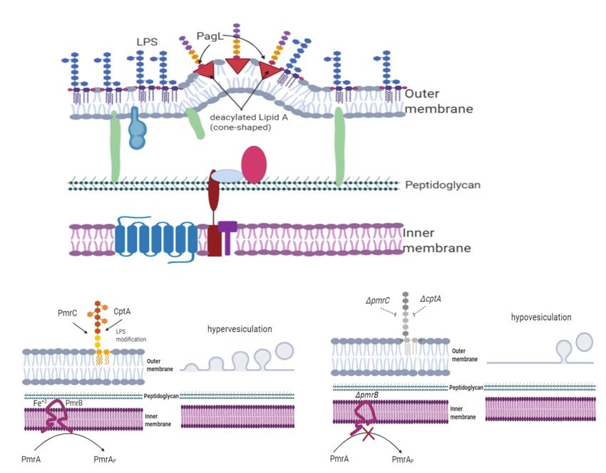
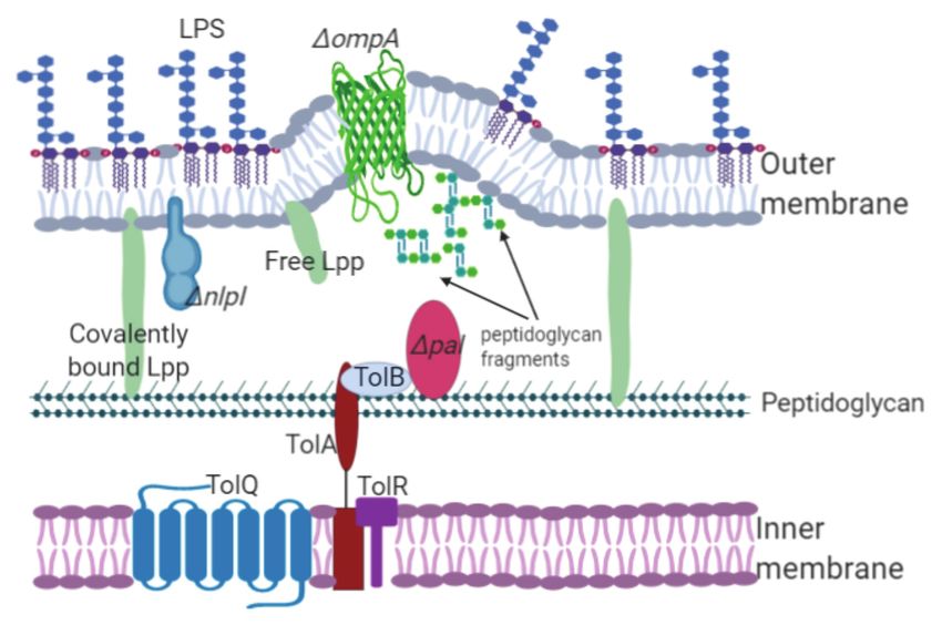
Avila-Calderón et al.                                                                                                                                OMVs Biogenesis

of a physiological phenomenon but not the product of cell                             observed in the results obtained by different research groups.
lysis or membrane instability.                                                        In 2009, Deatherage et al. analyzed the effect on OMV formation
   In 1976 and 1978, the first molecular evidence regarding                           in S. typhimurium LT2 caused by specific mutations in the
OMV biogenesis was revealed by Weigand et al. whose results                           lpp and ompA genes. Further experiments on the S. typhimurium
were obtained using Salmonella enterica serovar Typhimurium                           LT2 tolA, tolB, and pal mutants suggested that they could not
(hereafter referred to as Salmonella typhimurium) and Suzuki                          form outer-membrane-peptidoglycan-inner membrane linkages,
et al. whose work focused on E. coli. Their works showed that                         and vesicle formation was observed around the cell body.
the E. coli lpo mutant and S. typhimurium lkyD mutant lacking                         Weigand et al. (1976) reported OMV formation in the septa
the murein lipoprotein (the product of these genes was later                          of the S. typhimurium lpp mutant. However, Deatherage et al.
designated Braun’s lipoprotein or Lpp) released “blebs” from                          (2009) observed vesicle formation in the cell bodies of different
the outer membrane. Although Suzuki et al. (1978) observed                            S. typhimurium lpp gene mutants. Differences observed in both
the production of blebs from Salmonella cells, Weigand et al.                         studies can be attributed to S. typhimurium harboring two
(1976) reported bleb production at the septal region in E. coli.                      copies of the lipoprotein gene that are separated by 82 bp
Bernadac et al. (1998) studied two mutated E. coli strains, the                       (lppA and lppB). Deatherage et al. (2009) mutated both lpp
lpp and ompA mutants, both of which exhibited a hypervesiculation                     genes, while Weigand mutated only one of them. These findings
phenotype. Similarly, Lpp was also associated with OMV                                showed two possible hypotheses: (i) the bonds between the
production in bacterial species such as Yersinia pestis (Eddy                         outer membrane and peptidoglycan layer dissociate, leading
et al., 2014). The hypervesiculation phenotype was also observed                      to OMV release into the periphery of the cell body (Deatherage
in Acinetobacter baumannii and Vibrio cholerae ompA (Moon                             et al., 2009); and (ii) during bacterial division, the bonds
et al., 2012; Valeru et al., 2014). A. baumannii, V. cholerae, and                    between the peptidoglycan layer and inner membrane decrease
other Gram-negative bacteria with a mutation in the ompA                              within the division septum, affecting the number of linkages
gene could not establish bonds between the peptidoglycan and                          among the cytoplasmic membrane, cell wall, and outer membrane,
the outer membrane, leading to OMV overproduction (Figure 1;                          and leading to membrane protrusion and vesicle release
Jin et al., 2011; Moon et al., 2012). These results demonstrate                       (Deatherage et al., 2009). OMVs are released from either the
that lipoproteins and the outer membrane proteins involved in                         septa or cell body likely because of specific interactions among
membrane stability are linked to OMV biogenesis.                                      Lpp, OmpA, and peptidoglycan. OmpA, as a monomer or
   The role of Lpp and OmpA in the formation of OMVs                                  homodimer, interacts with peptidoglycan and the outer membrane
has also been analyzed. However, some discrepancies were                              in the presence or absence of Lpp. Most likely, the presence

  FIGURE 1 | Lipoproteins and outer membrane proteins are involved in OMV biogenesis. The Lpp, NlpI, OmpA, and Tol-Pal members maintain the stability of
  cellular envelopes joining the peptidoglycan layer with the inner membrane. Interruption or deletion of the genes encoding these proteins decrease the number of
  linkages, inducing OMV formation. For example, mutation of the pal gene decreases linkage with the outer membrane, leading to membrane protrusion and OMV
  release. Additionally, the accumulation of components in the periplasm, such as peptidoglycan precursors, triggers vesicle formation.

Frontiers in Microbiology | www.frontiersin.org                                   3                                             March 2021 | Volume 12 | Article 557902
Avila-Calderón et al.                                                                                                           OMVs Biogenesis

of OmpA as a monomer or homodimer and its distribution                      Another lipoprotein found to be involved in OMV biogenesis
on the outer membrane determine the sites where vesicles                is NlpA, which is an inner membrane lipoprotein. Mutation
can form.                                                               of this protein decreases OMV production in E. coli (Yamaguchi
    Deatherage et al. (2009) and Nevermann et al. (2019) treated        and Inouye, 1988; McBroom et al., 2006; Schwechheimer and
S. typhi and S. typhimurium LT2 ompA mutants with deoxycholate          Kuehn, 2013). Furthermore, Schwechheimer et al. (2014) observed
together with vancomycin and evaluated bacterial viability.             that the single mutation of the inner-membrane-anchored
Nevermann et al. (2019) reported that the S. typhi ompA mutant          lipoprotein NlpA decreased vesiculation in E. coli, while a
was sensitive to deoxycholate and that the S. typhimurium               specific mutation of the ompA gene increased OMV production.
ATCC14028s ompA mutant was sensitive to vancomycin.                     The E. coli ΔnlpAΔompA double mutant exhibited heightened
However, Deatherage et al. (2009) found that the S. typhimurium         vesiculation (Schwechheimer et al., 2014). However, the E. coli
LT2 ompA mutant was not sensitive to deoxycholate. The                  ΔycfSΔybiSΔerfKΔnlpA mutant (defective in L,D-transpeptidases
sensitivity of the S. typhi and S. typhimurium ompA mutants             involved in the covalent crosslink between Lpp and peptidoglycan,
to deoxycholate suggests defective outer membrane stability.            and NlpA) showed increased vesiculation compared with that
Additionally, the hypervesiculating phenotype might be expressed        of the E. coli ΔycfSΔybiSΔerfK mutant. The heightened vesiculation
because of defective outer membrane permeability. Although              observed in both the E. coli ΔnlpAΔompA and E. coli
S. typhi and S. typhimurium are phylogenetically closely related,       ΔycfΔybiΔerfKΔnlpA mutants can be explained in terms of
differences observed in the membrane stability of the mutants           NlpA, which provides stabilization of sites of the bacterial
in the study suggest a different role for the OmpA lipoprotein          envelope necessary for Lpp and OmpA linkages (Schwechheimer
in OMV biogenesis.                                                      et al., 2014). In 1998, Bernadac et al. used a Tol-Pal system
    The accumulation of periplasmic compounds, including                mutated E. coli strain to purify and analyze OMVs using
proteins and peptidoglycan, induces membrane protrusion and             electron microscopy. The research team observed that the tolA,
vesicle release (Hayashi et al., 2002; McBroom and Kuehn,               tolQ, and tolR mutants displayed a hypervesiculation phenotype,
2007). Schwechheimer et al. (2014) analyzed the relationship            whereas the tolB and pal mutants showed a reduced vesicle
among the accumulation of periplasmic compounds, Lpp linkages,          production (Bernadac et al., 1998). The Tol-Pal system comprises
and OMV release. The effects of the periplasmic content on              proteins associated with outer membrane integrity. TolA, TolQ,
OMV formation were analyzed using three E. coli mutants:                and TolR link the inner membrane to peptidoglycan, whereas
(i) E. coli ΔampGΔamiD, a mutant defective in the transport             TolB and the peptidoglycan-associated lipoprotein (Pal) interact
of peptidoglycan-recycling residues; (ii) E. coli ΔrfaC, ΔrfaG,         with the outer membrane (Gerding et al., 2007). McBroom
and ΔrfaP mutants defective in the transport and assembly of            et al. (2006) phenotyped random transposon mutants and found
LPS; (iii) E. coli ΔdegP, a mutant that cannot degrade misfolded        that the E. coli tolA, tolB, and pal mutants showed a
peptides in the periplasm. The hypervesiculation phenotype              hypervesiculation phenotype. Similar results of hypervesiculation
was exhibited by the three E. coli mutants likely because of            were observed in the P. aeruginosa oprL and oprI mutants
the accumulation of peptidoglycan fragments, LPS, and proteins          (Wessel et al., 2013). OprL and OprI are homologs to the Pal
in the periplasm. However, the overall number of Lpp bonds              and Lpp proteins, respectively (Hancock et al., 1990). P. aeruginosa
were not affected because the levels of Lpp cross-linking were          oprI and oprF mutants produced more OMVs than wild-type
similar to those observed in the wild-type strain (Schwechheimer        P. aeruginosa (Wessel et al., 2013). A recent study by Nevermann
et al., 2014). By contrast, the E. coli ΔmepAΔdacBΔpbpG mutant,         et al. (2019) identified genes related to OMV biogenesis in
defective in three peptidoglycan hydrolases, showed a decreased         S. typhi employing transposon mutagenesis. Mutation of the
number of Lpp-peptidoglycan linkages and increased OMV                  tolR gene led to the expression of the hypervesiculation phenotype.
production (Schwechheimer et al., 2014). The vesiculation               The S. typhi tolR mutant was treated with deoxycholate together
process was analyzed for E. coli strains displaying deficient           with vancomycin. Subsequently, cell viability was measured to
genes encoding L,D-transpeptidases (ynhG and ycbB). ynhG                evaluate whether membrane integrity had been affected. Following
(now renamed ldtE) and ycbB (ldtD) encode enzymes that                  treatment with deoxycholate, neither cell growth nor
catalyze the formation of meso-diaminopimelyl → meso-                   hypervesiculation in the S. typhi tolR mutants was affected.
diaminopimelyl crosslinks (also called DAP→DAP or 3–3                   These results indicated that OMV production was due to
cross-links) in peptidoglycan (Magnet et al., 2007). The E. coli        defective lipoprotein linkages. Tol-Pal members are highly
ΔynhGΔycbB mutant produced fewer OMVs than the wild-type                homologous (possibly because of speciation events, gene
strain, and the number of Lpp cross-links in this mutant                duplication, and/or lipoprotein redundancy), and their distinct
increased (Schwechheimer et al., 2014). Therefore, the authors          functions may be essential to OMV biogenesis.
propose that the E. coli ΔynhGΔycbB mutant, lacking DAP-DAP                 NlpI is an outer-membrane-anchored lipoprotein whose
linkages, exhibits Lpp and peptidoglycan cross-links formed             function remains unknown. Although it was previously associated
randomly around the cell body, decreasing OMV release. These            with cell division in E. coli, it has also been related to biofilm
data revealed that the modulation of the peptidoglycan structure        formation in S. typhimurium (Ohara et al., 1999). Inactivation
leads to a decrease in Lpp linkages and an increase in OMV              of the nlpI gene in E. coli resulted in abnormal cell division
production, while a decrease in OMV production is related               and formation of membrane projections, whereas overexpression
to an increase in covalently bound Lpp to peptidoglycan                 of this gene inhibited cell growth (Rouf et al., 2011;
(Schwechheimer et al., 2014).                                           Banzhaf et al., 2020). The S. typhimurium ATCC14028s nlpI

Frontiers in Microbiology | www.frontiersin.org                     4                                      March 2021 | Volume 12 | Article 557902
Avila-Calderón et al. OMVs Biogenesis
mutant expressed the hypervesiculation phenotype and was more within the vesicles (Kadurugamuwa and Beveridge, 1995). The
resistant to vancomycin than the S. typhi nlpI mutant (Nevermann addition of gentamicin (a polycation) to P. aeruginosa cultures
et al., 2019). However, the E. coli nlpI mutant displayed the modified electric charges in the outer membrane, affecting
hypervesiculation phenotype (McBroom et al., 2006). LPS packing into the OMVs. The role of negatively charged
Schwechheimer et al. (2015) obtained an E. coli nlpI mutant LPS in the biogenesis of OMVs was confirmed using P. aeruginosa
displaying the hypervesiculation phenotype but no sensitivity CPA−, OSAB−, and CPA−OSA− mutants. In 2003, Nguyen et al.
to deoxycholate, concluding that the hypervesiculation phenotype confirmed that the OSA−LPS complex also contributes to OMV
observed was neither due to defects in membrane integrity nor formation because the P. aeruginosa CPA− mutant, which only
cell lysis. NlpI regulates the activity of the peptidoglycan hydrolases synthesizes negatively charged LPS, released larger OMVs than
Spr and PBP4, which play an essential role in cell wall renewal. those released by the OSA− mutant (Nguyen et al., 2003).
Mutation of the nlpI gene in E. coli increased Spr and PBP-4 These data showing that OMVs contain a higher concentration
activity, as well as peptidoglycan cleavage and peptidoglycan of negatively charged LPS suggest that OSA plays a role in
synthesis. The increased activity of Spr and PBP-4 hydrolyses vesicle formation.
observed in the E. coli nlpI mutant led to decreased linkages In 2015, Cahill et al. while working with the Klebsiella
between Lpp and peptidoglycan and vesicle overproduction. Most pneumoniae wbbO mutant, a glycosyltransferase-defective strain
likely, peptidoglycan dynamics (i.e., growth and renewal) are lacking the O-side chain, reported that the release of OMVs
regulated by NlpI in the E. coli wild-type strain. However, was not affected, whereas, the outer membrane and vesicles
peptidoglycan hydrolases Spr and PBP-4 decrease from the K. pneumoniae wbbO mutant exhibited a different
Lpp-peptidoglycan linkages, inducing outer membrane protrusion protein profile and were quite distinct from the vesicles of the
and subsequent OMV formation. Importantly, peptidoglycan wild-type strain. Furthermore, vesicles released by the
dynamics and/or Lpp-peptidoglycan linkages are also affected K. pneumoniae wbbO mutant contained a higher concentration
by different hydrolases that are not regulated by NlpI. Based of proteins associated with posttranslational modification, protein
on these findings, NlpI lipoprotein is likely closely associated turnover, and chaperones. However, OMVs produced by the
with OMV formation (Schwechheimer et al., 2015). K. pneumoniae wild-type strain contained proteins involved in
cell wall, membrane, and envelope biosynthesis (Cahill et al.,
Bacterial LPS Plays an Essential Role in 2015). P. gingivalis, an etiologic agent of chronic periodontitis,
OMV Production also expresses two types of LPS, neutrally charged LPS (O-LPS)
The outer membrane is an asymmetric structure comprising and negatively charged LPS (A-LPS; Paramonov et al., 2005,
an inner leaflet made up of phospholipids along with proteins 2009). Studies performed on P. gingivalis porS and waaL P
and an outer leaflet that contains phospholipids, proteins, and mutants demonstrated that neither A-LPS nor O-LPS is essential
LPS. LPS is an essential structure for Gram-negative bacteria for OMV biogenesis. The P. gingivalis porS mutant, lacking the
and is the most abundant antigen found on their cell surface; flippase PorS, did not display the O-antigen in lipid A. However,
for example, the outer membrane of E. coli and Salmonella the P. gingivalis waaL mutant lacks the O-antigen ligase WaaL,
genera can contain up to 75% LPS (Klein and Raina, 2019). generating rough cells. Nonetheless, the electrophoretic profile
The structure of smooth LPS comprises an O-side chain, of proteins associated with OMVs produced by the P. gingivalis
an intermediate region known as the core oligosaccharide and waaL mutant was different from that obtained from the wild-
a glycolipid (lipid A) anchored to the outer membrane. Although type strain (Haurat et al., 2011). These results suggest that
smooth strains contain the O-side chain in their LPS, rough negatively charged LPS influence protein packing into OMVs.
strains do not (Raetz and Whitfield, 2002). The core In 2014, Murphy et al. analyzed lipid concentrations in
oligosaccharide is divided into two sections: the inner core, OMVs released from P. aeruginosa CPA−, OSA−, and CPA−OSA−
proximal to lipid A, and the outer core, which is the attachment mutants. No differences were found in either the concentration
site for the O-antigen. Lipid A can become chemically modified. of lipids or number of vesicles released by all three mutants
For example, phosphoethanolamine, 4-amino-4-deoxy-L- (Murphy et al., 2014). Most likely, repulsion between the core
arabinose (L-Ara4N), and additional palmitate groups were and lipid A produced an inducing effect on the P. aeruginosa
added (Raetz and Whitfield, 2002). Such lipid A modifications CPA−OSA− double mutant but not on the CPA or OSA single
make the bacteria more resistant to cationic antibacterial peptides mutant. By contrast, the double mutant exhibited an increased
and polymyxin (Raetz and Whitfield, 2002). number of OMVs, but no change in size was observed. OMVs
P. aeruginosa produces two variants of the O-side chain from the P. aeruginosa CPA−OSA− mutant and wild-type strain
antigen: the common polysaccharide antigen, also called the contain proteins sharing similar functions. Nevertheless, OMVs
CPA or A-band (a short, neutrally charged molecule), together from the P. aeruginosa CPA− mutant showed the highest
with the O-specific antigen, also referred to as the OSA or accumulation of proteins involved in the transport of small
B-band (a highly immunogenic, negatively charged molecule; molecules. The P. aeruginosa OSA− mutant displayed the highest
Lam et al., 2011). Both OSA and CPA were detected in OMVs proportion of proteins involved in adaptation, protection, and
obtained from a P. aeruginosa strain cultured in the presence transcriptional regulation (Murphy et al., 2014). A high content
and absence of gentamicin. OMVs exhibited a higher of periplasmic proteins and a low number of OMPs were
concentration of OSA in the absence of gentamicin; however, detected in OMVs from the P. aeruginosa OSA− mutant.
in the presence of this antibiotic, CPA was hardly detected Conversely, high numbers of OMPs and periplasmic proteins
Frontiers in Microbiology | www.frontiersin.org 5 March 2021 | Volume 12 | Article 557902Avila-Calderón et al. OMVs Biogenesis
were observed in OMVs from the P. aeruginosa CPA−OSA− Fe+3 concentration. PmrAB regulates the expression of the
double mutant, CPA− mutant and wild-type strain (Murphy pmrD gene. PmrB senses high Fe+3 concentrations. Once PmrA
et al., 2014). These results suggest that OSA-LPS plays an is activated through phosphorylation, it triggers the differential
important role in the selection of proteins. Proteins interacting expression of pmrC (also known as eptA) and cptA; both
with specific LPS lead to OMVs with different contents due pmrC and cptA catalyze LPS modifications (Perez and Groisman,
to LPS composition. It is highly probable that the lack of the 2007). In Salmonella enterica, PmrC and CptA link
O-side chain modifies the electric charge on the surface of phosphoethanolamine, while ArnT links 4-amino-4-deoxy-L
the membrane, impairing electrostatic interactions between arabinose to lipid A and the LPS core; these bonds are
proteins and electrically charged LPS, and ultimately affecting regulated by PmrAB (Trent et al., 2001b; Lee et al., 2004;
the protein packing of OMVs. Tamayo et al., 2005). Once 4-amino-4-deoxy-L arabinose and
PagL is an enzyme that modifies lipid A by removing the phosphoethanolamine are linked to lipid A and the core, the
acyl chain at the 3-position of the disaccharide backbone synthesis of negatively charged LPS is decreased, and molecules
(King et al., 2009). In S. typhimurium, a deacylated lipid A attached to LPS enhance resistance to antimicrobial peptides
(i.e., penta-acylated lipid A) modified by PagL makes LPS and oxidative stress (Sinha et al., 2019). The C. rodentium
less detectable to Toll-like receptor 4 of the mouse B-cell ΔcptA, ΔpmrC, and ΔpmrAB mutants produce more OMVs
line (Kawasaki et al., 2004). PagL activity is regulated by a than the wild-type strain (Sinha et al., 2019). Furthermore,
two-component system (represented as PhoP/PhoQ), Mg+2 decreased vesiculation was observed in C. rodentium strains
and temperature, being less active at low Mg+2 concentrations overexpressing the pmrC (eptA) or cptA genes, as shown in
and temperatures (Trent et al., 2001a; Ernst et al., 2006). Figure 2B (Sinha et al., 2019). PmrC and CptA are putative
OMVs from P. gingivalis displayed higher concentrations of transferases in C. rodentium; hence, mutations of these genes
deacylated lipids (Haurat et al., 2011). Therefore, the effect could lead to decreased modification of LPS with
of lipid A deacylation on OMV biogenesis was evaluated phosphoethanolamine, increasing the overall negative charge
using the expression of the PagL enzyme. The pagL gene of LPS and causing outer-membrane curvature imbalance,
was cloned using a low-copy expression vector lacking a subsequent membrane protrusion, and OMV release.
control from the PhoP/PhoQ two-component system. S. Nevermann et al. (2019) demonstrated that the waaC and
typhimurium expressing PagL produced almost four times rfaE genes are also associated with OMV biogenesis in S. typhi.
more OMVs than the wild-type strain (Elhenawy et al., 2016). Additionally, RfaE has been associated with heptose (Hep)
The outer membrane of the S. typhimurium strain expressing precursors necessary for inner-core LPS assembly in E. coli
PagL showed that hexa-acylated lipid A was predominant, and S. typhimurium (Valvano et al., 2002). Heptoses are found
while OMVs mainly contained penta-acylated lipid A (Elhenawy in the oligosaccharide core, and they play a fundamental role
et al., 2016). The authors explained that these differences are in outer membrane stability and the crosslinking of adjacent
likely based on conformational changes in the lipid A structure LPS; they also interact with the positive charges of certain
because hexa-acylated lipid A has a conical shape, while proteins (Heinrichs et al., 1998). Heptosyltransferase I (HepI
deacylated lipid A has a cylindrical or inverted cone shape. or WaaC) catalyzes the addition of the first heptose to the
Thus, either the cylindrical or inverted-cone shape decreases inner Kdo (3-deoxy-D-manno-oct-2-ulosonic acid; Czyzyk et al.,
hydrophobic interactions, leading to membrane protrusion 2011). The S. typhi waaC mutant produced fewer vesicles than
and favoring OMV formation (Figure 2A; Schromm et al., the wild-type strain, while the OMV protein profiles from
2000; Elhenawy et al., 2016). The presence of deacylated lipid both the mutant and wild-type strains were similar. Additionally,
A in other Gram-negative bacteria should be investigated to the S. typhi rfaE mutant produced more vesicles; however,
determine whether this is a common molecule implicated in when comparing OMV protein profiles from both the mutant
OMV biogenesis. The quantitative comparison analysis of and wild-type strains, relevant differences could clearly
subtypes of LPS in OMVs and outer membranes of wild be observed. Lateral interactions with LPS molecules provide
type S. typhimurium undergoing environmental shift conditions a significant fraction of the driving force necessary to establish
that do not rely on PagL supported an additional role for lipid asymmetry in the outer membrane (Nikaido, 2003).
polymorphic regulation of membrane LPS composition in Regarding the S. typhi rfaE mutant, it is likely that a defective
vesiculation (Bonnington and Kuehn, 2016). Nonlamellar types link of heptose in the inner core alters its assembly, modifying
of lipids, which have an overall conical shape and a preference certain interactions between positively and negatively charged
for the hexagonal phase, are thought to aid in the formation LPS groups, and affecting the overall protein interaction with
of nonbilayer structures. Accordingly, both the geometry and LPS. This finding could explain why the protein composition
the propensity of individual membrane components to form of OMVs from the S. typhi rfaE mutant was different from
nonbilayer lipid phases in the membrane are both likely to that obtained for OMVs from the wild-type strain. WaaC and
be important to vesiculation processes. RfaE induced an opposite effect on vesiculation. Mutation of
Citrobacter rodentium, a murine enteric pathogen, lacks the waaC gene led to the expression of the hypovesiculation
the pagL gene. This bacterium uses two-component systems phenotype, while mutation of the rfaE gene induced a
to modify LPS, and these systems may indirectly regulate hypervesiculation phenotype. These data confirmed the same
OMV production, similar to PagL. One of these systems is effects on the OMV phenotypes by waaC and rfaE mutants
PmrAB, a two-component system regulated by low pH and found for E. coli (Kulp et al., 2015). Further research should
Frontiers in Microbiology | www.frontiersin.org 6 March 2021 | Volume 12 | Article 557902Avila-Calderón et al. OMVs Biogenesis
A
B
FIGURE 2 | LPS modifications are implicated in OMV release. Negatively charged LPS causes an outer membrane imbalance, subsequent membrane protrusion,
and OMV release. LPS modifications also contribute to OMV formation. (A) The S. typhimurium strain expressing PagL without the PhoPQ control produces more
OMVs than the wild-type strain. The outer membrane of S. typhimurium expressing PagL shows that hexa-acylated lipid A is predominant, while OMVs primarily
contain penta-acylated lipid A. Thus, either the cylindrical or inverted-cone shape decreases hydrophobic interactions, leading to membrane protrusion and favoring
OMV formation. (B) Citrobacter rodentium uses a two-component system to modify LPS. PmrB, a member of this system, senses high Fe+3 concentrations and
regulates the modification of lipid A. C. rodentium ΔpmrB cannot add groups to lipid A, leading to increased vesiculation.
focus on these enzymes, which are associated with inner core components, including PE, PG, and CL. OMVs from Actinobacillus
assembly, and how they contribute to OMV formation. actinomycetemcomitans displayed higher CL and PE concentrations
and a low proportion of unidentified lipids, which are not
Contribution of Phospholipids to OMV present in the outer membrane (Kato et al., 2002). OMVs
Biogenesis from P. aeruginosa showed high PG and stearic acid
Most reports concerning OMV biogenesis focus on the role concentrations, but a low percentage of unsaturated fatty acids
of lipoproteins and LPS. However, little is known about released from vesicles composed of a rigid membrane (Tashiro
phospholipids, which are the major components of OMVs. The et al., 2011). The orf5 and plsC1 genes of Shewanella livingstonesis
main phospholipids comprising the outer membrane of E. coli, play an essential role in eicosapentaenoic acid biosynthesis,
in order of proportion, are phosphatidylethanolamine (PE), while plsC1 and plsC4 code for acyltransferases that are involved
phosphatidylglycerol (PG), and cardiolipin (CL; Raetz and in the synthesis of membrane phospholipids. Mutation of orf5,
Dowhan, 1990). Using thin-layer chromatography, Horstman plsC1, and plsC4 in S. livingstonesis increased the production
and Kuehn (2000) determined that the phospholipid composition of vesicles (Yokoyama et al., 2017).
of the outer membranes of the E. coli HB101 and ETEC 2 Gram-negative bacteria maintain asymmetric distribution of
strains, as well as their respective vesicles, share similar phospholipids in their membranes through the translocation
Frontiers in Microbiology | www.frontiersin.org 7 March 2021 | Volume 12 | Article 557902Avila-Calderón et al. OMVs Biogenesis
of phospholipids from the inner membrane to the inner leaflet formation in the outer membrane, the transport of phospholipids
of the outer membrane (known as phospholipid anterograde from the inner membrane to the outer membrane should
transport) and inversely (known as phospholipid retrograde be significantly faster than that from the outer membrane to
transport; Shrivastava and Chng, 2019). Regarding anterograde the inner membrane. Disorganization in phospholipid transport
transport, the molecular determinants implicated in the process leads to phospholipid accumulation in the outer membrane,
are still not fully understood; however, the PbgA/YejM lipoprotein provoking OMV release (Figure 3).
is involved in CL transport from the inner to the outer Differences found in the composition of these phospholipids
membrane in Shigella flexneri and S. typhimurium (Dalebroux suggest that vesicles are released from the microdomains of
et al., 2015; Rossi et al., 2017). Regarding retrograde transport, diverse phospholipid compositions. However, the possibility of
two systems have been described: (i) the Tol-Pal system is a different mechanism that can regulate phospholipids in OMVs
involved in the movement of phospholipids from the inner should not be excluded. The composition of predominant
leaflet of the outer membrane to the inner membrane; (ii) the phospholipids in OMVs has already been determined. For
OmpC-Mla system is responsible for outer membrane asymmetry example, vesicles purified from E. coli ETEC exhibited high
by transporting misplaced phospholipids found in the outer concentrations of PG, CL, and PE. By contrast, OMVs from
leaflet of the outer membrane to the inner membrane (Chong A. actinomycetemcomitans contained high concentrations of CL
et al., 2015; Shrivastava et al., 2017). The MlaA lipoprotein and PE, whereas OMVs from P. aeruginosa only showed high
interacts with OmpC, which is embedded in the outer membrane concentrations of PG, and OMVs from H. influenzae mainly
and removes phospholipids in the outer leaflet of the outer comprised PE (Kato et al., 2002). The accumulation of certain
membrane to another component of the system the MlaC lipids, such as PG and PE, in OMVs is essential to decipher
protein. Subsequently, MlaC delivers these phospholipids to a mechanism for OMV biogenesis.
the MlaFEDB complex located in the inner membrane. This Tol-Pal helps maintain outer membrane stability through
complex can then reintegrate these phospholipids to the inner lipoprotein linkages. This system is involved in phospholipid
membrane (Malinverni and Silhavy, 2009; Chong et al., 2015). retrograde transport and contributes to OMV biogenesis
Mutation of mla in E. coli induces vesicle production at the (Masilamani et al., 2018; Shrivastava and Chng, 2019). Because
septa, increases hepta-acylated LPS, and promotes cell death the S. typhimurium tolA, tolQ, and tolR mutants display high
(Sutterlin et al., 2016). Vesicle overproduction decreases the levels of PG and PE in the outer membrane, the presence of
lipid levels in the outer membrane that are then replaced by glycerophospholipids may promote OMV release (Masilamani
lipids from the inner membrane. The decrease in lipids of the et al., 2018). Because Tol-Pal proteins lack a phospholipid-
inner membrane leads to cell lysis (Sutterlin et al., 2016). binding domain, they may stabilize unidentified phospholipid
Roier et al. (2016) mutated the vacJ and yrbE genes of transporters or act as phospholipid transporters from the inner
Haemophilus influenzae, which are homologous to the mlaA to the outer membrane (Shrivastava and Chng, 2019).
and mlaE genes (respectively) of E. coli and are involved in
lipid membrane asymmetry. Vesiculation increased 1.6‐ and Bacterial Flagellin Induces OMV Release
2.2-fold in the H. influenzae vacJ and yrbE mutants, respectively, Several studies have shown an association between the bacterial
while vesicle production decreased in the complemented mutants. flagellum and OMVs. The flagellum comprises a basal body, a
Vesicles from the H. influenzae vacJ and yrbE mutants exhibited flexible linker known as the hook, and a filament that drives
higher levels of PE and myristic acid (C14:0), similar to the the bacterium’s movement. The filament of the bacterial flagellum
outer membrane composition in the wild-type strain; however, comprises the flagellin protein (Chen et al., 2011). Using a
the PE and C14:0 fatty acid ratio increased only in vesicles density gradient protocol, OMVs from E. coli were purified,
produced by the mutants. Hypervesiculation in H. influenzae and flagellin (FliC) was identified using proteomics (Lee et al.,
vacJ and yrbE was directly due to phospholipid accumulation 2007). Manabe et al. analyzed vesicles from E. coli W3110 and
in the outer membrane because the genes involved in retrograde the E. coli ΔfliC mutant derived from the W3110 strain. OMVs
transport were mutated. The H. influenzae vacJ and yrbE mutants were obtained using a density gradient protocol. Both flagellin
showed the hypervesiculation phenotype, which could be caused and OMVs from the wild-type strain were found in the same
by the regulation of the transport of phospholipids from the fraction collected from the density gradient. Moreover, flagellin
inner leaflet to the outer leaflet of the outer membrane. Mutation was detected in the lumen of fractioned vesicles from the wild-
of the homologous genes vacJ and yrbE, which are associated type strain, and only in the mutant did OMV production decrease
with phospholipid transport in V. cholerae, E. coli, and (Manabe et al., 2013). In another study, researchers tried to
Campylobacter jejuni, increased vesiculation, confirming the correlate the presence of flagella and OMV production in Vibrio
hypothesis that phospholipid asymmetry (retrograde trafficking fischeri; in this bacterium, the flagellum shaft is encased by a
of phospholipids) plays a role in OMV biogenesis (Roier et al., membranous sheath (Millikan and Ruby, 2004; Aschtgen et al.,
2016; Davies et al., 2019). Homologous proteins to the phospholipid 2016). In this study, vesicle production was determined in the
ABC transport system were found in phylogenetically distant V. fischeri motB1 mutant (MotB1 is a sodium pump and the
species, such as Pasteurella multocida, P. aeruginosa, S. typhimurium, main motor protein for the flagellum machinery), V. fischeri
and Yersinia enterocolitica, among others. This finding suggests nonflagellated flrA mutant (flrA codes for a transcriptional
that the mechanism of OMV formation may be strongly linked activator), V. fischeri hyperflagellated swarmer strain (expresses
to phospholipid transport (Roier et al., 2016). To trigger vesicle 3‐ to 4-fold more flagella), and V. fischeri wild-type strain.
Frontiers in Microbiology | www.frontiersin.org 8 March 2021 | Volume 12 | Article 557902Avila-Calderón et al. OMVs Biogenesis
FIGURE 3 | Phospholipid transport regulates OMVs biogenesis. H. influenzae vacJ and yrbE mutants show a hypervesiculation phenotype because of the
accumulation of phospholipids in the outer membrane. The vacJ and yrbE genes are involved in retrograde transport, maintaining membrane asymmetry. To trigger
vesicle formation in the outer membrane, the transport of phospholipids from the inner membrane to the outer membrane should be significantly faster than
phospholipid transport from the outer membrane to the inner membrane. Disorganization in phospholipid transport leads to the accumulation of phospholipids in the
outer membrane, provoking OMV release.
However, the number of vesicles decreased in the V. fischeri P. aeruginosa produces a quinolone called the Pseudomonas
flrA mutant that lacks flagella. Vesicle production increased in quinolone signal (PQS). To regulate the expression of some
the hyperflagellated strain. The addition of phenamil (a sodium- genes, PQS binds to the PqsR receptor (Lin et al., 2018). The
pump blocking reagent) to the V. fischeri hyperflagellated strain synthesis of PQS is regulated by pqsABCDE along with other
decreased OMV production, suggesting that flagellum rotation genes, including phnAB and pqsH. PqsA triggers PQS synthesis.
improves vesicle release (Aschtgen et al., 2016). To demonstrate PqsB, PqsC, and PqsD then produce 2-heptyl-4-quinolone
the association of flagella with OMV production, the following (HHQ) or quinolone and other precursors. Finally, PqsH
organisms were analyzed: V. fischeri and V. parahaemolyticus catalyzes the conversion of HHQ into PQS. The P. aeruginosa
(which expresses several sheathed flagella), V. cholerae (which pqsH mutant synthesizes quinolones but not PQS (Lin et al., 2018).
contains unique polar sheathed flagella), E. coli (which expresses The quorum-sensing system in P. aeruginosa comprises
an unsheathed flagellum), and the V. fischeri nonflagellated flrA quinolone molecules, including 3-oxo-dodecanoyl homoserine
mutant. V. fischeri and V. parahaemolyticus released more vesicles lactone (3OC12-HSL), butyryl homoserine lactone (C4-HSL),
than V. cholerae and E. coli, while the V. cholerae nonflagellated and 2-heptyl-3-hydroxy-4-quinolone (PQS; Déziel et al., 2004).
mutant exhibited decreased OMV production (Aschtgen et al., OMVs from P. aeruginosa contain higher PQS (86%) than 12-HSL
2016). They concluded that species expressing flagella such as and C4-HSL, which were measured using thin-layer
E. coli and Vibrio released more vesicles. Clearly, unsheathed chromatography (TLC) and mass spectrometry (Mashburn-Warren
and sheathed flagella perturb the outer membrane and promote and Whiteley, 2005). Additionally, the P. aeruginosa pqsA mutant
OMV release. To better ascertain the relationship between flagella (displaying defective quinolone synthesis) showed a 10-fold
and OMV production, more flagellated strains along with their decrease in OMV production. Vesicle production was restored
corresponding nonflagellated mutants and hyperflagellated strains after treating mutants with a synthetic quinolone. Thus, PQS
should be studied. It is important to explore whether flagellum may also be involved in OMV release. However, when the P.
movement only promotes the release of vesicles from the aeruginosa mvfR mutant (which lacks the PQS mvfR regulator)
surrounding outer membrane area or promotes vesiculation from was treated with exogenous PQS, it yielded the same number
the whole cell body. of vesicles as that of the wild-type strain. This finding suggests
physical interactions among PQS and different undetermined
Pseudomonas Quinolone Signaling and Its molecules found in outer membrane sites where it begins to
Role in OMV Biogenesis bulge (Mashburn-Warren and Whiteley, 2005). Additionally, the
Bacterial species such as P. aeruginosa regulate the secretion interaction of LPS with either PQS or the alkyl chain and
of virulence factors by sensing bacterial density through the hydroxyl group (-OH) of PQS induced a curvature in the outer
quorum-sensing mechanism (Strateva and Mitov, 2011). membrane necessary to start vesicle formation. Hence, a synthetic
Frontiers in Microbiology | www.frontiersin.org 9 March 2021 | Volume 12 | Article 557902Avila-Calderón et al. OMVs Biogenesis compound similar to PQS (which lacks the alkyl chain and K. pneumoniae (Horspool and Schertzer, 2018). Molecules hydroxyl group) does not interact with LPS, while the natural present in the supernatants induced OMV production in PQS molecule interacts with LPS (Mashburn-Warren et al., P. aeruginosa, but these molecules have yet to be identified. 2008). Moreover, PQS could impair the interaction among Although Mashburn-Warren and Whiteley attributed OMV divalent cations (Mg+2 and Ca+2) and phosphate at the production in P. aeruginosa exclusively to PQS, they also 4-position of lipid A, affecting the motility of lipid A and observed a minimal concentration of acyl-homoserine lactone destabilizing the outer membrane, improving outer membrane packaged in OMVs. Therefore, exogenous acyl-homoserine protrusion and vesicle release (Bredenbruch et al., 2006; lactone from E. coli and K. pneumoniae induce membrane Mashburn-Warren et al., 2008). curvature, leading to OMV release in P. aeruginosa (Mashburn- In 2012, Schertzer and Whiteley proposed a novel bilayer- Warren and Warren, 2005; Schertzer and Whiteley, 2012). coupled model to explain OMV biogenesis, which they theorized could be mediated by PQS in P. aeruginosa (Schertzer and Whiteley, 2012). This model requires the presence of CONCLUDING REMARKS an amphiphilic molecule that, due to specific electric interactions, would concentrate in the outer leaflet of the Several works have focused on explaining the different possible membrane, leading to the expansion of the outer leaflet while OMV biogenesis mechanisms. Lipoproteins, outer membrane promoting membrane curvature (Sheetz and Singer, 1974). proteins, LPS, and flagellin have been proposed as the key To study the effect of small molecules on the cell membrane, pieces in different mechanisms of OMV biogenesis. a solution prepared with a low concentration of PQS was Lipoproteins and outer membrane proteins involved in added to red blood cells. The erythrocytes showed crenation linkages within the bacterial envelopes regulate membrane and membrane protrusions, an effect similar to that elicited protrusion and subsequent OMV release. Further investigation during vesicle production (Schertzer and Whiteley, 2012). is needed and should focus on the search for lipoproteins The addition of chlorpromazine, which induces cup formation and outer membrane proteins homologous to those reviewed in red blood cells, prevented the formation of protrusions here involved in OMV biogenesis. The interaction with the in the membrane. Such evidence indicates that PQS may peptidoglycan and/or outer membrane and null mutants contribute to OMV release, in accordance with the bilayer- were then analyzed to determine their role in vesicle formation. coupled model proposed by Schertzer and Whiteley (2012). It is possible that more than one molecule, such as LPS, P. aeruginosa PA01 strain was previously shown to produce outer membrane proteins, and phospholipids, could contribute a higher concentration of PQS in the inner membrane and to vesicle formation. Although LPS differs considerably among released fewer OMVs. By contrast, PQS production in bacterial species, it has also been associated with OMV P. aeruginosa PA14 was extracellular, and a higher OMV biogenesis. LPS charge and architecture are involved in OMV production was observed. These results suggest that a relevant formation and protein composition. In addition, it should difference between the strains could be defective PQS export be explored which LPS component (O-antigen, core, and/ (Florez et al., 2017). Temporal analyses showed PQS or lipid A) plays the major role in vesiculation. However, accumulation in the inner membrane of the P. aeruginosa retrograde phospholipid transport is involved in OMV release; PA01 strain due to PQS saturation. This saturation was phospholipid accumulation increases outer membrane observed after culturing the strain for 10 h in Luria-Bertani protrusion and subsequent vesicle release. Other molecules, medium (Florez et al., 2017). When the PA01 strain was including PQS or flagellin, could be involved in vesicle cultured using brain infusion broth, vesicle production formation. Although the secretion of PQS molecules or increased along with PQS export compared with that obtained presence of flagella in the cell body is restricted to a few in Luria-Bertani medium. These results were similar to those bacterial species, their role in vesiculation must be explored reported in clinical strains of P. aeruginosa isolated from in more detail. patients with cystic fibrosis. These strains showed an increase in OMV production and PQS export in brain infusion broth culture (Florez et al., 2017). Future research is required to AUTHOR CONTRIBUTIONS analyze PQS transport in Gram-negative clinical strains and define possible PQS-associated mechanisms in OMV All authors listed have made a substantial, direct and intellectual biogenesis. contribution to the work, and approved it for publication. In 2018, Horspool and Schertzer treated different strains of gammaproteobacteria, such as E. coli, Klebsiella pneumoniae, and Proteus mirabilis, with exogenous PQS. All the strains FUNDING increased vesicle production up to 3.5-fold. This effect was not observed in alphaproteobacteria such as Agrobacterium This work was funded by CONACYT 61529, SIP-IPN 20182152, tumefaciens and Caulobacter crescentus. Importantly, the 20195737, 20200594, and SAGARPA-CONACYT 2017-02-291311. authors observed an increase in OMV production when EA-C was supported by CONACYT and PIFI-IPN scholarships. using the P. aeruginosa strain that was stimulated with a MA-A, ER, ZG-L, and AC-R were supported by fellowships concentrated supernatant from cultures of E. coli and from COFAA-IPN, SIP-EDI, and SNI-CONACYT. Frontiers in Microbiology | www.frontiersin.org 10 March 2021 | Volume 12 | Article 557902
Avila-Calderón et al.                                                                                                                                            OMVs Biogenesis

REFERENCES                                                                                    Florez, C., Raab, J. E., Cooke, A. C., and Schertzer, J. W. (2017). Membrane
                                                                                                  distribution of the Pseudomonas quinolone signal modulates outer membrane
Aschtgen, M. S., Lynch, J. B., Koc, E., Schwartzman, J., McFall-Ngai, M., and                     vesicle production in Pseudomonas aeruginosa. MBio 8, e01034–e01117. doi:
   Ruby, E. (2016). Rotation of Vibrio fischeri flagella produces outer membrane                  10.1128/mBio.01034-17
   vesicles that induce host development. J. Bacteriol. 198, 2156–2165. doi:                  Gamazo, C., and Moriyón, I. (1987). Release of outer membrane fragments
   10.1128/JB.00101-16                                                                            by exponentially growing Brucella melitensis cells. Infect. Immun. 55, 609–615.
Avila-Calderón, E. D., Araiza-Villanueva, M. G., Cancino-Diaz, J. C.,                             doi: 10.1128/IAI.55.3.609-615.1987
   López-Villegas, E. O., Sriranganathan, N., Boyle, S. M., et al. (2015). Roles              Gerding, M. A., Ogata, Y., Pecora, N. D., Niki, H., and De Boer, P. A. (2007).
   of bacterial membrane vesicles. Arch. Microbiol. 197, 1–10. doi: 10.1007/                      The trans-envelope Tol-Pal complex is part of the cell division machinery
   s00203-014-1042-7                                                                              and required for proper outer-membrane invagination during cell constriction
Banzhaf, M., Yau, H. C., Verheul, J., Lodge, A., Kritikos, G., Mateus, A., et al.                 in E. coli. Mol. Microbiol. 63, 1008–1025. doi: 10.1111/j.1365-2958.2006.05571.x
   (2020). Outer membrane lipoprotein NlpI scaffolds peptidoglycan hydrolases                 Grenier, D., and Bélanger, M. (1991). Protective effect of Porphyromonas gingivalis
   within multi-enzyme complexes in Escherichia coli. EMBO J. 39:e102246.                         outer membrane vesicles against bactericidal activity of human serum. Infect.
   doi: 10.15252/embj.2019102246                                                                  Immun. 9, 3004–3008. doi: 10.1128/IAI.59.9.3004-3008.1991
Bernadac, A., Gavioli, M., Lazzaroni, J. C., Raina, S., and Lloubès, R. (1998).               Hancock, R. E., Siehnel, R., and Martin, N. (1990). Outer membrane proteins
   Escherichia coli tol-pal mutants form outer membrane vesicles. J. Bacteriol.                   of Pseudomonas. Mol. Microbiol. 4, 1069–1075. doi: 10.1111/j.1365-2958.1990.
   180, 4872–4878. doi: 10.1128/JB.180.18.4872-4878.1998                                          tb00680.x
Bonnington, K. E., and Kuehn, M. J. (2014). Protein selection and export via                  Haurat, M. F., Aduse-Opoku, J., Rangarajan, M., Dorobantu, L., Gray, R. M.,
   outer membrane vesicles. Biochim. Biophys. Acta. 1843, 1612–1619. doi:                         Curtis, M. A., et al. (2011). Selective sorting of cargo proteins into bacterial
   10.1016/j.bbamcr.2013.12.011                                                                   membrane vesicles. J. Biol. Chem. 286, 1269–1276. doi: 10.1074/jbc.
Bonnington, K. E., and Kuehn, M. J. (2016). Outer membrane vesicle production                     M110.185744
   facilitates LPS remodeling and outer membrane maintenance in Salmonella                    Hayashi, J., Hamada, N., and Kuramitsu, H. K. (2002). The autolysin of
   during environmental transitions. MBio 7, e01532–e01616. doi: 10.1128/                         Porphyromonas gingivalis is involved in outer membrane vesicle release.
   mBio.01532-16                                                                                  FEMS Microbiol. Lett. 216, 217–222. doi: 10.1111/j.1574-6968.2002.tb11438.x
Bredenbruch, F., Geffers, R., Nimtz, M., Buer, J., and Häussler, S. (2006). The               Heinrichs, D. E., Yethon, J. A., and Whitfield, C. (1998). Molecular basis for
   Pseudomonas aeruginosa quinolone signal (PQS) has an iron-chelating activity.                  structural diversity in the core regions of the lipopolysaccharides of Escherichia
   Environ. Microbiol. 8, 1318–1329. doi: 10.1111/j.1462-2920.2006.01025.x                        coli and Salmonella enterica. Mol. Microbiol. 30, 221–232. doi:
Cahill, B. K., Seeley, K. W., Gutel, D., and Ellis, T. N. (2015). Klebsiella                      10.1046/j.1365-2958.1998.01063.x
   pneumoniae O antigen loss alters the outer membrane protein composition                    Hoekstra, D., van der Laan, J. W., de Leij, L., and Witholt, B. (1976). Release
   and the selective packaging of proteins into secreted outer membrane vesicles.                 of outer membrane fragments from normally growing Escherichia coli. Biochim.
   Microbiol. Res. 180, 1–10. doi: 10.1016/j.micres.2015.06.012                                   Biophys. Acta 455, 889–899. doi: 10.1016/0005-2736(76)90058-4
Chen, S., Beeby, M., Murphy, G. E., Leadbetter, J. R., Hendrixson, D. H.,                     Horspool, A. M., and Schertzer, J. W. (2018). Reciprocal cross-species induction
   Briegel, A., et al. (2011). Structural diversity of bacterial flagellar motors.                of outer membrane vesicle biogenesis via secreted factors. Sci. Rep. 8:9873.
   EMBO J. 30, 2972–2981. doi: 10.1038/emboj.2011.186                                             doi: 10.1038/s41598-018-28042-4
Chong, Z. S., Woo, W. F., and Chng, S. S. (2015). Osmoporin OmpC forms                        Horstman, A. L., and Kuehn, M. J. (2000). Enterotoxigenic Escherichia coli
   a complex with MlaA to maintain outer membrane lipid asymmetry in                              secretes active heat-labile enterotoxin via outer membrane vesicles. J. Biol.
   Escherichia coli. Mol. Microbiol. 98, 1133–1146. doi: 10.1111/mmi.13202                        Chem. 275, 12489–12496. doi: 10.1074/jbc.275.17.12489
Czyzyk, D. J., Liu, C., and Taylor, E. A. (2011). Lipopolysaccharide biosynthesis             Jan, A. T. (2017). Outer membrane vesicles (OMVs) of gram-negative bacteria:
   without the lipids: recognition promiscuity of Escherichia coli heptosyltransferase            a perspective update. Front. Microbiol. 8:1053. doi: 10.3389/fmicb.2017.01053
   I. Biochemistry 50, 10570–10572. doi: 10.1021/bi201581b                                    Jin, J. S., Kwon, S. O., Moon, D. C., Gurung, M., Lee, J. H., Kim, S. I., et al.
Dalebroux, Z. D., Edrozo, M. B., Pfuetzner, R. A., Ressl, S., Kulasekara, B. R.,                  (2011). Acinetobacter baumannii secretes cytotoxic outer membrane protein
   Blanc, M. -P., et al. (2015). Delivery of cardiolipins to the Salmonella outer                 a via outer membrane vesicles. PLoS One 6:e17027. doi: 10.1371/journal.
   membrane is necessary for survival within host tissues and virulence. Cell                     pone.0017027
   Host Microbe 17, 441–451. doi: 10.1016/j.chom.2015.03.003                                  Kadurugamuwa, J. L., and Beveridge, T. J. (1995). Virulence factors are released
Davies, C., Taylor, A. J., Elmi, A., Winter, J., Liaw, J., Grabowska, A. D.,                      from Pseudomonas aeruginosa in association with membrane vesicles during
   et al. (2019). Sodium taurocholate stimulates Campylobacter jejuni outer                       normal growth and exposure to gentamicin: a novel mechanism of enzyme
   membrane vesicle production via down-regulation of the maintenance of                          secretion. J. Bacteriol. 177, 3998–4008. doi: 10.1128/jb.177.14.3998-4008.1995
   lipid asymmetry pathway. Front. Cell. Infect. Microbiol. 9:177. doi: 10.3389/              Kato, S., Kowashi, Y., and Demuth, D. R. (2002). Outer membrane-like vesicles
   fcimb.2019.00177                                                                               secreted by Actinobacillus actinomycetemcomitans are enriched in leukotoxin.
Deatherage, B. L., Lara, J. C., Bergsbaken, T., Rassoulian Barrett, S. L., Lara, S.,              Microb. Pathog. 32, 1–13. doi: 10.1006/mpat.2001.0474
   and Cookson, B. T. (2009). Biogenesis of bacterial membrane vesicles. Mol.                 Katsui, N., Tsuchido, T., Hiramatsu, R., Fujikawa, S., Takano, M., and Shibasaki, I.
   Microbiol. 72, 1395–1407. doi: 10.1111/j.1365-2958.2009.06731.x                                (1982). Heat-induced blebbing and vesiculation of the outer membrane of
Déziel, E., Lépine, F., Milot, S., He, J., Mindrinos, M. N., Tompkinset, R. G.,                   Escherichia coli. J. Bacteriol. 151, 1523–1531. doi: 10.1128/JB.151.3.1523-
   et al. (2004). Analysis of Pseudomonas aeruginosa 4-hydroxy-2-alkylquinolines                  1531.1982
   (HAQs) reveals a role for 4-hydroxy-2-heptylquinoline in cell-to-cell                      Kawasaki, K., Ernst, R. K., and Miller, S. I. (2004). 3-O-deacylation of lipid
   communication. Proc. Natl. Acad. Sci. U. S. A. 101, 1339–1344. doi: 10.1073/                   a by PagL, a PhoP/PhoQ-regulated deacylase of Salmonella typhimurium,
   pnas.0307694100                                                                                modulates signaling through toll-like receptor 4. J. Biol. Chem. 279, 20044–20048.
Eddy, J. L., Gielda, L. M., Caulfield, A. J., Rangel, S. M., and Lathem, W. W.                    doi: 10.1074/jbc.M401275200
   (2014). Production of outer membrane vesicles by the plague pathogen                       King, J. D., Kocíncová, D., Westman, E. L., and Lam, J. S. (2009). Review:
   Yersinia pestis. PLoS One 9:e107002. doi: 10.1371/journal.pone.0107002                         lipopolysaccharide biosynthesis in Pseudomonas aeruginosa. Innate Immun.
Elhenawy, W., Bording-Jorgensen, M., Valguarnera, E., Haurat, M. F., Wine, E.,                    15, 261–312. doi: 10.1177/1753425909106436
   and Feldman, M. F. (2016). LPS remodeling triggers formation of outer                      Klein, G., and Raina, S. (2019). Regulated assembly of LPS, its structural
   membrane vesicles in Salmonella. MBio 7, e00940–e01016. doi: 10.1128/                          alterations and cellular response to LPS defects. Int. J. Mol. Sci. 20:E356.
   mBio.00940-16                                                                                  doi: 10.3390/ijms20020356
Ernst, R. K., Adams, K. N., Moskowitz, S. M., Kraig, G. M., Kawasaki, K.,                     Knox, K. W., Vesk, M., and Work, E. (1966). Relation between excreted
   Stead, C. M., et al. (2006). The Pseudomonas aeruginosa lipid a deacylase:                     lipopolysaccharide complexes and surface structures of a lysine-limited culture
   selection for expression and loss within the cystic fibrosis airway. J. Bacteriol.             of Escherichia coli. J. Bacteriol. 92, 1206–1217. doi: 10.1128/JB.92.4.
   188, 191–201. doi: 10.1128/JB.188.1.191-201.2006                                               1206-1217.1966

Frontiers in Microbiology | www.frontiersin.org                                          11                                               March 2021 | Volume 12 | Article 557902
You can also read