Sonic hedgehog signaling in astrocytes
←
→
Page content transcription
If your browser does not render page correctly, please read the page content below
Cellular and Molecular Life Sciences
https://doi.org/10.1007/s00018-020-03668-8 Cellular and Molecular Life Sciences
REVIEW
Sonic hedgehog signaling in astrocytes
Steven A. Hill1 · Marissa Fu1 · A. Denise R. Garcia1,2
Received: 17 April 2020 / Revised: 2 September 2020 / Accepted: 5 October 2020
© The Author(s) 2020
Abstract
Astrocytes are complex cells that perform a broad array of essential functions in the healthy and injured nervous system.
The recognition that these cells are integral components of various processes, including synapse formation, modulation of
synaptic activity, and response to injury, underscores the need to identify the molecular signaling programs orchestrating
these diverse functional properties. Emerging studies have identified the Sonic hedgehog (Shh) signaling pathway as an
essential regulator of the molecular identity and functional properties of astrocytes. Well established as a powerful regulator
of diverse neurodevelopmental processes in the embryonic nervous system, its functional significance in astrocytes is only
beginning to be revealed. Notably, Shh signaling is active only in discrete subpopulations of astrocytes distributed throughout
the brain, a feature that has potential to yield novel insights into functional specialization of astrocytes. Here, we discuss
Shh signaling and emerging data that point to essential roles for this pleiotropic signaling pathway in regulating various
functional properties of astrocytes in the healthy and injured brain.
Keywords Astrocyte · Sonic hedgehog · Gli1 · Glia · Neuron-astrocyte communication
Introduction are also vital for neuronal circuit function through their regu-
lation of the extracellular ionic environment. Disruptions in
Astrocytes are the most abundant glial cells in the brain and astrocytic proteins, such as the inward rectifying potassium
are vital for normal brain function. Astrocytes are no longer channel Kir4.1, produce circuit abnormalities throughout
relegated to the sidelines as monolithic “support cells”, and the CNS, including in the lateral habenula, striatum, and
crosstalk between astrocytes and neurons is now known to spinal cord, which are implicated in neurological diseases,
be required for a number of important processes [1]. Astro- such as depression, Huntington’s disease, and amyotrophic
cytes are required for synapses to form between neurons, and lateral sclerosis, respectively [8–10]. There is also evidence
a number of studies have identified astrocyte-derived mole- that astrocytes sense and respond to neurons via calcium-
cules that are required for the formation and function of indi- mediated mechanisms that result in secreted neuromodula-
vidual synapses [2]. These include both astrocyte-secreted tors to regulate circuit activity [11, 12]. These are, but a few
molecules, such as SPARC, hevin, thrombospondins, and examples of the myriad functions of astrocytes that have
chordin-like 1, whose presence regulates the insertion of been recently elucidated. However, while there have been a
specific receptors into developing synapses, and direct astro- number of important insights into the functional diversity of
cyte–neuron contact via neuroligin/neurexin linkages, which astrocytes, the particular molecular profiles that govern this
also regulates astrocyte morphogenesis [3–7]. In addition to diversity remain elusive [13–18].
their roles in synapse formation and maturation, astrocytes Insight into the unique molecular profiles of different
astrocyte populations has been achieved from a number
of recent studies, and there is now substantial evidence
* A. Denise R. Garcia that astrocytes are a diverse and heterogeneous cell type
adg82@drexel.edu in the brain. Although early transcriptomic studies identi-
1
Department of Biology, Drexel University, Philadelphia,
fied genes shared in all astrocytes throughout the brain or
PA 19104, USA changes in gene expression in a particular region over time,
2
Department of Neurobiology and Anatomy, Drexel
the recent studies have identified transcriptional differences
University College of Medicine, Philadelphia, PA 19129, between astrocytes of different regions and point to inherent
USA
13
Vol.:(0123456789)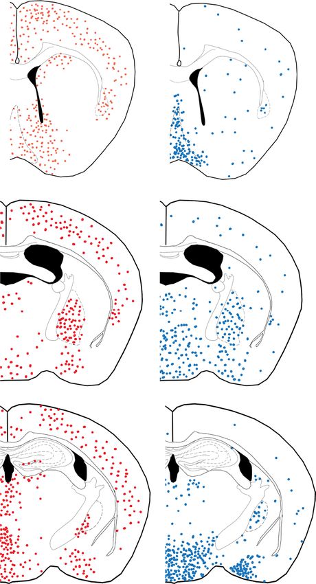
S. A. Hill et al.
heterogeneity between different astrocyte populations [19, cerebellar neural precursor cells, and induces granule cell
20]. Unique gene profiles have been identified between astro- progenitor division and migration from the external germi-
cytes of the cortex, striatum, brainstem, and hippocampus nal layer to the inner granule layer [33–35]. In addition to
[21–23]. In addition to region-specific differences in gene its mitogenic roles, Shh signaling also plays a critical role
expression, astrocyte diversity has also been shown within in axon pathfinding, where it acts as both a chemoattractant
single brain regions. At least 5 different astrocyte popula- and repellant for commissural axons in the developing spi-
tions were identified in the cortex and hippocampus using nal cord [36–38]. Likewise, in the developing retina, SHH
single-cell RNA sequencing, and transcriptional diversity guides proper wiring of retinal ganglion axons through the
between astrocytes in the cortex suggest that astrocytes optic chiasm [39]. Thus, Shh signaling exerts powerful influ-
exhibit a cortical layering pattern defined by distinct gene- ence over diverse processes that are central to establishing
expression profiles [17, 18]. Together, these studies provide the organization of the CNS.
evidence for astrocyte diversity both between and within A number of excellent reviews have covered the molecu-
regions and highlight the need to identify specific molecular lar mechanisms of Shh signaling but we will provide a brief
signaling programs to identify the functional significance overview here [29, 40, 41]. Shh signaling is transduced and
conferred by this remarkable transcriptional diversity. orchestrated within the primary cilium, a small microtubule-
The Sonic hedgehog (Shh) signaling pathway represents rich protrusion present on all cells, including astrocytes [42,
an exciting opportunity to interrogate molecularly defined 43]. The canonical pathway includes the twelve-pass trans-
populations of astrocytes and investigate their unique func- membrane receptor, patched1 (PTCH1), its obligate seven-
tional properties. Although best understood for its essential pass transmembrane co-receptor, smoothened (SMO), and
roles in patterning the nervous system during development, its effector proteins, the zinc finger transcription factors
in the adult brain, Shh activity is found in discrete popula- known as GLIs (glioblastoma gene products 1–3; Fig. 1). In
tions of mature astrocytes [24]. Cells actively transducing the absence of SHH, PTCH1 inhibits the activity of SMO
SHH are identified by the expression of the transcription through mechanisms that are not yet well understood. GLI3
factor GLI1. Interestingly, SHH is produced by neurons, sug- is processed into its repressor form (GLI3R), which inhibits
gesting it mediates neuron–astrocyte communication in the transcriptional activation of Shh pathway genes [44–49].
adult brain [24, 25]. The recent studies have demonstrated Binding of SHH to PTCH1 relieves inhibition of SMO trig-
important roles for Shh signaling in multiple processes, gering signal transduction within the cell [50]. Upon SMO
including astrocyte modulation of neuronal activity and activation, GLI2 acts as the primary transcriptional activa-
defining specific molecular signatures, suggesting that Shh tor of the pathway and initiates transcription of downstream
regulates key functional properties of astrocytes [26, 27]. Shh target genes, including Gli1 [49, 51]. GLI1 is a tran-
Essential features of Shh signaling in astrocytes, including scriptional activator, and its presence is indicative of active
the neuronal source of ligand and its distribution in discrete Shh activity within the cell [44, 52–54]. Importantly, SHH
subpopulations of astrocytes, have the potential to provide is absolutely required for GLI1 expression, as SHH mutant
novel insight into many open questions in astrocyte biology, embryos fail to transcribe Gli1 [55]. Transcriptional activa-
such as astrocyte heterogeneity and astrocyte-neuron inter- tion of Gli1 is therefore a reliable read-out of active Shh
actions. Here, we discuss the emerging body of literature signaling within the cell.
on the diverse roles of Shh signaling in astrocyte function.
Shh signaling in astrocytes
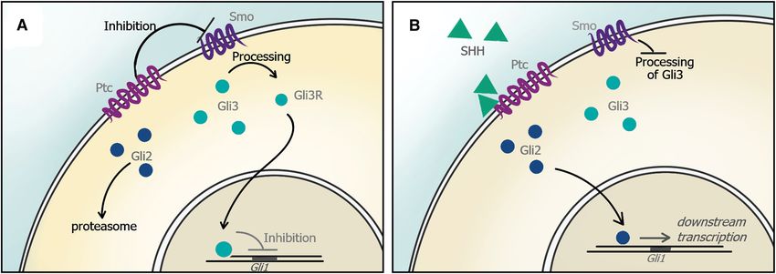
Overview of Sonic hedgehog signaling
Early studies identified the persistence of Shh signaling
Shh signaling is best understood for its diverse roles dur- throughout the adult mammalian CNS using in situ hybridi-
ing embryonic and early postnatal development where it zation. Ptch1, Smo, and Shh transcripts were found in the
mediates a broad range of developmental processes [28, spinal cord and various brain regions, including the hypo-
29]. SHH exhibits a ventral–dorsal gradient in the develop- thalamus, cortex, and the cerebellum [56, 57]. One limita-
ing neural tube in which SHH released from the notochord tion of the in situ hybridization studies is the inability to
and ventral floor plate specifies the fate of neural precursor identify cell type. However, the development of molecu-
cells in a concentration-dependent manner. Both neurons lar genetic tools to label cells with Gli1 activity identified
and oligodendrocytes are specified through SHH-dependent astrocytes as the predominant cell type actively transduc-
regulation of specific transcription factors, including Nkx2.2 ing Shh signaling in the adult mouse brain [24]. Astrocytes
and Olig2, whose precise expression defines progenitor express Ptch1, Smo, Gli2, and Gli3, demonstrating that they
cell domains and identity [30–32]. In the cerebellum, Shh possess the key machinery to transduce SHH [19]. Nota-
signaling regulates both the foliation and proliferation of bly, transduction of SHH is restricted to regionally defined
13Sonic hedgehog signaling in astrocytes
Fig. 1 The Shh pathway. a In the absence of SHH, SMO activ- SHH to PTC alleviates inhibition of SMO, and GLI2 is proteolyti-
ity is inhibited by PTC. Cytosolic GLI3 is processed as a repressor cally processed as an activator and is translocated to the nucleus, acti-
(GLI3R) and inhibits transcription of Shh target genes. b Binding of vating downstream transcription of SHH target genes, including GLI1
subpopulations, suggesting that these cells may possess spe- below [27]. Further studies should explore whether this dis-
cific molecular signatures and functional properties that dis- tribution is specific to the somatosensory cortex or whether
tinguish them from other astrocytes. Astrocytes that express this is found in other cortical regions. More recently, it has
Gli1 are distributed throughout the forebrain, including in also been shown that mature oligodendrocytes may also be
the hypothalamus, thalamus, globus pallidus, and deep lay- a source of SHH in the adult brain [61]. The extent to which
ers of the cortex. In contrast, the striatum and white matter transduction of Shh signaling in individual astrocytes or
tracts are largely devoid of Gli1-expressing astrocytes, as is specific astrocyte populations is due to neuronal or oligo-
the hippocampus, with the exception of adult neural stem dendrocyte sources of SHH requires further study.
cells in the dentate gyrus [58] (Fig. 2). Notably, the propor-
tion of astrocytes exhibiting active Shh signaling in differ-
ent brain regions varies, with ventral regions, such as the The functional significance of Shh signaling
hypothalamus harboring a larger fraction of Gli1-expressing in astrocytes
astrocytes than the cortex [24]. This distribution suggests
that astrocytes may possess functional specialization both The gene expression programs regulated by Shh signaling
between disparate brain regions, as well as within a given in neural precursor cells are well characterized and include
region, that may be regulated by Shh signaling. programs associated with cell fate and proliferation [62]. In
Interestingly, while Gli1 is expressed primarily in contrast, the Shh-dependent gene expression programs in
astrocytes, Shh in the healthy, adult mammalian brain is differentiated astrocytes of the mature brain are only begin-
expressed by neurons, suggesting that Shh signaling medi- ning to be elucidated. Growing evidence, however, points to
ates neuron-astrocyte communication [24, 26, 59, 60]. As an essential role in modulating synaptic function and neu-
in the developing embryo, SHH in the adult brain is found ronal activity. In the cortex, Shh signaling between layer
predominantly in ventral regions, such as the hypothalamus, V neurons and neighboring astrocytes is required for the
which harbors a large number of Shh-expressing neurons refinement of synapses during neural circuit development.
and Gli1-expressing astrocytes (Fig. 2) [24]. Shh + neurons Conditional mutants in which Smo is selectively deleted in
are also found in the cortex, where genetic marking studies astrocytes exhibit impairments in structural plasticity and
show that they correspond mostly to pyramidal cells [25]. In organization and concomitant reduction in expression of
the somatosensory cortex, there is an apparent laminar dis- Kir4.1 [27]. In these mutants, layer V cortical neurons show
tribution such that Shh + neurons are found predominantly in significant changes in spine density and long-term synapse
layer V while astrocytes expressing Gli1 are found primarily plasticity. These structural phenotypes are accompanied by
in layers IV and V [27] (Fig. 3). The functional significance heightened neuronal excitability, suggesting that Shh sign-
of this precise distribution pattern is not well understood, but aling initiates reciprocal interactions between neurons and
a recent study suggests that Shh-dependent local interactions astrocytes that exert functional and structural regulation
between neurons and astrocytes mediates synaptic function of developing neural circuits. Importantly, these pheno-
and organization of layer V neurons, as discussed further types are not observed in mutants lacking Smo in neurons,
13S. A. Hill et al.
A B layer I
cx
layer II/III
cc
CPu layer IV
sep
cl layer V
layer VI
Fig. 3 Laminar distribution of Shh and Gli1-expressing cells in the
cortex. In the somatosensory cortex, astrocytes expressing Gli1 (red)
are localized predominantly in layers IV and V. Neurons expressing
gp Shh (blue) are found largely in layer V
thal
hypo to the Purkinje cell layer and extend their processes into
the molecular layer toward the apical surface of the cer-
ebellar cortex, where they associate with the dendrites
of Purkinje neurons. They exhibit robust expression of
Ptch1, and Smo transcripts and transduce SHH derived
hp from neighboring Purkinje cells [26]. Genetic deletion
experiments demonstrate that Shh signaling regulates
expression of Kir4.1 in Bergmann glia, as observed in
the cortex [26, 27]. Interestingly, Shh activity also regu-
ic lates expression of AMPA receptor subunits, GluA1 and
GluA4 in Bergmann glia, further supporting the role of
amg
Shh signaling in astrocyte modulation of synaptic activity
[26]. Velate astrocytes, on the other hand, reside in the
Fig. 2 Distribution of Shh and Gli1-expressing cells in the adult fore- granule cell layer, below Purkinje cells, and receive lower
brain. a Schematics depicting the distribution of astrocytes express- levels of SHH. In contrast to Bergmann glia, velate astro-
ing Gli1 (red) across three levels of the anterior/posterior axis. Note
that these cells are found in high abundance in various regions includ- cytes express low levels of GluA1 and GluA4. However
ing the hypothalamus, globus pallidus and cortex, but are noticeably constitutive activation of Shh activity in velate astrocytes
absent in other regions, such as the white matter and hippocampus. upregulates expression of these proteins [26]. These obser-
b The distribution of Shh-expressing neurons (blue), as observed vations demonstrate that Shh signaling is both necessary
from genetic labeling experiments. cx cortex, hypo hypothalamus, gp
globus pallidus, cc corpus callosum, CPu caudate putamen, hp hip- and sufficient to regulate molecular characteristics of dis-
pocampus, amg amygdala tinct astrocyte classes.
In both the cortex and cerebellum, Shh signaling regu-
lates expression of genes in astrocytes that are required to
highlighting the requirement for Shh signaling in astrocytes mediate their fundamental role as essential regulators of
on synapse activity and function. Interestingly, Shh signal- the extracellular synaptic environment. Specifically, Shh
ing between Layer V and Layer II/III neurons mediates the signaling affects the ability of astrocytes to clear K + and
precise wiring between these cells during postnatal cortical glutamate, both of which must be tightly regulated to ensure
development [25]. Because neurons do not express Gli1, this appropriate synaptic activity and neuronal survival [64–67].
suggests that these actions are GLI-independent and medi- In addition to its role in mediating astrocyte modulation of
ated by non-canonical Shh signaling [63]. This suggests that synaptic activity, there is also evidence that Shh signal-
Shh signaling exerts essential regulation of synaptic con- ing also influences the ability of astrocytes to communi-
nectivity, through both heterotypic and homotypic cellular cate with neurons via gliotransmission [68–70]. Applica-
interactions. tion of SHH on cultured astrocytes initiates an intracellular
In the cerebellum, Shh signaling drives expression of calcium response and subsequent release of both ATP and
genes that confer specific molecular identities to different glutamate, and blocking Shh activity or chelating intracel-
classes of astroglial cells [26]. Bergmann glia are localized lular calcium inhibited their release [68, 70]. Taken together,
13Sonic hedgehog signaling in astrocytes
these demonstrate that SHH influences astrocyte-synapse astrogliosis includes proliferation of reactive astrocytes
interactions in diverse ways, and point to Shh signaling in [72, 80]. Consistent with its role as a key regulator of pro-
astrocytes as an important mediator of bidirectional com- liferation in neural precursor cells, Shh signaling promotes
munication between neurons and astrocytes. proliferation of cells isolated from the cortex following an
acute invasive injury [81]. Cultures prepared from cortical
stab wound tissue generate reactive astrocyte-derived neu-
Shh signaling in reactive astrocytes rospheres in a SHH-dependent manner [81]. Application
of SHH or its agonist SAG increase neurosphere forma-
A growing body of evidence shows that Shh signaling tion whereas the SHH inhibitor, cyclopamine, blocks neu-
exerts neuroprotective influence on the injury microen- rosphere formation, demonstrating that Shh activation is
vironment by limiting inflammation. Shh activity lowers necessary and sufficient to stimulate proliferation in vitro
permeability of the blood brain barrier (BBB), restricting [81]. However genetic inactivation studies demonstrate
entry of peripheral proteins and blood-borne macrophages that reactive astrocyte proliferation in vivo occurs inde-
into the CNS parenchyma that can elicit toxic inflamma- pendently of Shh signaling [75]. Conditional mutants lack-
tory signaling [71, 72]. Importantly, this effect has been ing Smo in astrocytes show no difference in proliferating
observed in diverse injury environments, including spinal cells at the lesion site following a cortical stab, despite
cord injury contusion, ischemia, and cortical stab wound, reduced proliferation of GFAP-expressing adult neural
suggesting a central role of the pathway in neuroinflam- stem cells in both the dentate gyrus of the hippocampus
mation [73–75]. Astrocytes are key cellular mediators of and in the subventricular zone [75, 82–84]. However as
the anti-inflammatory action of Shh signaling. Application discussed above, this study also demonstrated a dramatic
of the SHH agonist, SAG, limits accumulation of leuko- loss of Shh activity in reactive astrocytes during the first
cytes in CNS parenchyma following cortical stab wound. week following insult, the time during which reactive
SAG acts directly on SMO, the obligatory co-receptor that astrocytes proliferate [72]. Thus, although endogenous
activates the pathway. Genetic deletion of Smo selectively Shh activity is suppressed during the initial acute stages
in astrocytes abolishes the effect of SAG on leukocyte of injury, the observations from the neurosphere assay sug-
accumulation [75]. Notably, despite the powerful anti- gest that activation of the pathway in vivo can promote
inflammatory activity of the pathway, these mutants show proliferation of reactive astrocytes and other proliferating
no signs of a weakened BBB in the absence of injury, cell populations. Indeed, across various injury models,
suggesting that Shh activity may play a role in repressing including spinal cord injury, ischemia, and demyelination,
injury-induced cytokine production by reactive astrocytes Shh signaling promotes proliferation of neural precursor
[75]. In addition to its action on astrocytes, Shh signaling cells, including oligodendrocyte progenitor cells as well as
has also been shown to act on endothelial cells, regulat- adult neural stem cells in the lateral ventricles or dentate
ing expression of tight junction proteins. Shh activity pro- gyrus [73, 74, 77, 85–88].
motes expression of the junctional proteins occludin and While these studies suggest that Shh may be a promising
claudin-5 in vitro and in vivo, promoting BBB integrity target for mitigating CNS damage following various types
[71]. Conversely, permeability assays in cultured endothe- of trauma, conflicting reports on the direction of endogenous
lial cells show that the pro-inflammatory cytokine, IL-1β, Shh activity following injury, as well as the precise cellular
suppresses Shh and downregulates expression of tight sources and targets of the pathway, leave open the ques-
junction proteins [76]. This suggests that inflammation tion of the actions of Shh signaling in vivo in the injury
suppresses Shh signaling. Consistent with this, genetic environment. Whereas early studies reported an increase in
labeling studies show that Gli1 expression is lost in reac- Shh activity following injury, and identified astrocytes as
tive astrocytes during the acute stages of injury, when cellular sources of SHH, later studies using genetic labeling
inflammation is high, but is restored to baseline levels by approaches demonstrate a loss of Shh activity. Using trans-
14 days after the insult, as inflammation is resolved [75, genic mice carrying a Gli-luciferase reporter, an increase
77]. This is accompanied by a concomitant reduction, in bioluminescence was observed up to 1 week after both
and subsequent restoration, of Shh gene expression [75]. a cortical stab wound and kainic-acid induced lesion, sug-
Taken together, these studies point to Shh signaling as a gesting an increase in Shh activity following both an acute
potential target for mitigating injury-induced neurotoxic invasive injury model and acute neurodegeneration [85, 89].
inflammation. Similar observations were reported in ischemic models, in
Following virtually all types of insults, astrocytes which middle cerebral artery occlusion increases expression
undergo pronounced changes in physiology, morphol- of various components of the Shh pathway, as measured
ogy, and gene expression, collectively referred to as by qPCR or antibody labeling [74, 81, 90, 91]. Antibody
reactive astrogliosis [3, 56, 57]. In its most severe form, labeling has shown that astrocytes are the source of SHH
13S. A. Hill et al. in several studies and injury models, whereas other studies labeling for cell-type specific markers, such an approach report neurons and cerebrospinal fluid as the source of SHH would provide a reliable and robust strategy for resolving [71, 81, 85, 89–91]. these conflicting observations, and move towards a bet- In more recent studies, the application of genetic ter understanding of the precise role and mechanisms of labeling strategies to mark and identify cells expressing action of Shh signaling in injury. Gli1 instead demonstrate loss of Shh activity immedi- ately following the insult that persists for up to 2 weeks [75, 77]. Studies using double transgenic mice carrying Outstanding questions the tamoxifen-inducible CreER at the Gli1 locus and a Cre-dependent reporter (Gli1 CreER/+ ;R26R) show that How is SHH released? tamoxifen administered to mice within 3 days after a mild cortical contusion injury or stab injury show fewer or no The mechanism by which SHH is released from cells in Gli1-expressing astrocytes at the lesion site, compared the postnatal and adult CNS is not well understood. In the to contralateral or uninjured controls [75, 77]. Chemical embryonic CNS, SHH is secreted from the notochord and demyelination in Gli1CreER/+;R26R mice fed cuprizone for floor plate, and diffuses dorsally across the ventral neural 6 weeks similarly show fewer labeled cells in the cortex tube where interactions with extracellular matrix proteins, when tamoxifen is administered in the fifth week, suggest- such as heparin sulfate proteoglycan (HSPG), help estab- ing that the loss of Gli1 activity persists in environments lish a gradient [41, 94]. Such concentration-dependent experiencing sustained injury [88]. These observations are transduction of Shh signaling in astrocytes has not been supported by direct measurements of Shh by qPCR show- shown. However, examination of the relative distributions ing reductions in gene expression following stab injury of Shh-expressing neurons and Gli1-expressing astrocytes or cuprizone-mediated demyelination [75, 92]. While dif- suggests that passive diffusion of ligand may mediate local ferent injury models produce distinct microenvironments interactions between neighboring neurons and astrocytes. that trigger differential gene expression programs [78, 79], In the cerebellum, SHH expressed by Purkinje neurons the observations that Gli1 expression is both increased is transduced by neighboring Bergman glial cells [26]. and decreased following a cortical stab wound suggest Likewise, regions, such as the septum, hypothalamus and that injury type alone cannot account for these conflicting globus pallidus possess a high proportion of Gli1-express- observations. Notably, a similar injury-induced reduction ing astrocytes that are associated with local populations in Shh activity is found in lung tissues where Shh is found of Shh-expressing neurons [24] (Fig. 2). Indeed, genetic in epithelial cells and Gli1 is expressed in adjacent mes- marking experiments in adult Gli1CreER mice show that enchymal cells [93]. Using Cre-mediated labeling of Gli1- approximately 80% of astrocytes in the hypothalamus expressing cells, Peng et al. [93] observed that chemical express Gli1, suggesting that these cells are responding to injury to the lung produces a transient downregulation of local availability of high amounts of SHH [24]. Shh signaling that is associated with epithelial expansion In contrast, the cortex displays an apparent mismatch and regeneration. As in the brain, Shh signaling is restored between the relative numbers of Gli1-expressing astro- following resolution of the injury, supporting an essential cytes and Shh-expressing neurons that are observed by role for context-dependent cues that regulate activity of genetic marking [24] (Fig. 2). Despite the considerable the pathway. number of astrocytes in the adult cortex that express Gli1, These conflicting reports of the direction and sources of the number of Shh + neurons identified by genetic labe- SHH after injury, together with the growing evidence that ling experiments is relatively small. One possibility is that Shh signaling exerts powerful anti-inflammatory actions additional sources of SHH are available from cells that fail in the injured environment, underscore the need for further to undergo Cre-mediated recombination in genetic labeling studies. Nevertheless, whether reactive astrocytes are the experiments. Indeed, both astrocytes and oligodendrocytes source or the effectors of Shh signaling, these studies point have been shown to express SHH by antibody labeling, to astrocytes as key cellular mediators in the neuroprotec- though this has not been observed in genetic labeling tive actions of Shh activity. Because SHH is a secreted experiments [61, 81, 89, 91]. Gli1 expression in corti- protein, identifying the precise cellular source responsible cal astrocytes may occur independent of SHH. However for production of the protein can be difficult using anti- loss of one copy of Shh leads to a significant reduction body labeling in which localization of the antibody on cell in Gli1 expression arguing against this possibility [24]. surfaces cannot be ruled out. Genetic labeling experiments Alternatively, an intriguing possibility is that astrocytes using transgenic mice carrying inducible Cre-dependent may also be responding to SHH from distal neurons pro- reporters would permit direct readouts of transcriptional jecting to the cortex. There is evidence that SHH is trans- activity within an individual cell. Coupled with double ported axonally and is released at synapses [95–97]. SHH 13
Sonic hedgehog signaling in astrocytes
has been observed in retinal ganglion cell (RGCs) axons Does Shh signaling have a role in astrocyte
and in axons originating in ventral forebrain neurons that development?
project to the SVZ [59, 95, 98–100]. In the basal gan-
glia, dopaminergic neurons in the substantia nigra express While the role of Shh signaling in oligodendrocyte develop-
SHH that is required for normal physiology and survival ment is well-established, considerably less is known about
of GABAergic neurons in the striatum [101]. Moreover, its role in astrocyte development. Astrocyte production
SHH is found within vesicles in presynaptic terminals of occurs primarily during the first two postnatal weeks [108].
neurons, and in vitro studies have demonstrated SNAREs- They are derived from radial glia and progenitor cells resid-
dependent (soluble n-ethylmaleimide-sensitive fusion ing in the subventricular zone, as well as from local prolif-
protein attachment protein-receptors) release of SHH fol- eration of differentiated cells [109–111]. In the developing
lowing high frequency stimulation [96, 102]. Uncovering optic nerve, SHH released from retinal ganglion cell axons
whether astrocytes transduce SHH from neighboring cells, regulates proliferation of astrocyte precursors [98]. Because
or whether astrocytes can respond to SHH from region- Gli1 expression is not found in white matter tracts in the
ally distant cells is an important question that should be adult forebrain, this suggests that Shh signaling in astrocyte
explored in future experiments. precursors is developmentally regulated and that activity
of the pathway in precursor populations does not predict
Does SHH confer functional specialization to specific activity in mature cells [24]. In the mature cortex, astro-
astrocyte populations? cytes expressing Gli1 are found predominantly in deep lay-
ers. Whether Shh signaling plays a role in the production of
The idea that astrocytes represent a heterogeneous population these, or other Gli1-expressing astrocytes, remains an open
of cells with distinct molecular signatures and functional spe- question. Loss of Shh activity in postnatal glial progenitor
cialization has emerged as an exciting topic of intense interest cells does not impair the total number of cortical astrocytes
over the past decade [14, 15, 103, 104]. Though classically in the mature brain, suggesting that their production does
regarded as a uniform cell population, advances in our under- not require Shh signaling [24]. Nevertheless, expression of
standing of the regional and functional diversity of astrocytes Kir4.1 is dramatically reduced in these conditional mutants,
are re-shaping this view [18, 21, 22, 105, 106]. Astrocytes suggesting that Shh activity is important for gene expression
across brain regions exhibit diversity in calcium activity, programs associated with astrocyte function, but not their
synaptic coverage, and expression of channels and trans- production [27]. Future studies employing fate mapping and
porters that regulate synaptic activity [13, 14, 16, 107]. The intersectional genetics approaches would be a powerful strat-
observation that transduction of SHH is restricted to discrete egy for elucidating the relationship between Shh signaling
subpopulations of astrocytes, distributed in region-specific and astrocyte development.
ways, presents an opportunity to discover novel insights into
astrocyte heterogeneity. One possibility is that SHH regulates
gene expression programs that confer specific molecular and Conclusions and future perspectives
functional identities to specific astrocyte populations, consist-
ent with its role in cell specification of uncommitted neural There has been a remarkable paradigm shift in our under-
precursor cells during embryonic development. Indeed, differ- standing of the vital roles astrocytes play in CNS function.
ential transduction of Shh between Bergmann glia and velate No longer relegated to passive, functionally homogeneous
astrocytes in the cerebellum drives the unique molecular pro- cells, astrocytes have gained recognition as a complex class
files of these two astroglial cells. It would be interesting to of cells with diverse roles in a broad range of CNS functions.
examine whether all astrocytes expressing Gli1 throughout The features that characterize Shh signaling in astrocytes,
the brain share the same molecular signatures. Alternatively, including the selective activation in specific subpopulations
Shh signaling may cooperate with local environmental cues to and the neuronal source of the initiating signal, present excit-
facilitate context-dependent gene expression, enabling astro- ing opportunities to interrogate broad questions about astro-
cytes to perform region-specific functions. Recent evidence cyte biology including molecular identity and functional
suggests that astrocytes in different brain regions exhibit vast specialization of these cells, as well as reciprocal interac-
transcriptomic, proteomic, and functional differences [22]. In tions between astrocytes and neurons. Given the potent abil-
this framework, Shh signaling functions less as a driver of spe- ity of Shh to regulate gene expression during development,
cific cellular identity, but rather as a powerful tool with which it is plausible that Shh activity regulates gene expression
the nervous system can instruct specific astrocyte populations programs that confer specific functional specialization onto a
to meet local needs. Understanding how astrocytes interpret molecularly distinct class of astrocytes. Consequently, astro-
SHH in different regions will facilitate a better understanding cytes expressing Gli1 may be markedly different from those
of astrocyte functional specialization. that do not. Alternatively, Shh signaling may be dynamic,
13S. A. Hill et al.
with Gli1 expression reflecting Shh activity that is regulated 10. Kelley KW, Ben Haim L, Schirmer L et al (2018) Kir4.1-depend-
by local environmental cues. Indeed, there is evidence that ent astrocyte-fast motor neuron interactions are required for peak
strength. Neuron 98:306-319.e7. https://doi.org/10.1016/j.neuro
neuronal activity stimulates release of SHH, and this may n.2018.03.010
be used by neurons to recruit nearby astrocytes to regulate 11. Ma Z, Stork T, Bergles DE, Freeman MR (2016) Neuromodula-
the extracellular environment during times of high activity tors signal through astrocytes to alter neural circuit activity and
[102]. Further studies elucidating the roles of Shh signaling behaviour. Nature 539:428–432. https://doi.org/10.1038/natur
e20145
in astrocytes in healthy and pathological states hold promise 12. Lines J, Martin ED, Kofuji P et al (2020) Astrocytes modulate
to yield novel insight and advancements in our understand- sensory-evoked neuronal network activity. Nat Commun. https
ing of astrocyte function, development, and heterogeneity. ://doi.org/10.1038/s41467-020-17536-3
13. Ben Haim L, Rowitch DH (2016) Functional diversity of astro-
Acknowledgements The work was funded by the National Institute cytes in neural circuit regulation. Nat Rev Neurosci 18:31–41.
of Neurological Disorders and Stroke (Grant number R01NS096100). https://doi.org/10.1038/nrn.2016.159
14. Khakh BS, Sofroniew MV (2015) Diversity of astrocyte func-
tions and phenotypes in neural circuits. Nat Neurosci 18:942–
Open Access This article is licensed under a Creative Commons Attri- 952. https://doi.org/10.1038/nn.4043
bution 4.0 International License, which permits use, sharing, adapta- 15. Khakh BS, Deneen B (2019) The emerging nature of astro-
tion, distribution and reproduction in any medium or format, as long cyte diversity. Annu Rev Neurosci 42:187–207. https://doi.
as you give appropriate credit to the original author(s) and the source, org/10.1146/annurev-neuro-070918-050443
provide a link to the Creative Commons licence, and indicate if changes 16. Dallérac G, Zapata J, Rouach N (2018) Versatile control of syn-
were made. The images or other third party material in this article are aptic circuits by astrocytes: where, when and how? Nat Rev Neu-
included in the article’s Creative Commons licence, unless indicated rosci 19:729–743. https://doi.org/10.1038/s41583-018-0080-6
otherwise in a credit line to the material. If material is not included in 17. Batiuk MY, Martirosyan A, Wahis J et al (2020) Identification of
the article’s Creative Commons licence and your intended use is not region-specific astrocyte subtypes at single cell resolution. Nat
permitted by statutory regulation or exceeds the permitted use, you will Commun 11:1–15. https://doi.org/10.1038/s41467-019-14198-8
need to obtain permission directly from the copyright holder. To view a 18. Bayraktar OA, Bartels T, Holmqvist S et al (2020) Astrocyte
copy of this licence, visit http://creativecommons.org/licenses/by/4.0/. layers in the mammalian cerebral cortex revealed by a single-cell
in situ transcriptomic map. Nat Neurosci 23:500–509. https: //doi.
org/10.1038/s41593-020-0602-1
19. Cahoy JD, Emery B, Kaushal A et al (2008) A transcriptome
References database for astrocytes, neurons, and oligodendrocytes: a
new resource for understanding brain development and func-
tion. J Neurosci 28:264–278. https://doi.org/10.1523/JNEUR
1. Allen NJ, Barres BA (2009) Neuroscience: Glia—more than just OSCI.4178-07.2008
brain glue. Nature 457:675–677. https://doi.org/10.1038/45767 20. Srinivasan R, Lu TY, Chai H et al (2016) New transgenic mouse
5a lines for selectively targeting astrocytes and studying calcium
2. Ullian EM, Sapperstein SK, Christopherson KS, Barres BA signals in astrocyte processes in situ and in vivo. Neuron
(2001) Control of synapse number by Glia. Science 291:657– 92:1181–1195. https://doi.org/10.1016/j.neuron.2016.11.030
661. https://doi.org/10.1126/science.291.5504.657 21. John Lin C-CC, Yu K, Hatcher A et al (2017) Identification of
3. Christopherson KS, Ullian EM, Stokes CCA et al (2005) Throm- diverse astrocyte populations and their malignant analogs. Nat
bospondins are astrocyte-secreted proteins that promote CNS Neurosci 20:396–405. https://doi.org/10.1038/nn.4493
synaptogenesis. Cell 120:421–433. https://doi.org/10.1016/j. 22. Chai H, Diaz-Castro B, Shigetomi E et al (2017) Neural circuit-
cell.2004.12.020 specialized astrocytes: transcriptomic, proteomic, morphologi-
4. Jones EV, Bernardinelli Y, Tse YC et al (2011) Astrocytes con- cal, and functional evidence. Neuron 95:531-549.e9. https://doi.
trol glutamate receptor levels at developing synapses through org/10.1016/j.neuron.2017.06.029
SPARC—β-integrin interactions. J Neurosci 31:4154–4165. https 23. Huang AY-S, Woo J, Sardar D et al (2020) Region-specific tran-
://doi.org/10.1523/JNEUROSCI.4757-10.2011 scriptional control of astrocyte function oversees local circuit
5. Kucukdereli H, Allen NJ, Lee AT et al (2011) Control of excita- activities. Neuron. https://doi.org/10.1016/j.neuron.2020.03.025
tory CNS synaptogenesis by astrocyte-secreted proteins Hevin 24. Garcia ADR, Petrova R, Eng L, Joyner AL (2010) Sonic hedge-
and SPARC. Proc Natl Acad Sci USA 108:E440–E449. https:// hog regulates discrete populations of astrocytes in the adult
doi.org/10.1073/pnas.1104977108 mouse forebrain. J Neurosci 30:13597–13608. https: //doi.
6. Stogsdill JA, Ramirez J, Liu D et al (2017) Astrocytic neuroli- org/10.1523/JNEUROSCI.0830-10.2010
gins control astrocyte morphogenesis and synaptogenesis. Nature 25. Harwell CC, Parker PR, Gee SM et al (2012) Sonic hedgehog
551:192–197. https://doi.org/10.1038/nature24638 expression in corticofugal projection neurons directs corti-
7. Blanco-Suarez E, Liu T-F, Kopelevich A, Allen NJ (2018) Astro- cal microcircuit formation. Neuron 73:1116–1126. https://doi.
cyte-secreted chordin-like 1 drives synapse maturation and limits org/10.1016/j.neuron.2012.02.009
plasticity by increasing synaptic GluA2 AMPA receptors. Neu- 26. Farmer WT, Abrahamsson T, Chierzi S et al (2016) Neurons
ron. https://doi.org/10.1016/j.neuron.2018.09.043 diversify astrocytes in the adult brain through sonic hedgehog
8. Cui Y, Yang Y, Ni Z et al (2018) Astroglial Kir4.1 in the lateral signaling. Science 351:849–854. https://doi.org/10.1126/scien
habenula drives neuronal bursts in depression. Nature 554:323– ce.aab3103
327. https://doi.org/10.1038/nature25752 27. Hill SA, Blaeser AS, Coley AA et al (2019) Sonic hedgehog sign-
9. Tong X, Ao Y, Faas GC et al (2014) Astrocyte Kir4.1 ion chan- aling in astrocytes mediates cell type-specific synaptic organiza-
nel deficits contribute to neuronal dysfunction in Huntington’s tion. Elife 8:2013–2016. https: //doi.org/10.7554/eLife. 45545. 001
disease model mice. Nat Neurosci 17:694–703. https://doi.
org/10.1038/nn.3691
13Sonic hedgehog signaling in astrocytes
28. Ingham PW, McMahon AP (2001) Hedgehog signaling in animal 47. Sasaki H, Nishizaki Y, Hui CC et al (1999) Regulation of Gli2
development: paradigms and principles. Genes Dev 15:3059– and Gli3 activities by an amino-terminal repression domain:
3087. https://doi.org/10.1101/gad.938601 Implication of Gli2 and Gli3 as primary mediators of Shh sign-
29. Fuccillo M, Joyner AL, Fishell G (2006) Morphogen to mito- aling. Development 126:3915–3924
gen: the multiple roles of hedgehog signalling in vertebrate 48. Persson M, Stamataki D, Te WP et al (2002) Dorsal-ventral pat-
neural development. Nat Rev Neurosci 7:772–783. https://doi. terning of the spinal cord requires Gli3 transcriptional repres-
org/10.1038/nrn1990 sor activity. Genes Dev 16:2865–2878. https://doi.org/10.1101/
30. Jessell TM (2000) Neuronal specification in the spinal cord: gad.243402
Inductive signals and transcriptional codes. Nat Rev Genet 49. Pan Y, Bai CB, Joyner AL, Wang B (2006) Sonic hedgehog sign-
1:20–29. https://doi.org/10.1038/35049541 aling regulates Gli2 transcriptional activity by suppressing its
31. Stamataki D, Ulloa F, Tsoni SV et al (2005) A gradient of Gli processing and degradation. Mol Cell Biol 26:3365–3377. https
activity mediates graded sonic hedgehog signaling in the neural ://doi.org/10.1128/mcb.26.9.3365-3377.2006
tube. Genes Dev 19:626–641. https: //doi.org/10.1101/gad.32590 50. Tukachinsky H, Petrov K, Watanabe M, Salic A (2016) Mech-
5 anism of inhibition of the tumor suppressor patched by sonic
32. Ribes V, Briscoe J (2009) Establishing and interpreting graded hedgehog. Proc Natl Acad Sci USA 113:E5866–E5875. https://
sonic hedgehog signaling during vertebrate neural tube pattern- doi.org/10.1073/pnas.1606719113
ing: the role of negative feedback. Cold Spring Harb Perspect 51. Bai CB, Stephen D, Joyner AL (2004) All mouse ventral spi-
Biol. https://doi.org/10.1101/cshperspect.a002014 nal cord patterning by Hedgehog is Gli dependent and involves
33. Corrales JMD, Rocco GL, Blaess S et al (2004) Spatial pattern an activator function of Gli3. Dev Cell 6:103–115. https://doi.
of sonic hedgehog signaling through Gli genes during cerebel- org/10.1016/S1534-5807(03)00394-0
lum development. Development 131:5581–5590. https://doi. 52. Hynes M, Stone DM, Dowd M et al (1997) Control of cell pat-
org/10.1242/dev.01438 tern in the neural tube by the zinc finger transcription factor and
34. Corrales JMD, Blaess S, Mahoney EM, Joyner AL (2006) The oncogene Gli-1. Neuron 19:15–26. https: //doi.org/10.1016/S0896
level of sonic hedgehog signaling regulates the complexity of -6273(00)80344-X
cerebellar foliation. Development 133:1811–1821. https://doi. 53. Dai P, Akimaru H, Tanaka Y et al (1999) Sonic hedgehog-
org/10.1242/dev.02351 induced activation of the Gli1 promoter is mediated by
35. Wechsler-Reya RJ, Scott MP (1999) Control of neuronal precur- GLI3. J Biol Chem 274:8143–8152. https://doi.org/10.1074/
sor proliferation in the cerebellum by sonic hedgehog. Neuron jbc.274.12.8143
22:103–114. https://doi.org/10.1016/S0896-6273(00)80682-0 54. Bai CB, Joyner AL (2001) Gli1 can rescue the in vivo function
36. Charron F, Stein E, Jeong J et al (2003) The morphogen sonic of Gli2. Development 128:5161–5172
hedgehog is an axonal chemoattractant that collaborates with 55. Bai CB, Auerbach W, Lee JS et al (2002) Gli2, but not Gli1,
Netrin-1 in midline axon guidance. Cell 113:11–23. https://doi. is required for initial Shh signaling and ectopic activation of
org/10.1016/S0092-8674(03)00199-5 the Shh pathway. Development 129:4753–4761. https://doi.
37. Bourikas D, Pekarik V, Baeriswyl T et al (2005) Sonic hedgehog org/10.1242/dev.00115
guides commissural axons along the longitudinal axis of the spi- 56. Traiffort E, Charytoniuk DA, Faure H, Ruat M (1998) Regional
nal cord. Nat Neurosci 8:297–304. https: //doi.org/10.1038/nn139 distribution of sonic hedgehog, patched, and smoothened mRNA
6 in the adult rat brain. J Neurochem 70:1327–1330. https: //doi.org
38. Ferent J, Giguère F, Jolicoeur C et al (2019) Boc acts via numb as /10.1046/j.1471-4159.1998.70031327.x
a Shh-dependent endocytic platform for Ptch1 internalization and 57. Traiffort E, Charytoniuk D, Watroba L et al (1999) Discrete
Shh-mediated axon guidance. Neuron 102:1157-1171.e5. https: // localizations of hedgehog signalling components in the develop-
doi.org/10.1016/j.neuron.2019.04.003 ing and adult rat nervous system. Eur J Neurosci 11:3199–3214.
39. Trousse F, Martí E, Gruss P et al (2001) Control of retinal gan- https://doi.org/10.1046/j.1460-9568.1999.00777.x
glion cell axon growth: a new role for sonic hedgehog. Develop- 58. Ahn S, Joyner AL (2005) In vivo analysis of quiescent adult
ment 128:3927–3936 neural stem cells responding to sonic hedgehog. Nature 437:894–
40. Briscoe J, Thérond PP (2013) The mechanisms of Hedgehog 897. https://doi.org/10.1038/nature03994
signalling and its roles in development and disease. Nat Rev Mol 59. Ihrie RA, Shah JK, Harwell CC et al (2011) Persistent sonic
Cell Biol 14:416–429. https://doi.org/10.1038/nrm3598 hedgehog signaling in adult brain determines neural stem cell
41. Ramsbottom SA, Pownall ME (2016) Regulation of hedgehog positional identity. Neuron 71:250–262. https: //doi.org/10.1016/j.
signalling inside and outside the cell. J Dev Biol. https://doi. neuron.2011.05.018
org/10.3390/jdb4030023 60. Courchet J, Polleux F (2012) Sonic hedgehog, BOC, and
42. Di Pietro C, Marazziti D, La Sala G et al (2017) Primary cilia synaptic development: new players for an old game. Neuron
in the murine cerebellum and in mutant models of medulloblas- 73:1055–1058
toma. Cell Mol Neurobiol 37:145–154. https://doi.org/10.1007/ 61. Tirou L, Russo M, Faure H et al (2020) C9C5 positive mature
s10571-016-0354-3 oligodendrocytes are a source of sonic hedgehog in the mouse
43. Sterpka A, Chen X (2018) Neuronal and astrocytic primary cilia brain. PLoS ONE 15:e0229362. https://doi.org/10.1371/journ
in the mature brain. Pharmacol Res 137:114–121. https://doi. al.pone.0229362
org/10.1016/j.phrs.2018.10.002 62. Belgacem YH, Hamilton AM, Shim S et al (2016) The many hats
44. Lee J, Platt KA, Censullo P, i Altaba AR (1997) Gli1 is a target of sonic hedgehog signaling in nervous system development and
of sonic hedgehog that induces ventral neural tube development. disease. J Dev Biol. https://doi.org/10.3390/jdb4040035
Development 124:2537–2552 63. Brennan D, Chen X, Cheng L et al (2012) Noncanonical Hedge-
45. i Altaba AR (1998) Combinatorial Gli gene function in floor hog signaling. Vitam Horm 88:55–72. https://doi.org/10.1016/
plate and neuronal inductions by sonic hedgehog. Development B978-0-12-394622-5.00003-1
125:2203–2212 64. Olsen ML, Sontheimer H (2008) Functional implications for
46. Ding Q, Motoyama J, Gasca S et al (1998) Diminished sonic Kir4.1 channels in glial biology: from K + buffering to cell dif-
hedgehog signaling and lack of floor plate differentiation in Gli2 ferentiation. J Neurochem 107:589–601
mutant mice. Development 125:2533–2543
13S. A. Hill et al.
65. Chever O, Djukic B, McCarthy KD, Amzica F (2010) Implica- hedgehog. Cell Stem Cell 12:426–439. https://doi.org/10.1016/j.
tion of Kir4.1 channel in excess potassium clearance: an in vivo stem.2013.01.019
study on anesthetized glial-conditional Kir4.1 knock-out mice. 82. Petrova R, Garcia ADR, Joyner AL (2013) Titration of GLI3
J Neurosci 30:15769–15777. https://doi.org/10.1523/JNEUR repressor activity by sonic hedgehog signaling is critical for
OSCI.2078-10.2010 maintaining multiple adult neural stem cell and astrocyte func-
66. Djukic B, Casper KB, Philpot BD et al (2007) Conditional knock- tions. J Neurosci 33:17490–17505. https://doi.org/10.1523/
out of Kir4.1 leads to glial membrane depolarization, inhibition JNEUROSCI.2042-13.2013
of potassium and glutamate uptake, and enhanced short-term 83. Balordi F, Fishell G (2007a) Hedgehog signaling in the subven-
synaptic potentiation. J Neurosci 27:11354–11365. https://doi. tricular zone is required for both the maintenance of stem cells
org/10.1523/jneurosci.0723-07.2007 and the migration of newborn neurons. J Neurosci 27:5936–
67. Rothstein JD, Dykes-Hoberg M, Pardo CA et al (1996) Knock- 5947. https://doi.org/10.1523/JNEUROSCI.1040-07.2007
out of glutamate transporters reveals a major role for astroglial 84. Balordi F, Fishell G (2007b) Mosaic removal of hedgehog signal-
transport in excitotoxicity and clearance of glutamate. Neuron ing in the adult SVZ reveals that the residual wild-type stem cells
16:675–686. https://doi.org/10.1016/S0896-6273(00)80086-0 have a limited capacity for self-renewal. J Neurosci 27:14248–
68. Okuda H, Tatsumi K, Morita-Takemura S et al (2016) Hedgehog 14259. https://doi.org/10.1523/JNEUROSCI.4531-07.2007
signaling modulates the release of gliotransmitters from cultured 85. Amankulor NM, Hambardzumyan D, Pyonteck SM et al
cerebellar astrocytes. Neurochem Res 41:278–289. https://doi. (2009) Sonic hedgehog pathway activation is induced by acute
org/10.1007/s11064-015-1791-y brain injury and regulated by injury-related inflammation. J
69. Araque A, Carmignoto G, Haydon PG et al (2014) Gliotransmit- Neurosci 29:10299–10308. https: //doi.org/10.1523/JNEUR
ters travel in time and space. Neuron 81:728–739. https://doi. OSCI.2500-09.2009
org/10.1016/j.neuron.2014.02.007 86. Bambakidis NC, Miller RH (2004) Transplantation of oligo-
70. Adachi C, Kakinuma N, Jo SH et al (2019) Sonic hedgehog dendrocytes precursors and sonic hedgehog results in improved
enhances calcium oscillations in hippocampal astrocytes. J Biol function and white matter sparing in the cords of adult rats after
Chem 294:16034–16048. https://doi.org/10.1074/jbc.RA119 contusion. Spine J 4:16–26. https://doi.org/10.1016/j.spine
.007883 e.2003.07.004
71. Alvarez JI, Dodelet-Devillers A, Kebir H et al (2011) The 87. Ferent J, Ruat M, Traiffort E (2013) Investigation of the pro-
hedgehog pathway promotes blood-brain barrier integrity and teolipid protein promoter activity during demyelination and
CNS immune quiescence. Science 334:1727–1731. https://doi. repair. Differentiation 85:182–189. https://doi.org/10.1016/j.
org/10.1126/science.1206936 diff.2013.05.002
72. Sofroniew MV (2015) Astrocyte barriers to neurotoxic inflam- 88. Sanchez MA, Armstrong RC (2018) Postnatal sonic hedgehog
mation. Nat Rev Neurosci 16:249–263. https://doi.org/10.1038/ (Shh) responsive cells give rise to oligodendrocyte lineage cells
nrn3898 during myelination and in adulthood contribute to remyelina-
73. Bambakidis NC, Wang RZ, Franic L, Miller RH (2003) Sonic tion. Exp Neurol 299:122–136. https://doi.org/10.1016/j.expne
hedgehog-induced neural precursor proliferation after adult urol.2017.10.010
rodent spinal cord injury. J Neurosurg 99:70–75. https://doi. 89. Pitter KL, Tamagno I, Feng X et al (2014) The SHH/Gli pathway
org/10.3171/spi.2003.99.1.0070 is reactivated in reactive glia and drives proliferation in response
74. Chechneva OV, Mayrhofer F, Daugherty DJ et al (2014) A to neurodegeneration-induced lesions. Glia 62:1595–1607. https
smoothened receptor agonist is neuroprotective and promotes ://doi.org/10.1002/glia.22702
regeneration after ischemic brain injury. Cell Death Dis 5:e1481– 90. Sims JR, Lee S-W, Topalkara K et al (2009) Sonic hedgehog
e1481. https://doi.org/10.1038/cddis.2014.446 regulates ischemia/hypoxia-induced neural progenitor prolif-
75. Allahyari RV, Clark KL, Shepard KA, Garcia ADR (2019) eration. Stroke 40:3618–3626. https://doi.org/10.1161/STROK
Sonic hedgehog signaling is negatively regulated in reactive EAHA.109.561951
astrocytes after forebrain stab injury. Sci Rep 9:565. https://doi. 91. Jin Y, Raviv N, Barnett A et al (2015) The Shh signaling pathway
org/10.1038/s41598-018-37555-x is upregulated in multiple cell types in cortical ischemia and
76. Wang Y, Jin S, Sonobe Y et al (2014) Interleukin-1β induces influences the outcome of stroke in an animal model. PLoS ONE
blood-brain barrier disruption by downregulating sonic hedge- 10:e0124657. https://doi.org/10.1371/journal.pone.0124657
hog in astrocytes. PLoS ONE. https://doi.org/10.1371/journ 92. Hibbits N, Yoshino J, Le TQ, Armstrong RC (2012) Astrogliosis
al.pone.0110024 during acute and chronic cuprizone demyelination and impli-
77. Mierzwa AJ, Sullivan GM, Beer LA et al (2014) Comparison of cations for remyelination. ASN Neuro 4:393–408. https://doi.
cortical and white matter traumatic brain injury models reveals org/10.1042/an20120062
differential effects in the subventricular zone and Divergent sonic 93. Peng T, Frank DB, Kadzik RS et al (2015) Hedgehog actively
hedgehog signaling pathways in neuroblasts and oligodendrocyte maintains adult lung quiescence and regulates repair and regen-
progenitors. ASN Neuro. https://doi.org/10.1177/1759091414 eration. Nature 526:578–582. https: //doi.org/10.1038/nature 1498
551782 4
78. Liddelow SA, Barres BA (2017) Reactive astrocytes: production, 94. Yan D, Lin X (2009) Shaping morphogen gradients by proteo-
function, and therapeutic potential. Immunity 46:957–967 glycans. Cold Spring Harb Perspect Biol 1:a002493. https://doi.
79. Sofroniew MV (2009) Molecular dissection of reactive astroglio- org/10.1101/cshperspect.a002493
sis and glial scar formation. Trends Neurosci 32:638–647. https 95. Traiffort E, Moya KL, Faure H et al (2001) High expression and
://doi.org/10.1016/j.tins.2009.08.002 anterograde axonal transport of aminoterminal sonic hedgehog
80. Wanner IB, Anderson MA, Song B et al (2013) Glial scar borders in the adult hamster brain. Eur J Neurosci 14:839–850. https://
are formed by newly proliferated, elongated astrocytes that inter- doi.org/10.1046/j.0953-816X.2001.01708.x
act to corral inflammatory and fibrotic cells via STAT3-depend- 96. Petralia RS, Schwartz CM, Wang Y-XX et al (2011) Subcellular
ent mechanisms after spinal cord injury. J Neurosci 33:12870– localization of patched and smoothened, the receptors for sonic
12886. https://doi.org/10.1523/JNEUROSCI.2121-13.2013 hedgehog signaling, in the hippocampal neuron. J Comp Neurol
81. Sirko S, Behrendt G, Johansson PA et al (2013) Reactive glia in 519:3684–3699. https://doi.org/10.1002/cne.22681
the injured brain acquire stem cell properties in response to sonic
13You can also read