Dual roles of the Sterol Recognition Region in Hedgehog protein modification
←
→
Page content transcription
If your browser does not render page correctly, please read the page content below
bioRxiv preprint first posted online Jan. 1, 2020; doi: http://dx.doi.org/10.1101/2019.12.31.892067. The copyright holder for this
preprint (which was not peer-reviewed) is the author/funder, who has granted bioRxiv a license to display the preprint in perpetuity.
 All rights reserved. No reuse allowed without permission.

 Dual roles of the Sterol Recognition Region in Hedgehog protein modification

 Rahul Purohit1, Daniel S. Peng1, Erika Vielmas1, and Alison E. Ondrus*1
 1
 Division of Chemistry & Chemical Engineering, California Institute of Technology, Pasadena,
 California, USA

 Abstract
 Nature provides a number of mechanisms to encode dynamic information in biomolecules.
 In metazoans, there exist rare chemical modifications that occur through entirely unique
 mechanistic regimes. One such example occurs in the Hedgehog (Hh) morphogens, proteins
 singular across all domains of life for the nature of their covalent ligation to cholesterol. The
 isoform- and context-specific efficiency of the ligation reaction has profound impact on the activity
 of Hh morphogens and represents an unexplored aspect of Hh ligand-dependent cancers. To
 elucidate the chemical mechanism of this modification, we have defined roles of the
 uncharacterized sterol recognition region (SRR) in Hh proteins. We use a combination of
 sequence conservation, directed mutagenesis, and biochemical assays to specify residues of the
 SRR that are responsible for cellular and biochemical processes in Hh cholesterolysis. Our
 investigations offer the first functional template of this region, providing opportunities to identify
 parallel reactivity in nature and revealing new mechanisms that can be exploited as tools in
 chemical biology.

 Introduction
 The Hedgehog (Hh) morphogens undergo an auto-catalytic modification that cleaves the
 translated protein approximately in half and covalently appends cholesterol to the last residue of
 the N-terminal fragment.1 To date, this intramolecular small molecule transfer activity has been
 observed only in Hh proteins and in no other domains of life.2 Cholesterol modification is
 fundamental to the activity of the Hh morphogen;3,4 mutation of the catalytic residues are lethal,
 and mutations that reduce the efficiency of the reaction give rise to profound congenital disorders
 (Fig. S1A).5,6 This unique modification appears to have co-evolved with cholesterol-sensing
 mechanisms that coordinate Hh signal transduction, chemically linking embryogenesis to
 cholesterol homeostasis.7,8,9
 The initially translated, full-length Hh proteins (typically 350-475 residues in length) consist
 of an N-terminal signaling domain, an internal catalytic domain, and a C-terminal region known
 as the Sterol Recognition Region (SRR, Fig. 1A).10 During post-translational modification, the
 full-length protein is cleaved at the end of the N-terminal signaling domain; concomitantly, the last
 residue of the N-terminus is released as a cholesterol ester. Independently, the N-terminal
 residue of the Hh morphogen is palmitoylated by dedicated acyltransferase enzyme in the ER.11,12
 A crystal structure of the first 145 residues of D. melanogaster Hh-C provided by Beachy and
 coworkers immediately revealed the basis for proteolytic activity of the protein: an intein splicing
 domain remarkably similar in tertiary structure to those found in bacteria and fungi.13,14 Inteins

 1
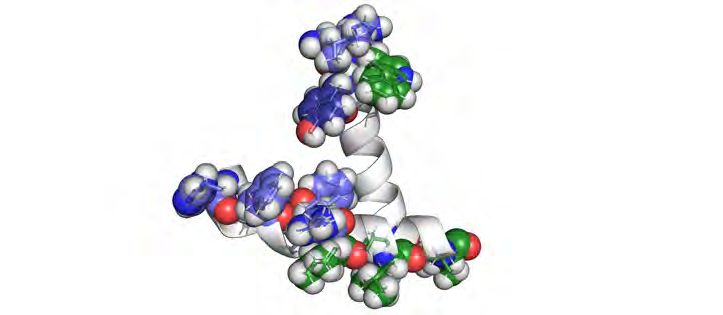
bioRxiv preprint first posted online Jan. 1, 2020; doi: http://dx.doi.org/10.1101/2019.12.31.892067. The copyright holder for this
preprint (which was not peer-reviewed) is the author/funder, who has granted bioRxiv a license to display the preprint in perpetuity.
 All rights reserved. No reuse allowed without permission.

 catalyze intramolecular attack of a cysteine, serine, or threonine side chain on the backbone
 carbonyl of the preceding residue, effecting N-to-S/O acyl transfer and creating a labile (thio)ester
 bond in the protein backbone. In conventional inteins, a nucleophilic residue in the C-terminus of
 the protein reacts with this (thio)ester in a transesterification reaction, ultimately ligating the two
 exteins.15,16 However, Hh proteins lack catalytic intein residues required for protein re-ligation.
 Instead, the thioester formed during the splicing reaction is intercepted by cholesterol, co-opting
 this mechanism for small molecule adduction.
 As in many other intein-containing proteins, the N-terminus of Hh does not participate in
 the cleavage reaction.10 Thioester formation and cholesteroylation activities inhere solely in the
 intein and SRR regions, respectively. Accordingly, cholesterol ligation can occur in vitro and in
 cells when the Hh N-terminus is replaced by fluorescent proteins or simply by a His-tag.17,18,19,20
 However, the molecular details of the SRR that facilitate cholesterol transfer remain unknown.
 The SRR, which spans 50-100 residues in different species, bears no significant homology to
 known proteins in any domain of life. While nematodes possess Hh intein-domain (Hint)
 homologues that likewise lack a C-terminal nucleophilic residue for transesterification, these
 domains do not couple small molecule transfer to splicing activity.2 The structure of the SRR has
 never been established crystallographically, and to date presents an unsolved challenge for high-
 resolution analysis. Moreover, no static conformation can fully capture the dynamic activity of
 these fascinating proteins.
 To define structural requirements for cholesterol transfer by the Hh SRR, we used a
 combination of tools at the interface of chemistry and biology. We demonstrate that the process
 of Hh cholesterolysis encompasses two main activities: 1) engagement of cholesterol as a
 nucleophile, and 2) cellular localization to cholesterol-containing membranes. We use sequence
 conservation and structure prediction to reveal a helix-loop-helix arrangement that is broadly
 represented among Hh SRRs. Guided by this structural analysis, we used site-directed
 mutagenesis of these motifs within human Sonic hedgehog (hSHH) expressed in mammalian
 cells to identify loss of function mutations that abolish Hh autoprocessing. Biochemical analysis
 of full-length protein purified from mammalian cells enabled us to distinguish conserved
 hydrophobic residues that are essential for cholesterolysis in cells but not in vitro. Cellular
 localization of a fluorescent SRR fusion protein revealed a novel Golgi targeting motif that may
 bias interaction of full-length hSHH with cellular membranes for cholesterol recruitment. By
 defining the structural requirements and elementary steps in this unconventional modification, this
 work expands our understanding of Nature’s chemical biology toolkit and facilitates rational design
 of tools for small molecule ligation in cells.

 Results
 A helix-loop-helix motif is conserved across SRRs
 To identify the residues within the SRR that participate in cholesterol transfer we
 considered the SRR as V366-S462 of the hSHH protein, which begins after the last corresponding
 residue in the crystal structure of the D. melanogaster intein domain.10 Cleavage of the D.
 melanogaster protein at this residue renders the protein soluble in the absence of detergent and

 2
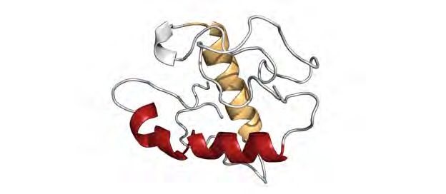
bioRxiv preprint first posted online Jan. 1, 2020; doi: http://dx.doi.org/10.1101/2019.12.31.892067. The copyright holder for this
preprint (which was not peer-reviewed) is the author/funder, who has granted bioRxiv a license to display the preprint in perpetuity.
 All rights reserved. No reuse allowed without permission.

 eliminates cholesterol ligation during proteolysis. As protein- and nucleotide-based alignment
 programs show no significant homology between the hSHH SRR and other non-Hh proteins, we
 aligned 710 manually curated, diverse Hh proteins to identify common features across species
 and isoforms (Fig. 1B and S2).21 This alignment revealed two sections of conserved residues
 before and after a long intervening sequence that is unique to a subset of Hh proteins (e.g. hSHH).
 Secondary structure analysis revealed that the conserved segments are predicted to form α-
 helices,22 while the glycine-rich intervening segment is predicted to be disordered.
 To guide our hypotheses regarding the residue-level contributions of the SRR to
 cholesterolysis, we used ab initio fragment assembly to generate a 3D structure of the last 100
 residues of hSHH, which encompasses the hSHH SRR (Fig. 1C).23 In line with our secondary
 structure predictions, a lowest energy hSHH SRR structure possesses helices at residues from
 W372-L390 (1st helix) and I432-L447 (2nd helix), which are linked by a disordered loop comprising
 residues A391-G431. The structure contains a “kink” introduced by proline residue P379 in the
 1st helix, which separates the 1st helix into to angled segments from W372-A378 and P379-L390.24
 Helical wheel analysis of the two regions, revealed that each segment of the helices possess a
 face enriched in hydrophobic residues (Fig. 2D). This arrangement is characteristic of
 amphipathic peripheral membrane helices;25 further, none of our analyses predict these
 sequences to span the bilayer.
 To gain empirical support for our alignment-based model, we synthesized peptides
 comprising the 1st and 2nd helices and examined their secondary structures by Circular Dichroism
 (CD) spectroscopy (Fig. 1E). As anticipated, these hydrophobic peptides are insoluble in the
 absence of liposomes. CD spectra of each helix in multidisperse POPC liposomes clearly
 displayed helical character in both the presence and absence of cholesterol. Grounded by
 conservation analysis and secondary structure analysis, we used this model as a scaffold for
 experimental studies to define residue-level structural elements of the SRR that link intein
 proteolysis to cholesterol transfer.

 Conserved SRR residues are required for hSHH cholesterolysis in cells
 As a relatively hindered, hydrophobic nucleophile with a pKa of ~18,26 the cholesterol
 hydroxyl group imposes strict requirements on the residues involved in nucleophilic activation and
 trajectory for attack. Whereas an intein-SRR unit catalyzes cholesterolysis both in cells and in
 vitro, Hh mutants lacking the SRR undergo proteolysis without attachment of cholesterol.10 These
 observations demonstrate that the SRR is necessary and sufficient for biochemical Hh
 cholesterolysis. In cells, however, the marked insolubility of cholesterol in aqueous solution (
bioRxiv preprint first posted online Jan. 1, 2020; doi: http://dx.doi.org/10.1101/2019.12.31.892067. The copyright holder for this
preprint (which was not peer-reviewed) is the author/funder, who has granted bioRxiv a license to display the preprint in perpetuity.
 All rights reserved. No reuse allowed without permission.

 processed and/or lipidated forms of the hSHH protein partition into cell lysates or secreted media.
 The cholesteroylated and dually lipidated (cholesteroylated and palmitoylated) N-terminal
 proteins remain membrane-associated and are retained in cell lysates. By contrast, hSHH
 bearing the palmitate modification alone is secreted into cell media.11,29,30,31,32 Accordingly, we
 used production of cell-associated hSHH-N in lysates (hSHH-NL) as a measure of
 cholesteroylation for a given SRR mutant (Fig. S3).
 We first generated deletion mutants that lacked components of the helix-loop-helix motif
 identified in our protein alignment and biochemical studies (Fig. 2A). Consistent with previous
 observations, a construct lacking the SRR failed to produce any cell-associated hSHH-N.10
 Specific deletion of the 1st helix, 2nd helix, and loop region demonstrated that hSHH-N was likewise
 absent in lysate of mutants lacking the 1st or 2nd helices. Strikingly, the ∆loop construct that lacked
 a non-conserved 32 residue connector functioned identically to the wild-type protein. This result
 defines the 1st and 2nd helices as a minimal required unit, consistent with the absence of this loop
 region in Hh proteins known to undergo Hh cholesterolysis.33
 To more explicitly probe the residues of the SRR that participate in cholesterolysis in cells,
 we performed alanine scanning of both the 1st and 2nd helices (Fig. 2B and 2C). Mutation of highly
 conserved 1st helix residues H374, F377, and R381 drastically reduced production of cell-
 associated hSHH-N. Interestingly, mutation of the absolutely conserved P379 residue did not
 reduce the proportion of cell-associated hSHH-N relative to WT but lowered the total amount of
 protein in cell lysate, likely due to an effect on biochemical and/or cellular protein stability. Based
 on our helical wheel analysis (Fig. 1D), we envisioned that the three conserved leucine residues
 on the same face of the 1st helix, L382, L386, and L390 might interact with the lipid tails of the
 membrane. Consistent with this hypothesis, Monte Carlo simulations of the interaction between
 the 1st helix peptide and a phospholipid bilayer predict that these three leucines will reside at the
 membrane interior (Fig. 2D).34 While single L382A, L386A, and L390A mutants enabled the
 production of cell-associated hSHH-N, a L382A/L386A/L390A triple mutant drastically reduced
 cell-associated hSHH-N, indicating that a stretch of alanine residues is insufficient to support the
 production of cholesteroylated hSHH-N in cells (Fig. 2E). Consistent with a model involving
 membrane association, replacement of either of the individual L382 or L386 residues with a
 negatively charged residue (glutamate) abolished production of cell-associated hSHH-N. An
 L390E mutation showed a significant but attenuated reduction of cell-associated hSHH-N,
 whereas glutamate mutation of L387, which is perpendicular to the hydrophobic leucine surface,
 shows only slight reduction in cell-associated hSHH-N versus wild-type protein. This model also
 suggests an electrostatic interaction between the basic residues H374 and R381 and
 phospholipid head groups, which may contribute to membrane association.25
 The 2nd helix of the hSHH SRR contains a HWY motif that is largely conserved among
 species and Hh isoforms. While this motif has been demonstrated to regulate post-cleavage
 trafficking of hSHH-C,35 a contribution to cholesterolysis has not been tested.36 Alanine mutation
 of each residue in this motif drastically reduced production of cell-associated hSHH-N (Fig. 2H).
 In particular, hSHH-N was undetectable in the lysates of cells expressing a Y435A mutant. We
 envisioned that this conserved tyrosine residue might participate in a hydrogen bond interaction

 4
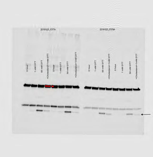
bioRxiv preprint first posted online Jan. 1, 2020; doi: http://dx.doi.org/10.1101/2019.12.31.892067. The copyright holder for this
preprint (which was not peer-reviewed) is the author/funder, who has granted bioRxiv a license to display the preprint in perpetuity.
 All rights reserved. No reuse allowed without permission.

 with the C3-OH of cholesterol. Surprisingly, mutation of this residue to an alternative hydrogen
 bond donor (aspartate) failed to rescue production of cell-associated hSHH-N. Interestingly,
 mutation to phenylalanine reduced total protein in lysate but restored the fraction of cell-
 associated hSHH-N relative to wild-type. The exclusive recovery of cell-associated hSHH-N by
 an aromatic residue suggests a potential mechanistic contribution of the Y435 -system to
 cholesterolysis.37 By contrast, the lower overall expression of Y435F suggests that the hydroxyl
 group is essential to biochemical and/or cellular protein stability.
 The segment of the 2nd helix following the HWY motif consists of a number of polar
 residues interspersed with four conserved leucine residues (L438, L439, and L446, and L447).
 Surprisingly, while alanine replacement of the conserved L447 residue significantly decreased
 cell-associated hSHH-N, mutation of the less conserved residues L438, I442, and L446 to alanine
 had an attenuated effect (Fig. 2C). Alanine scanning of the remaining positions in the 2nd helix
 revealed that substitution of the polar residues S436 and T444 and the hydrogen bond donating
 residue Y440 did not significantly reduce production of hSHH-N in lysates. Likewise, mutation of
 glutamine residues Q437 and Q441 did not affect levels of cell-associated hSHH-N. Taken
 together, these studies provide a roadmap of the SRR residues involved in cholesterol transfer
 by Hh proteins in human cells.

 A subset of SRR residues are required for hSHH cholesterolysis in vitro
 To distinguish between biochemical and cellular factors that contribute to cholesterolysis,
 we investigated the reactivity of SRR mutants toward cholesterol in vitro. To do so, we appended
 a Myc-DDK tag to the C-terminus of our hSHH construct (Fig. S4). Overexpression of tagged
 protein followed by mild lysis and affinity purification with agarose conjugated to an anti-FLAG
 antibody enabled us to isolate active unprocessed hSHH-FL free from N-terminal protein
 generated in cells. Consistent with previous observations, exposure of this protein to 0.5 mM
 cholesterol and 1 mM DTT resulted in production of cholesteroylated hSHH-N (hSHH-NC), which
 could be distinguished from un-cholesteroylated hSHH-N by apparent molecular weight (Fig. 3A,
 Fig. S5).28,30,31,38,39
 For all mutants, exposure of the purified protein to 50 mM DTT effected production of
 hSHH-N, confirming formation of an active thioester. While a mutant lacking the catalytic cysteine
 residue (C198A) was unreactive, a construct lacking the entire SRR (E368*, ∆SRR) was
 susceptible to in vitro DTT cleavage but not cholesterolysis (Fig. 3B). Consistent with our cellular
 assays, neither the ∆1st helix nor the ∆2nd helix mutants were capable of undergoing
 cholesterolysis in vitro. Interestingly, a ∆2nd helix mutant underwent non-cholesteroylative
 cleavage in the presence of DTT and cholesterol, whereas a ∆1st helix mutant was unreactive
 under these conditions. This result suggests that an interaction between the 1st helix and
 cholesterol is sufficient to promote cleavage but not cholesterolysis. As anticipated, a ∆loop
 mutant functioned equivalently to wild-type hSHH.
 We next sought to query the in vitro reactivity of 1st helix SRR mutants that failed to
 produce cell-associated hSHH-N. All purified mutant proteins were capable of undergoing robust
 cleavage at high (50 mM) but not low concentrations (1 mM) of DTT (Fig. S6). Consistent with

 5
bioRxiv preprint first posted online Jan. 1, 2020; doi: http://dx.doi.org/10.1101/2019.12.31.892067. The copyright holder for this
preprint (which was not peer-reviewed) is the author/funder, who has granted bioRxiv a license to display the preprint in perpetuity.
 All rights reserved. No reuse allowed without permission.

 our cellular assays, expression levels of the H374A and R381A mutants were lower than wild-
 type hSHH, and protein that was produced did not undergo cholesterolysis. While an F377A
 mutant expressed high quantities of protein, trace hSHH-N protein that was produced in the
 presence of 1 mM DTT and cholesterol likewise did not exhibit a shift characteristic of
 cholesteroylated hSHH-N. Strikingly, while neither L382A/L386A/L390A, nor L386E mutants
 were capable of producing cell-associated hSHH-N, both exhibited a migration shift to indicate
 cholesterolysis in vitro. This observation suggests that the L382/L386/L390 residues act primarily
 to facilitate cellular aspects of cholesterolysis, potentially through an interaction involving this
 hydrophobic face within the 1st helix.
 We then sought to characterize the biochemical contributions of the 2nd helix HWY motif
 to cholesterolysis in vitro. Purified H433A nor Y435A mutant proteins failed to produce
 cholesteroylated hSHH-N, consistent with their observed lack of this reactivity in cells. As in
 cells, mutation of the conserved Y435 residue to an alternative aromatic (Y435F) but not an
 aliphatic (Y435A) residue preserved biochemical cholesterolysis. By contrast, a W434A mutant,
 which was also incapable of producing cell-associated hSHH-N, showed restored reactivity
 toward cholesterolysis in vitro. The unique restoration of cholesterolysis by a W434A mutant
 indicates that this residue contributes predominantly to cellular aspects of hSHH cholesterolysis.
 The observation that specific SRR mutants can undergo cholesteroylation in vitro but not
 in cells implies that hSHH-N cholesteroylation entails separable biochemical and cellular
 processes. Within our structural model, this analysis points to a cluster of conserved aromatic
 residues that is required for biochemical cholesterol adduction opposite to a hydrophobic face
 that is required for cellular cholesterolysis (Fig. 3C). Thereby, this functional portrait provides a
 launching point for identification of related motifs in nature and a blueprint for redesign.

 The SRR contains a Golgi localization motif
 Due to its fleetingly low (nanomolar) concentrations in aqueous media, essentially all
 cellular cholesterol is sequestered in membranes. Moreover, specific lipid and protein
 compositions within a given membrane control the fraction of active cholesterol that is available
 to interact with other biomolecules.40,41,42,43 While hSHH-N release has been investigated both
 functionally and biochemically in native hSHH-N secreting cells,44,45 only a handful of studies have
 tracked the intra- and extracellular trajectories of the hSHH-C fragment.28,46 Whether the SRR
 has inherent cellular targeting activity is unknown.
 Because the Hh intein domain itself is soluble, we hypothesized that the SRR might
 localize the full-length hSHH protein to microdomains within the ER and or/other membranes for
 cholesterolysis. To ascertain preferences for subcellular localization inherent to the SRR, we
 created a fusion protein comprising N-terminal EGFP fused to SRR-encompassing residues
 A365-S462 of hSHH (Fig. 3D). To survey whether the SRR could bias the localization of the intein
 domain, we also created EGFP-intein(C198A), EGFP-intein(C198A)-SRR fusions. When EGFP
 alone was expressed from a pCMV6-EGFP construct, it assumed a diffuse cellular distribution
 that included the nucleus. By contrast, when expressed as a fusion with the hSHH SRR, EGFP
 fluorescence was observed as discrete puncta clustered at the periphery of the nucleus, and

 6
bioRxiv preprint first posted online Jan. 1, 2020; doi: http://dx.doi.org/10.1101/2019.12.31.892067. The copyright holder for this
preprint (which was not peer-reviewed) is the author/funder, who has granted bioRxiv a license to display the preprint in perpetuity.
 All rights reserved. No reuse allowed without permission.

 nuclear fluorescence was eliminated. Time course studies revealed that this distribution was
 established immediately upon expression and at various levels of overexpression. Likewise,
 overexpression of an EGFP-intein-SRR but not an EGFP-intein construct was sufficient to
 establish punctate, extranuclear distribution. Finally, while deletion of the 1st and 2nd helices in an
 EGFP-SRR construct preserved extranuclear fluorescence, deletion of the 1st helix dramatically
 reduced puncta formation and enhanced cytoplasmic fluorescence, while deletion of the 2nd helix
 attenuated, but did not abolish, wild-type EGFP-SRR distribution. This observation is consistent
 with a significant role for the 1st helix in subcellular trafficking, and reinforces a hypothesis that
 the 1st helix engages in functional membrane interactions during cellular cholesterolysis.
 To determine which cellular compartments colocalize with EGFP-SRR, we co-expressed
 the EGFP-SRR construct with red fluorescent protein-labeled organelle markers (Fig. 3E). These
 studies revealed that SRR constructs colocalized extensively with the Golgi, as demonstrated
 using mCherry markers bearing either Golgin or β-galactosyltransferase Golgi-targeting
 sequences (Fig. S6). Importantly, the EGFP-SRR construct showed no indication of lysosomal
 localization to indicate misfolded or aberrantly processed protein. Partial colocalization was also
 observed with ER and caveolin markers, whereas no significant colocalization was observed with
 the nucleus, peroxisomes, mitochondria, actin, or the plasma membrane (Fig. S7). Together
 these studies reveal that the SRR imparts a distinct Golgi localization to fusion proteins.

 Discussion
 To elucidate the molecular features of the SRR that ligate cholesterol to Hh proteins, we
 have defined residues that are required for cellular and biochemical Hh cholesterolysis. We used
 sequence conservation to identify common regions of secondary structure, which revealed a
 helix-loop-helix motif shared by all Hh proteins. Site-directed mutagenesis provided a blueprint
 of residues essential for Hh cholesterolysis in human cells and established minimal criteria for
 biochemical Hh cholesteroylation. This cellular and biochemical analysis was coupled to our
 identification of a new motif that induces Golgi localization in living cells.
 Our data suggest possible mechanisms by which the SRR achieves this cholesterol-
 protein ligation in cells (Fig. 4). In one scenario, the SRR may tether the full-length Hh protein to
 cholesterol-rich membranes, enabling direct recruitment of a cholesterol nucleophile to the intein
 active site. Alternatively, the SRR might itself extract cholesterol from the membrane and shuttle
 it to the reactive site within the intein. Finally, the SRR could form a hydrophobic conduit with the
 intein domain, and together this unit could orient cholesterol for nucleophilic attack.
 These studies can now provide a chemical basis for a number of SRR mutations that lead
 to developmental disease and can shed light on dysregulated Hh ligand activity in cancer (Fig.
 S1). We are currently building upon this basis set of functional residues to re-engineer the SRR
 ligase activity for alternative substrates and proteins of interest. Future studies will investigate
 the contribution of intein-SRR interactions to reaction efficiency to inform construct design. We
 anticipate that this work will lead to a general means to repurpose Nature’s small molecule ligation
 technology as an invaluable tool in the chemical biology of living systems.

 7
bioRxiv preprint first posted online Jan. 1, 2020; doi: http://dx.doi.org/10.1101/2019.12.31.892067. The copyright holder for this
preprint (which was not peer-reviewed) is the author/funder, who has granted bioRxiv a license to display the preprint in perpetuity.
 All rights reserved. No reuse allowed without permission.

 Acknowledgements
 We thank Divya Kolli for preparation of the EGFP-SRR fusion construct and Colin Lee for
 performing Western blot control experiments. We are grateful to Prof. Peter Dervan and Prof.
 James K. Chen for valuable feedback. This work was supported by the Margaret E. Early Medical
 Research Trust.

 Author contributions
 R. P. and A. E. O. designed experiments. R. P. performed CD spectroscopy, hSHH mutagenesis,
 cellular hSHH protein analysis, hSHH mutant protein isolation and in vitro cholesterolysis assays,
 and analyzed data. D. S. P. and E. V. performed hSHH mutagenesis, and E. V. performed
 Western blot experiments. A. E. O. performed EGFP-SRR mutagenesis, conducted microscopy
 experiments, and analyzed data. R. P. and A. E. O. wrote the manuscript.

 Competing interests
 The authors declare no competing interests.

 *Corresponding author: Alison E. Ondrus, email: aondrus@caltech.edu

 8
bioRxiv preprint first posted online Jan. 1, 2020; doi: http://dx.doi.org/10.1101/2019.12.31.892067. The copyright holder for this
preprint (which was not peer-reviewed) is the author/funder, who has granted bioRxiv a license to display the preprint in perpetuity.
 All rights reserved. No reuse allowed without permission.

 References

 1
 Porter, J., Young, K. & Beachy, P. Cholesterol modification of Hedgehog signaling proteins in
 animal development. Science 274, 255–259 (1996).
 2
 T. Burglin, The Hedgehog protein family. Genome Biol. 9, 241 (2008).
 3
 Lewis, P. M. et al. Cholesterol modification of Sonic Hedgehog is required for long-range
 signaling activity and effective modulation of signaling by Ptc1. Cell 105, 599–612 (2001).
 4
 Li, Y., Zhang, H., Litingtung, Y. & Chiang, C. Cholesterol modification restricts the spread of Shh
 gradient in the limb bud. Proc. Natl. Acad. Sci. USA 103, 6548–6553 (2006).
 5
 Roessler, E. et al. Mutations in the C-terminal domain of Sonic Hedgehog cause
 holoprosencephaly. Hum. Mol. Genet. 6, 1847–1853 (1997).
 6
 Odent, S. et al. Expression of the Sonic hedgehog (SHH) gene during early human development
 and phenotypic expression of new mutations causing holoprosencephaly. Hum. Mol. Genet. 8,
 1683–1689 (1999).
 7
 Cooper, M. et al. A defective response to Hedgehog signaling in disorders of cholesterol
 biosynthesis. Nat. Genet. 33, 508–13 (2003).
 8
 Hausmann, G., von Mering, C. & Basler, K. The Hedgehog signaling pathway: Where did it come
 from? Plos Biol. 7, e1000146 (2009).
 9
 Kowatsch, C., Woolley, R. E., Kinnebrew, M., Rohatgi, R. & Siebold, C. Structures of vertebrate
 Patched and Smoothened reveal intimate links between cholesterol and Hedgehog signalling.
 Curr. Opin. Struc. Biol. 57, 204–214 (2019).
 10
 Hall, T. et al. Crystal structure of a Hedgehog autoprocessing domain: Homology between
 Hedgehog and self-splicing proteins. Cell 91, 85–97 (1997).
 11
 Pepinsky, B. R. et al. Identification of a palmitic acid-modified form of human Sonic hedgehog.
 J. Biol. Chem. 273, 14037–14045 (1998).
 12
 Chamoun, Z. et al. Skinny Hedgehog, an acyltransferase required for palmitoylation and activity
 of the Hedgehog signal. Science 293, 2080–2084 (2001).
 13
 Perler, F. B. Homing Endonucleases and Inteins. vol. 16 193–210 (2005).
 14
 Novikova, O. et al. Intein clustering suggests functional importance in different domains of life.
 Mol. Biol. Evol. 33, 783–799 (2016).
 15
 Beachy, P. et al. Multiple roles of cholesterol in hedgehog protein biogenesis and signaling.
 Cold Spring Harb. Symp. 62, 191–204 (1997).
 16
 Shah, N. H. & Muir, T. W. Inteins: Nature’s gift to protein chemists. Chem. Sci. 5, 446–461
 (2013).
 17
 Zhang, X. et al. Protein–nucleic acid conjugation with sterol linkers using Hedgehog
 autoprocessing. Bioconj. Chem. 30, 2799–2804.
 18
 Vincent, S., Thomas, A., Brasher, B. & Benson, J. D. Targeting of proteins to membranes
 through hedgehog auto-processing. Nat. Biotechnol. 21, nbt844 (2003).
 19
 Mozhdehi, D. et al. Genetically encoded cholesterol-modified polypeptides. J. Am. Chem. Soc.
 141, 945–951 (2019).

 9
bioRxiv preprint first posted online Jan. 1, 2020; doi: http://dx.doi.org/10.1101/2019.12.31.892067. The copyright holder for this
preprint (which was not peer-reviewed) is the author/funder, who has granted bioRxiv a license to display the preprint in perpetuity.
 All rights reserved. No reuse allowed without permission.

 20
 Jiang, S. & Paulus, H. A high-throughput, homogeneous, fluorescence polarization assay for
 inhibitors of Hedgehog protein autoprocessing. J. Biomol. Screen 15, 1082–1087 (2010).
 21
 Pei, J. & Grishin, N. V. PROMALS: towards accurate multiple sequence alignments of distantly
 related proteins. Bioinformatics 23, 802–808 (2007).
 22
 Drozdetskiy, A., Cole, C., Procter, J. & Barton, G. J. JPred4: a protein secondary structure
 prediction server. Nucleic Acids Res. 43, W389–W394 (2015).
 23
 Raman, S. et al. Structure prediction for CASP8 with all-atom refinement using Rosetta. Prot.
 Struct. Funct. Bioinform. 77, 89–99 (2009).
 24
 Gautier, R., Douguet, D., Antonny, B. & Drin, G. HELIQUEST: a web server to screen
 sequences with specific α-helical properties. Bioinformatics 24, 2101–2102 (2008).
 25
 Prévost, C. et al. Mechanism and determinants of amphipathic helix-containing protein targeting
 to lipid droplets. Dev. Cell 44, 73-86.e4 (2018).
 26
 Dixon, S. L. & Jurs, P. C. Estimation of pKa for organic oxyacids using calculated atomic
 charges. J. Comput. Chem. 14, 1460–1467 (1993).
 27
 Saad, H. Y. & Higuchi, W. I. Water solubility of cholesterol. J. Pharm. Sci. 54, 1205–1206
 (1965).
 28
 Simons, K. & Ikonen, E. How cells handle cholesterol. Science 290, 1721–1726 (2000).
 29
 Peters, C., Wolf, A., Wagner, M., Kuhlmann, J. & Waldmann, H. The cholesterol membrane
 anchor of the Hedgehog protein confers stable membrane association to lipid-modified proteins.
 Proc. Natl. Acad. Sci. USA 101, 8531–8536 (2004).
 30
 Guy, K. R. Inhibition of sonic hedgehog autoprocessing in cultured mammalian cells by sterol
 deprivation. Proc. Natl. Acad. Sci. USA 97, 7307–7312 (2000).
 31
 Chen, X. et al. Processing and turnover of the Hedgehog protein in the endoplasmic reticulum.
 J. Cell. Biol. 192, 825–838 (2011).
 32
 Zeng, X. et al. A freely diffusible form of Sonic hedgehog mediates long-range signalling. Nature
 411, 716–720 (2001).
 33
 Pettigrew, C. A., Asp, E. & Emerson, C. P. A new role for Hedgehogs in juxtacrine signaling.
 Mech. Dev. 131, 137–149 (2014).
 34
 Gofman, Y., Haliloglu, T. & Ben-Tal, N. Monte Carlo simulations of peptide–membrane
 interactions with the MCPep web server. Nucleic Acids Res. 40, W358–W363 (2012).
 35
 Daniele, J. R., Chu, T. & Kunes, S. A novel proteolytic event controls Hedgehog intracellular
 sorting and distribution to receptive fields. Biol. Open 6, 540–550 (2017).
 36
 Banavali, N. K. The mechanism of cholesterol modification of Hedgehog ligand. J. Comput.
 Chem. https://doi.org/10.1002/jcc.26097 (2019).
 37
 Dougherty, D. A. Cation-π interactions in chemistry and biology: A new view of benzene, Phe,
 Tyr, and Trp. Science 271, 163–168 (1996).
 38
 Porter, J. A. et al. The product of hedgehog autoproteolytic cleavage active in local and long-
 range signalling. Nature 374, 363–366 (1995).
 39
 Porter, J. A. et al. Hedgehog patterning activity: Role of a lipophilic modification mediated by
 the carboxy-terminal autoprocessing domain. Cell 86, 21–34 (1996).

 10
bioRxiv preprint first posted online Jan. 1, 2020; doi: http://dx.doi.org/10.1101/2019.12.31.892067. The copyright holder for this
preprint (which was not peer-reviewed) is the author/funder, who has granted bioRxiv a license to display the preprint in perpetuity.
 All rights reserved. No reuse allowed without permission.

 40
 Maxfield, F. R. & van Meer, G. Cholesterol, the central lipid of mammalian cells. Curr. Opin.
 Cell Biol. 22, 422–429 (2010).
 41
 Menon, A. K. Sterol gradients in cells. Curr. Opin. Cell Biol. 53, 37–43 (2018).
 42
 Ikonen, E. Cellular cholesterol trafficking and compartmentalization. Nat. Rev. Mol. Cell Bio. 9,
 125–138 (2008).
 43
 Gatta, A. T. & Levine, T. P. Piecing together the patchwork of contact sites. Trends Cell Biol.
 27, 214–229 (2017).
 44
 Guerrero, I. & Kornberg, T. B. Hedgehog and its circuitous journey from producing to target
 cells. Semin. Cell Dev. Biol. 33, 52–62 (2014).
 45
 Thérond, P. P. Release and transportation of Hedgehog molecules. Curr. Opin. Cell Biol. 24,
 173–180 (2012).
 46
 Zhu, L. et al. An IL-12/Shh-C domain fusion protein-based IL-12 autocrine loop for sustained
 natural killer cell activation. Int. J. Oncol. 41, 661–669 (2012).

 11
bioRxiv preprint first posted online Jan. 1, 2020; doi: http://dx.doi.org/10.1101/2019.12.31.892067. The copyright holder for this
preprint (which was not peer-reviewed) is the author/funder, who has granted bioRxiv a license to display the preprint in perpetuity.
 All rights reserved. No reuse allowed without permission.

 Materials and Methods

 Antibodies

 Primary antibodies: Shh Antibody (E-1) (N-terminus), (Santa Cruz Biotechnology, sc-365112),
 Sonic Hedgehog/Shh C-Terminus Antibody (R&D Systems, AF445), Loading control:
 Karyopherin β1 (H-7) (Santa Cruz Biotechnology, sc-137016). Secondary antibodies: Donkey
 anti-Goat IgG (H+L) Cross-Adsorbed Secondary Antibody, Alexa Fluor 647 (Invitrogen, A-
 21447), Donkey anti-Mouse IgG (H+L) Cross-Adsorbed Secondary Antibody, Alexa Fluor 488
 (Invitrogen, A-21202).

 Constructs

 Myc-DDK-tagged human Sonic Hedgehog (hSHH) in a pCMV6 vector was obtained from
 Origene (RC222175). For analysis of proteins from cell lysates and media, a stop codon was
 introduced after the last residue of hSHH (S462) to obtain untagged hSHH. Untagged and Myc-
 DDK tagged hSHH constructs were used for site-directed mutagenesis to create the
 corresponding SRR mutants. Briefly, mutagenesis reactions were performed using overlapping
 or non-overlapping primers as required. Truncation mutants were generated by inserting stop
 codons via site-directed mutagenesis. SRR deletion mutants were generated by PCR extension
 of the parent hSHH sequence using phosphorylated reverse primers to exclude specified
 residues, then ligating the resulting PCR product using T4 ligase (NEB, M0318). To generate
 EGFP and EGFP-hSHH constructs used for microscopy experiments, an EGFP insert
 (Clontech, Addgene vdb2487) followed by the linker sequence (GGGS)2 was cloned into the
 pCMV6 backbone before the corresponding hSHH fragment using Gibson assembly. All PCR
 reactions were performed using Phusion High Fidelity polymerase (NEB, M0530) in the
 presence of 10% DMSO. Parent construct was digested with DpnI (NEB, R0176). The full
 hSHH insert sequence for each mutant was verified by Sanger sequencing.

 Sequence alignment and ab initio structure prediction

 Secondary structure prediction for human hSHH SRR (368-462) was performed using the online
 server JPred4 (http://www.compbio.dundee.ac.uk/jpred). Three dimensional models for the
 hSHH residues 363-462 were predicted by ab initio modelling using the Robetta online server
 (http://robetta.bakerlab.org). Helical wheel diagrams for the 1st helix (W372-A378 and P379-
 L390) and the 2nd helix (I432-L447) were generated using the Heliquest online server
 (http://heliquest.ipmc.cnrs.fr/). Hedgehog proteins were identified in the Uniprot repository using

 1
bioRxiv preprint first posted online Jan. 1, 2020; doi: http://dx.doi.org/10.1101/2019.12.31.892067. The copyright holder for this
preprint (which was not peer-reviewed) is the author/funder, who has granted bioRxiv a license to display the preprint in perpetuity.
 All rights reserved. No reuse allowed without permission.

 the query: name:hedgehog NOT name:skinny NOT name:acyltransferase NOT name:interacting
 NOT name:interfering NOT name:interference NOT name:hhat NOT name:like NOT
 name:receptor NOT name:cysteine NOT name:smoothened NOT name:sufu NOT
 name:tetratricopeptide NOT name:similar NOT name:putative NOT name:patched NOT
 name:domain NOT name:hint NOT name:pyruvate NOT name:amino NOT name:peptidase
 NOT name:quality NOT name:containing NOT name:partial length:[356 TO 518], and then by
 manual inspection. Protein sequences were aligned using the PROMALS3D algorithm
 (http://prodata.swmed.edu/promals3d) and visualized as a sequence logo generated by the
 WebLogo online server (http://weblogo.threeplusone.com/).

 Liposome preparation and CD spectroscopy

 Peptides for residues encompassing the 1st helix (368-391) and 2nd helix (431-449) of the hSHH
 SRR were obtained from Genscript Inc. at >90% purity. Crude soybean phospholipids
 containing L-α-phosphatidylcholine (PC, Sigma-Aldrich, P5638) were used to prepare liposomes
 for CD analysis. Briefly, phospholipids in the presence or absence of cholesterol were dissolved
 in chloroform and dried to a thin layer on the sides of a glass vial using a rotary evaporator.
 Dried lipids were resuspended in CD buffer (specified below) and mixed to homogeneity using a
 vortex to yield a 10 mM PC ± 2 mM cholesterol stock solution. Unilamellar vesicles were
 prepared by disruption on ice using a tip sonicator, and liposomes were centrifuged at 4400 × g
 for 10 minutes to remove undissolved lipids. Supernatant containing unilamellar vesicles was
 diluted to 0.5 mg/mL and analyzed by dynamic light scattering (Wyatt DynaPro PlateReader-II)
 to assess size distribution.

 CD spectra were collected on 430 Series Aviv Biomedical Inc. spectrophotometer with a rotating
 sample chamber at 25 ºC using a 0.1 cm path length quartz cuvette. CD scans were collected
 from 190–250 nm with a 1 nm bandwidth and 10 s averaging at each data point. Stock
 liposomes were diluted to a concentration of 0.5 mM PC ± 0.1 mM cholesterol in CD sample
 buffer. 1st helix peptide: A stock solution of peptide in aqueous 50 mM KF was diluted to 50-100
 uM in liposomes ± cholesterol in 10 mM sodium borate (pH 9.6) and 50 mM KF. 2nd helix
 peptide: A solution of 50-100 uM peptide was prepared directly in liposomes ± cholesterol in 10
 mM sodium phosphate (pH 7.4) and 50 mM KF. Signal for liposomes-only in the corresponding
 buffer was subtracted from the peptide signal for all measurements.

 2
bioRxiv preprint first posted online Jan. 1, 2020; doi: http://dx.doi.org/10.1101/2019.12.31.892067. The copyright holder for this
preprint (which was not peer-reviewed) is the author/funder, who has granted bioRxiv a license to display the preprint in perpetuity.
 All rights reserved. No reuse allowed without permission.

 Gel electrophoresis and Western blot analysis

 Samples were loaded on a 4-15% SDS-PAGE gel (Bio-Rad, 5678084) in Tris/Glycine/SDS
 running buffer (Bio-Rad, 1610772), and proteins were resolved at a current of 150 V for 1 h at
 room temperature. After electrophoresis, proteins were transferred to a 0.2 µM PVDF
 membrane (Bio-Rad, 1704157) using a Bio-Rad Trans-Blot Turbo Transfer System.
 Membranes were incubated with primary antibodies at a 1:1000 dilution in 2% milk in PBST (1×
 PBS and 0.1% Tween-20) for 6-12 h at 4 ºC, washed three times for 5 min each with fresh
 PBST, then incubated with secondary antibodies at room temperature for 1-4 h. Fluorescence
 signal was collected on Bio-Rad Chemidoc MP Imaging System. Band intensities were
 quantified using Bio-Rad Image Lab Software v6.0. Briefly, bands corresponding to full length
 and/or processed hSHH proteins in each lane were identified by the software and visually
 inspected for accuracy, and relative pixel intensities were measured for each band.

 Cell culture

 All hSHH proteins were expressed in adherent HEK-293T cells (ATCC, CRL-3216). Cells were
 cultured in high-glucose DMEM (Gibco, 11965118) containing 10% fetal bovine serum (FBS,
 Gibco, 26140079), 100 U/mL of penicillin-streptomycin (Gibco, 15140163), 1 mM sodium
 pyruvate, and 2 mM L-glutamine. Cells were seeded at an initial confluence of ~20%, passaged
 every 3-4 days upon reaching 85-90% confluence, and maintained in an atmosphere of 5% CO2
 and 95% humidity at 37 ºC. Transfection mixtures were prepared in low serum media that
 contained 0.5 % FBS instead of 10% FBS. Polyethylenimine (PEI, PolyScience, 23996-1) (1
 mg/mL stock) was used as a transfection reagent.

 Analysis of hSHH protein from cells

 HEK-293T cells were seeded in a 12-well plate 24 h before transfection at a density of 200,000
 cells/well. Upon reaching 80-90% confluence, cells were transfected with 0.75 µg hSHH
 plasmid DNA (untagged) and PEI at a ratio of 3:1 PEI:DNA (w/w), which were pre-incubated for
 30 min in Opti-MEM (Invitrogen, 51985034) and diluted in low serum media. After media
 replacement, cells were incubated for 42-48 hours. After rinsing with with 200 µL PBS, 125 µL
 ice-cold lysis buffer (50 mM Tris-HCl (pH 7.4), 250 mM NaCl, 1% IGEPAL CA-630, 1× cOmplete
 EDTA-free Protease Inhibitor Cocktail (Roche, 11836170001) and 1 mM PMSF) was added to
 each well and the plate was incubated at 4 ºC with rocking for 10-15 minutes. Cell material was
 collected by scraping and transferred to microcentrifuge tubes. Cells were then disrupted using

 3
bioRxiv preprint first posted online Jan. 1, 2020; doi: http://dx.doi.org/10.1101/2019.12.31.892067. The copyright holder for this
preprint (which was not peer-reviewed) is the author/funder, who has granted bioRxiv a license to display the preprint in perpetuity.
 All rights reserved. No reuse allowed without permission.

 tip a sonicator at 60% amplitude for 14 seconds (2 s ON, 1 s OFF). Cell lysate was heated to 98
 ºC for 5 minutes on a heating block, supplemented with SDS-PAGE sample buffer, boiled for
 additional 5 minutes, and analyzed or stored at -20 ºC.

 To detect secreted hSHH proteins, transfection media was collected before cell lysis and protein
 was precipitated with 30% trichloroacetic acid (final) and pelleted by centrifugation. The pellet
 was washed with ddH2O to remove excess trichloroacetic acid, resuspended in 1× SDS-PAGE
 sample buffer, boiled for 5 min, and analyzed or stored at -20 ºC.

 Purification of hSHH-FL

 10 million HEK-293T cells were seeded in a 15 cm dish 24 h before transfection. Upon reaching
 80-90% confluence, cells were transfected with 20 µg hSHH plasmid DNA (Myc-DDK tagged)
 and PEI at a ratio of 3:1 PEI:DNA (w/w), which were pre-incubated for 30 min in Opti-MEM
 (Invitrogen, 51985034) and diluted in low serum media. Cells were incubated for 40-48 hours,
 then dissociated with trypsin (TripLE, Thermo Fisher, 12605036), resuspended in culture media,
 and collected by centrifugation at 750 × g for 5 min at room temperature. Cell pellets were flash
 frozen in liquid nitrogen and purified directly or stored at -80 ºC.

 All protein isolation procedures were performed on ice or at 4 ºC. To isolate Myc-DDK tagged
 hSHH-FL, the cell pellet from each 15 cm plate was resuspended in 1 mL resuspension buffer
 (2× PBS (pH 7.4) + 5% glycerol + 1% Triton-X100 + 1 mM PMSF + 1× Roche EDTA free
 Complete Protease Inhibitor Cocktail). Cells were disrupted using a tip sonicator for 20 sec (2
 sec ON and 1 sec OFF), then lysate was solubilized by addition of Fos-Choline-12 (Anatrace,
 F308S) to final concentration of 0.5%. Lysate was incubated in at 4 ºC for 30 minutes with
 gentle rocking to solubilize membrane-associated proteins, then clarified by centrifugation at
 21,000 × g for 10 minutes at 4 ºC. Supernatant was carefully transferred to a new
 microcentrifuge tube and incubated with 100 µL pre-equilibrated Anti-Flag M2 Affinity Gel
 (Sigma, A2220) at 4 ºC with gentle rocking for 2-3 hours. Unbound proteins were removed by
 centrifugation and the beads were washed twice (4× resin volume, 15 minutes each) with wash
 buffer (2× PBS (pH 7.4) + 5% glycerol) containing 0.1% Fos-Choline-12. Bound proteins were
 eluted by incubating the resin for 2 hours with 0.15 mg/mL FLAG-peptide in a final volume of
 250 µL wash buffer containing 0.1% Fos-Choline12. Beads were removed from eluate

 4
bioRxiv preprint first posted online Jan. 1, 2020; doi: http://dx.doi.org/10.1101/2019.12.31.892067. The copyright holder for this
preprint (which was not peer-reviewed) is the author/funder, who has granted bioRxiv a license to display the preprint in perpetuity.
 All rights reserved. No reuse allowed without permission.

 containing hSHH-FL-Myc-DDK by centrifugation at 10,000 × g and used immediately or flash
 frozen in liquid nitrogen and stored at -80 ºC.

 Analysis of hSHH protein reactivity in vitro

 Isolated hSHH-FL-MycDDK protein was thawed on ice and divided into 30 µL aliquots in a PCR
 strip. For each experiment, 30 µL of the protein sample was boiled immediately after thawing to
 serve as an input control. For thiolysis, 1 M DTT was added to a final concentration of 50 mM.
 For cholesterolysis, 1 mM DTT was added from the 50 mM DTT stock, then 50 mM stock
 cholesterol in 2-propanol was added immediately with constant gentle vortexing to avoid
 precipitation of cholesterol, to a final concentration of 0.5 mM. A 1 mM DTT-only reaction was
 performed similarly without cholesterol. The processing reactions were incubated at room
 temperature for 5 hours with gentle rocking. Reactions were quenched by heating the samples
 to 98 ºC for 5 min and adding 1× SDS-PAGE sample buffer, then samples were analyzed or
 stored at -20 ºC.

 Fluorescence colocalization studies

 HEK-293T cells were seeded 24 h before transfection at 60,000 cells per well on sterile, acid
 cleared coverslips in a 24-well plate. Upon reaching 80-90% confluence, cells in each well were
 transfected with 0.2 µg GFP-hSHH SRR plasmid DNA and PEI at a ratio of 3:1 PEI:DNA (w/w),
 which was pre-incubated for 30 min in Opti-MEM (Invitrogen, 51985034) and diluted in low
 serum media. When present, the marker plasmids were included at 0.1 µg/well. After media
 replacement, cells were incubated for 6-8 hours, then rinsed with 1× PBS and fixed with ice cold
 4% paraformaldehyde for 10 minutes. Coverslips were rinsed three times for 5 minutes each
 with 1× PBS before a final rinse in ddH2O, then mounted on clean glass slides in DAPI-free
 mounting media (Invitrogen, ProLong Diamond Antifade Mountant, P36965). Mounting media
 was cured in the dark for 8-12 h and coverslips were sealed with nail polish. Images were
 obtained on a Zeiss 700 confocal microscope using a 63× oil immersion objective.

 5
Plasmids used
Human Sonic Hedgehog and EGFP plasmids
Name Source
hSHH Origene RC222175
hSHH untagged Origene RC222175, stop after hSHH(S462)
pEGFP-c1 Addgene vdb2487

Organelle marker protein plasmids
Addgene # Plasmid Origin of localization sequence Organelle
55838 DsRed2-Mito-7 COX8A mitochondria
55073 mCherry-Lysosomes-20 LAMP1 lysosomes
55110 mCherry-Nucleus-7 SV40 T-Antigen nucleus
27705 CAV1-mCherry caveolin caveolae
55052 mCherry-Golgi-7 β-galactosyltransferase Golgi
54520 mCherry-Peroxisomes-2 SGSKL peroxisomes
55011 mCherry-CD36-C-10 CD36 plasma membrane
54967 mCherry-Actin-C-18 actin cytoskeleton
55859 mRuby-Endo-14 RHOB endosomes
85048 pmScarlet-Giantin-C1 Giantin Golgi

 6
Primers used
Deletions and truncations
Cloning method: Inverse PCR
Construct Forward Primer Reverse primer
hSHH(∆369-391) (∆1st helix) GCTACGCGGTCATCCCCGCGCGCACGGA GCGCGCGGGGATGACCGCAGAACCC
hSHH(∆431-447) (∆2nd helix) TGCGGGGGCCACCGCGGACAGCGAGGC GCCTCGCTGTCCGCGGTGGCCCCCGCACCCG
 CCTGC GA
hSHH(∆393-424) (∆loop) GCTGCACTGGCGCCCGGTGCGGGGG GGTGGCCCCCGCACCGGGCGCCAGT
hSHH(E368*) (∆SRR-Myc- TCGTGCTACGCGGTCATCACGCGTACGC CGAGCGGCCGCGTACGCGTGATGACCGCGTA
DDK) GGCCGCTCGAGCAGAA GCACGAGGCCA

Single amino acid mutations
Cloning method: Site directed mutagenesis

Intein domain mutants
Construct Forward Primer Reverse primer
hSHH(C198A) CAAATCGGGAGGCGCCTTCCCGGGCTCGG CCGAGCCCGGGAAGGCGCCTCCCGATTTG
hSHH(D243A) TTCCTCACTTTCCTGGCCCGCGACGACGGCGCC GGCGCCGTCGTCGCGGGCCAGGAAAGTGAGGAA
hSHH(T267A) CGCCTGCTGCTCGCCGCCGCGCACCTG CAGGTGCGCGGCGGCGAGCAGCAGGCG
hSHH(H270A) GCTCACCGCCGCGGCCCTGCTCTTTGTGG CCACAAAGAGCAGGGCCGCGGCGGTGAGC
hSHH(C363A) GGTGCTGGCCTCGGCC TACGCGGTCATCG CGATGACCGCGTAGGCCGAGGCCAGCACC
hSHH(E368A) GCGGTCATCGCGGAGCACAGCTGGGCGCACCGG GCTGTGCTCCGCGATGACCGCGTAGCACGAGGC
hSHH(E368*) GCTACGCGGTCATCTGAGAGCACAGCTGGG CCCAGCTGTGCTCTCAGATGACCGCGTAGC

 7
1st helix mutants
Construct Forward Primer Reverse primer
hSHH(W372A) GAGGAGCACAGCGCGGCGCACCGGGC GCCCGGTGCGCCGCGCTGTGCTCCTC
 GTCATCGAGGAGCACAGCTGGGCGGCCCGGGCC GCTGTGCTCCTCGATGACCGCGTAGCACGAGGC
hSHH(H374A) TT CAGCA
hSHH(R375A) TGGGCGCACGCGGCCTTCGCGCCCTTCCGCCTG CGCGAAGGCCGCGTGCGCCCAGCTGTGCTCCTC
 GAAGGGCGCGGCGGCCCGGTGCGCCCAGCTGT
hSHH(F377A) CACCGGGCCGCCGCGCCCTTCCGCCTGGCGCAC G
 CAGGCGGAAGGCCGCGAAGGCCCGGTGCGCCC
hSHH(P379A) GCCTTCGCGGCCTTCCGCCTGGCGCACGCGCTC A
hSHH(F380A) GCCTTCGCGCCCGCCCGCCTGGCGCA TGCGCCAGGCGGGCGGGCGCGAAGGC
hSHH(R381A) TTCGCGCCCTTCGCCCTGGCGCACGCG CGCGTGCGCCAGGGCGAAGGGCGCGAA
 CGCGTGCGCCGCGCGGAAGGGCGCGAAGGCCC
hSHH(L382A) CCCTTCCGCGCGGCGCACGCGCTCCTGGCTGCA G
hSHH(L382E) TTCCGCGAAGCGCACGCGCTCCTGGCTGCACT TGCGCTTCGCGGAAGGGCGCGAAGGCCCGGT
hSHH(H384A) TTCCGCCTGGCGGCCGCGCTCCTGGC GCCAGGAGCGCGGCCGCCAGGCGGAA
hSHH(L386A) GCCTGGCGCACGCGGCCCTGGCTGCACTGGC GCCAGTGCAGCCAGGGCCGCGTGCGCCAGGC
hSHH(L386E) TTCCGCCTGGCGCACGCGGAACTGGCTGCACT TGCGCCAGGCGGAAGGGCGCGAAGGCCCGGT
hSHH(L387A) GCGCACGCGCTCGCGGCTGCACTGGC GCCAGTGCAGCCGCGAGCGCGTGCGC
hSHH(L387E) GCGCACGCGCTCGAGGCTGCACTGGC GAGCGGCCGCGTACGCGTGCTGGACTTGACCGC
hSHH(L390A) CGCTCCTGGCTGCAGCGGCGCCCGCGCGCAC GTGCGCGCGGGCGCCGCTGCAGCCAGGAGCG
 CACGCGCTCCTGGCTGCAGAAGCGCCCGCGCGC AGCCAGGAGCGCGTGCGCCAGGCGGAAGGGCG
hSHH(L390E) A CGA
hSHH(L382A/L386 Sequential mutagenesis; L382A-L386A-L390A

 8
A/L390A)

2nd helix mutants
Construct Forward Primer Reverse primer
hSHH(I432A) GGCCACCGCGGGCGCCCACTGGTACTCGCA TGCGAGTACCAGTGGGCGCCCGCGGTGGCC
hSHH(H433A) CACCGCGGGCATCGCCTGGTACTCGCAGC GCTGCGAGTACCAGGCGATGCCCGCGGTG
hSHH(W434A) CGCGGGCATCCACGCGTACTCGCAGCTGC GCAGCTGCGAGTACGCGTGGATGCCCGCG
hSHH(Y435A) GGGCATCCACTGGGCCTCGCAGCTGCTCT AGAGCAGCTGCGAGGCCCAGTGGATGCCC
 GCGGGCATCCACTGGGACTCGCAGCTGCTCTA
hSHH-Y435D C GTAGAGCAGCTGCGAGTCCCAGTGGATGCCCGC
hSHH-Y435F GCGGGCATCCACTGGTTCTCGCAGCTGCTCTAC GTAGAGCAGCTGCGAGAACCAGTGGATGCCCGC
 CATCCACTGGTACTCGGCGCTGCTCTACCAAAT CTATTTGGTAGAGCAGCGCCGAGTACCAGTGGAT
hSHH-Q437A AG G
hSHH-L438A CACTGGTACTCGCAGGCCCTCTACCAAATAGGC GCCTATTTGGTAGAGGGCCTGCGAGTACCAGTG
hSHH-L439A GTACTCGCAGCTGGCCTACCAAATAGGCACC GGTGCCTATTTGGTAGGCCAGCTGCGAGTAC
hSHH-Y440A CTCGCAGCTGCTCGCCCAAATAGGCACCT AGGTGCCTATTTGGGCGAGCAGCTGCGAG
 CCACTGGTACTCGCAGCTGCTCTACGCAATAGG
hSHH-Q441A C CGCTGTCCAGGAGCCAGGTGCCTATTGCGTAGAG
 CTGGTACTCGCAGCTGCTCTACCAAGCAGGCAC
hSHH-I442A C CCTCGCTGTCCAGGAGCCAGGTGCCTGCTTGGTA
 CAAATAGCCACCTGGCTCCTGGACAGCGAGGC
hSHH(G443A) CC CCAGGTGGCTATTTGGTAGAGCAGCTGCGAGTAC
hSHH-T444A CTCTACCAAATAGGCGCCTGGCTCCTGGACAGC GCTGTCCAGGAGCCAGGCGCCTATTTGGTAGAG
hSHH-W445A TACCAAATAGGCACCGCCCTCCTGGACAGCGAG CTCGCTGTCCAGGAGGGCGGTGCCTATTTGGTA

 9
CAAATAGGCACCTGGGCCCTGGACAGCGAGGC
hSHH-L446A C GGCCTCGCTGTCCAGGGCCCAGGTGCCTATTTG
 ATAGGCACCTGGCTCGCGGACAGCGAGGCCCT
hSHH-L447A G CAGGGCCTCGCTGTCCGCGAGCCAGGTGCCTAT
hSHH(D448A) CACCTGGCTCCTGGCCAGCGAGGCCCTGCA TGCAGGGCCTCGCTGGCCAGGAGCCAGGTG

Primers to remove stop codons
 TCCAGCACGCGTACGCGGCCGCTCGAGCAGAAA CGTACGCGTGCTGGACTTGACCGCCATGCCCAG
hSHH-Unstop-1 C C
hSHH-Unstop-2 CGGTCAAGTCCAGCACGCGTACGCGGCCGCT AGCGGCCGCGTACGCGTGCTGGACTTGACCG

EGFP fusion proteins
Cloning method: Gibson Assembly

 Template Forward primer Reverse primer
EGFP-SRR
Insert pEGFP-c1 GATCTGCCGCCGCGATCGCCATGGT CTGTGCTCCTCGATGACCGCAGAACC
 GAGCAAGGGCGAG CCCACCACCGCTGCCACCACCGCCC
 TTGTACAGCTCGTCCATGC
Backbone pCMV6-hSHH GCGGTCATCGAGGAGCACAGCTGGG GGCGATCGCGGCGGCAGATCTCCTC
 untagged CGCAC GGTAC
EGFP-intein(C198A)
Insert pCMV6- GCAGCGGTGGTGGGGGTTCTTCGGT GCCTCGTGCTACGCGGTCATCTGAC
 hSHH(C198A) GAAAGCAGAGAACTCGGTG GTACGCGGCCGCTC

 10
untagged
Backbone EGFP-SRR TGACGTACGCGGCCGCTCGAGCAGA AGAACCCCCACCACCGCTGCCACCA
 AACTCAT CC
EGFP-
intein(C198A)-SRR
Insert pCMV6- GCAGCGGTGGTGGGGGTTCTTCGGT GAGCGGCCGCGTACGTCAGCTGGAC
 hSHH(C198A) GAAAGCAGAGAACTCGGTG TTGACCGCCATGCC
 untagged
Backbone EGFP-SRR TGACGTACGCGGCCGCTCGAGCAGA AGAACCCCCACCACCGCTGCCACCA
 AACTCAT CC

 11
bioRxiv preprint first posted online Jan. 1, 2020; doi: http://dx.doi.org/10.1101/2019.12.31.892067. The copyright holder for this
preprint (which was not peer-reviewed) is the author/funder, who has granted bioRxiv a license to display the preprint in perpetuity.
 All rights reserved. No reuse allowed without permission.

 Main Figure Captions

 Figure 1. The SRR is a helix-loop-helix motif. A. The intein domain within the C-terminus of
 full-length Hedgehog (Hh) proteins catalyzes formation of a thioester intermediate in the peptide
 backbone. The SRR facilitates cholesterol attack on the thioester, cleaving the Hh protein and
 ligating cholesterol to the last residue of the N-terminus. In a separate process, palmitate is
 attached to the first residue of the N-terminus after cleavage of the signal peptide. Conventional
 intein numbering is shown; R = palmitoyl. B. Sequence logo for PROMALS3D alignment (ref. 21)
 of the SRR of 710 manually annotated Hedgehog protein sequences above the corresponding
 sequence of hSHH (SRR, residues 363-462). Heatmap below shows the percent helical character
 predicted by the JNet4 algorithm (ref. 22). C. Model of the SRR of hSHH (residues 363-462),
 created using ab initio Rosetta prediction (ref. 23). D. Helical wheel diagrams of the 1st and 2nd
 SRR helices from HELIQUEST (ref. 24). The 1st helix is twisted at P379; axial views of each
 segment are shown. Residues are colored according to the Kyte Doolittle hydrophobicity scale
 (white = hydrophobic, blue = hyrophilic). E. Circular dichroism (CD) spectra of peptides
 encompassing the 1st helix (residues 368-391) and 2nd helix (residues 431-449) in liposomes.

 Figure 2. Conserved residues in the SRR helices enable cellular hSHH cholesterolysis. A.
 Left: Schematic of deletion mutants lacking individual SRR secondary structure elements. Middle:
 Western blot analysis of cell-associated hSHH-N protein (hSHH-NL) produced by SRR deletion
 mutants in overexpressing HEK293T cells. Right: Plot of relative hSHH-NL production versus
 wild-type protein for each mutant. B and C. Alanine scanning of the 1st and 2nd helices,
 respectively, showing % hSHH-NL versus wild-type produced by each mutant. Above: sequence
 logos for the corresponding regions (from Fig. 1B). D. Monte Carlo simulation of 1st helix residues
 W372-L390 in a phospholipid bilayer, showing leucine residues L382, L386, and L389 embedded
 in the membrane (ref. 34). E. Relative hSHH-NL produced by alanine and glutamate mutants of
 the three predicted membrane-embedded (L382, L386, L389A) residues and one surface (L387A)
 residue in the 1st helix of the SRR. F. Relative hSHH-NL produced by alanine, aspartate, and
 phenylalanine mutants of Y435 in the 2nd helix of the SRR. For A, B, C, E, and F: The ratio of
 pixel intensity of hSHH-NL to hSHH-FL for each mutant was compared to the same ratio for wild-
 type protein and expressed as %WT. A biological replicate for wild-type protein was analyzed in
 each blot. Symbols represent the mean of n > 3 biological replicates for each mutant ± s.d.
 Mutants that produced < 50% hSHH-NL protein relative to wild-type protein are indicated in red.

 Figure 3. Specific SRR residues control biochemical cholesterolysis and cellular
 localization. A. Wild-type hSHH-FL isolated from HEK293T cells shows no cleavage in 1 mM
 DTT, non-cholesteroylative cleavage with 50 mM DTT, and cholesteroylative cleavage with 1 mM
 DTT + 0.5 mM cholesterol. Cholesteroylated hSHH-N (hSHH-NC) shows a characteristic
 migration shift relative to non-cholesteroylated hSHH-N, as demonstrated by protein generated
 from a construct expressing hSHH-N only (hSHH-N*, residues 1-197). B. Cleavage and/or
 cholesterolysis hSHH mutants by 50 mM DTT or 1 mM DTT + 0.5 mM cholesterol. For additional

 1
You can also read