Awake fMRI Reveals Brain Regions for Novel Word Detection in Dogs - bioRxiv
←
→
Page content transcription
If your browser does not render page correctly, please read the page content below
bioRxiv preprint first posted online Aug. 20, 2017; doi: http://dx.doi.org/10.1101/178186. The copyright holder for this preprint
      (which was not peer-reviewed) is the author/funder, who has granted bioRxiv a license to display the preprint in perpetuity.
                                         All rights reserved. No reuse allowed without permission.

                                                                                                                                  Prichard 1

 1      Awake fMRI Reveals Brain Regions for Novel Word Detection in Dogs
 2

 3      Ashley Prichard1, Peter F. Cook2, Mark Spivak3, Raveena Chhibber1, & Gregory S. Berns1*
        1
 4       Psychology Department, Emory University, Atlanta, GA 30322; 2Psychology Department, New
 5      College of Florida, Sarasota, FL 34243; and 3Comprehensive Pet Therapy, Atlanta, GA, 30328
        *
 6          E-mail: gberns@emory.edu

 7      Abstract

 8      How do dogs understand human words? At a basic level, understanding would require the
 9      discrimination of words from non-words. To determine the mechanisms of such a discrimination,
10      we trained 12 dogs to retrieve two objects based on object names, then probed the neural basis
11      for these auditory discriminations using awake-fMRI. We compared the neural response to these
12      trained words relative to “oddball” pseudowords the dogs had not heard before. Consistent with
13      novelty detection, we found greater activation for pseudowords relative to trained words
14      bilaterally in the parietotemporal cortex. To probe the neural basis for representations of trained
15      words, searchlight multivoxel pattern analysis (MVPA) revealed that a subset of dogs had
16      clusters of informative voxels that discriminated between the two trained words. These clusters
17      included the left temporal cortex and amygdala, left caudate nucleus, and thalamus. These results
18      demonstrate that dogs’ processing of human words utilizes basic processes like novelty
19      detection, and for some dogs, may also include auditory and hedonic representations.

20      Introduction

21      Because dogs can learn basic verbal commands, it is obvious that they have the capacity for
22      discriminative processing of some aspects of human language. For humans, words represent
23      symbolic placeholders for a multitude of people, objects, actions, and other attributes. However,
24      just because a dog can match a word with an action, like ‘fetch,’ does not mean that the dog
25      understands the word has meaning in the same way humans do. For example, dogs may rely on
26      other cues to follow verbal commands such as gaze, gestures and emotional expressions, as well
27      as intonation (Fukuzawa et al., 2005;Mills, 2015;Müller et al., 2015;Persson et al.,
28      2015;D'Aniello et al., 2016). This raises the question of what cognitive mechanisms dogs use to
29      differentiate between words, or even what constitutes a word to a dog.

30      Part of the problem in studying word comprehension in dogs is the necessity of a behavioral
31      response to demonstrate understanding. Some dogs can retrieve a named object based on a
32      command combined with the name of the object, but this often requires months of training.
33      Examples include Chaser, the border collie who learned over one thousand object-word pairings,
34      and the border collie Rico, who demonstrated the ability to select a novel object among familiar
35      objects based on a novel label (Kaminski et al., 2004;Pilley and Reid, 2011;Zaine et al., 2014).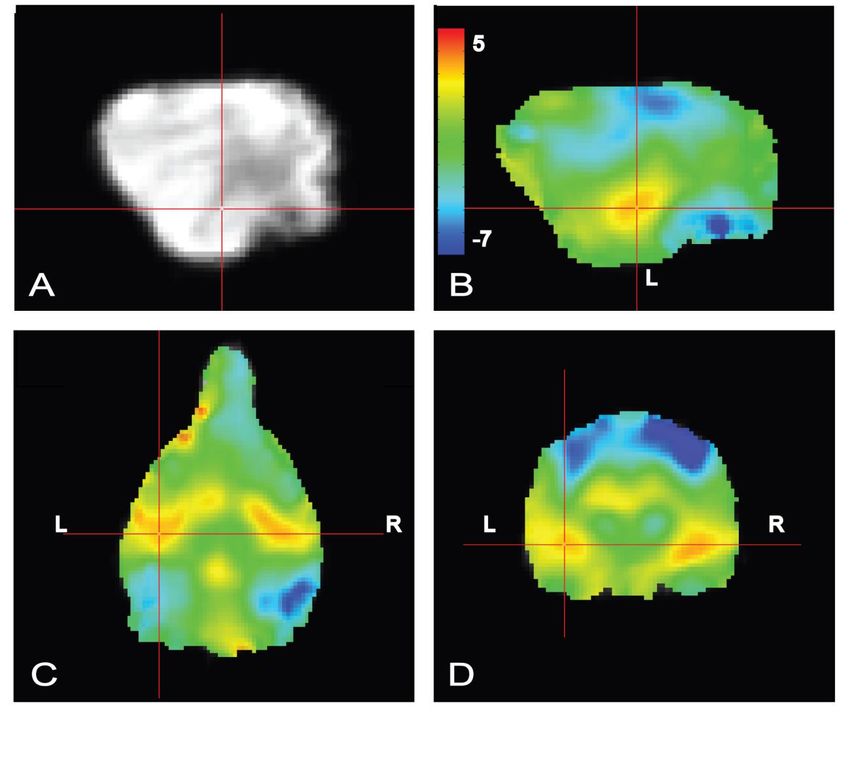
bioRxiv preprint first posted online Aug. 20, 2017; doi: http://dx.doi.org/10.1101/178186. The copyright holder for this preprint
      (which was not peer-reviewed) is the author/funder, who has granted bioRxiv a license to display the preprint in perpetuity.
                                         All rights reserved. No reuse allowed without permission.

                                                                                                                                  Prichard 2

36      But these dogs may have been exceptional. Few other dogs have been documented to have this
37      level of expertise. It may be that most dogs rely on simple mechanisms of discrimination – like
38      novelty detection – coupled with other cues from the human to figure out an appropriate
39      behavioral response.

40      The auditory oddball task, where subjects behaviorally discriminate between target and novel
41      acoustic stimuli, is a well-established task used to measure the processing of target detection and
42      decision-making in humans and nonhumans. The neural regions responsible for target detection
43      and novelty processing not only include primary sensory areas associated with the stimulus
44      modality, but also recruit broader areas such as the posterior cingulate, inferior and middle
45      frontal gyri, superior and middle temporal gyri, amygdala, thalamus, and lateral occipital cortex
46      (Linden et al., 1999;Kiehl et al., 2001;Brazdil et al., 2005;Goldman et al., 2009;Cacciaglia et al.,
47      2015). This suggests that differentiating between target versus novel sounds requires primary
48      auditory cortex as well as an additional attentional network to discriminate between competing
49      sensory stimuli. At least one event-related potential study in dogs suggested similar mechanisms
50      might be at work, finding mismatch negativity to deviant tones (Howell et al., 2012).

51      Recent advances in awake neuroimaging in dogs have provided a means to investigate many
52      aspects of canine cognition using approaches similar to those in humans. Since 2012, pet dogs
53      have been trained using positive reinforcement to lie still during fMRI scans in order to explore a
54      variety of aspects of canine cognition (Berns et al., 2012;Berns et al., 2013). These studies have
55      furthered our understanding of the dog’s neural response to expected reward, identified
56      specialized areas in the dog brain for processing faces, observed olfactory responses to human
57      and dog odors, and linked prefrontal function to inhibitory control (Cook et al., 2014;Berns et al.,
58      2015;Dilks et al., 2015;Cook et al., 2016a;Cuaya et al., 2016). In one fMRI study, dogs listened
59      to human and dog vocalizations through headphones and showed differential activation within
60      regions of the temporal and parietal cortex (Andics et al., 2014). A follow-up study suggested a
61      hemispheric bias for praise words versus neutral words, a finding that was interpreted as proof of
62      semantic processing in dogs. However, a subsequent correction in which left and right were
63      reversed raised questions about the interpretability of this finding (Andics et al., 2016).

64      To examine auditory processing in dogs, we used fMRI to measure activity in dogs’ brains in
65      response to both trained words and novel pseudowords. Over several months prior to scanning,
66      owners trained their dogs to select two objects based on the objects’ names. During the fMRI
67      session, the owner spoke the names of the trained objects as well as novel pseudowords the dog
68      had never heard before. If dogs discriminate target words from novel words as humans do, they
69      should show differential activity in the parietal and temporal cortex in response to trained words
70      relative to pseudowords (Friederici et al., 2000;Humphries et al., 2006;Raettig and Kotz, 2008).
71      In addition, if dogs use hedonic mechanisms to associate reward value with trained words, then
72      differential activity should also be observed in the caudate.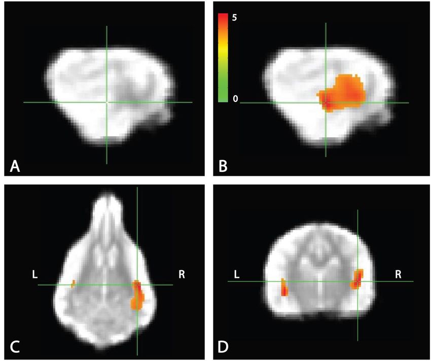
bioRxiv preprint first posted online Aug. 20, 2017; doi: http://dx.doi.org/10.1101/178186. The copyright holder for this preprint
      (which was not peer-reviewed) is the author/funder, who has granted bioRxiv a license to display the preprint in perpetuity.
                                         All rights reserved. No reuse allowed without permission.

                                                                                                                                  Prichard 3

73      Methods

74      Ethics Statement

75      This study was performed in accordance with the recommendations in the Guide for the Care and
76      Use of Laboratory Animals of the National Institutes of Health. The study was approved by the
77      Emory University IACUC (Protocol DAR-2002879-091817BA), and all owners gave written
78      consent for their dog’s participation in the study.

79      Participants

80      Participants were pet dogs from the Atlanta community volunteered by their owners for fMRI
81      training and experiments (Table 1). All dogs had previously completed one or more scans for the
82      project and had demonstrated the ability to remain still during training and scanning (Berns et al.,
83      2012).

84      Table 1. Dogs and their object names.
                                                                          Years with
        Dog          Breed                                 Age        Sex fMRI            Object 1      Object 2
                                                                          project
        Caylin      Border Collie                 8       Spayed F             4         Monkey       Blue
        Eddie       Golden Retriever-Lab mix      6       Neutered M           2         Piggy        Monkey
        Kady        Golden Retriever–Lab mix      7       Spayed F             4         Taffy        Yellow
        Libby       Pit mix                       11      Spayed F             4         Duck         Hedge Hog
        Ninja       Australian Cattle dog- mix    2       Spayed F             1         Block        Monkey
        Ohana       Golden Retriever              7       Spayed F             3         Blue         Star
        Pearl       Golden Retriever              7       Spayed F             3         Duck         Elephant
        Stella      Bouvier                       6       Spayed F             3         Stick        Tuxy
        Truffles Pointer mix                      12      Spayed F             2         Pig          Blue
        Velcro      Viszla                        8       Intact M             3         Rhino        Beach Ball
        Zen         Golden Retriever– Lab mix     8       Neutered M           4         Teddy        Duck
        Zula        Lab-Mastiff mix               4       Spayed F             1         Goldie       Bluebell
85      Dog’s names, breed, age in years when undergoing scanning, sex, years participating in fMRI experiments, and
86      training objects (S+) are listed
87
88      Word-Object Training

89      In the current experiment, dogs were trained to reliably fetch or select a trained object given the
90      matching verbal name for the object. The dogs were trained by implementing the “Chaser
91      Protocol” in which object names were used as verbal referents to retrieve a specific object (Pilley
92      and Reid, 2011). To keep the task simple, each dog had a set of two objects, selected by the
93      owner from home or from dog toys provided by the experimenters. One object had a soft texture,
94      such as a stuffed animal, whereas the other was of a different texture such as rubber or squeaked,
95      to facilitate discrimination (Fig. 1).

96      Each dog was trained by his or her owner at home, approximately 10 minutes per day, over 2 to
97      6 months, as well as at biweekly practices located at a dog training facility. Initial shaping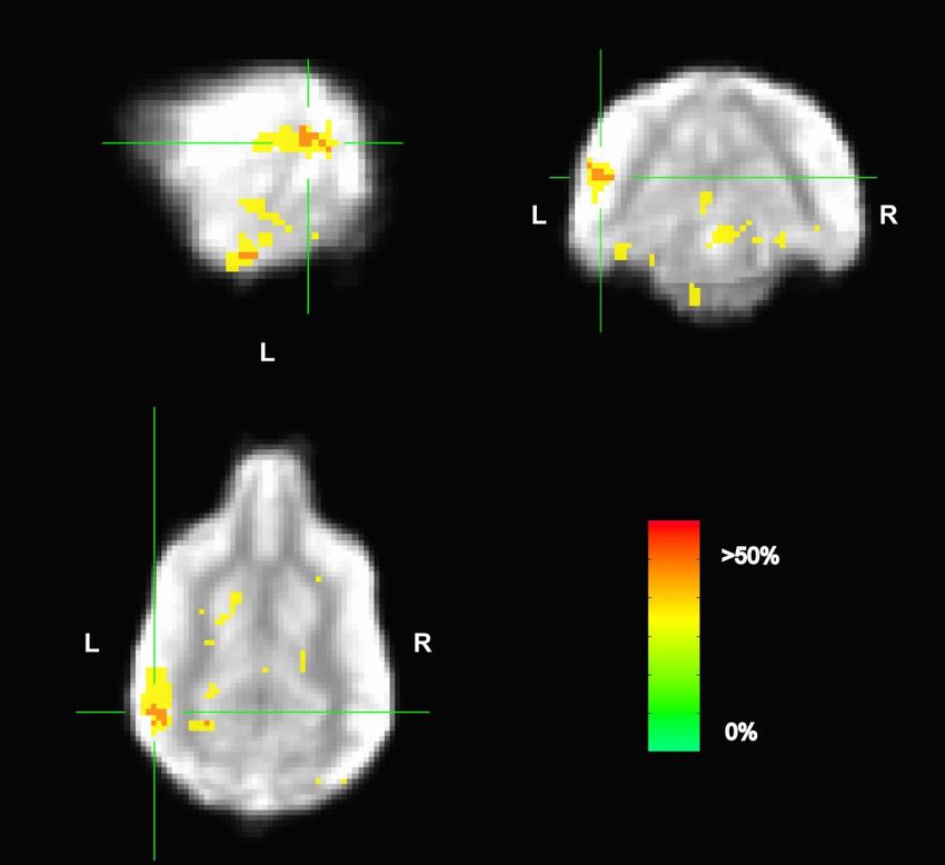
bioRxiv preprint first posted online Aug. 20, 2017; doi: http://dx.doi.org/10.1101/178186. The copyright holder for this preprint
       (which was not peer-reviewed) is the author/funder, who has granted bioRxiv a license to display the preprint in perpetuity.
                                          All rights reserved. No reuse allowed without permission.

                                                                                                                                   Prichard 4

 98      involved the owner playing “tug” or “fetch” with her dog and one object while verbally
 99      reinforcing the name of the object. Later, the objects were placed at a distance (four feet on
100      average) and the owner instructed the dog to “go get [object]” or “where is [object]?” or
101      “[object]!” The dog was reinforced with food or praise (varied per dog) for retrieving or nosing
102      the object. Next, the object was placed beside a novel object roughly two feet apart, at least 4
103      feet from the dog, and the command repeated. The dog was reinforced only for correctly
104      selecting the trained object if it was her first selection. Otherwise, if the dog selected the wrong
105      object, the owner made no remark and a new trial began. Regardless of the selection, objects
106      were rearranged before each trial to limit learning by position. If the dog failed to approach an
107      object, the trial was repeated. This training was repeated for each dog’s second object against a
108      different comparison object, to limit the possibility of learning by exclusion. Owners were
109      instructed to train one object per day, alternating between objects every other day until they
110      showed the ability to discriminate between the trained and novel object, at which point they
111      progressed to discrimination training between the 2 trained objects.

112      All dogs in the current study participated in training for previous fMRI experiments. As
113      described in previous experiments (Berns et al., 2012;Berns et al., 2013;Cook et al., 2014;Cook
114      et al., 2016b), each dog had participated in a training program involving behavior shaping,
115      desensitization, habituation and behavior chaining to prepare for the loud noise and physical
116      confines of the MRI bore inherent in fMRI studies.

117      Word-Object Discrimination Tests

118      Two weeks after progressing to two-object discrimination training, and every two weeks
119      thereafter, each dog was tested on her ability to discriminate between the two trained objects.
120      Discrimination between the two named objects was chosen as the measure of performance, as
121      both objects had a similar history of reinforcement, and this precluded the possibility that
122      performance was based on familiarity. Discrimination testing consisted of the observer placing
123      both trained objects 2-3 feet apart, and at least 4 feet from the dog (Ann Young, 1991), though
124      the number of distractor objects was sometimes increased during training to maximize
125      discriminatory performance. With the dog positioned next to the owner in the heel position, the
126      owner gave the dog the command to “go get [object]” or “[object]!” The dog was reinforced only
127      for correctly selecting the trained object if it was her first selection. If the dog selected the
128      incorrect object, the owner made no remark. After each trial, the objects were rearranged, and the
129      test progressed to the next trial. A performance criterion to move forward to the MRI scan was
130      set at 80% correct for at least one of the objects, with the other object at or above 50%.

131      During training, owners were asked to report if their dog showed a preference for one object over
132      the other. For the majority of the dogs, the preference was for the softer object of the two, and
133      both the preferred word and the object were consistently labeled as word 1 and object 1. Though
134      Zula passed the discrimination test, she was unable to complete the MRI scan and was excluded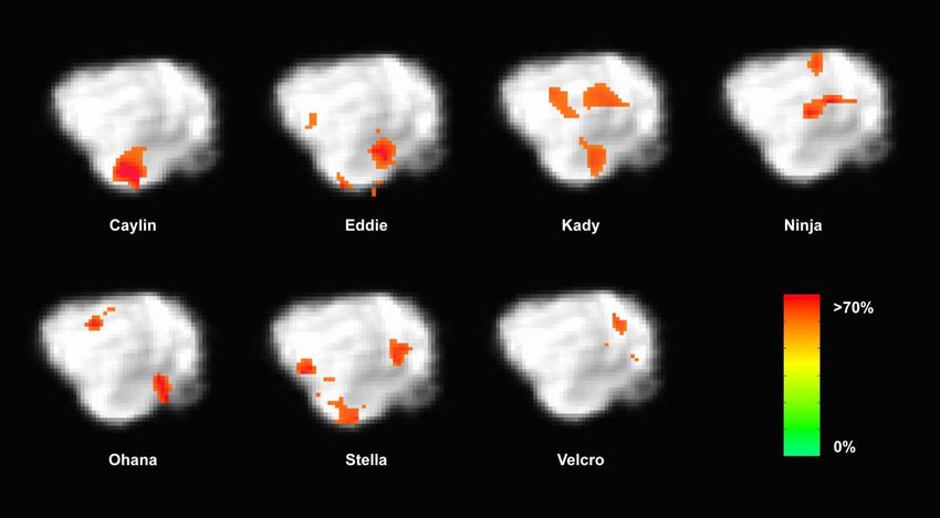
bioRxiv preprint first posted online Aug. 20, 2017; doi: http://dx.doi.org/10.1101/178186. The copyright holder for this preprint
       (which was not peer-reviewed) is the author/funder, who has granted bioRxiv a license to display the preprint in perpetuity.
                                          All rights reserved. No reuse allowed without permission.

                                                                                                                                   Prichard 5

135      from the remainder of the study. Individuals varied on the amount of time needed to train both
136      objects.

137      Scan Day Discrimination Test

138      Scan day tests were conducted in a neighboring room to the MRI room, and were typically
139      conducted prior to the MRI scan. Test procedure was identical to the word-object discrimination
140      test as described above, although the number of trials was increased from 10 to 12 trials if the
141      dog failed to make a response during one or more trials.

142      fMRI Stimuli

143      The stimuli consisted of the two trained words and the corresponding objects. Pseudowords were
144      included as a control condition. Pseudowords were matched to the group of trained words based
145      on the number of syllables and bigram frequency where possible using a pseudoword generator
146      (Keuleers and Brysbaert, 2010) (Table 2). Phoneme substitution was necessary in some cases to
147      ensure that trained words and pseudowords did not overlap at onset or coda. During the scan,
148      pseudowords were followed by the presentation of novel objects with which the dogs had no
149      previous experience. The novel objects included a bubble wand, Barbie doll, stuffed caterpillar,
150      wooden train whistle, plastic gumball dispenser, yellow hat, watermelon seat cushion, Nerf ball
151      launcher, etc.

152                                                Table 2. List of pseudowords per run.
                                                      Run 1 Run 2 Run 3
                                                      prang       cal      Cloft
                                                      risnu       o gri    Sowt
                                                      doba        ropp     bodmick
                                                      bobbu       prel     Fons
                                                      zelve       thozz    Stru
153

154      fMRI Experimental Design

155      As in previous studies, dogs were stationed in the magnet bore using custom chin rests. All
156      words were spoken by the dog’s primary owner, who stood directly in front of the dog at the
157      opening of the magnet bore. Both owners and dogs wore ear plugs, which reduced scanner noise
158      by approximately 30 decibels, but allowed for intelligible human speech over the sound of the
159      scanner. The spoken words were intelligible to the experimenters, who also wore ear plugs while
160      next to the MRI during scanning, as well as human operators in the control room via the
161      intercom. At the onset of each trial, a word was projected onto the surface of the scanner, directly
162      above the owner’s head. An experimenter stood next to the owner, out of view of the dog. The
163      experimenter controlled the timing and presentation of the words to the owner via a four-button
164      MRI-compatible button box (Fig. 2A). Onset of words and objects were controlled by the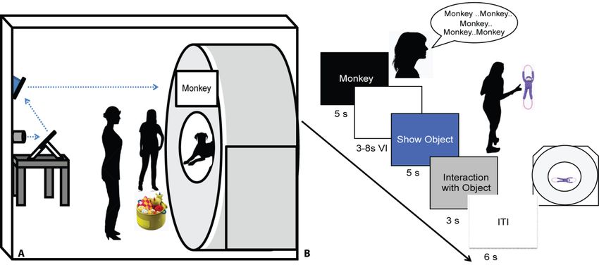
bioRxiv preprint first posted online Aug. 20, 2017; doi: http://dx.doi.org/10.1101/178186. The copyright holder for this preprint
       (which was not peer-reviewed) is the author/funder, who has granted bioRxiv a license to display the preprint in perpetuity.
                                          All rights reserved. No reuse allowed without permission.

                                                                                                                                   Prichard 6

165      simultaneous presentation and press of the button box by the experimenter marking the onset and
166      duration of presentation. This was controlled manually by the experimenter during each dog’s
167      scan, as opposed to a scripted presentation as in human fMRI studies, because dogs may leave
168      the MRI at any time and data for absentee trials would be lost.

169      An event-based design was used, consisting of four trial types presented semi-randomly:
170      expected, unexpected, pseudoword, and reward. On expected trials, the owner repeated a trained
171      object’s name five times, once per second. Words were repeated to ensure a robust hemodynamic
172      response on each trial and spoken loudly to be heard above the scanner noise. After a variable 3
173      to 8 s delay, the dog was shown the corresponding object for 5 s and was subsequently allowed
174      to interact with the object. During unexpected trials, the owner repeated the name for a trained
175      object as above, but following the delay period a novel object was presented instead of the
176      corresponding object. In pseudoword trials, the owner repeated a pseudoword, and the delay was
177      followed by a novel object. Reward trials were interspersed throughout each run, during which
178      the owner rewarded the dog’s continued down-stay with food. Trials were separated by a 6 s
179      inter-trial interval, and each dog received the same trial sequence (Fig. 2B). Each of three runs
180      consisted of 26 trials, for a total of 78 trials. The trial types included: 30 expected (15 each of
181      word1 and word2), 15 unexpected (7 or 8 of word1 and word2), 15 pseudowords, and 18 food
182      rewards.

183      Imaging

184      Scanning for the current experiment was conducted with a Siemens 3 T Trio whole-body scanner
185      using procedures described previously (Berns et al., 2012;Berns et al., 2013). During previous
186      experiments, a T2-weighted structural image of the whole brain was acquired using a turbo spin-
187      echo sequence (25-36 2mm slices, TR = 3940 ms, TE = 8.9 ms, flip angle = 131˚, 26 echo trains,
188      128 x 128 matrix, FOV = 192 mm). The functional scans used a single-shot echo-planar imaging
189      (EPI) sequence to acquire volumes of 22 sequential 2.5 mm slices with a 20% gap (TE = 25 ms,
190      TR = 1200 ms, flip angle = 70˚, 64 x 64 matrix, 3 mm in-plane voxel size, FOV = 192 mm).
191      Slices were oriented dorsally to the dog’s brain (coronal to the magnet, as in the sphinx position
192      the dogs’ heads were positioned 90 degrees from the prone human orientation) with the phase-
193      encoding direction right-to-left. Sequential slices were used to minimize between-plane offsets
194      from participant movement, while the 20% slice gap minimized the “crosstalk” that can occur
195      with sequential scan sequences. Three runs of up to 700 functional volumes were acquired for
196      each participant, with each run lasting 10 to 14 minutes.

197      Analysis

198      Preprocessing

199      Data preprocessing included motion correction, censoring and normalization using AFNI (NIH)
200      and its associated functions. Two-pass, six-parameter affine motion correction was used with a
201      hand-selected reference volume for each dog that best reflected their average position within the
bioRxiv preprint first posted online Aug. 20, 2017; doi: http://dx.doi.org/10.1101/178186. The copyright holder for this preprint
(which was not peer-reviewed) is the author/funder, who has granted bioRxiv a license to display the preprint in perpetuity.
All rights reserved. No reuse allowed without permission.
Prichard 7
202 scanner. All volumes were aligned to the reference volume. Aggressive censoring (i.e., removing
203 bad volumes from the fMRI time sequence) was used because dogs can move between trials,
204 when interacting with the object, and when consuming rewards. Data were censored when
205 estimated motion was greater than 1 mm displacement scan-to-scan and based on outlier voxel
206 signal intensities. Smoothing, normalization, and motion correction parameters were identical to
207 those described previously (Cook et al., 2016b). A high-resolution canine brain atlas was used as
208 the template space for individual spatial transformations (Datta et al., 2012). The atlas resolution
209 was 1 mm x 1 mm x 1 mm. Thus voxel volumes are in mm3.
210 General Linear Model
211 For a priori hypotheses, each participant’s motion-corrected, censored, smoothed images were
212 analyzed with a general linear model (GLM) for each voxel in the brain using 3dDeconvolve
213 (part of the AFNI suite). Nuisance regressors included motion time courses generated through
214 motion correction, constant, linear, quadratic, and cubic drift terms. The drift terms were
215 included for each run to account for baseline shifts between runs as well as slow drifts unrelated
216 to the experiment. Task related regressors included: (1) spoken word1; (2) spoken word2; (3)
217 spoken pseudowords; (4) presentation of object1; (5) presentation of object2; (6) presentation of
218 unexpected objects (novel object following either word1 or word2); and (7) presentation of novel
219 objects following a pseudoword. The object on which each dog performed best during the day of
220 the MRI scan as well as the object owners reported as being the preferred of the two was labeled
221 as word1 and object1 when creating the GLM regressors. Stimulus onset and duration were
222 modeled using the dmUBLOCK function, with the 5 utterances treated as a block.
223 Whole Brain Analysis
224 Contrasts focused on the dogs’ response to words and pseudowords. Auditory novelty detection
225 was probed with the contrast: [pseudowords – (word1 + word2)/2]. Low-level aspects of
226 language processing (including acoustic and hedonic representations) were probed with the
227 contrast [word1 – word2] and expectation violation with [novel objects – unexpected objects].
228 Each participant’s individual-level contrast from the GLM was normalized to template space as
229 described in (Berns et al., 2012;Cook et al., 2014) via the Advanced Normalization Tools
230 (ANTs) software (Avants et al., 2011). Spatial transformations included a rigid-body mean EPI
231 to structural image, affine structural to template, and diffeomorphic structural to template. These
232 spatial transformations were concatenated and applied to individual contrasts from the GLM to
233 compute group level statistics. 3dttest++, part of the AFNI suite, was used to compute a t-test
234 across dogs against the null hypothesis that each voxel had a mean value of zero. All contrasts
235 mentioned above as part of the GLM were included.
236 As there is spatial heterogeneity within fMRI data, the average smoothness of the residuals from
237 each dog’s time series regression model was calculated using AFNI’s non-Gaussian spatial
238 autocorrelation function 3dFWHMx –acf. The acf option leads to greatly reduced FPRs clusteredbioRxiv preprint first posted online Aug. 20, 2017; doi: http://dx.doi.org/10.1101/178186. The copyright holder for this preprint
(which was not peer-reviewed) is the author/funder, who has granted bioRxiv a license to display the preprint in perpetuity.
All rights reserved. No reuse allowed without permission.
Prichard 8
239 around 5 percent across all voxelwise thresholds (Cox et al., 2017). AFNI’s 3dClustsim was then
240 used to estimate the significance of cluster sizes across the whole brain after correcting for
241 familywise error (FWE). Similar to human fMRI studies, a voxel threshold of p ≤ 0.005 was
242 used, and a cluster was considered significant if it exceeded the critical size estimated by
243 3dClustsim for a FWER ≤ 0.01, using two-sided thresholding and a nearest-neighbor of 1.
244 Multivoxel Pattern Analysis (MVPA)
245 In previous fMRI studies of the oddball task, it was noted that attentional differences occurring
246 trial-by-trial may go undetected in the univariate analysis (Goldman et al., 2009). As an
247 exploratory analysis, we used searchlight MVPA to identify regions potentially involved in the
248 representation of words that were not captured in the univariate analysis. We were primarily
249 interested in the representation of word1 vs. word2.
250 We used a linear support vector machine (SVM) for a classifier because of its previously
251 demonstrated robust performance (Misaki et al., 2010;Mahmoudi et al., 2012). Unsmoothed
252 volumes were censored for motion and outlier count as in the univariate GLM. We then made a
253 model for the unsmoothed data using AFNI’s 3dDeconvolve stim_times_IM function. This
254 model yielded trial-by-trial estimates (betas) for each repetition of word1 and word2, regardless
255 of which object followed. Although it is common in the human literature to use each scan
256 volume as a data point in MVPA (for training and testing), we have found this approach to be
257 problematic with dogs, who move more than humans, resulting in spurious volumes that should
258 be censored. Estimating the beta for each trial affords an additional level of robustness with less
259 sensitivity to potential outlier volumes due to motion. As an additional check for outliers, masks
260 were drawn of the left and right caudate on each dogs’ T2-weighted structural image. Average
261 beta values were extracted from both the left and right caudate for each trial of word1 and word2.
262 Trials with beta values greater than |3%| were assumed to be non-physiological and were
263 removed prior to MVPA. Finally, these trial-dependent estimates were then used as inputs to a
264 whole-brain searchlight MVPA for each individual dog using PyMVPA2 (Hanke et al., 2009).
265 The classifier was trained on the fMRI dataset for each dog by training on 2 runs and testing on
266 the third using the NFoldPartitioner. We used the Balancer function to retain the same number of
267 trials for word1 and word2 across training and testing for 100 repetitions. For the searchlight, we
268 used a 3-voxel radius sphere. This yielded a map of classification accuracies throughout each
269 dog’s brain.
270 Given the difficulty in finding significant effects in small datasets using cross-validation and
271 parametric methods, we used a permutation approach outlined by Stelzer et al. (2013) to
272 determine the significance of any cluster of common voxels across dogs (Stelzer et al.,
273 2013;Varoquaux, 2017). Briefly, we permuted the order of attributes—but not their
274 corresponding data—and ran the searchlight in individual space for all dogs. This created a null
275 distribution of accuracies. The mean of these distributions was noted to be very close to 0.5,
276 confirming that the classifiers wasn’t biased or skewed. The cumulative distribution of that anbioRxiv preprint first posted online Aug. 20, 2017; doi: http://dx.doi.org/10.1101/178186. The copyright holder for this preprint
(which was not peer-reviewed) is the author/funder, who has granted bioRxiv a license to display the preprint in perpetuity.
All rights reserved. No reuse allowed without permission.
Prichard 9
277 accuracy ≥ 0.63 corresponded to the top 5% of voxels, and this was used as a cut-off threshold
278 for the individual maps. These binarized maps were transformed into template space and the
279 average computed across dogs. The resultant group map represented the locations of potentially
280 informative voxels and served as qualitative representation of the relative consistency versus
281 heterogeneity of word-processing in the dogs’ brains. Somewhat arbitrarily, we only considered
282 locations in which at least two dogs had informative voxels.
283
284 Results
285
286 Scan Day Discrimination Tests
287 Scans were scheduled as close as possible to the day on which object identification criterion was
288 met (M = 9.33 days, SD =4.92 days) based on owner availability. On the day of the scheduled
289 MRI scan, each dog was tested on her ability to behaviorally differentiate between the two
290 trained objects out of 5 trials each. With the exception of Eddie, each dog correctly selected
291 object 1 on 80 to 100 percent of the trials [M=85.73%, SE =3.87%], and object 2 on 60 to 100
292 percent of the trials [M=64.27%, SE=5.91%] (Fig. 3). The percent correct performance
293 (subtracting 50 percent for chance levels of responding) on scan days for each object was
294 compared in a mixed-effect linear model and showed that performance was significantly greater
295 than chance [T(17.1) = 3.00, P = 0.008] and that there was a significant difference in
296 performance between word1 and word2 [T(11) = 4.67, P < 0.001].
297 Primary Auditory and Visual Activation
298 To confirm that the dogs clearly heard the words during scanning, a simple contrast subtracting
299 activation to objects (trained and novel) from activation to words (trained and pseudowords) was
300 performed. In human fMRI, the MRI operator may ask the participant whether they can hear
301 auditory stimuli, which is not necessarily possible in dog fMRI, so this was included as a quality
302 check. We opted for an unthresholded image not only to highlight the activation in bilateral
303 auditory cortex but, just as important, to show what was not activated. Notably in the contrast
304 [Words—Objects] positive activation was localized to the auditory cortex for words and negative
305 activation for presentation objects in parietal cortex (Fig. 4), confirming that the dogs heard the
306 words and saw the objects.
307 Whole Brain Analyses
308 Whole brain analysis of the contrasts of interest revealed significant activation only within the
309 right parietotemporal cortex for the contrast [pseudowords – trained words]. With a voxel-level
310 significance threshold of P ≤ 0.005, the cluster size in the right hemisphere (839 voxels) was
311 statistically significant at P ≤ 0.005 after correction for whole-brain FWE (although activation
312 appeared bilaterally) (Fig. 5). Whole brain analysis of the contrasts of [word1– word2] andbioRxiv preprint first posted online Aug. 20, 2017; doi: http://dx.doi.org/10.1101/178186. The copyright holder for this preprint
(which was not peer-reviewed) is the author/funder, who has granted bioRxiv a license to display the preprint in perpetuity.
All rights reserved. No reuse allowed without permission.
Prichard 10
313 [novel – unexpected] were not significant as no cluster survived thresholding at the voxel
314 significance mentioned above.
315
316 MVPA
317
318 Because the univariate analysis of word1 vs. word2 did not reveal any region with a significant
319 difference, we used MVPA to explore potential regions that may code for different
320 representations of the words. The searchlight map of word1 vs. word2, which identified regions
321 involved in the discrimination of the trained words, showed four clusters of informative voxels
322 (Fig. 6): posterior thalamus/brainstem; amygdala; left temporoparietal junction (TPJ); and left
323 dorsal caudate nucleus. Seven dogs shared informative voxels in or near the left temporal cortex
324 that passed the 0.63 accuracy threshold (Fig. 7).
325 Discussion
326 Using awake-fMRI in dogs, we found neural evidence for auditory novelty detection in the
327 domain of human speech. The hallmark of this finding was greater activation in parietotemporal
328 cortex to novel pseudowords relative to trained words. Thus, even in the absence of a behavioral
329 response, we demonstrate that dogs process human speech at least to the extent of differentiating
330 words they have heard before from those they have not. The mechanism of such novelty
331 detection may be rooted in either the relatively less frequent presentation of the pseudowords
332 (oddball detection) or the lack of meaning associated with them (lexical processing).
333 The activation observed in the parietotemporal cortex to pseudowords relative to trained words
334 meets current standards of human fMRI analyses concerning up-to-date methods for cluster
335 thresholds. Specifically, to address concerns raised by Eklund et al. (2016), present analyses for
336 cluster inferences address the former Gaussian-shaped assumption about spatial structure in the
337 residuals of fMRI data and provide more accurate false positive rates compared to previous
338 methods (Eklund et al., 2016;Cox et al., 2017;Slotnick, 2017). As the identified cluster was
339 significant at P ≤ 0.005, corrected for whole-brain FWE, the result does not appear to be a false
340 positive. However, as the study was limited to 11 participants, future studies with an increased
341 number of participants could produce a more robust finding.
342 In humans, greater BOLD response to pseudowords versus real words has been noted in the
343 superior temporal gyrus – an area potentially analogous to the one we identified in dogs (Kotz,
344 2002;Raettig and Kotz, 2008). In humans, stronger activation to pseudowords depends on
345 whether the pseudoword strongly resembles a known word or is so unlike known words as to
346 prevent any semantic retrieval. When the pseudoword is similar to a known word, more
347 processing has been observed in the superior temporal gyri, presumably to disambiguate it from
348 known words (Raettig and Kotz, 2008). Thus, in dogs, the greater activation to the pseudowords
349 could be due to the acoustic similarity between pseudowords and words that the dogs “knew”
350 and their attempt to resolve the ambiguity. This would be a form of low-level lexical processing.bioRxiv preprint first posted online Aug. 20, 2017; doi: http://dx.doi.org/10.1101/178186. The copyright holder for this preprint
(which was not peer-reviewed) is the author/funder, who has granted bioRxiv a license to display the preprint in perpetuity.
All rights reserved. No reuse allowed without permission.
Prichard 11
351 However, previous research has shown that dogs can discriminate between altered phonemes of
352 well-known commands (Fukuzawa et al., 2005), suggesting that it is unlikely that the dogs in our
353 study were confused by acoustic similarity of words and pseudowords.
354 More likely, a novel word resulted in increased auditory processing to facilitate learning the
355 association with the novel object that followed. A dog’s behavioral bias for novelty is often
356 described as an explanation for performance otherwise labeled as learning by exclusion (Bloom,
357 2004;Markman and Abelev, 2004;Zaine et al., 2014). As such, a dog may select a novel item
358 because it is novel among other stimuli, but not because she has learned all other stimuli and
359 associated a new word with the novel item. A bias for novelty would therefore be reflected in the
360 dog’s brain as with her behavior.
361 Auditory stimuli can be difficult to discriminate in the scanner. We used a continuous scanning
362 protocol because that is what the dogs were accustomed to. The simple contrast of all words vs.
363 all objects showed bilateral activation of the superior temporal lobe, indicating that the dogs
364 heard something. However, the main effect of pseudowords vs. trained words showed that the
365 majority of dogs discriminated well enough to tell the difference. The predominant location in
366 the auditory pathway also suggests that the effect wasn’t based on non-verbal cues from the
367 handler (i.e. Clever Hans effect).
368 The manner in which dogs learn words is different than humans do, and this undoubtedly affects
369 their performance on behavioral tests and the patterns of brain activation we observed. Humans
370 acquire nouns as early as six months of age and differentiate between nouns prior to their ability
371 to use verbs (Bergelson and Swingley, 2012;Waxman et al., 2013). In contrast, dogs do not
372 typically have much experience with nouns because humans tend to train them on actions/verbs
373 (e.g. sit and fetch). Consequently, even the trained words in our study were novel for the dogs in
374 comparison to years of experience with verbs as commands. Prior studies have shown only three
375 dogs that consistently retrieved objects given a verbal referent (Kaminski et al., 2004;Pilley and
376 Reid, 2011). Additionally, those dogs had been trained to retrieve from a young age (bioRxiv preprint first posted online Aug. 20, 2017; doi: http://dx.doi.org/10.1101/178186. The copyright holder for this preprint
(which was not peer-reviewed) is the author/funder, who has granted bioRxiv a license to display the preprint in perpetuity.
All rights reserved. No reuse allowed without permission.
Prichard 12
389 Lastly, dogs might have habituated to the continued presentation of trained words followed by
390 trained objects, as opposed to the single trial presentations of pseudowords and the
391 accompanying novel objects.
392 So what do words mean to dogs? Even though our findings suggest a prominent role for novelty
393 in dogs’ processing of human words, this leaves the question of what the words represent. One
394 possibility is that the words had no further representation than the relative hedonic value of the
395 objects. While some dogs showed a behavioral preference for one object over the other, this
396 preference was not reflected in whole brain analyses. Admittedly, the somewhat arbitrary
397 designation of word1 / word2 and object1 / object2 could explain the nonsignificant results in the
398 univariate analysis. Indeed, the MVPA of word1 vs. word2, which identified regions that
399 classified the words above chance regardless of directionality, showed one cluster in the left
400 caudate. However, the MVPA also identified clusters in the left TPJ, amygdala, and posterior
401 thalamus. The TPJ was located just posterior to the region in the univariate analysis, which
402 would take it out of the area of cortex associated with low-level acoustic processing. Its location
403 appears similar to human angular gyrus – aka Wernicke’s area. If so, this could be a potential site
404 for receptive word processing in dogs (e.g. the Dog Wernicke’s Area), but future work would
405 need to verify this.
406 Evaluating classifier performance for MVPA remains a complex task. We used MVPA as an
407 exploratory analysis to identify brain regions that potentially discriminate between trained words
408 across dogs. But classification using the whole brain may result in a high classification accuracy
409 that is not generalizable across subjects. Indeed, the regions identified using MVPA were of
410 marginal statistical significance, especially given the small sample size. Further, it should be
411 noted that only a subset of dogs contained informative voxels in the TPJ region. Although all
412 dogs had informative voxels somewhere in the brain, only seven dogs had informative voxels in
413 the TPJ area. Thus, even though all the dogs were cleared for scanning by reaching performance
414 criterion, they may have used different mechanisms to process the words. Like our previous
415 fMRI studies, heterogeneity seems to be the rule (Cook et al., 2016a;Cook et al., 2016b). Even
416 so, the accuracy of the classifier was not correlated with a dog’s performance. This suggests that
417 performance on such tasks may be influenced by factors other than word discrimination alone.
418 These results highlight potential mechanisms by which dogs process words. Word novelty
419 appears to play an important role. The strong response of the parietotemporal region to
420 pseudowords suggests that dogs have some basic ability to differentiate words with associations
421 from those that do not. Future studies may reveal whether these representations remain in the
422 auditory domain or whether such representations are invariant to modality.
423 Acknowledgments
424 Thanks to Dr. Kate Revill for advice about language processing and generating pseudowords.
425 Thank you to all of the owners who trained their dogs over the course of 6 months for this study:bioRxiv preprint first posted online Aug. 20, 2017; doi: http://dx.doi.org/10.1101/178186. The copyright holder for this preprint
(which was not peer-reviewed) is the author/funder, who has granted bioRxiv a license to display the preprint in perpetuity.
All rights reserved. No reuse allowed without permission.
Prichard 13
426 Lorrie Backer, Darlene Coyne, Vicki D’Amico, Diana Delatour, Marianne Ferraro, Patricia
427 King, Cecilia Kurland, Claire Mancebo, Cathy Siler, Lisa Tallant, Nicole & Sairina Merino Tsui,
428 and Nicole Zitron.
429 Funding:
430 This work was supported by the Office of Naval Research (N00014-16-1-2276). M.S. is the
431 owner of Comprehensive Pet Therapy (CPT). ONR provided support in the form of salaries for
432 authors [PC, MS, & GSB], scan time, and volunteer payment, but did not have any additional
433 role in the study design, data collection and analysis, decision to publish, or preparation of the
434 manuscript.
435 Competing Interests:
436 G.B. and M.S. own equity in Dog Star Technologies and developed technology used in some of
437 the research described in this paper. The terms of this arrangement have been reviewed and
438 approved by Emory University in accordance with its conflict of interest policies. MS is the
439 owner of Comprehensive Pet Therapy (CPT) but no CPT technology or IP was used in this
440 research.
441 Author Contributions
442 A.P., P.C., and G.B. designed and performed research; A.P, R.C., and G.B. analyzed data; A.P.,
443 P.C., G.B., & M. S. trained dogs; and A.P., G.B., P.C., R. C., and M.S. wrote the paper.
444 References
445 Andics, A., Gabor, A., Gacsi, M., Farago, T., Szabo, D., and Miklosi, A. (2016). Neural
446 mechanisms for lexical processing in dogs. Science 353, 1030-1032.
447 Andics, A., Gacsi, M., Farago, T., Kis, A., and Miklosi, A. (2014). Voice-sensitive regions in the
448 dog and human brain are revealed by comparative fMRI. Curr Biol 24, 574-578.
449 Ann Young, C. (1991). Verbal commands as discriminative stimuli in domestic dogs (Canis
450 familiaris). Applied Animal Behaviour Science 32, 75-89.
451 Avants, B.B., Tustison, N.J., Song, G., Cook, P.A., Klein, A., and Gee, J.C. (2011). A
452 reproducible evaluation of ANTs similarity metric performance in brain image
453 registration. Neuroimage 54, 2033-2044.
454 Bergelson, E., and Swingley, D. (2012). At 6-9 months, human infants know the meanings of
455 many common nouns. Proc Natl Acad Sci U S A 109, 3253-3258.
456 Berns, G.S., Brooks, A., and Spivak, M. (2013). Replicability and heterogeneity of awake
457 unrestrained canine FMRI responses. PLoS One 8, e81698.
458 Berns, G.S., Brooks, A.M., and Spivak, M. (2012). Functional MRI in awake unrestrained dogs.
459 PLoS One 7, e38027.
460 Berns, G.S., Brooks, A.M., and Spivak, M. (2015). Scent of the familiar: An fMRI study of
461 canine brain responses to familiar and unfamiliar human and dog odors. Behav Processes
462 110, 37-46.
463 Bloom, P. (2004). Behavior. Can a dog learn a word? Science 304, 1605-1606.bioRxiv preprint first posted online Aug. 20, 2017; doi: http://dx.doi.org/10.1101/178186. The copyright holder for this preprint
(which was not peer-reviewed) is the author/funder, who has granted bioRxiv a license to display the preprint in perpetuity.
All rights reserved. No reuse allowed without permission.
Prichard 14
464 Brazdil, M., Dobsik, M., Mikl, M., Hlustik, P., Daniel, P., Pazourkova, M., Krupa, P., and
465 Rektor, I. (2005). Combined event-related fMRI and intracerebral ERP study of an
466 auditory oddball task. Neuroimage 26, 285-293.
467 Cacciaglia, R., Escera, C., Slabu, L., Grimm, S., Sanjuan, A., Ventura-Campos, N., and Avila, C.
468 (2015). Involvement of the human midbrain and thalamus in auditory deviance detection.
469 Neuropsychologia 68, 51-58.
470 Cook, P.F., Brooks, A., Spivak, M., and Berns, G.S. (2016a). Regional brain activations in
471 awake unrestrained dogs. Journal of Veterinary Behavior-Clinical Applications and
472 Research 16, 104-112.
473 Cook, P.F., Prichard, A., Spivak, M., and Berns, G.S. (2016b). Awake canine fMRI predicts
474 dogs' preference for praise vs food. Soc Cogn Affect Neurosci 11, 1853-1862.
475 Cook, P.F., Spivak, M., and Berns, G.S. (2014). One pair of hands is not like another: caudate
476 BOLD response in dogs depends on signal source and canine temperament. PeerJ 2,
477 e596.
478 Cox, R.W., Chen, G., Glen, D.R., Reynolds, R.C., and Taylor, P.A. (2017). FMRI Clustering in
479 AFNI: False-Positive Rates Redux. Brain Connect 7, 152-171.
480 Cuaya, L.V., Hernandez-Perez, R., and Concha, L. (2016). Our Faces in the dog's brain:
481 Functional imaging reveals temporal cortex activation during perception of human faces.
482 PLoS One 11, e0149431.
483 D'aniello, B., Scandurra, A., Alterisio, A., Valsecchi, P., and Prato-Previde, E. (2016). The
484 importance of gestural communication: a study of human-dog communication using
485 incongruent information. Anim Cogn 19, 1231-1235.
486 Datta, R., Lee, J., Duda, J., Avants, B.B., Vite, C.H., Tseng, B., Gee, J.C., Aguirre, G.D., and
487 Aguirre, G.K. (2012). A digital atlas of the dog brain. PLoS One 7, e52140.
488 Dilks, D.D., Cook, P., Weiller, S.K., Berns, H.P., Spivak, M., and Berns, G.S. (2015). Awake
489 fMRI reveals a specialized region in dog temporal cortex for face processing. PeerJ 3,
490 e1115.
491 Eklund, A., Nichols, T.E., and Knutsson, H. (2016). Cluster failure: Why fMRI inferences for
492 spatial extent have inflated false-positive rates. Proc Natl Acad Sci U S A 113, 7900-
493 7905.
494 Friederici, A.D., Meyer, M., and Von Cramon, D.Y. (2000). Auditory language comprehension:
495 An event-related fMRI study on the processing of syntactic and lexical information.
496 Brain Lang 74, 289-300.
497 Fukuzawa, M., Mills, D.S., and Cooper, J.J. (2005). The effect of human command phonetic
498 characteristics on auditory cognition in dogs (Canis familiaris). J Comp Psychol 119,
499 117-120.
500 Goldman, R.I., Wei, C.Y., Philiastides, M.G., Gerson, A.D., Friedman, D., Brown, T.R., and
501 Sajda, P. (2009). Single-trial discrimination for integrating simultaneous EEG and fMRI:
502 identifying cortical areas contributing to trial-to-trial variability in the auditory oddball
503 task. Neuroimage 47, 136-147.
504 Hanke, M., Halchenko, Y.O., Sederberg, P.B., Hanson, S.J., Haxby, J.V., and Pollmann, S.
505 (2009). PyMVPA: A python toolbox for multivariate pattern analysis of fMRI data.
506 Neuroinformatics 7, 37-53.
507 Howell, T.J., Conduit, R., Toukhsati, S., and Bennett, P. (2012). Auditory stimulus
508 discrimination recorded in dogs, as indicated by mismatch negativity (MMN). Behav
509 Processes 89, 8-13.bioRxiv preprint first posted online Aug. 20, 2017; doi: http://dx.doi.org/10.1101/178186. The copyright holder for this preprint
(which was not peer-reviewed) is the author/funder, who has granted bioRxiv a license to display the preprint in perpetuity.
All rights reserved. No reuse allowed without permission.
Prichard 15
510 Humphries, C., Binder, J.R., Medler, D.A., and Liebenthal, E. (2006). Syntactic and semantic
511 modulation of neural activity during auditory sentence comprehension. J Cogn Neurosci
512 18, 665-679.
513 Kaminski, J., Call, J., and Fischer, J. (2004). Word learning in a domestic dog: evidence for "fast
514 mapping". Science 304, 1682-1683.
515 Keuleers, E., and Brysbaert, M. (2010). Wuggy: A multilingual pseudoword generator. Behav
516 Res Methods 42, 627-633.
517 Kiehl, K.A., Laurens, K.R., Duty, T.L., Forster, B.B., and Liddle, P.F. (2001). An event-related
518 fMRI study of visual and auditory oddball tasks. Journal of Psychophysiology 15, 221-
519 240.
520 Kotz, S. (2002). Modulation of the lexical–semantic network by auditory semantic priming: An
521 event-related functional MRI study. NeuroImage 17, 1761-1772.
522 Linden, D.E., Prvulovic, D., Formisano, E., Vollinger, M., Zanella, F.E., Goebel, R., and Dierks,
523 T. (1999). The functional neuroanatomy of target detection: an fMRI study of visual and
524 auditory oddball tasks. Cereb Cortex 9, 815-823.
525 Mahmoudi, A., Takerkart, S., Regragui, F., Boussaoud, D., and Brovelli, A. (2012). Multivoxel
526 pattern analysis for FMRI data: A review. Comput Math Methods Med 2012, 961257.
527 Markman, E.M., and Abelev, M. (2004). Word learning in dogs? Trends Cogn Sci 8, 479-481;
528 discussion 481.
529 Mills, D.S. (2015). What's in a word? A review of the attributes of a command affecting the
530 performance of pet dogs. Anthrozoös 18, 208-221.
531 Misaki, M., Kim, Y., Bandettini, P.A., and Kriegeskorte, N. (2010). Comparison of multivariate
532 classifiers and response normalizations for pattern-information fMRI. Neuroimage 53,
533 103-118.
534 Müller, C.A., Schmitt, K., Barber, A.L.A., and Huber, L. (2015). Dogs can discriminate
535 emotional expressions of human faces. Current Biology 25, 601-605.
536 Persson, M.E., Roth, L.S., Johnsson, M., Wright, D., and Jensen, P. (2015). Human-directed
537 social behaviour in dogs shows significant heritability. Genes Brain Behav 14, 337-344.
538 Pilley, J.W., and Reid, A.K. (2011). Border collie comprehends object names as verbal referents.
539 Behav Processes 86, 184-195.
540 Raettig, T., and Kotz, S.A. (2008). Auditory processing of different types of pseudo-words: an
541 event-related fMRI study. Neuroimage 39, 1420-1428.
542 Slotnick, S.D. (2017). Cluster success: fMRI inferences for spatial extent have acceptable false-
543 positive rates. Cogn Neurosci 8, 150-155.
544 Stelzer, J., Chen, Y., and Turner, R. (2013). Statistical inference and multiple testing correction
545 in classification-based multi-voxel pattern analysis (MVPA): random permutations and
546 cluster size control. Neuroimage 65, 69-82.
547 Varoquaux, G. (2017). Cross-validation failure: Small sample sizes lead to large error bars.
548 Neuroimage.
549 Waxman, S., Fu, X., Arunachalam, S., Leddon, E., Geraghty, K., and Song, H.J. (2013). Are
550 nouns learned before verbs? Infants provide insight into a longstanding debate. Child Dev
551 Perspect 7, 155-159.
552 Zaine, I., Domeniconi, C., and Costa, A.R.A. (2014). Exclusion performance in visual simple
553 discrimination in dogs (Canis familiaris). Psychology & Neuroscience 7, 199-206.
554bioRxiv preprint first posted online Aug. 20, 2017; doi: http://dx.doi.org/10.1101/178186. The copyright holder for this preprint
(which was not peer-reviewed) is the author/funder, who has granted bioRxiv a license to display the preprint in perpetuity.
All rights reserved. No reuse allowed without permission.
Prichard 16
555 FIGURES
556
557 Fig. 1. Individual dogs and their trained objects. All 12 dogs successfully trained to retrieve
558 two objects using object names as verbal referents.bioRxiv preprint first posted online Aug. 20, 2017; doi: http://dx.doi.org/10.1101/178186. The copyright holder for this preprint
(which was not peer-reviewed) is the author/funder, who has granted bioRxiv a license to display the preprint in perpetuity.
All rights reserved. No reuse allowed without permission.
Prichard 17
559
560 Fig. 2. Experimental design. A) Experimental setup with mirror relay projected words onto
561 MRI surface. Owner is facing the projected word and her dog while the experimenter controls
562 the presentation of words and objects to the owner. B) Trial timeline indicating spoken word
563 over 5 s, 3-8 s delay, 5 s presentation of object, 3 s for the dog to interact with the object,
564 followed by a 6 s intertrial interval.
565
566 Fig. 3. Individual performance on two object discrimination tests. Tests were conducted on
567 the day of the fMRI scan. Each dog’s Object 1 is in black, object 2 is in grey. All dogs performed
568 significantly greater than chance, with the dog’s greater performance or owner’s report of their
569 preference for one object over the other designating object 1.bioRxiv preprint first posted online Aug. 20, 2017; doi: http://dx.doi.org/10.1101/178186. The copyright holder for this preprint
(which was not peer-reviewed) is the author/funder, who has granted bioRxiv a license to display the preprint in perpetuity.
All rights reserved. No reuse allowed without permission.
Prichard 18
570
571 Fig 4 Whole brain group map showing unthresholded activation to all words versus all
572 objects. A) Location of crosshairs in superior temporal lobe on average image of all dogs. B)
573 Sagittal view of left hemisphere. Colors represent T-statistics. The primary auditory region
574 extending into the parietotemporal area showed greater activation to words (red), whereas
575 parietal and occipital areas showed greater activation to objects (blue). C) Dorsal view. D)
576 Transverse view.bioRxiv preprint first posted online Aug. 20, 2017; doi: http://dx.doi.org/10.1101/178186. The copyright holder for this preprint
(which was not peer-reviewed) is the author/funder, who has granted bioRxiv a license to display the preprint in perpetuity.
All rights reserved. No reuse allowed without permission.
Prichard 19
577
578 Fig. 5. Whole brain response to [pseudowords – words] contrast. Whole brain analysis
579 revealed significant activation within a parietotemporal region including primary auditory cortex
580 and neighboring regions. A) Location of crosshairs on average image of all dogs without overlay.
581 B) Sagittal view of right hemisphere. Colors represent T-statistics. With a single voxel
582 significance of 0.005, the clusterwise significance (Right: 839 voxels; Left: 43 voxels) corrected
583 across the whole brain was P = 0.005 for the right hemisphere, though activation seemed
584 bilateral. C) Dorsal view. D) Transverse view.bioRxiv preprint first posted online Aug. 20, 2017; doi: http://dx.doi.org/10.1101/178186. The copyright holder for this preprint
(which was not peer-reviewed) is the author/funder, who has granted bioRxiv a license to display the preprint in perpetuity.
All rights reserved. No reuse allowed without permission.
Prichard 20
585
586 Fig. 6. Aggregate performance of searchlight MVPA classifier for word1 and word2 across
587 dogs. Color intensity indicates fraction of dogs with informative voxels at each location. The
588 image is thresholded such that only voxels that were informative for more than one dog are
589 shown. This map showed four clusters: posterior thalamus/brainstem; amygdala; left
590 temporoparietal junction; and left dorsal caudate nucleus. The temporoparietal junction appears
591 similar to human angular gyrus and could be a potential site for receptive language processing in
592 dogs.bioRxiv preprint first posted online Aug. 20, 2017; doi: http://dx.doi.org/10.1101/178186. The copyright holder for this preprint
(which was not peer-reviewed) is the author/funder, who has granted bioRxiv a license to display the preprint in perpetuity.
All rights reserved. No reuse allowed without permission.
Prichard 21
593
594 Fig. 7. Dogs with informative voxels for word1 and word2 in the left temporal and parietal
595 lobes. Color intensity indicates classification accuracy at each location, thresholded ≥ 0.63.
596 Seven dogs displayed clusters in the left temporal and parietal lobes, suggesting some
597 heterogeneity in the location underlying word discrimination.You can also read