Lateralization and Bodily Patterns of Segmental Signs and Spontaneous Pain in Acute Visceral Disease: Observational Study - XSL FO
←
→
Page content transcription
If your browser does not render page correctly, please read the page content below
JOURNAL OF MEDICAL INTERNET RESEARCH                                                                                              Shaballout et al

     Original Paper

     Lateralization and Bodily Patterns of Segmental Signs and
     Spontaneous Pain in Acute Visceral Disease: Observational Study

     Nour Shaballout1*, MD, PhD; Anas Aloumar1,2*, MD; Jorge Manuel1,3, MSc; Marcus May1, MD; Florian Beissner1,4,
     MSc, PhD
     1
      Somatosensory and Autonomic Therapy Research, Institute for Neuroradiology, Hannover Medical School, Hannover, Germany
     2
      Department of Internal Medicine, Klinikum Region Hannover, Großburgwedel, Hannover, Germany
     3
      Institute of Aerospace Medicine, German Aerospace Centre, Cologne, Germany
     4
      Insula Institute for Integrative Therapy Research, Hannover, Germany
     *
      these authors contributed equally

     Corresponding Author:
     Florian Beissner, MSc, PhD
     Insula Institute for Integrative Therapy Research
     Brabeckstraße 177e
     Hannover, 30539
     Germany
     Phone: 49 16095543423
     Email: f.beissner@insula-institut.org

     Abstract
     Background: The differential diagnosis of acute visceral diseases is a challenging clinical problem. Older literature suggests
     that patients with acute visceral problems show segmental signs such as hyperalgesia, skin resistance, or muscular defense as
     manifestations of referred visceral pain in somatic or visceral tissues with overlapping segmental innervation. According to these
     sources, the lateralization and segmental distribution of such signs may be used for differential diagnosis. Segmental signs and
     symptoms may be accompanied by spontaneous (visceral) pain, which, however, shows a nonsegmental distribution.
     Objective: This study aimed to investigate the lateralization (ie, localization on one side of the body, in preference to the other)
     and segmental distribution (ie, surface ratio of the affected segments) of spontaneous pain and (referred) segmental signs in acute
     visceral diseases using digital pain drawing technology.
     Methods: We recruited 208 emergency room patients that were presenting for acute medical problems considered by triage as
     related to internal organ disease. All patients underwent a structured 10-minute bodily examination to test for various segmental
     signs and spontaneous visceral pain. They were further asked their segmental symptoms such as nausea, meteorism, and urinary
     retention. We collected spontaneous pain and segmental signs as digital drawings and segmental symptoms as binary values on
     a tablet PC. After the final diagnosis, patients were divided into groups according to the organ affected. Using statistical image
     analysis, we calculated mean distributions of pain and segmental signs for the heart, lungs, stomach, liver/gallbladder, and
     kidneys/ureters, analyzing the segmental distribution of these signs and the lateralization.
     Results: Of the 208 recruited patients, 110 (52.9%) were later diagnosed with a single-organ problem. These recruited patients
     had a mean age of 57.3 (SD 17.2) years, and 40.9% (85/208) were female. Of these 110 patients, 85 (77.3%) reported spontaneous
     visceral pain. Of the 110, 81 (73.6%) had at least 1 segmental sign, and the most frequent signs were hyperalgesia (46/81, 57%),
     and muscle resistance (39/81, 48%). While pain was distributed along the body midline, segmental signs for the heart, stomach,
     and liver/gallbladder appeared mostly ipsilateral to the affected organ. An unexpectedly high number of patients (37/110, 33.6%)
     further showed ipsilateral mydriasis.
     Conclusions: This study underlines the usefulness of including digitally recorded segmental signs in bodily examinations of
     patients with acute medical problems.

     (J Med Internet Res 2021;23(8):e27247) doi: 10.2196/27247

     KEYWORDS
     digital pain drawings; visceral referred pain; referred pain; head zones; mydriasis; chest pain; clinical examination; differential
     diagnosis; digital health; digital drawings; pain; health technology; image analysis
     https://www.jmir.org/2021/8/e27247                                                              J Med Internet Res 2021 | vol. 23 | iss. 8 | e27247 | p. 1
                                                                                                                   (page number not for citation purposes)
XSL• FO
RenderX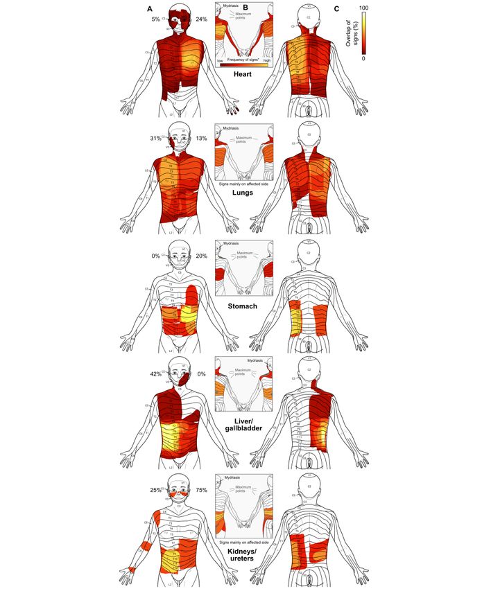
JOURNAL OF MEDICAL INTERNET RESEARCH                                                                                             Shaballout et al

                                                                           anatomy a “wrongly forgotten science” [29]. Only rarely do
     Introduction                                                          clinicians know that the transmitted signs are not limited to
     The differential diagnosis of acute visceral diseases is a common     hyperalgesia of the skin but instead show a plethora of
     but challenging clinical problem. Since pain originating from         manifestations, including sensory disturbances such as allodynia
     visceral organs (ie, visceral pain) often exhibits characteristic     and deep hyperalgesia (ie, Mackenzie zones); motor disturbances
     patterns [1-8], many textbooks assign pain location a                 such as increased resistance of the skin, muscular defense, and
     discriminative role in the differential diagnosis [9,10]. However,    resistance to passive joint movement; and, finally, signs of
     many studies have also reported negative results when testing         sympathetic activation such as vasomotor changes, asymmetric
     the predictive power of pain location [11,12]. For example, pain      hyperhidrosis (ie, asymmetric sweating between left and right
     localization in patients with coronary heart disease does not         side of body), piloerection, and anisocoria (ie, unequal pupil
     significantly differ from chest pain patients without coronary        size). As such, they are not limited to the dermatomes but
     heart disease [13].                                                   include the myotomes, sclerotomes, and other parts of the
                                                                           segmental innervation [4]. Furthermore, segmental signs may
     While primary visceral pain itself is a poorly defined, midline       be accompanied by symptoms of viscero-visceral reflexes such
     sensation, it starts to be referred or “transferred” to somatic       as nausea, vomiting, diarrhea, constipation, meteorism, and
     structures when it persists for several minutes or longer [14,15].    urinary retention [33].
     These somatic structures can include skin, subcutaneous tissue,
     and muscle and are characterized by an overlapping segmental          To our knowledge, a systematic evaluation of simultaneously
     innervation with that of the diseased organ [16-29]. In these         collected segmental signs and symptoms in patients has never
     instances, referred visceral pain manifests as hyperalgesia, a        been published in the English scientific literature. In Germany,
     phenomenon first described by Ross and Sturge in the 1880s            however, Karl Hansen (1893-1962) and Hans Schliack
     [30,31] and subsequently studied in depth by Head and                 (1919-2008) had studied a wide variety of segmental signs over
     Mackenzie [16-19]. Head mapped out the cutaneous zones of             several decades. While their results have only been published
     referred hyperalgesia for all major organs and compared them          in German [33], the essence of their work has recently been
     with the location of skin lesions in herpes zoster [16]. The result   made available in book form and extended by the work of other
     is still considered one of the most precise maps of segmental         clinicians [29]. In a large sample of internal medicine patients,
     innervation [32,33].                                                  Hansen and Schliack [33] confirmed many of Head’s
                                                                           observations and greatly extended them to include all of the
     To the present day, zones of referred hyperalgesia in visceral        above-mentioned segmental signs and symptoms. Even more
     disease carry Head’s name in many European countries, such            than Head, the authors emphasized the importance of sign
     as France, Germany, and Spain. In other parts of the world,           lateralization by defining a side rule, according to which
     however, clinicians mainly speak of “dermatomes,” and                 segmental signs are most likely to appear ipsilateral to the
     clinicians hardly know the term “Head zones” as well as Head’s        affected organ (Table 1).
     work, in general. Some authors have even called segmental

     https://www.jmir.org/2021/8/e27247                                                             J Med Internet Res 2021 | vol. 23 | iss. 8 | e27247 | p. 2
                                                                                                                  (page number not for citation purposes)
XSL• FO
RenderX
JOURNAL OF MEDICAL INTERNET RESEARCH                                                                                                          Shaballout et al

     Table 1. Lateralization of segmental signs for individual organs according to Hansen and Schliack [33].
         Organ                                        Segmental signs by side of body and part of organ (yes, possible,a or nob)
                                                      Right                                               Left
         Heart                                        Possible                                            Yes
         Pericardium                                  No                                                  Yes
         Aorta                                        Possible                                            Yes
         Lung and bronchi                             Yes                                                 Yes
         Pleura                                       Yes                                                 Yes
         Stomach                                      Yes (pylorus)                                       Yes (corpus, fundus)
         Small intestine                              Yes (duodenum, ileum)                               Yes (jejunum)
         Pancreas                                     No                                                  Yes
         Liver                                        Yes                                                 No
         Gallbladder                                  Yes                                                 No
         Spleen                                       No                                                  Yes
         Large intestine                              Yes (caecum, appendix, ascending colon, proximal Yes (distal part of transverse colon, descending
                                                      part of transverse colon)                        colon, sigmoid colon, rectum)
         Kidney                                       Yes                                                 Yes
         Ureter                                       Yes                                                 Yes
         Testis and ovary                             Yes (testis, ovary, salpinx)                        Yes (testis, ovary, salpinx)

     a
         Possible indicates a possible but unlikely occurrence of signs from that organ.
     b
         No indicates that segmental signs from a particular organ were never observed on that side.

     A methodological problem that has hampered clinical research                     the Declaration of Helsinki. All patients provided written
     of segmental signs for a long time is the difficulty in adequately               informed consent after they were informed about the purpose
     measuring bodily signs. However, recent developments in the                      of the study.
     field of digital pain drawings offer new and exciting possibilities
     to systematically record not only pain sensations but also
                                                                                      Study Population
     segmental signs and analyze them using methods of statistical                    Our study population consisted of patients from the emergency
     image analysis [34].                                                             department of Hannover Medical School who were referred to
                                                                                      internal medicine physicians between March 2017 and October
     Here, we report the results of a study that investigated both the                2017. Eligible patients were adults (age ≥18 years in Germany),
     bodily patterns and lateralization of segmental signs and                        presenting with an acute medical problem and with the ability
     spontaneous pain in acute visceral diseases. We aimed to derive                  to provide written informed consent. Furthermore, patients
     mean distributions of spontaneous pain and segmental signs for                   needed to be oriented as to place, time, and person. Exclusion
     as many internal organs as possible and to analyze their                         criteria comprised refusal or inability to provide written consent,
     segmental content and lateralization. To achieve this, we                        previously known or acutely diagnosed spinal cord injury,
     combined digital pain drawing technology and a structured,                       pregnancy, acute or past ocular illnesses, acute or past central
     10-minute bodily examination in a sample of emergency room                       or peripheral nervous disease, uncooperative patients, and
     patients.                                                                        patients who only presented to the emergency room for
                                                                                      educational purposes or to receive a prescription. For a
     Methods                                                                          flowchart, see Figure 1.
     Ethics
     The study was approved by the Ethics Committee of Hannover
     Medical School (number 2987-2017) and was conducted under

     https://www.jmir.org/2021/8/e27247                                                                          J Med Internet Res 2021 | vol. 23 | iss. 8 | e27247 | p. 3
                                                                                                                               (page number not for citation purposes)
XSL• FO
RenderX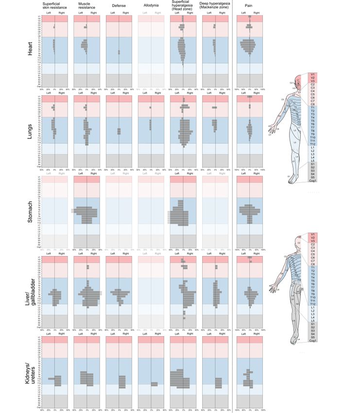
JOURNAL OF MEDICAL INTERNET RESEARCH                                                                                      Shaballout et al

     Figure 1. Flowchart of the study.

     We recruited 208 patients (85, 40.9% women) for participation    characteristics of the final study population can be found in
     in our study. Nine drawings were lost due to technical failure   Table 2, and their final diagnoses can be found in Multimedia
     of a tablet PC during the physical examination. The              Appendix 1.

     https://www.jmir.org/2021/8/e27247                                                      J Med Internet Res 2021 | vol. 23 | iss. 8 | e27247 | p. 4
                                                                                                           (page number not for citation purposes)
XSL• FO
RenderX
JOURNAL OF MEDICAL INTERNET RESEARCH Shaballout et al
Table 2. Demographics of the study population (n=208).
Characteristic Value
Age (years), mean (SD) 57.3 (17.2)
Age range, n (%)
18-39 34 (17.1)
40-59 66 (33.2)
60-79 80 (40.2)
≥80 19 (9.5)
Gender, n (%)
Women 85 (40.9)
Men 123 (59.1)
Main complaint, n (%)
Chest pain 88 (44.2)
Abdominal pain 55 (27.6)
Dyspnea 22 (11.1)
Other 34 (17.1)
First, distributed findings were spontaneous pain, allodynia,
Procedures superficial hyperalgesia, deep hyperalgesia, superficial skin
All clinical data were collected by 2 of the authors (AA and resistance, muscle resistance, defense, asymmetric hyperhidrosis,
NS), henceforth called examiners. AA is an internal medicine piloerection, vasomotor changes, herpes zoster, and resistance
specialist, and NS is a physician with 4 years of training for an to passive movement of the limbs. Distributed findings were
internal medicine specialization. The examiners were fully recorded by the examiners in the form of electronic drawings
informed about the study purpose and trained to do the physical on a body template, thus capturing their exact location and
examination for segmental signs and symptoms according to extent.
the protocol described below. Prior to the study, the examiners
trained intensively together to ensure their physical examinations Second, lateralized findings were anisocoria, glossy eye, eyelid
were standardized. This was also necessary to ensure that all separation, tense facial muscles, asymmetric posture, and
procedures could be completed in a very limited timeframe. reduced respiration movements. These findings were recorded
by choosing from a list of the abovementioned findings in
During recruitment, the examiners screened the emergency conjunction with a side label (eg, “glossy eye right,” “mydriasis
dashboard to identify patients who were referred to internal left,” etc).
medicine specialists. They approached all eligible patients,
informed them about the study, and obtained written informed Third, other findings were symptoms potentially related to
consent. viscero-visceral reflexes, namely, nausea, vomiting,
constipation, diarrhea, meteorism, and urinary retention. These
The examination took place directly after triage and before any findings were recorded by choosing from a simple list of the
medical intervention, diagnosis, or treatment. The examination abovementioned symptoms.
lasted between 7 and 15 minutes, depending on the patient’s
compliance (ie, general motivation to be examined, speed of Tablet Computer and Software Application
undressing and answering the examiner’s questions, precision All findings were recorded on Galaxy Note 10.1 (2014) tablet
of the answers given, unrelated conversation, etc) and PCs with an electronic stylus based on inductive digitizing
interruptions by nurses and physicians (as routine diagnostics technology (Samsung, Seoul, South Korea). The tablets had a
and medical interventions had priority over the scientific 10.1-inch touch screen with a resolution of 800×1280 pixels
investigation). Directly after the physical examination, all and were running Android 5.1.1 (Open Handset Alliance,
findings were recorded on a tablet computer running the app Mountain View, CA, United States). The stylus was used for
“SymptomMapper” (described in the section “Tablet Computer all data entry, hence allowing for a higher resolution while
and Software Application”). eliminating unwanted activation of the screen, for example, by
the palm. The tablet and stylus were disinfected after every
Categories of Findings patient using disinfectant wipes.
The clinical findings we were interested in can be divided into
3 groups, according to the ways they were recorded in the tablet: We used a modified version of the SymptomMapper app
(1) distributed findings (ie, those with a bodily pattern), (2) developed by our group (Somatosensory and Autonomic
lateralized findings (ie, those without a bodily pattern but with Therapy Research, Institute for Neuroradiology, Hannover
clear lateralization), and (3) other findings. Medical School, Hannover, Germany) to acquire electronic pain
drawings [35]. Its usability for doctors and the reliability of its
https://www.jmir.org/2021/8/e27247 J Med Internet Res 2021 | vol. 23 | iss. 8 | e27247 | p. 5
(page number not for citation purposes)
XSL• FO
RenderXJOURNAL OF MEDICAL INTERNET RESEARCH Shaballout et al
symptom-drawing approach have recently been shown [35]. Complete Examination
The app allowed the examiners to enter all findings from the The complete examination program had the following steps
bodily examination quickly. They could either draw distributed carried out in the exact order specified here: examination of (1)
findings on a body template or choose from a list of lateralized asymmetric posture, (2) pain and segmental symptoms, (3) the
or other findings. For the electronic drawings, examiners had head, (4) the neck and chest, (5) the abdomen, and (6) the limbs.
a front and back view of the body available, and each newly
added sign or symptom was displayed in a semitransparent way First, for asymmetric posture, a general visual inspection of the
and in a different color. patient’s posture was carried out directly after entering the
examination room to check side differences in muscle tone.
Bodily Examination
Second, pain and segmental symptoms were collected by asking
Distributed Segmental Sign Examination patients the following questions: (1) “Do you have pain—where,
Our approach to the bodily examination was based entirely on exactly? Do you have a headache?” and, when the patient
Hansen and Schliack (p140-176) [33]. Its primary purpose was reported pain, the painful region was drawn; (2) “Do you have
to check for the presence and record the extent and lateralization nausea? Did you vomit since the onset of symptoms?”; (3) “Do
of pain and segmental signs and symptoms. We start by you have diarrhea or constipation?”; (4) “Do you feel that your
describing how distributed segmental signs were collected, as abdomen is full of gas?”; and (4) “Did you have any problem
this was the same for different body regions (see below). These with urination since the onset of symptoms?”
collection methods were (1) visual inspection and (2) palpation. Third, the head was examined with (1) special tests and (2) tests
First, for visual inspection, the skin was visually inspected for of distributed segmental signs. For the head, first, the special
the following signs: shingles (as a potential sign of Zoster tests examined (1) the pupils, (2) the eyes/eyelids, and (3) tense
reactivation), vasomotor changes (ie, skin color changing to facial muscles. For these special tests, first, for pupils, we tested
red, pale, or blue, as a sign of sympathetic reflexes); piloerection for mydriasis, a sign of sympathetic activation, by equally
(ie, any hair erection or “goosebumps,” as a sign of sympathetic exposing both eyes to light after instructing the patient to relax
reflexes), and muscular asymmetries (eg, asymmetric posture, and look far away. The examiner used one hand to shadow the
tense facial muscles, respiratory chest movement, etc). eyes and compared pupil diameters on both sides. This was
repeated 3-5 times. In the case of a striking side difference, the
Second, for palpation, the body was palpated with warm hands test was considered positive for mydriasis. Second, for
to test for the following signs of sympathetic reflexes, increased eyes/eyelids, eyelid separation (due to eyelid retraction) and
muscle tone, or sensory disturbance: (1) asymmetric eye gloss (due to excessive lacrimation) are both signs of
hyperhidrosis, (2) superficial hyperalgesia, (3) deep sympathetic activation and were assessed by visually comparing
hyperalgesia, (4) allodynia, (5) superficial skin resistance, and the visible area and gloss of both eyes. In the case of a striking
(6) muscle resistance. Among palpation, first, for asymmetric side difference, the more open and glossier eye was noted. Third,
hyperhidrosis, the skin was observed and palpated for any local for tense facial muscles, a potential asymmetry of facial features
differences in the amount of sweating. Second, for superficial caused by side differences in muscle tone was checked visually.
hyperalgesia, the patient was informed that the examination It was considered positive when the upper lip was noticeably
could cause a little twinge and then was asked if the tip of a higher, the nasolabial fold deeper, and the cheeks more retracted
neurological examination needle (Healthstar, Lakewood, NJ, on one side than on the other. The test was repeated once under
United States), when passed vertically over the skin in long and provocation by applying pressure with the index and middle
slow strokes, caused a different sensation in any area. Third, fingers on a point between the 2 heads of the
for deep hyperalgesia, folds of skin were held gently between sternocleidomastoid muscle. In terms of head tests overall,
the thumb and index finger or the region was tapped on. The second, the distributed segmental signs were tested, including
test was considered positive if this procedure caused dull pain zoster, vasomotor changes, piloerection, asymmetric
that lasted longer in some part of the body than in other parts hyperhidrosis, superficial hyperalgesia, allodynia, and superficial
of the body. Fourth, for allodynia, patients were asked if their skin resistance.
clothes caused an unpleasant sensation somewhere on the body.
Then, they were asked if a medical cotton swab passed over the Fourth, as part the complete examination, was the neck and
skin in long and slow strokes caused a different sensation in chest, where the patient’s front was examined while the patient
any area. Fifth, superficial skin resistance was tested by was in a supine position after freeing the chest from clothes.
superficial palpation of the trunk skin using the palm with very Then, the back was examined with the patient sitting or lying
soft pressure. If the examiner felt either resistance or a rubbery on one side. Similar as for the head, the neck and chest included
membrane in any area, the test was considered positive in this special tests (ie, the patient's chest movement during inspiration
area. Sixth, for muscle resistance, deep palpation of the trunk and expiration was observed during the visual inspection over
wall was performed on the front and back sides with the palm several respiratory cycles, and any striking side differences were
to detect the guarding of the trunk’s wall muscles (ie, anterior noted as a sign of increased muscle tone) and distributed
thoracic muscles, anterolateral abdominal wall muscles, segmental sign tests (ie, tests for zoster, vasomotor changes,
posterior superficial muscles, and posterior deep muscles). piloerection, asymmetric hyperhidrosis, superficial hyperalgesia,
deep hyperalgesia, allodynia, superficial skin resistance, and
muscle resistance).
https://www.jmir.org/2021/8/e27247 J Med Internet Res 2021 | vol. 23 | iss. 8 | e27247 | p. 6
(page number not for citation purposes)
XSL• FO
RenderXJOURNAL OF MEDICAL INTERNET RESEARCH Shaballout et al
Fifth, for the abdomen, the patient was examined on the front and each finding, the percentage of patients that had that finding.
in a supine position after freeing the abdomen. Then, the back For lateralized findings, we calculated the percentage for each
was examined with the patient sitting or lying on one side. side individually, treating front and back as one surface. Finally,
Again, special tests were performed; mainly, the defense was we calculated the mean frequency of each finding (ie, how often
examined by applying sudden deep palpation over the painful it was observed), irrespective of the specific organ.
areas of the abdomen. If the examiner felt a reflex of the
abdominal wall, it was considered positive. Next, distributed
Distributed Findings
segmental signs were tested: zoster, vasomotor changes, Digital drawings from the app were converted to Nifti format
piloerection, asymmetric hyperhidrosis, superficial hyperalgesia, (Neuroimaging Informatics Technology Initiative, 2017) with
deep hyperalgesia, allodynia, superficial skin resistance, and a custom-written Python script (Python 2.7, Python Software
muscle resistance. Foundation, 2018) and analyzed using tools from the Functional
Magnetic Resonance Imaging (FMRIB) Software Library (FSL)
Sixth, for limbs, as for the other body parts, there were special version 5.0 (FMRIB Analysis Group, Oxford University, United
tests and testing of distributed segmental signs. For special tests, Kingdom). Figures were prepared using VINCI (Volume
passive movements of the joints were examined to detect any Imaging in Neurological Research, Co-Registration and Region
resistance due to increased muscle tone. The distributed of Interest (ROI)s Included) 4.86.0 (Max Planck Institute for
segmental signs test included zoster, vasomotor changes, Metabolism Research, Cologne, Germany) and GNU Image
piloerection, superficial hyperalgesia, and allodynia. Manipulation Program (GIMP; version 2.8.16, The GIMP
Patient Selection Team).
Medical reports of all recruited patients were followed up First, to derive the bodily distribution of all segmental signs,
through Hannover Medical Schools’ electronic health records all distributed signs were superimposed and the result binarized.
by 3 of the authors (NS, AA, and MM), to identify those patients In the resulting map, a pixel of value 1 on the body template
with a definite diagnosis of visceral disease. All information meant that at least 1 sign had been found at that particular point
regarding the acute complaint, previous diagnoses, and on the body, in that particular patient. Binarization meant that
diagnostic procedures (electrocardiogram, laboratory, radiology, we disregarded the number of signs that each patient showed
etc) were reviewed, and the most likely etiology for each patient and instead only considered their bodily location.
was discussed. Patients without a definite visceral diagnosis
We then analyzed distributed signs individually to assess the
were excluded from further analysis (n=50). The remaining
segmental distribution for each sign according to the segmental
cases were divided into those where a single organ was affected
scheme of Hansen and Schliack [33], which is largely based on
(n=110) and those with multi-organ problems (n=39). Only the
Head’s scheme [14,32]. To do this assessment, we calculated,
single-organ cases were included in the final analysis, and only
for each segment, the percentage of the segment covered by the
organs with at least 4 patients in the sample were included in
sign. For this calculation, we divided the pixel count by the total
any organ-specific analyses (Figure 1).
number of pixels of the respective segment. Only segments with
Data Analysis at least 5% coverage were included. This arbitrary threshold
was set to ensure that segments with marginal coverage (eg,
General Considerations on Lateralization due to drawing imperfections) were excluded. To assess the
According to Hansen and Schliack [33], the majority of lateralization of findings, we further divided segments into left
segmental signs are lateralized and appear on specific sides of and right body halves, calculating the percentage for each of
the body defined by the innervation of the individual organs them. This resulted in a list of half segments covered by each
(Table 1). In particular, the lateralization of signs for paired sign. Finally, we calculated, for each organ, the mean number
organs such as lungs and kidneys depends on which side is of segmental signs per half segment and the mean frequency of
affected. Due to the nature of this study, it was not possible to each sign across all organs.
conduct separate analyses for the left and right side in diseases
Spontaneous pain was analyzed in the same way but separately
of the lungs and kidneys/ureters. Furthermore, many lung cases
from all other signs.
were bilateral affections. Information about the lateralization
of segmental signs for lungs and kidneys/ureters is, therefore,
of little value and only shown for the sake of completeness. Results
Lateralization and Other Findings The Overall Frequency of Signs and Symptoms
We extracted all lateralized findings (ie mydriasis, glossy eye, Of the 110 patients in our final sample, 85 (77.3%) had
eyelid separation, tense facial muscles, asymmetric posture, and spontaneous pain, 81 (73.6%) showed at least 1 segmental sign,
reduced respiration movements) and other findings (nausea, and 52 (47.3%) showed at least 1 segmental symptom. On
vomiting, constipation, diarrhea, meteorism, and urinary average, each patient had a mean of 1.80 (SD 1.86) segmental
retention) from SymptomMapper’s JavaScript Object Notation signs and 0.77 (SD 1.00) segmental symptoms. The most
files using a custom-written Python script (Python 2.7, Python frequent signs and symptoms are shown in Table 3.
Software Foundation, 2018). Then, we calculated, for each organ
https://www.jmir.org/2021/8/e27247 J Med Internet Res 2021 | vol. 23 | iss. 8 | e27247 | p. 7
(page number not for citation purposes)
XSL• FO
RenderXJOURNAL OF MEDICAL INTERNET RESEARCH Shaballout et al
Table 3. Frequency of segmental signs and symptoms in our patient sample (N=110).
Segmental sign or symptom Value, n (%)
Segmental sign
Superficial hyperalgesia (Head zone) 46 (41.8)
Muscle resistance 39 (35.5)
Mydriasis 37 (33.6)
Defense 13 (11.8)
Deep hyperalgesia (Mackenzie zone) 13 (11.8)
Superficial skin resistance 12 (10.9)
Tense facial muscles 11 (10.0)
Vasomotor changes 10 (9.1)
Glossy eye/wide eyelid 8 (7.3)
Asymmetric posture 7 (6.4)
Reduced respiration movements 3 (2.7)
Allodynia 2 (1.8)
Piloerection 1 (0.9)
Asymmetric hyperhidrosis 1 (0.9)
Zoster 0 (0)
At least 1 segmental sign 84 (76.4)
Segmental symptoms
Nausea 45 (40.9)
Vomiting 18 (16.4)
Diarrhea 10 (9.1)
Meteorism 8 (7.3)
Constipation 5 (4.5)
Urinary retention 0 (0)
At least 1 segmental symptom 52 (47.3)
Spontaneous pain 85 (77.3)
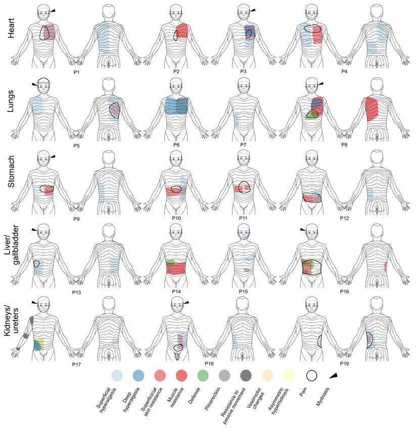
Segmental Signs and Spontaneous Pain in Individual
Frequency of Lateralization of Signs and Symptoms
Patients
All lateralization of signs and segmental symptoms are shown
in Multimedia Appendix 2. As predicted by the side rule (see Bodily maps of segmental signs and spontaneous pain for a
[33] and Table 1), the majority of lateralization signs were representative selection of individual patients are shown in
ipsilateral to the affected organ for the unpaired organs heart, Figure 2. The cases shown in Figure 2 reflect the entire
stomach, and liver/gallbladder. The most striking finding was bandwidth of segmental signs encountered in patients presenting
the high number of patients showing ipsilateral mydriasis as a to the emergency room. It ranges from “textbook cases” (eg,
potential sign of unilateral sympathetic activation. This patients 2, 3, 6, 9, 13, 18, and 19), where segmental signs alone
lateralization was 100% ipsilateral for diseases of the allow for a preliminary diagnosis, to those where segmental
liver/gallbladder (5 right vs 0 left), 100% ipsilateral for stomach signs are hardly helpful or even misleading (eg, patients 7, 12,
diseases (1 left vs 0 right), and 83% ipsilateral for heart diseases and 15). Their primary diagnoses and demographic information
(15 left vs 3 right). are summarized in Multimedia Appendix 3.
https://www.jmir.org/2021/8/e27247 J Med Internet Res 2021 | vol. 23 | iss. 8 | e27247 | p. 8
(page number not for citation purposes)
XSL• FO
RenderXJOURNAL OF MEDICAL INTERNET RESEARCH Shaballout et al
Figure 2. Segmental signs and spontaneous pain in individual patients with acute visceral diseases. P: patient.
Hansen and Schliack [33]. The lungs were the only exception,
Bodily Maps and Segmental Patterns of Distributed which showed a more widespread distribution than predicted.
Signs Concerning lateralization, segmental signs from the unpaired
Bodily maps of all distributed segmental signs are shown in organs showed a clear side difference, with more signs appearing
Figure 3, while Figure 4 contains detailed segmental information ipsilateral to the affected organ, thus supporting the “side rule”
concerning the distribution of the individual signs and represented in Table 1. For the lungs and kidneys/ureters,
spontaneous pain. In general, the observed distributions of however, this rule could not be tested, since results for these
segmental signs were largely consistent with those reported by organs reflected a mixture of left, right, and bilateral affections.
https://www.jmir.org/2021/8/e27247 J Med Internet Res 2021 | vol. 23 | iss. 8 | e27247 | p. 9
(page number not for citation purposes)
XSL• FO
RenderXJOURNAL OF MEDICAL INTERNET RESEARCH Shaballout et al
Figure 3. Distributed segmental signs in acute visceral diseases. Columns A and C show a front and back body map of all distributed segmental signs.
The inserts in column B show the segmental distributions for each organ as reported by Hansen and Schliack [33], for comparison. Percentage values
at the sides of the head indicate the frequency of unilateral mydriasis in affections of the respective organ.
Within-organ comparison showed that the different segmental Regarding lateralization, segmental signs for the unpaired organs
signs but also spontaneous pain had a similar segmental mostly obeyed the side rule, according to which signs should
distribution (Figure 4). Between organs, these distributions appear on the body half where the organ is located. Pain differed
showed considerable overlap. Superficial hyperalgesia (Head markedly in that respect and, instead, showed a rather symmetric
zone) exhibited the greatest spread in terms of segments. pattern.
https://www.jmir.org/2021/8/e27247 J Med Internet Res 2021 | vol. 23 | iss. 8 | e27247 | p. 10
(page number not for citation purposes)
XSL• FO
RenderXJOURNAL OF MEDICAL INTERNET RESEARCH Shaballout et al
Figure 4. Segmental patterns and lateralization of segmental signs and pain in acute visceral diseases. Each row contains the results for an individual
organ, while columns represent the different most-common segmental signs and pain, respectively. Graphs without any occurrence were blanked. Please
note the different axis scaling for the latter. For the sake of clarity, segmental sections have been color-coded according to the body template shown on
the right. This body template shows the different segments: trigeminal (V1-3), cervical (C2-8), thoracal (T1-12), lumbar (L1-5), sacral (S1-5), and
coccygeal (Cog1).
For cardiac-related conditions, segmental signs were mostly distributed signs were strongly left-dominated with deep
located in the thoracic segments and, to a lesser extent, in hyperalgesia (Mackenzie zone), showing complete
cervical segments. Superficial hyperalgesia (Head zone) was left-lateralization (Figure 4). In heart patients, defense and
also detected in the trigeminal segments. The maximum of the allodynia were rare and nonexistent, respectively.
averaged signs was in the T3-T5 region (Figure 3B), as predicted
by Hansen and Schliack [33]. In terms of lateralization, all
https://www.jmir.org/2021/8/e27247 J Med Internet Res 2021 | vol. 23 | iss. 8 | e27247 | p. 11
(page number not for citation purposes)
XSL• FO
RenderXJOURNAL OF MEDICAL INTERNET RESEARCH Shaballout et al
For diseases of the lungs, segmental signs were very widespread the characteristic shoulder presentation in segments C3-C5 [33].
and covered a range from V2 to L2. Signs were generally less In terms of lateralization, muscle resistance, defense, and
focused than for the heart, and no clear maximum was superficial and deep hyperalgesia were predominantly
discernible. In this, the distribution deviated from Hansen and right-lateralized, with superficial hyperalgesia (Head zone)
Schliack’s [33], who reported T9 as the lower margin of showing almost complete right-lateralization.
segmental signs in lung diseases. As was to be expected due to
Finally, segmental signs of the kidneys had the narrowest
the mixture of left, right, and bilateral organ diseases, no
distribution, starting at T6 and extending down to L2, once
lateralization could be seen.
again, showing a rather high similarity with the predicted
For the stomach, the segmental distribution was almost strictly distribution by Hansen and Schliack [33].
thoracic, from T2 to T12, with a maximum at T6-9 on the front
and on the back. Similar to the heart, superficial hyperalgesia
Comparison of Spontaneous Pain and Segmental Signs
for the stomach was lateralized to the left, as predicted by the The segmental distributions of spontaneous pain and segmental
side rule. The comparison with Hansen and Schliack [33] signs are shown in Figure 5. It is evident that spontaneous pain
showed that the maximum of signs in T6-9 fell in the expected differed markedly from segmental signs. It spanned fewer
range in the front view. On the back, however, there was only segments but extended to the head region (V1) in cardiac,
a partial overlap, with Hansen and Schliack [33] predicting respiratory, and liver/gallbladder affections. Furthermore,
higher thoracic segments than were found in our study. spontaneous pain was much less lateralized than segmental
signs and, instead, rather localized in the body midline. In
The similarity with Hansen and Schliack’s [33] results was general, the pain was less widespread and showed much weaker
much higher for patients with liver/gallbladder diseases. Here, lateralization than segmental signs in the unpaired organs (ie,
segmental signs showed a largely thoracic distribution but with heart, stomach, and liver/gallbladder).
https://www.jmir.org/2021/8/e27247 J Med Internet Res 2021 | vol. 23 | iss. 8 | e27247 | p. 12
(page number not for citation purposes)
XSL• FO
RenderXJOURNAL OF MEDICAL INTERNET RESEARCH Shaballout et al
Figure 5. Direct comparison between segmental signs (A and B) and spontaneous pain (C and D) in acute visceral diseases. Column A shows the mean
number of segmental signs per segment for the individual organs, and column B shows the joint distribution of segmental signs (cf Figure 2). The
symptom of spontaneous pain is shown as mean distributions of pain in column C, and their exact segmental content is shown in column D.
liver/gallbladder, and kidneys/ureters by combining digital pain
Discussion drawing technology and a structured 10-minute bodily
Principal Findings examination in patients presenting to the emergency room. We
extracted precise information on the segmental content and
In this study, we investigated bodily patterns and lateralization lateralization and compared the results with the slightly outdated
of segmental signs and spontaneous pain in acute visceral but authoritative German work of Hansen and Schliack [33].
diseases. We derived mean distributions of spontaneous pain Although purely descriptive by design, our study is the first in
and segmental signs for the heart, lungs, stomach,
https://www.jmir.org/2021/8/e27247 J Med Internet Res 2021 | vol. 23 | iss. 8 | e27247 | p. 13
(page number not for citation purposes)
XSL• FO
RenderXJOURNAL OF MEDICAL INTERNET RESEARCH Shaballout et al
the English language to provide a detailed account of resistance, and defense), and visceromotor signs (ie, vasomotor
simultaneously collected segmental signs and symptoms for changes, piloerection, and asymmetric hyperhidrosis). Of these,
visceral diseases in the clinical setting. superficial hyperalgesia (ie, Head zones), muscle resistance,
defense, and deep hyperalgesia (ie, Mackenzie zones) were the
Lateralization of Segmental Signs most frequently observed in our sample of patients, while others,
The lateralization of segmental signs is important, as it allows such as allodynia, piloerection, asymmetric hyperhidrosis, or
one to quickly identify the affected body side (ie, the side zoster, were exceedingly rare.
hosting the affected organ; Table 1). This side rule may be useful
in the differential diagnosis, such as in differentiating gastritis There was a close similarity between the original maps of
from hepatitis or pancreatitis, or acute coronary syndrome from segmental signs by Hansen and Schliack [33] and our mean
a pulmonary embolism or esophagitis. Due to our study design distributions of all signs (Figure 3). For a prospective evaluation,
and the very mixed patient sample, our results regarding however, future studies should aim to quantify this similarity
lateralization were limited to the heart, stomach, and (eg, by using spatial similarity measures).
liver/gallbladder. Although segmental signs of the lungs and Several groups have studied individual segmental signs or
kidneys/ureters are also expected to be found ipsilateral to the groups of signs since the days of Hansen and Schliack. For
affected side, a separate analysis for the individual sides was example, Nicholas and colleagues [39] found that patients with
not possible for these organs due to the limited number of cases, myocardial infarction showed characteristic paravertebral soft
many of which showed bilateral affections. tissue changes readily detected by palpation. Compared with
Although our data were not analyzed prospectively, it appears, patients without diagnosed cardiovascular diseases, patients
for the heart, stomach, and liver/gallbladder, that they may with myocardial infarction had a significantly higher incidence
support the findings of Hansen and Schliack [33] that segmental of increased firmness, warmth, ropiness, oedematous changes,
signs appear ipsilateral to the affected organ. While this was and heavy musculature, almost entirely confined to cardiac
evident for the mean bodily distributions of segmental signs, segments T1-4. In a follow-up 3 years after the infarction, these
we also found an ipsilateral occurrence of mydriasis, a finding signs had regressed in the majority of patients [40]. Vecchiet
rarely raised outside the neurological setting. It results from a and colleagues [41] found ipsilateral superficial and deep
reflex mediated by the ciliospinal center, which conducts hyperalgesia of the first lumbar (L1) segment in patients after
impulses from the entire body to the sympathetically innervated renal/ureteral calculosis.
dilator pupillae muscle (p271) [28] and, more than 100 years For the gallbladder, Stawowy and colleagues [42] found that
ago, was first described to occur in affections of the lungs [36] all patients with acute cholecystitis reported referred pain in the
and the heart [37]. More recently, Rosenberg [38] has shown epigastrium and under the right curvature. Segmental signs
that anisocoria (ie, unequal pupil size) under physiological inside this area were quantitatively evaluated using von Frey
conditions is a manifestation of sympathetic asymmetry. hairs, warm and cold metal rollers, and a constant current
We found ipsilateral (ie, right-sided) mydriasis in 42% of our stimulator to test for the different forms of hypersensitivity or
liver/gallbladder patients and not a single case of contralateral allodynia. The authors reported that 20% of the patients showed
mydriasis. For the heart, mydriasis was less frequent (24% hypersensitivity or allodynia to mechanical, 53% to cold, 40%
ipsilateral vs 5% contralateral), yet this means that patients to warmth, and 63% to electrical stimulation [42]. The same
showing the sign had it on the ipsilateral side in almost 83% of authors reported that 50% to 56% of patients with acute
the cases. Hansen and Schliack [33] reported qualitatively appendicitis showed segmental signs over the right abdominal
similar but generally higher numbers for mydriasis. In their quadrant, with the maximum located approximately at
sample of 28 heart patients, 27 (96%) had mydriasis, and this McBurney point [43]. These findings were recently confirmed
was ipsilateral in 26 patients (96%). In 56 liver/gallbladder by Roumen and colleagues [44], who reported that 39% of
patients, 54 (96%) had mydriasis, of which 50 cases (93%) were patients with acute appendicitis demonstrated at least one
ipsilateral (ie, right-sided). segmental sign (ie, hyperalgesia, hypoesthesia, altered cool
perception, or positive pinch test) over the lower right abdomen.
The generally higher numbers of mydriasis in heart diseases Finally, a large number of smaller studies and case reports have
found in Hansen and Schliack’s [33] work may be explained been published, which have been reviewed by Beal [45].
by the fact that these authors used dark adaptation and infrared
photographs in many of their patients, which made their Segmental Signs Versus Spontaneous Pain
examination less subjective, while our examiners were restricted The majority of our patients with visceral diseases reported
to visual inspection under normal light. Clinicians interested in spontaneous pain (Table 3). In 85% of the cases, it was, by far,
this phenomenon should consider using a portable infrared the most frequent finding, followed by superficial hyperalgesia
pupilometer. (46%), nausea (45%), and muscle resistance (39%).
Localization and Distribution of Segmental Signs Many textbooks assign pain location a discriminative role in
A subset of the findings collected in our study was further the differential diagnosis (eg, retrosternal chest pain that radiates
analyzed to extract detailed segmental information. We called to the left arm or lower jaw usually refers to acute coronary
this group of findings “distributed signs.” It comprised a number heart disease. However, such predictive power of pain location
of somatosensory (ie, superficial and deep hyperalgesia and has been a matter of debate for decades [11-13]. Here, we found,
allodynia), somatomotor (ie, superficial skin resistance, muscle by direct comparison of spontaneous pain and segmental signs,
https://www.jmir.org/2021/8/e27247 J Med Internet Res 2021 | vol. 23 | iss. 8 | e27247 | p. 14
(page number not for citation purposes)
XSL• FO
RenderXJOURNAL OF MEDICAL INTERNET RESEARCH Shaballout et al
that the two were rather dissimilar in their bodily patterns and This is due to the unique situation in the emergency room, where
segmental distributions (Figure 5). Irrespective of the affected the vital role of the specialist is to rule out life-threatening
organ, spontaneous pain was less widespread than segmental conditions. A further one-quarter of the remaining patients had
signs (ie, it included fewer segments). Furthermore, spontaneous to be excluded from the analysis because they suffered from
pain appeared mostly in the body midline, thus lacking the diseases affecting multiple organs. Secondly, while we took
diagnostically relevant ipsilateral distribution seen in the great care to include only patients with single-organ problems,
majority of segmental signs. As Figure 5 shows, patients with it is likely that affections of other organs were present but
lung, stomach, and liver/gallbladder diseases all showed overlooked in some of the patients. This means that some
spontaneous pain in the epigastric region (T5-9), thus rendering patients who seemed to only present with cardiac disease may
this symptom unsuitable for differential diagnosis. have had another underlying disease affecting other organs.
Thirdly, findings collected by means of palpation are naturally
The substantial differences found between pain and segmental
more subjective than, for example, laboratory results. While
signs regarding their location and lateralization underline the
there are ways to measure segmental signs more quantitatively,
importance of making a clear distinction between visceral pain,
we did not do so to keep the examination time to an absolute
(referred) hyperalgesia, and other segmental signs.
minimum, as required by the clinical setting. Finally, we did
Despite the purely descriptive design of this study, our results not differentiate explicitly between signs and symptoms that
(Figures 4 and 5) regarding the benefit of using spontaneous patients had only during their acute problem from patient
pain or segmental signs seem to favor the latter over the former. symptoms that occurred usually. This may have introduced
Future studies should test this in a prospective way (eg, by some bias.
letting a blinded assessors predict the affected organ from the
distribution of spontaneous pain or from that of segmental
Conclusions
signs). This study underlines the usefulness of including segmental
signs in the bodily examination of patients with acute medical
Limitations problems. As we have shown, capturing the location of
Our study had several limitations that need to be discussed. segmental signs on a digital body map may assist in the clinical
Firstly, our patient sample was relatively small, as we could decision-making process in some acute visceral conditions.
only analyze approximately half of the included patients. There Segmental information and lateralization from the 3
were two reasons for this. On the one hand, approximately most-frequent signs (superficial hyperalgesia, muscle resistance,
one-quarter of our patients had to be excluded from the analysis, and mydriasis) can be quickly acquired and may help physicians
since they left the hospital without a confirmed final diagnosis. narrow the differential diagnosis.
Acknowledgments
The authors would also like to thank Christoph Duesberg, former Head of the Central Emergency Room at Hannover Medical
School, for his support in planning and carrying out the study. The authors would like to thank the Horst Görtz Foundation for
providing financial support.
Conflicts of Interest
None declared.
Multimedia Appendix 1
Supplementary table 1.
[PDF File (Adobe PDF File), 42 KB-Multimedia Appendix 1]
Multimedia Appendix 2
Supplementary figure 1.
[ File , 125 KB-Multimedia Appendix 2]
Multimedia Appendix 3
Supplementary table 2.
[ File , 54 KB-Multimedia Appendix 3]
References
1. Bennett J, Atkinson M. The Differentiation Between Œsophageal and Cardiac Pain. The Lancet 1966
Nov;288(7473):1123-1127. [doi: 10.1016/s0140-6736(66)92209-4]
2. Henderson RD, Wigle ED, Sample K, Marryatt G. Atypical chest pain of cardiac and esophageal origin. Chest 1978
Jan;73(1):24-27. [doi: 10.1378/chest.73.1.24] [Medline: 620553]
https://www.jmir.org/2021/8/e27247 J Med Internet Res 2021 | vol. 23 | iss. 8 | e27247 | p. 15
(page number not for citation purposes)
XSL• FO
RenderXJOURNAL OF MEDICAL INTERNET RESEARCH Shaballout et al
3. Berger J, Buclin T, Haller E, Van Melle G, Yersin B. Right arm involvement and pain extension can help to differentiate
coronary diseases from chest pain of other origin: a prospective emergency ward study of 278 consecutive patients admitted
for chest pain. J Intern Med 1990 Mar;227(3):165-172. [doi: 10.1111/j.1365-2796.1990.tb00138.x] [Medline: 2313224]
4. Giamberardino MA, Vecchiet L. Pathophysiology of visceral pain. Current Review of Pain 1997 Mar;1(1):23-33. [doi:
10.1007/bf02938147]
5. Yamamoto W, Kono H, Maekawa M, Fukui T. The relationship between abdominal pain regions and specific diseases: an
epidemiologic approach to clinical practice. J Epidemiol 1997 Mar;7(1):27-32 [FREE Full text] [doi: 10.2188/jea.7.27]
[Medline: 9127570]
6. Kalloo A. Overview of differential diagnoses of abdominal pain. Gastrointest Endosc 2002 Dec;56(6 Suppl):S255-S257.
[doi: 10.1067/mge.2002.129004] [Medline: 12447278]
7. Bates C, Plevris J. Clinical evaluation of abdominal pain in adults. Medicine 2013 Feb;41(2):81-86. [doi:
10.1016/j.mpmed.2012.11.005]
8. Wilson SJ, MacLeod DC. Chest pain in acute medicine. Medicine 2013 Feb;41(2):113-117. [doi:
10.1016/j.mpmed.2012.11.010]
9. Kasper D, Fauci A, Hauser S, Longo D, Jameson J, Loscalzo J. Harrison's principles of internal medicine, 19e (Vol. In: 1,
No. 2). Mcgraw-Hill. New York: McGraw Hill Education Medical; 2015.
10. Mahadevan S, Garmel G. An introduction to clinical emergency medicine. New York: Cambridge University Press; 2012.
11. Eriksson B, Vuorisalo D, Sylvén C. Diagnostic potential of chest pain characteristics in coronary care. J Intern Med 1994
May;235(5):473-478. [doi: 10.1111/j.1365-2796.1994.tb01105.x] [Medline: 8182404]
12. Everts B, Karlson BW, Währborg P, Hedner T, Herlitz J. Localization of pain in suspected acute myocardial infarction in
relation to final diagnosis, age and sex, and site and type of infarction. Heart & Lung 1996 Nov;25(6):430-437. [doi:
10.1016/s0147-9563(96)80043-4]
13. Bösner S, Bönisch K, Haasenritter J, Schlegel P, Hüllermeier E, Donner-Banzhoff N. Chest pain in primary care: is the
localization of pain diagnostically helpful in the critical evaluation of patients?--A cross sectional study. BMC Fam Pract
2013 Oct 18;14(1):154 [FREE Full text] [doi: 10.1186/1471-2296-14-154] [Medline: 24138299]
14. Ness T, Gebhart G. Visceral pain: a review of experimental studies. Pain 1990;41(2):167-234
https://www.sciencedirect.com/science/article/pii/0304395990900215. [doi: 10.1016/0304-3959(90)90021-5]
15. Giamberardino M, Vecchiet L. Visceral pain, referred hyperalgesia and outcome: new concepts. European journal of
anaesthesiology.Supplement 1995;10:61. [doi: 10.1016/b0-12-226870-9/01739-1]
16. Head H. On Disturbances of Sensation With Especial Reference to the Pain of Visceral Disease. Brain 1893;16(1-2):1-133.
[doi: 10.1093/brain/16.1-2.1]
17. HEAD H. On Disturbances of Sensation With Especial Reference to the Pain of Visceral Disease: Part II. Brain
1894;17(3):339-480. [doi: 10.1093/brain/17.3.339]
18. HEAD H. On Disturbances of Sensation With Especial Reference to the Pain of Visceral Disease: Part III. Brain
1896;19(2-3):153-276. [doi: 10.1093/brain/19.2-3.153]
19. Mackenzie J. Some Points Bearing on the Association of Sensory Disorders and Visceral Disease. Brain 1893;16(3):321-354.
[doi: 10.1093/brain/16.3.321]
20. Sherren J. On the Occurence and Significance of Cutaneous Hyperalgesia in Appendictitis. The Lancet 1903
Sep;162(4177):816-821. [doi: 10.1016/s0140-6736(01)51715-8]
21. Elsberg CA, Neuhof H. The Diagnostic Value of Cutaneous Hyperalgesia (Head's Zones) in Abdominal Disease. The
American Journal of the Medical Sciences 1908;135(5):690-711. [doi: 10.1097/00000441-190811000-00007]
22. Langstroth L. An Attempt to Determine the Diagnostic Importance of Head's Zones of Hyperalgesia. Arch Intern Med 1915
Aug 01;XVI(2):149. [doi: 10.1001/archinte.1915.00080020003001]
23. Bullowa JG. Segmental Hyperalgesia and Segmental Increased Muscle Tone in Diseases of the Lungs and Heart. Bull N
Y Acad Med 1929 Jun;5(6):538-551 [FREE Full text] [Medline: 19311681]
24. Bolton C. Observations on Referred Pain. Brain 1934;57(3):211-226. [doi: 10.1093/brain/57.3.211]
25. Boas EP, Levy H. Extracardiac determinants of the site and radiation of pain in angina pectoris with special reference to
shoulder pain. American Heart Journal 1937 Nov;14(5):540-554. [doi: 10.1016/s0002-8703(37)90278-3]
26. Brown FR. The Problem of Abdominal Pain. Br Med J 1942 May 02;1(4243):543-546 [FREE Full text] [doi:
10.1136/bmj.1.4243.543] [Medline: 20784206]
27. Kennard M, Haugen F. The relation of subcutaneous focal sensitivity to referred pain of cardiac origin. Anesthesiology:
The Journal of the American Society of Anesthesiologists 1955;16(3):297-311. [doi: 10.1097/00000542-195505000-00001]
28. Schliack H. Klinische und theoretische Bedeutung der Headschen Zonen. Acta Neurovegetativa 1966 Mar;28(1-4):437-449.
[doi: 10.1007/bf01227404]
29. Wancura-Kampik I. Segmental anatomy: the key to mastering acupuncture, neural therapy, and manual therapy. MUNICH,
Germany: Urban & Fischer/Elsevier; 2012.
30. Sturge WA. The Phenomena of Angina Pectoris, and their Bearing Upon the Theory of Counter-Irritation. Brain
1883;5(4):492-510. [doi: 10.1093/brain/5.4.492]
31. Ross J. On the Segmental Distribution of sensory Disorders. Brain 1888;10(4):333-361. [doi: 10.1093/brain/10.4.333]
https://www.jmir.org/2021/8/e27247 J Med Internet Res 2021 | vol. 23 | iss. 8 | e27247 | p. 16
(page number not for citation purposes)
XSL• FO
RenderXJOURNAL OF MEDICAL INTERNET RESEARCH Shaballout et al
32. Lee M, McPhee R, Stringer M. An evidence-based approach to human dermatomes. Clin Anat 2008 Jul;21(5):363-373.
[doi: 10.1002/ca.20636] [Medline: 18470936]
33. Hansen K, Schliack H. Segmentale Innervation: ihre Bedeutung für Klinik und Praxis. Stuttgart, Germany: Thieme; 1962.
34. Shaballout N, Neubert T, Boudreau S, Beissner F. From Paper to Digital Applications of the Pain Drawing: Systematic
Review of Methodological Milestones. JMIR Mhealth Uhealth 2019 Sep 05;7(9):e14569 [FREE Full text] [doi:
10.2196/14569] [Medline: 31489841]
35. Neubert T, Dusch M, Karst M, Beissner F. Designing a Tablet-Based Software App for Mapping Bodily Symptoms:
Usability Evaluation and Reproducibility Analysis. JMIR Mhealth Uhealth 2018 May 30;6(5):e127 [FREE Full text] [doi:
10.2196/mhealth.8409] [Medline: 29848470]
36. Roque. Note sur l'inégalité des pupilles dans les affections unilatérales des poumons. Gaz. méd (1869)., Paris 48 1869:639.
37. Gibson GA. Some Hitherto Undescribed Symptoms in Agina Pectoris. Brain 1905;28(1):52-64. [doi: 10.1093/brain/28.1.52]
38. Rosenberg ML. Physiologic Anisocoria: A Manifestation of a Physiologic Sympathetic Asymmetry. Neuro-Ophthalmology
2009 Jul 08;32(3):147-149. [doi: 10.1080/01658100802115254]
39. Nicholas AS, DeBias DA, Ehrenfeuchter W, England KM, England RW, Greene CH, et al. A somatic component to
myocardial infarction. Br Med J (Clin Res Ed) 1985 Jul 06;291(6487):13-17 [FREE Full text] [doi: 10.1136/bmj.291.6487.13]
[Medline: 3926040]
40. Nicholas AS, DeBias DA, Greene CH. Somatic component to myocardial infarction: three year follow up. BMJ 1991 Jun
29;302(6792):1581-1581 [FREE Full text] [doi: 10.1136/bmj.302.6792.1581] [Medline: 1855044]
41. Vecchiet L, Giamberardino M, Dragani L, Albe-Fessard D. Pain from renal/ureteral calculosisvaluation of sensory thresholds
in the lumbar area. Pain 1989;36(3):e. [doi: 10.1016/0304-3959(89)90087-0]
42. Stawowy M, Bluhme C, Arendt-Nielsen L, Drewes AM, Funch-Jensen P. Somatosensory changes in the referred pain area
in patients with acute cholecystitis before and after treatment with laparoscopic or open cholecystectomy. Scand J
Gastroenterol 2004 Oct 08;39(10):988-993. [doi: 10.1080/00365520410003425] [Medline: 15513339]
43. Stawowy M, Rössel P, Bluhme C, Funch-Jensen P, Arendt-Nielsen L, Drewes AM. Somatosensory changes in the referred
pain area following acute inflammation of the appendix. Eur J Gastroenterol Hepatol 2002 Oct;14(10):1079-1084. [doi:
10.1097/00042737-200210000-00008] [Medline: 12362098]
44. Roumen RMH, Vening W, Wouda R, Scheltinga MM. Acute Appendicitis, Somatosensory Disturbances ("Head Zones"),
and the Differential Diagnosis of Anterior Cutaneous Nerve Entrapment Syndrome (ACNES). J Gastrointest Surg 2017
Jun 14;21(6):1055-1061. [doi: 10.1007/s11605-017-3417-y] [Medline: 28411350]
45. Beal M. Viscerosomatic reflexes: a review. J Am Osteopath Assoc 1985 Dec;85(12):786-801. [Medline: 3841111]
Edited by R Kukafka; submitted 19.01.21; peer-reviewed by T Ots, M Galve Villa, Y Cai; comments to author 13.02.21; revised version
received 02.05.21; accepted 14.06.21; published 27.08.21
Please cite as:
Shaballout N, Aloumar A, Manuel J, May M, Beissner F
Lateralization and Bodily Patterns of Segmental Signs and Spontaneous Pain in Acute Visceral Disease: Observational Study
J Med Internet Res 2021;23(8):e27247
URL: https://www.jmir.org/2021/8/e27247
doi: 10.2196/27247
PMID:
©Nour Shaballout, Anas Aloumar, Jorge Manuel, Marcus May, Florian Beissner. Originally published in the Journal of Medical
Internet Research (https://www.jmir.org), 27.08.2021. This is an open-access article distributed under the terms of the Creative
Commons Attribution License (https://creativecommons.org/licenses/by/4.0/), which permits unrestricted use, distribution, and
reproduction in any medium, provided the original work, first published in the Journal of Medical Internet Research, is properly
cited. The complete bibliographic information, a link to the original publication on https://www.jmir.org/, as well as this copyright
and license information must be included.
https://www.jmir.org/2021/8/e27247 J Med Internet Res 2021 | vol. 23 | iss. 8 | e27247 | p. 17
(page number not for citation purposes)
XSL• FO
RenderXYou can also read