Accurately Differentiating Between Patients With COVID-19, Patients With Other Viral Infections, and Healthy Individuals: Multimodal Late Fusion ...
←
→
Page content transcription
If your browser does not render page correctly, please read the page content below
JOURNAL OF MEDICAL INTERNET RESEARCH                                                                                                                  Xu et al

     Original Paper

     Accurately Differentiating Between Patients With COVID-19,
     Patients With Other Viral Infections, and Healthy Individuals:
     Multimodal Late Fusion Learning Approach

     Ming Xu1,2,3*, MD, PhD; Liu Ouyang4*, MD; Lei Han1,2*, MD, PhD; Kai Sun5*, MD; Tingting Yu6*, MD; Qian Li7*,
     MD; Hua Tian8*, MSc; Lida Safarnejad9,10, MSc, PhD; Hengdong Zhang1,2, MD, PhD; Yue Gao1, MSc; Forrest Sheng
     Bao11, PhD; Yuanfang Chen12, MSc; Patrick Robinson3, MD, FACP, FIDSA; Yaorong Ge10, PhD; Baoli Zhu2, MSc;
     Jie Liu13, MD, PhD; Shi Chen3,14, PhD
     1
      Department of Occupational Disease Prevention, Jiangsu Provincial Center for Disease Control and Prevention, Nanjing, China
     2
      Center for Global Health, School of Public Health, Nanjing Medical University, Nanjing, China
     3
      Department of Public Health Sciences, College of Health and Human Services, University of North Carolina Charlotte, Charlotte, NC, United States
     4
      Department of Orthopedics, Union Hospital, Tongji Medical College, Huazhong University of Science and Technology, Wuhan, China
     5
      Department of Emergency Medicine, The First Hospital of Nanjing Medical University, Nanjing, China
     6
      Department of Medical Genetics, School of Basic Medical Science Jiangsu Key Laboratory of Xenotransplantation, Nanjing Medical University,
     Nanjing, China
     7
      Department of Pediatrics, Affiliated Kunshan Hospital of Jiangsu University, Kunshan, China
     8
      Department of Acute Infectious Disease Control and Prevention, Jiangsu Provincial Center for Disease Control and Prevention, Nanjing, China
     9
      School of Medicine, Stanford University, Stanford, CA, United States
     10
      Department of Software and Information Systems, College of Computing and Informatics, University of North Carolina Charlotte, Charlotte, NC,
     United States
     11
          Department of Computer Science, Iowa State University, Ames, IA, United States
     12
          Institute of HIV/AIDS/STI Prevention and Control, Jiangsu Provincial Center for Disease Control and Prevention, Nanjing, China
     13
          Department of Radiology, Union Hospital, Tongji Medical College, Huazhong University of Science and Technology, Wuhan, China
     14
          School of Data Science, University of North Carolina Charlotte, Charlotte, NC, United States
     *
      these authors contributed equally

     Corresponding Author:
     Jie Liu, MD, PhD
     Department of Radiology
     Union Hospital, Tongji Medical College
     Huazhong University of Science and Technology
     1277 Jiefang Avenue
     Wuhan
     China
     Phone: 86 13469981699
     Email: liu_jie0823@163.com

     Abstract
     Background: Effectively identifying patients with COVID-19 using nonpolymerase chain reaction biomedical data is critical
     for achieving optimal clinical outcomes. Currently, there is a lack of comprehensive understanding in various biomedical features
     and appropriate analytical approaches for enabling the early detection and effective diagnosis of patients with COVID-19.
     Objective: We aimed to combine low-dimensional clinical and lab testing data, as well as high-dimensional computed tomography
     (CT) imaging data, to accurately differentiate between healthy individuals, patients with COVID-19, and patients with non-COVID
     viral pneumonia, especially at the early stage of infection.
     Methods: In this study, we recruited 214 patients with nonsevere COVID-19, 148 patients with severe COVID-19, 198 noninfected
     healthy participants, and 129 patients with non-COVID viral pneumonia. The participants’ clinical information (ie, 23 features),
     lab testing results (ie, 10 features), and CT scans upon admission were acquired and used as 3 input feature modalities. To enable
     the late fusion of multimodal features, we constructed a deep learning model to extract a 10-feature high-level representation of

     http://www.jmir.org/2021/1/e25535/                                                                      J Med Internet Res 2021 | vol. 23 | iss. 1 | e25535 | p. 1
                                                                                                                           (page number not for citation purposes)
XSL• FO
RenderX
JOURNAL OF MEDICAL INTERNET RESEARCH Xu et al
CT scans. We then developed 3 machine learning models (ie, k-nearest neighbor, random forest, and support vector machine
models) based on the combined 43 features from all 3 modalities to differentiate between the following 4 classes: nonsevere,
severe, healthy, and viral pneumonia.
Results: Multimodal features provided substantial performance gain from the use of any single feature modality. All 3 machine
learning models had high overall prediction accuracy (95.4%-97.7%) and high class-specific prediction accuracy (90.6%-99.9%).
Conclusions: Compared to the existing binary classification benchmarks that are often focused on single-feature modality, this
study’s hybrid deep learning-machine learning framework provided a novel and effective breakthrough for clinical applications.
Our findings, which come from a relatively large sample size, and analytical workflow will supplement and assist with clinical
decision support for current COVID-19 diagnostic methods and other clinical applications with high-dimensional multimodal
biomedical features.
(J Med Internet Res 2021;23(1):e25535) doi: 10.2196/25535
KEYWORDS
COVID-19; machine learning; deep learning; multimodal; feature fusion; biomedical imaging; diagnosis support; diagnosis;
imaging; differentiation; testing; diagnostic
Currently, CT scans can be analyzed to differentiate patients
Introduction with COVID-19, especially those in a severe clinical state, from
COVID-19 is an emerging major biomedical challenge for the healthy people or patients with non-COVID infections. Patients
entire health care system [1]. Compared to severe acute with COVID-19 usually present the typical ground-glass opacity
respiratory syndrome (SARS) and Middle East respiratory (GGO) characteristic on CT images of the thoracic region. A
syndrome (MERS), COVID-19 has much higher infectivity. recent study has reported a 98% COVID-19 detection rate based
COVID-19 has also spread much faster across the globe than on a 51-patient sample without a comparison group [14].
other coronavirus diseases. Although COVID-19 has a relatively Detection rates that ranged between 60% and 93% were also
lower case fatality rate than SARS and MERS, the reported in another study on 1014 participants with a comparison
overwhelmingly large number of diagnosed COVID-19 cases, group [15]. Furthermore, the recent advances in data-driven
as well as the many more undiagnosed COVID-19 cases, has deep learning (DL) methods, such as convolutional neural
endangered health care systems and vulnerable populations networks (CNNs), have demonstrated the ability to detect
during the COVID-19 pandemic. Therefore, the early and COVID-19 in patients. On February 2020, Hubei, China adopted
accurate detection and intervention of COVID-19 are key in CT scans as the official clinical COVID-19 diagnostic method
effectively treating patients, protecting vulnerable populations, in addition to molecular confirmatory diagnostic methods for
and containing the pandemic at large. COVID-19, in accordance with the nation’s diagnosis and
treatment guidance [2]. However, the effectiveness of using DL
Currently, the gold standard for the confirmatory diagnosis of methods to further differentiate SARS-CoV-2 infection from
COVID-19 is based on molecular quantitative real-time clinically similar non-COVID viral infections still needs to be
polymerase chain reaction (qRT-PCR) and antigen testing for explored and evaluated.
the disease-causing SARS-CoV-2 virus [2-4]. Although these
tests are the gold standard for COVID-19 diagnosis, they suffer With regard to places where molecular confirmatory diagnoses
from various practical issues, including reliability, resource are not immediately available, symptoms are often used for
adequacy, reporting lag, and testing capacity across time and quickly evaluating presumed patients’ conditions and supporting
space [5]. To help frontline clinicians diagnose COVID-19 more triage [13,16,17]. Checklists have been developed for
effectively and efficiently, other diagnostic methods have also self-evaluating the risk of developing COVID-19. These
been explored and used, including medical imaging (eg, X-ray checklists are based on clinical information, including
scans and computed tomography [CT] scans [6]), lab testing symptoms, preexisting comorbidities, and various demographic,
(eg, various blood biochemistry analyses [7-10]), and identifying behavioral, and epidemiological factors. However, these clinical
common clinical symptoms [11]. However, these methods do data are generally used for qualitative purposes (eg, initial
not directly detect the disease-causing SARS-CoV-2 virus or assessment) by both the public and clinicians [18]. Their
the SARS-CoV-2 antigen. Therefore, these methods do not have effectiveness in providing accurate diagnostic decision support
the same conclusive power that confirmatory molecular is largely underexplored and unknown.
diagnostic methods have. Nevertheless, these alternative In addition to biomedical imaging and clinical information,
methods help clinicians with inadequate resources detect recent studies on COVID-19 have shown that laboratory testing,
COVID-19, differentiate patients with COVID-19 from patients such as various blood biochemistry analyses, is also a feasible
without COVID-19 and noninfected individuals, and triage method for detecting COVID-19 in patients, with reasonably
patients to optimize health care system resources [12,13]. When high accuracy [19,20]. The rationale is that the human body is
applied appropriately, these supplementary methods, which are a unity. When people are infected with SARS-CoV-2, the
based on alternative biomedical evidence, can help mitigate the clinical consequences can be observed not only from apparent
COVID-19 pandemic by accurately identifying patients with symptoms, but also from hematological biochemistry changes.
COVID-19 as early as possible. Due to the challenge of asymptomatic SARS-CoV-2 infection,
http://www.jmir.org/2021/1/e25535/ J Med Internet Res 2021 | vol. 23 | iss. 1 | e25535 | p. 2
(page number not for citation purposes)
XSL• FO
RenderXJOURNAL OF MEDICAL INTERNET RESEARCH Xu et al
other types of biomedical information, such as lab testing results, patients with non-COVID viral infection, and healthy
can be used to provide alternative and complementary diagnostic individuals, all at once, rather than a system that analyzes several
decision support evidence. It is possible that our current independent binary classifiers in parallel [28].
definition and understanding of asymptomatic infection can be
The third major challenge addresses the computational aspect
extended with more intrinsic, quantitative, and subtle medical
of harnessing the power of various biomedical data. Due to the
features, such as blood biochemistry characteristics [21,22].
novelty of the COVID-19 pandemic, human clinicians have
Despite the tremendous advances in obtaining alternative and varying degrees of understanding and experience with regard
complementary diagnostic evidence for COVID-19 (eg, CT to COVID-19, which can lead to inconsistencies in clinical
scans, chest X-rays, clinical information, and various blood decision making. Harnessing the power of multimodal
biochemistry characteristics), there are still substantial clinical biomedical information from combined imaging, clinical, and
knowledge gaps and technical challenges that hinder our efforts lab testing data can be the basis of a more objective, data-driven,
in harnessing the power of various biomedical data. First, most analytical framework. In theory, such a framework can provide
recent studies have usually focused on one of the multiple a more comprehensive understanding of COVID-19 and a more
modalities of diagnostic data, and these studies have not accurate decision support system that can differentiate between
considered the potential interactions between and added patients with severe or nonsevere COVID-19, patients with
interpretability of these modalities. For example, can we use non-COVID viral infection, and healthy individuals all at once.
both CT scan and clinical information to develop a more However, biomedical imaging data, such as CT data, with a
accurate COVID-19 decision support system [23]? As stated high-dimensional feature space do not integrate well with
earlier, the human body acts as a unity against SARS-CoV-2 low-dimensional clinical and lab testing data. Current studies
infection. Biomedical imaging and clinical approaches can be have usually only described the association between biomedical
used to evaluate different aspects of the clinical consequences imaging and clinical features [15,29-33], and the potential power
of COVID-19. By combining the different modalities of of an accurate decision support tool has not been reported.
biomedical information, a more comprehensive characterization Technically, CT scans are usually processed with DL methods,
of COVID-19 can be achieved. This is referred to as multimodal including the CNN method, independently from other types of
biomedical information research. biomedical data processing methods. Low-dimensional clinical
and lab testing data are usually analyzed with traditional
Second, while there are ample accurate DL algorithms/models/
hypothesis-driven methods (eg, binary logistic regression or
tools, especially in biomedical imaging, most of them focus on
multinomial classification) or other non-DL machine learning
differentiating patients with COVID-19 from noninfected
(ML) methods, such as the random forest (RF), support vector
healthy individuals. A moderately trained radiologist can
machine (SVM), and k-nearest neighbor (kNN) methods. The
differentiate CT scans of patients with COVID-19 from those
huge discrepancy of feature space dimensionality between CT
of healthy individuals with high accuracy, making current efforts
scan and clinical/lab testing data makes multimodal fusion (ie,
in developing supplicated DL algorithms not clinically useful
the direct combination of the different aspects of biomedical
for solving the binary classification problem [14]. The more
information) especially challenging [34].
critical and urgent clinical issue is not only being able to
differentiate patients with COVID-19 from noninfected healthy To fill these knowledge gaps and overcome the technical
individuals, but also being able to differentiate SARS-CoV-2 challenge of effectively analyzing multimodal biomedical
infection from non-COVID viral infections [24,25]. Patients information, we propose the following study objective: we
with non-COVID viral infection present with GGO in their CT aimed to clinically and accurately differentiate between patients
scans of the thoracic region as well. Therefore, the specificity with nonsevere COVID-19, patients with severe COVID-19,
of GGO as a diagnostic criterion of COVID-19 is low [15]. In patients with non-COVID viral pneumonia, and healthy
addition, patients with nonsevere COVID-19 and patients with individuals all at once. To successfully fulfill this
non-COVID viral infection share several common symptoms, much-demanded clinical objective, we developed a novel hybrid
which are easy to confuse [26]. Therefore, for frontline DL-ML framework that harnesses the power of a wide array of
clinicians, effectively differentiating nonsevere COVID-19 from complex multimodal data via feature late fusion. The clinical
non-COVID viral infection is a challenging task without readily objective and technical approach of this study synergistically
available and reliable confirmatory molecular tests at admission. complements each other to form the basis of an accurate
Incorrectly diagnosing severe COVID-19 as nonsevere COVID-19 diagnostic decision support system.
COVID-19 may result in missing the critical window of
intervention. Similarly, differentiating asymptomatic and Methods
presymptomatic patients, including those with nonsevere
COVID-19, from noninfected healthy individuals is another Participant Recruitment
major clinical challenge [27]. Incorrectly diagnosing patients We recruited a total of 362 patients with confirmed COVID-19
without COVID-19 or healthy individuals and treating them from Wuhan Union Hospital between January 2020 and March
alongside patients with COVID-19 will substantially increase 2020 in Wuhan, Hubei Province, China. COVID-19 was
their risk of exposure to the virus and result in health confirmed based on 2 independent qRT-PCR tests. For this
care–associated infections. There is an urgent need for a study, we did not aggregate patients with COVID-19 under the
multinomial classification system that can detect patients with same class because the clinical characteristics of nonsevere and
COVID-19, including patients with asymptomatic COVID-19, severe COVID-19 were distinct. Patients’ COVID-19 status
http://www.jmir.org/2021/1/e25535/ J Med Internet Res 2021 | vol. 23 | iss. 1 | e25535 | p. 3
(page number not for citation purposes)
XSL• FO
RenderXJOURNAL OF MEDICAL INTERNET RESEARCH Xu et al
was confirmed upon admission. The recruited patients were clinical information, including preexisting comorbidities,
further categorized as being in severe (n=148) or nonsevere symptoms, demographic characteristics, epidemiological
(n=214) clinical states based on their prognosis at 7-14 days characteristics, and other clinical data, were recorded. For the
after initial admission. This step ensured the development of noninfected healthy class, participants’ clinical data were
an early detection system for when the initial conditions of extracted from the Hubei Provincial Centers for Disease Control
patients with COVID-19 were not severe upon admission. and Prevention physical examination record system.
Patients in the severe state group were identified by having 1 Patient-level sensitive information, including name and exact
of the following 3 clinical features: (1) respiratory rate>30 residency, were completely deidentified. After comparing the
breaths per minute, (2) oxygen saturationJOURNAL OF MEDICAL INTERNET RESEARCH Xu et al
scan, and each image had the following characteristics: slice obtained in this study was over 30,000. CT images were
thickness=2 mm, voxel size=1.5 mm, and image archived and presented as DICOM (Digital Imaging and
resolution=512×512 pixels. Each participant underwent an Communications in Medicine) images for DL.
average of 50 CT scans. The total number of CT images
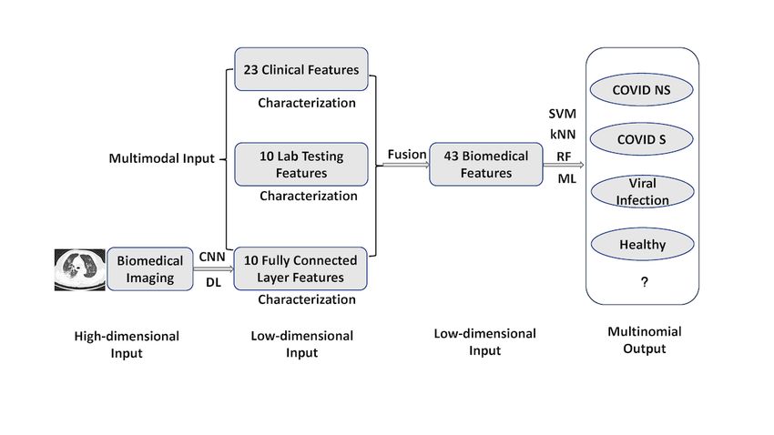
Figure 1. Multimodal feature late fusion and multinomial classification workflow. A deep learning convolutional neural network was applied to
computed tomography images for representation learning and extracting 10 features from a customized fully connected layer. These 10 features were
merged with other modality data through feature late fusion. In the machine learning stage of the workflow, each of the 3 machine learning models (ie,
the support vector machine, k-nearest neighbor, and random forest models) worked independently to provide their respective outputs. kNN: k-nearest
neighbor; ML: machine learning; RF: random forest; SVM; support vector machine.
are a type of deep neural network). In this study, we colloquially
The Multinomial Classification Objective used the term “machine learning” to refer to more traditional,
The main research goal of this study was to accurately non-DL types of ML (eg, RF ML), in contrast with DL that
differentiate between patients with severe COVID-19, patients focuses on deep neural networks. An important consideration
with nonsevere COVID-19, patients with non-COVID viral in the successful late fusion of multimodality features is the
infection, and noninfected healthy individuals from a total of representation learning of the high-dimensional CT features.
N participants all at once. Therefore, a formula was developed
to address the multinomial output classification problem. The For each CT scan of each participant, we constructed a
following equation uses 1 of the 4 mutually exclusive output customized residual neural network (ResNet) [36-39], which is
classes (ie, H=noninfected healthy, V=non-COVID viral a specific architecture for DL CNNs. A ResNet is considered a
pneumonia, NS=nonsevere COVID-19, and S=severe mature CNN architecture with relatively high performance
COVID-19) of an individual (ie, i), as follows: across different tasks. Although other CNN architectures exist
(eg, EfficientNet, VGG-16, etc), the focus of this study was not
f(Xc,Xl,Xm)i = {H,V,NS,S}, i = 1...N (1) to compare different architectures. Instead, we wanted to deliver
In this equation, the inputs were individuals’ (ie, i) multimodal the best performance possible with a commonly used CNN
features of binary clinical information (ie, Xc), continuous lab architecture (ie, ResNet) for image analysis.
test results (ie, Xl), and CT imaging (ie, Xm). The major By constructing a ResNet, we were able to transform the
advantage of our study was that we were able to classify 4 voxel-level imaging data into a high-level representation with
classes all at once, instead of developing several binary significantly fewer features. After several convolution and max
classifiers in parallel. pooling layers, the ResNet reached a fully connected (FC; ie,
FC1 layer) layer before the final output layer, thereby enabling
The Hybrid DL-ML Approach: Feature Late Fusion the delivery of actual classifications. In the commonly used
As stated earlier, the voxel level of CT imaging data does not ResNet architecture, the FC layer is a 1×512 vector, which is
integrate well with low-dimensional clinical and lab testing relatively closer in dimensionality to clinical information (ie,
features. In this study, we proposed a feature late fusion 1×23 vector) and lab testing (ie, 1×10 vector) feature modalities.
approach via the use of hybrid DL and ML models. Technically, However, the original FC layer from the ResNet was still much
DL is a type of ML that uses deep neural networks (eg, CNNs larger than the other 2 modalities. Therefore, we added another
http://www.jmir.org/2021/1/e25535/ J Med Internet Res 2021 | vol. 23 | iss. 1 | e25535 | p. 5
(page number not for citation purposes)
XSL• FO
RenderXJOURNAL OF MEDICAL INTERNET RESEARCH Xu et al
FC layer (ie, FC2 layer) after the FC1 layer, but before the final for the test set. We reported the overall performance of the ML
output layer. In this study, the FC2 layer was set to have a 1×10 models first. These different metrics evaluated ML models based
vector dimension (ie, 10 elements in the vector) to match the on different aspects. In this study, we also considered 3 different
dimensionality of the other 2 feature modalities. approaches for calculating the overall performance of
Computationally, the FC2 layer served as the low-dimensional, multinomial outputs, as follows: a micro approach (ie, the
high-level representation of the original CT scan data. The one-vs-all approach), a macro approach (ie, unweighted
distributions of the 10 features extracted from the ResNet in the averages; each of the 4 classes were given the same 25%
FC2 layer were compared across the 4 classes with the weights), and a weighted average approach based on the
Kolmogorov-Smirnov test. The technical details of this percentage of each class in the entire sample.
customized ResNet architecture are provided in Multimedia
In addition, because the output in this study was multinomial
Appendix 2.
instead of binary, each class had its own performance metrics.
Once low-dimensional high-level features were extracted from We aggregated these performance metrics across 100
CT data via the ResNet CNN, we performed multimodal feature independent runs, determined each metric’s distribution, and
fusion. The clinical information, lab testing, and FC2 layer evaluated model robustness based on these distributions. If ML
features of each participant (ie, i) were combined into a single performance metrics in the testing set had a small variation (ie,
1× 43 (ie, 1×[23+10+10]) row vector. The true values of the small standard errors), then the model was considered robust
output were the true observed classes of the participants. against model input changes, thereby allowing it to reveal the
Technically, the model would try to predict the outcome as intrinsic pattern of the data. This was because in each run, a
accurately as it could, based on the observed classes. different randomly selected dataset (ie, 80% of the original data)
was selected to train the model.
The Hybrid DL-ML Approach: Modeling
After deriving the feature matrix, we applied ML models for An advantage that the RF model had over SVM and kNN models
the multinomial classification task. In this study, 3 different was that it had relatively clearer interpretability, especially when
types of commonly used ML models were considered, as interpreting feature importance. After developing the RF model
follows: the RF, SVM, and kNN models. An RF model is a based on the training set, we were able to rank the importance
decision-tree–based ML model, and the number of tree of input features based on their corresponding Gini impurity
hyperparameters was set at 10, which is a relatively small score from the RF model [40,41]. It should be noted that only
number compared to the number of input features needed to the training set was used to compute Gini impurity, not the test
avoid potential model overfitting. Other RF hyperparameters set. We then assessed the top contributing features’ clinical
in this study included the Gini impurity score to determine tree relevance to COVID-19.
split, at least 2 samples to split an internal tree, and at least 1 We also developed and evaluated the performance of
sample at a leaf node. All default hyperparameter settings, single-modality (ie, using clinical information, lab testing, and
including those of the SVM and RF models, were based on the CT features individually) ML models. The performance results
scikit-learn library in Python. An SVM model is a model of were used as baseline conditions. The models’ performance
maximum hyperplane and L-2 penalty; radial basis function results were then compared to the multimodal classifications to
kernels and a gamma value of 1/43 (ie, the inverse of the total demonstrate the potential performance gain of the feature fusion
number of features) were used as hyperparameter values in this of different feature modalities. In this study, each individual
study. kNN is a nonparametric instance-based model; the ML model (ie, the RF, SVM, and kNN models) was
following hyperparameter values were used in this study: k=5, independently evaluated, and the respective results were
uniform weights, tree leaf size=30, and p=2. These 3 models reported, without combining the prediction of the final output
are technically distinct types of ML models. We aimed to class.
investigate whether specific types of ML models and multimodal
feature fusion would contribute to developing an accurate The deep learning CNN and late fusion machine learning codes
COVID-19 classifier for clinical decision support. were developed in Python with various supporting packages,
such as scikit-learn.
We evaluated each respective ML model with 100 independent
runs. Each run used a different randomly selected dataset Results
comprised of 80% of the original data for training, and the
remaining 20% of data were used to test and validate the model. Clinical Characterization of the 4 Classes
Performing multiple runs instead of a single run revealed how Detailed demographic, clinical, and lab testing results among
robust the model was, despite system stochasticity. The four classes were provided in supplementary Table S1. We
80%-20% split of the original data for separate training and compared clinical features across the 4 classes. The prevalence
testing sets also ensured that potential model overfitting and of each feature in all 4 classes is shown in Multimedia Appendix
increased model generalizability could be avoided. In addition, 3. In general, most clinical features varied substantially between
RF models use bagging for internal validation based on the nonsevere COVID-19, severe COVID-19, and non-COVID
out-of-bag errors (ie, how the “tree” would split out in the viral pneumonia classes. It should be noted that all symptom
“forest” model). feature values, except gender and age group (ie, >50 years)
After each run, important ML performance metrics, including values, in the noninfected healthy class were set to 0, so that
accuracy, sensitivity, precision, and F1 score, were computed they could be used as a reference. Based on the 2-sample z-test
http://www.jmir.org/2021/1/e25535/ J Med Internet Res 2021 | vol. 23 | iss. 1 | e25535 | p. 6
(page number not for citation purposes)
XSL• FO
RenderXJOURNAL OF MEDICAL INTERNET RESEARCH Xu et al
of proportions, the nonsevere COVID-19 and severe COVID-19 ratios and 95% confidence intervals for the forest plot are shown
classes differed significantly (PJOURNAL OF MEDICAL INTERNET RESEARCH Xu et al
distribution of hemoglobin levels is shown in Figure 3. Although testing features. The distribution of neutrophil count is shown
the noninfected healthy class differed significantly from the in Figure 3. All pairwise comparisons and the overall
nonsevere COVID-19, severe COVID-19, and non-COVID Kruskal-Wallis test showed significant differences between
viral infection class in terms of hemoglobin level, the other 3 classes in terms of lab testing features.
pairs did not show statistically significant differences in lab
Figure 3. Multiple comparisons of the top differentiating lab testing features. hs-CRP: high-sensitivity C-reactive protein.
COVID-19 and non-COVID viral infection classes and features
CT Differences Between the 4 Classes Based on 1, 4, and 5 between the noninfected healthy and non-COVID
High-Level CNN Features viral infection classes (Multimedia Appendix 6). Based on the
We analyzed the FC2 layer features from the ResNet CNN in RF model results, features 1, 6, and 10 were the 3 most critical
relation to the 4 classes. The corresponding boxplot is shown features in the FC2 layer with regard to multinomial
in Multimedia Appendix 5. The 2-sided Kolmogorov-Smirnov classification. Further Kruskal-Wallis tests were performed for
tests showed significant differences between every pair of these 3 features, and the results are shown in Figure 4. These
classes in almost all 10 CT features in the FC2 layer. The only results showed that developing an accurate classifier based on
exceptions were feature 6 (ie, CNN6) between the severe the CNN representation of high-level features is possible.
Figure 4. Multiple comparisons of the top differentiating CT features in the CNN. CNN: convolutional neural network; CT: computed tomography.
performance across all 4 classes based on the different
Accurate Multimodal Model for COVID-19 approaches for calculating the overall performance, including
Multinomial Classification the micro approach (ie, the one-vs-all approach), macro
We developed and validated 3 different types of ML models, approach (ie, unweighted averages across all 4 classes), and
as follows: the kNN, RF, and SVM models. With regard to weighted average approach (ie, based on percentage of each
training data, the average overall multimodal classification class in the entire sample). It should be noted that overall
accuracy of the kNN, RF, and SVM models was 96.2% (SE accuracy did not depend on sample size, so there was only 1
0.5%), 99.8% (SE 0.3%), and 99.2% (SE 0.2%), respectively. approach for calculating accuracy. The F1 score, sensitivity,
With regard to test data, the average overall multimodal and precision were quantified via each approach (ie, the micro,
classification accuracy of the 3 models was 95.4% (SE 0.2%), macro, and weighted average approaches). The F1 scores that
96.9% (SE 0.2%), and 97.7% (SE 0.1%), respectively (Figure were calculated using the macro approach were 95.9% (SE
5). These 3 models also achieved consistent and high 0.1%), 98.8% (SEJOURNAL OF MEDICAL INTERNET RESEARCH Xu et al
RF, and SVM models, respectively. The F1 scores that were approaches) were minimal (Figure 5). In addition, the
calculated using the micro approach was 96.2% (SEJOURNAL OF MEDICAL INTERNET RESEARCH Xu et al
With regard to each individual class, the noninfected healthy accuracy, 92.9%-96.8% F1 score, 95.4%-98.8% sensitivity, and
class had a 95.2%-99.9% prediction accuracy, 95.5%-98.4% 90.6%-95.0% precision. The non-COVID viral infection class
F1 score, 91.4%-97.3% sensitivity, and 97.5%-99.9% precision was relatively more challenging to differentiate from the other
in the testing set, depending on the specific ML model used. It 3 classes, but the difference was not substantial. Therefore, the
should be noted these are ranges, not standard errors, as shown potential clinical use of the ML models is still justified. Similar
in Figure 6. The approach to computing class-specific model to the results of overall model performance (Figure 5),
performance was the one-vs-all approach. With regard to the class-specific performance metrics also had relatively small
nonsevere COVID-19 class, ML models achieved a standard errors, indicating that the training of models was
95.8%-97.4% accuracy, 97.8%-98.6% F1 score, 99.8%-99.9% consistent and robust against randomly selected inputs. Except
sensitivity, and 95.8%-97.4% precision. With regard to the for a few classes and model performance metrics, the SVM
severe COVID-19 class, ML models achieved a 92.4%-99.0% model performed slightly better than the RF and kNN models.
accuracy, 93.4%-96.6% F1 score, 94.3%-94.7% sensitivity, and The complete class-specific results are shown in Figure 6. The
92.4%-99.0% precision. With regard to the non-COVID viral complete class-specific performance metrics across the 3 ML
pneumonia infection class, ML models achieved a 90.6%-95.0% models are shown in Multimedia Appendix 8.
Figure 6. Class-specific performance of machine learning models. kNN: k nearest neighbor; RF: random forest; SVM: support vector machine.
All 3 ML multinomial classification models, which were based overall performance (Figure 5, Table S3) and high performance
on different computational techniques, had consistently high for each specific class (Figure 6, Multimedia Appendix 8). Of
http://www.jmir.org/2021/1/e25535/ J Med Internet Res 2021 | vol. 23 | iss. 1 | e25535 | p. 10
(page number not for citation purposes)
XSL• FO
RenderXJOURNAL OF MEDICAL INTERNET RESEARCH Xu et al
the 3 types of ML models developed and evaluated, the SVM C-reactive protein level, hemoglobin level, and absolute
model was marginally better than the RF and kNN models. As neutrophil count. The distribution of these 3 features across the
a result, the ML multinomial classification models were able 4 classes and the results of multiple comparisons are shown in
to accurately differentiate between the 4 classes all at once, Figure 3. Although high-sensitivity C-reactive protein level is
provide accurate and detailed class-specific predictions, and act a known factor for COVID-19 severity and prognosis [42], we
as reliable decision-making tools for clinical diagnostic support showed that it could also differentiate patients with COVID-19
and the triaging of patients with suspected COVID-19, who from patients with non-COVID viral pneumonia and healthy
might or might not be infected with a clinically similar type of individuals. In addition, we learned that different hemoglobin
virus other than SARS-CoV-2. and neutrophil levels were novel features for accurately
distinguishing between patients with clinical COVID-19,
In addition to the multimodal classification that incorporated
patients with non-COVID viral pneumonia, and healthy
all 3 different feature sets (ie, binary clinical, continuous lab
individuals. These results shed light on which set of clinical
testing, and CT features in the ResNet CNN; Figure 1), we also
and lab testing features are the most critical in identifying
tested how each specific feature modality performed without
COVID-19, which will help guide clinical practice. With regard
feature fusion (ie, unimodality). By using each of the 23
to the CT features extracted from the CNN, the RF models
symptom features alone, the RF, kNN, and SVM models
identified the top 3 influential features, which were CT features
achieved an average accuracy of 74.5% (SE 0.3%), 73.3% (SE
6, 10, and 1 in the 10-element FC2 layer (Figure 4). Although
0.3%), and 75.5% (SE 0.3%) with the testing set, respectively.
the actual clinical interpretation of CT features was not clear at
By using each of the 10 lab testing features alone, the RF, kNN,
the time of this study due to the nature of DL models, including
and SVM models achieved an average accuracy of 67.7% (SE
the ResNet CNN applied in this study, these features showed
0.4%), 56.2% (SE 0.4%), and 59.5% (SE 0.3%) with the testing
promise in accurately differentiating between multinomial
set, respectively.
classes all at once via CT scans, instead of training several
The overall accuracy of the CNN with CT scan data alone was CNNs for binary classifications between each class pair. Future
90.8% (SE 0.3%) across the 4 classes. With regard to each pair research might reveal the clinical relevance of these features in
of classes, the CNN was able to accurately differentiate between a more interpretable way with COVID-19 pathology data.
the severe COVID-19 and noninfected healthy classes with
99.9% (SE50 years). The forest and understanding with the disease may vary substantially.
plots of odds ratios for these features are provided in Figure 2,
which shows the exact influence that these features had across Upon the further examination of comprehensive patient
classes. With regard to lab testing features, the top 3 most symptom data, we believed that our current understanding and
influential features, in descending order, were high-sensitivity definition of asymptomatic COVID-19 would be inadequate.
http://www.jmir.org/2021/1/e25535/ J Med Internet Res 2021 | vol. 23 | iss. 1 | e25535 | p. 11
(page number not for citation purposes)
XSL• FO
RenderXJOURNAL OF MEDICAL INTERNET RESEARCH Xu et al
Of the 214 patients with nonsevere COVID-19, 60 (28%) had participants [38]. In addition, professionally trained human
no fever (ie, 97%) in the testing set.
diabetes and COVID-19 actually mutually influence each other
We compared the performance of our model, which was based and result in undesirable clinical prognoses. Future studies can
on the multimodal biomedical data of 683 participants, against use data-driven methods to further investigate the causality of
the performance of state-of-the-art benchmarks in COVID-19 comorbidities and COVID-19.
classification studies. A DL study that involved thoracic CT
There are some limitations in this study and potential
scans for 87 participants claimed to have >99% accuracy [37],
improvements for future research. For instance, to perform
and another study with 200 participants claimed to have
multinomial classification across the 4 classes, we had to discard
86%-99% accuracy in differentiating between individuals with
a lot of features, especially those in the lab testing modality.
and without COVID-19 [36]. Another study reported a 95%
The non-COVID viral pneumonia class used a different
area under the curve for differentiating between COVID-19 and
electronic health record system that collected different lab
other community-acquired pneumonia diseases in 3322
testing features from participants in Wuhan (ie, participants in
participants [39]. Furthermore, a 92% area under the curve was
the severe COVID-19, nonsevere COVID-19, and noninfected
achieved in a study of 905 participants with and without
healthy classes). Many lab testing features were able to
COVID-19 by using multimodal CT, clinical, and lab testing
accurately differentiate between severe and nonsevere
information [44]. A study that used CT scans to differentiate
COVID-19 in our preliminary study, such as high-sensitivity
between 3 multinomial classes (ie, the COVID [no clinical state
Troponin I level, D-dimer level, and lactate dehydrogenase
information], non-COVID viral pneumonia, and healthy classes)
level. However, these features were not present, or largely
achieved an 89%-96% accuracy based on a total of 230
missing, in the non-COVID viral infection class. Eventually,
http://www.jmir.org/2021/1/e25535/ J Med Internet Res 2021 | vol. 23 | iss. 1 | e25535 | p. 12
(page number not for citation purposes)
XSL• FO
RenderXJOURNAL OF MEDICAL INTERNET RESEARCH Xu et al
only 10 lab testing features were included, which is small different in people of other races and ethnicities, or people with
compared to the average of 20-30 features that are usually other confounding factors. The cross-validation of the findings
available in different electronic health record systems. This is in this study based on other ethnicity groups and larger sample
probably the reason why the lab testing feature modality alone sizes is needed for future research.
was not able to provide accurate classifications (ie, the highest
This study used a common CNN architecture (ie, a ResNet).
accuracy achieved was 67.7% with the RF model) across all 4
The 10 CT features extracted from the FC2 layer of the ResNet
classes in this study. In addition, although we had a reasonably
were used to match the dimensionality of the other 2
large participant pool of 638 individuals, more participants are
low-dimensional feature modalities. Future research on different
needed to further validate the findings of this study.
disease systems can explore and compare other architectures
Another potential practical pitfall was that not all feature that use different biomedical imaging data (eg, CT, X-ray, and
modalities were readily available at the same time for feature histology data). The actual dimensionality of the FC2 layer can
fusion and multimodal classification. With regard to also be optimized to deliver better performance. Finally, this
single-modality features, CT had the best performance in study presented the results of individual classification models.
generating accurate predictions. However, CT is usually To achieve even higher performance, the combination of
performed in the radiology department. Lab testing may be multiple models can be explored in future studies.
outsourced, and obtaining lab test results takes time.
Consequently, there might be lags in data availability among
Conclusion
different feature modalities. We believe that when multimodal In summary, different biomedical information across different
features are not available all at once, single-modality features modalities, such as clinical information, lab testing results, and
can be used to perform first-round triaging. Multimodal features CT scans, work synergistically to reveal the multifaceted nature
are needed when accuracy is a must. of COVID-19 pathology. Our ML and DL models provided a
feasible technical method for working directly with multimodal
It should be noted that although the participants in this study biomedical data and differentiating between patients with severe
came from different health care facilities, the majority of them COVID-19, patients with nonsevere COVID-19, patients with
were of Chinese Han ethnicity. The biomedical features among non-COVID viral infection, and noninfected healthy individuals
the different COVID-19 and non-COVID classes may be at the same time, with >97% accuracy.
Acknowledgments
This study is dedicated to the frontline clinicians and other supporting personnel who have fought against COVID-19 worldwide.
This study is supported by the National Science Foundation for Young Scientists of China (81703201, 81602431, and 81871544),
the North Carolina Biotechnology Center Flash Grant on COVID-19 Clinical Research (2020-FLG-3898), the Natural Science
Foundation for Young Scientists of Jiangsu Province (BK20171076, BK20181488, BK20181493, and BK20201485), the Jiangsu
Provincial Medical Innovation Team (CXTDA2017029), the Jiangsu Provincial Medical Youth Talent program (QNRC2016548
and QNRC2016536), the Jiangsu Preventive Medicine Association program (Y2018086 and Y2018075), the Lifting Program of
Jiangsu Provincial Scientific and Technological Association, and the Jiangsu Government Scholarship for Overseas Studies.
Authors' Contributions
JL (liu_jie0823@163.com), BZ (zhubl@jscdc.cn), and SC (schen56@uncc.edu) serve as corresponding authors of this study
equally.
Conflicts of Interest
None declared.
Multimedia Appendix 1
Demographic, clinical, and lab testing results of the 4 Classes.
[DOCX File , 18 KB-Multimedia Appendix 1]
Multimedia Appendix 2
Architecture of the customized ResNet-18 CNN and sample computed tomography scans of the 4 classes. CNN: convolutional
neural networkl ResNet: residual neural network.
[PNG File , 433 KB-Multimedia Appendix 2]
Multimedia Appendix 3
Comparison of clinical features across the 4 classes.
[PNG File , 122 KB-Multimedia Appendix 3]
http://www.jmir.org/2021/1/e25535/ J Med Internet Res 2021 | vol. 23 | iss. 1 | e25535 | p. 13
(page number not for citation purposes)
XSL• FO
RenderXJOURNAL OF MEDICAL INTERNET RESEARCH Xu et al
Multimedia Appendix 4
Comparison of lab testing features across the 4 classes.
[PNG File , 97 KB-Multimedia Appendix 4]
Multimedia Appendix 5
Computed tomography features extracted via a deep learning convolutional neural network and compared across the 4 classes.
[PNG File , 131 KB-Multimedia Appendix 5]
Multimedia Appendix 6
z-test and Kolmogorov-Smirnov test results of significance for each biomedical feature among the 4 classes.
[DOCX File , 21 KB-Multimedia Appendix 6]
Multimedia Appendix 7
Overall machine learning model performance comparison.
[DOCX File , 15 KB-Multimedia Appendix 7]
Multimedia Appendix 8
Class-specific machine learning model performance comparison.
[DOCX File , 15 KB-Multimedia Appendix 8]
References
1. Coronavirus Disease 2019 (COVID-19) Situation Report - 203. World Health Organization. URL: https://www.who.int/
docs/default-source/coronaviruse/situation-reports/20200810-covid-19-sitrep-203.pdf?sfvrsn=aa050308_2 [accessed
2020-12-22]
2. Novel coronavirus pneumonia diagnosis and treatment plan. National Health Commission of China. 2020. URL: http:/
/www.nhc.gov.cn/yzygj/s7652m/202003/a31191442e29474b98bfed5579d5af95.shtml [accessed 2020-12-22]
3. Xiao SY, Wu Y, Liu H. Evolving status of the 2019 novel coronavirus infection: Proposal of conventional serologic assays
for disease diagnosis and infection monitoring. J Med Virol 2020 May;92(5):464-467 [FREE Full text] [doi:
10.1002/jmv.25702] [Medline: 32031264]
4. Wang Y, Kang H, Liu X, Tong Z. Combination of RT-qPCR testing and clinical features for diagnosis of COVID-19
facilitates management of SARS-CoV-2 outbreak. J Med Virol 2020 Jun;92(6):538-539 [FREE Full text] [doi:
10.1002/jmv.25721] [Medline: 32096564]
5. Böger B, Fachi MM, Vilhena RO, Cobre AF, Tonin FS, Pontarolo R. Systematic review with meta-analysis of the accuracy
of diagnostic tests for COVID-19. Am J Infect Control. Epub ahead of print 2020 Jul 10 [FREE Full text] [doi:
10.1016/j.ajic.2020.07.011] [Medline: 32659413]
6. Zhou L, Li Z, Zhou J, Li H, Chen Y, Huang Y, et al. A Rapid, Accurate and Machine-Agnostic Segmentation and
Quantification Method for CT-Based COVID-19 Diagnosis. IEEE Trans Med Imaging 2020 Aug;39(8):2638-2652. [doi:
10.1109/TMI.2020.3001810] [Medline: 32730214]
7. Fu Y, Zhu R, Bai T, Han P, He Q, Jing M, et al. Clinical Features of COVID-19-Infected Patients With Elevated Liver
Biochemistries: A Multicenter, Retrospective Study. Hepatology. Epub ahead of print 2020 Jun 30 [FREE Full text] [doi:
10.1002/hep.31446] [Medline: 32602604]
8. Shi H, Han X, Jiang N, Cao Y, Alwalid O, Gu J, et al. Radiological findings from 81 patients with COVID-19 pneumonia
in Wuhan, China: a descriptive study. Lancet Infect Dis 2020 Apr;20(4):425-434 [FREE Full text] [doi:
10.1016/S1473-3099(20)30086-4] [Medline: 32105637]
9. Ojha V, Mani A, Pandey NN, Sharma S, Kumar S. CT in coronavirus disease 2019 (COVID-19): a systematic review of
chest CT findings in 4410 adult patients. Eur Radiol 2020 Nov;30(11):6129-6138 [FREE Full text] [doi:
10.1007/s00330-020-06975-7] [Medline: 32474632]
10. Lyu P, Liu X, Zhang R, Shi L, Gao J. The Performance of Chest CT in Evaluating the Clinical Severity of COVID-19
Pneumonia: Identifying Critical Cases Based on CT Characteristics. Invest Radiol 2020 Jul;55(7):412-421 [FREE Full text]
[doi: 10.1097/RLI.0000000000000689] [Medline: 32304402]
11. Brooks M. How accurate is self-testing? New Sci 2020 May 16;246(3282):10 [FREE Full text] [doi:
10.1016/S0262-4079(20)30909-X] [Medline: 32501335]
12. Wu Z, McGoogan JM. Characteristics of and Important Lessons From the Coronavirus Disease 2019 (COVID-19) Outbreak
in China: Summary of a Report of 72 314 Cases From the Chinese Center for Disease Control and Prevention. JAMA 2020
Apr 07;323(13):1239-1242. [doi: 10.1001/jama.2020.2648] [Medline: 32091533]
http://www.jmir.org/2021/1/e25535/ J Med Internet Res 2021 | vol. 23 | iss. 1 | e25535 | p. 14
(page number not for citation purposes)
XSL• FO
RenderXJOURNAL OF MEDICAL INTERNET RESEARCH Xu et al
13. Truog RD, Mitchell C, Daley GQ. The Toughest Triage - Allocating Ventilators in a Pandemic. N Engl J Med 2020 May
21;382(21):1973-1975. [doi: 10.1056/NEJMp2005689] [Medline: 32202721]
14. Fang Y, Zhang H, Xie J, Lin M, Ying L, Pang P, et al. Sensitivity of Chest CT for COVID-19: Comparison to RT-PCR.
Radiology 2020 Aug;296(2):E115-E117 [FREE Full text] [doi: 10.1148/radiol.2020200432] [Medline: 32073353]
15. Menni C, Valdes AM, Freidin MB, Sudre CH, Nguyen LH, Drew DA, et al. Real-time tracking of self-reported symptoms
to predict potential COVID-19. Nat Med 2020 Jul;26(7):1037-1040. [doi: 10.1038/s41591-020-0916-2] [Medline: 32393804]
16. Timmers T, Janssen L, Stohr J, Murk JL, Berrevoets MAH. Using eHealth to Support COVID-19 Education, Self-Assessment,
and Symptom Monitoring in the Netherlands: Observational Study. JMIR Mhealth Uhealth 2020 Jun 23;8(6):e19822 [FREE
Full text] [doi: 10.2196/19822] [Medline: 32516750]
17. Nair A, Rodrigues JCL, Hare S, Edey A, Devaraj A, Jacob J, et al. A British Society of Thoracic Imaging statement:
considerations in designing local imaging diagnostic algorithms for the COVID-19 pandemic. Clin Radiol 2020
May;75(5):329-334 [FREE Full text] [doi: 10.1016/j.crad.2020.03.008] [Medline: 32265036]
18. Sun Y, Koh V, Marimuthu K, Ng O, Young B, Vasoo S, National Centre for Infectious Diseases COVID-19 Outbreak
Research Team. Epidemiological and Clinical Predictors of COVID-19. Clin Infect Dis 2020 Jul 28;71(15):786-792 [FREE
Full text] [doi: 10.1093/cid/ciaa322] [Medline: 32211755]
19. Brinati D, Campagner A, Ferrari D, Locatelli M, Banfi G, Cabitza F. Detection of COVID-19 Infection from Routine Blood
Exams with Machine Learning: A Feasibility Study. J Med Syst 2020 Jul 01;44(8):135 [FREE Full text] [doi:
10.1007/s10916-020-01597-4] [Medline: 32607737]
20. Daniells JK, MacCallum HL, Durrheim DN. Asymptomatic COVID-19 or are we missing something? Commun Dis Intell
(2018) 2020 Jul 09;44:1-5 [FREE Full text] [doi: 10.33321/cdi.2020.44.55] [Medline: 32640950]
21. Gandhi M, Yokoe DS, Havlir DV. Asymptomatic Transmission, the Achilles' Heel of Current Strategies to Control Covid-19.
N Engl J Med 2020 May 28;382(22):2158-2160 [FREE Full text] [doi: 10.1056/NEJMe2009758] [Medline: 32329972]
22. Shi F, Yu Q, Huang W, Tan C. 2019 Novel Coronavirus (COVID-19) Pneumonia with Hemoptysis as the Initial Symptom:
CT and Clinical Features. Korean J Radiol 2020 May;21(5):537-540 [FREE Full text] [doi: 10.3348/kjr.2020.0181] [Medline:
32174057]
23. Li X, Fang X, Bian Y, Lu J. Comparison of chest CT findings between COVID-19 pneumonia and other types of viral
pneumonia: a two-center retrospective study. Eur Radiol 2020 Oct;30(10):5470-5478 [FREE Full text] [doi:
10.1007/s00330-020-06925-3] [Medline: 32394279]
24. Altmayer S, Zanon M, Pacini GS, Watte G, Barros MC, Mohammed TL, et al. Comparison of the computed tomography
findings in COVID-19 and other viral pneumonia in immunocompetent adults: a systematic review and meta-analysis. Eur
Radiol 2020 Dec;30(12):6485-6496 [FREE Full text] [doi: 10.1007/s00330-020-07018-x] [Medline: 32594211]
25. Qu J, Chang LK, Tang X, Du Y, Yang X, Liu X, et al. Clinical characteristics of COVID-19 and its comparison with
influenza pneumonia. Acta Clin Belg 2020 Oct;75(5):348-356. [doi: 10.1080/17843286.2020.1798668] [Medline: 32723027]
26. Ooi EE, Low JG. Asymptomatic SARS-CoV-2 infection. Lancet Infect Dis 2020 Sep;20(9):996-998 [FREE Full text] [doi:
10.1016/S1473-3099(20)30460-6] [Medline: 32539989]
27. Baltruschat IM, Nickisch H, Grass M, Knopp T, Saalbach A. Comparison of Deep Learning Approaches for Multi-Label
Chest X-Ray Classification. Sci Rep 2019 Apr 23;9(1):6381 [FREE Full text] [doi: 10.1038/s41598-019-42294-8] [Medline:
31011155]
28. Ai T, Yang Z, Hou H, Zhan C, Chen C, Lv W, et al. Correlation of Chest CT and RT-PCR Testing for Coronavirus Disease
2019 (COVID-19) in China: A Report of 1014 Cases. Radiology 2020 Aug;296(2):E32-E40 [FREE Full text] [doi:
10.1148/radiol.2020200642] [Medline: 32101510]
29. Song S, Wu F, Liu Y, Jiang H, Xiong F, Guo X, et al. Correlation Between Chest CT Findings and Clinical Features of
211 COVID-19 Suspected Patients in Wuhan, China. Open Forum Infect Dis 2020 Jun;7(6):ofaa171 [FREE Full text] [doi:
10.1093/ofid/ofaa171] [Medline: 32518804]
30. Wu J, Wu X, Zeng W, Guo D, Fang Z, Chen L, et al. Chest CT Findings in Patients With Coronavirus Disease 2019 and
Its Relationship With Clinical Features. Invest Radiol 2020 May;55(5):257-261 [FREE Full text] [doi:
10.1097/RLI.0000000000000670] [Medline: 32091414]
31. Meng H, Xiong R, He R, Lin W, Hao B, Zhang L, et al. CT imaging and clinical course of asymptomatic cases with
COVID-19 pneumonia at admission in Wuhan, China. J Infect 2020 Jul;81(1):e33-e39 [FREE Full text] [doi:
10.1016/j.jinf.2020.04.004] [Medline: 32294504]
32. Cheng Z, Lu Y, Cao Q, Qin L, Pan Z, Yan F, et al. Clinical Features and Chest CT Manifestations of Coronavirus Disease
2019 (COVID-19) in a Single-Center Study in Shanghai, China. AJR Am J Roentgenol 2020 Jul;215(1):121-126. [doi:
10.2214/AJR.20.22959] [Medline: 32174128]
33. Zhu Y, Liu YL, Li ZP, Kuang JY, Li XM, Yang YY, et al. Clinical and CT imaging features of 2019 novel coronavirus
disease (COVID-19). J Infect. Epub ahead of print 2020 Mar 03 [FREE Full text] [doi: 10.1016/j.jinf.2020.02.022] [Medline:
32142928]
34. Baltrusaitis T, Ahuja C, Morency L. Multimodal Machine Learning: A Survey and Taxonomy. IEEE Trans Pattern Anal
Mach Intell 2019 Feb;41(2):423-443. [doi: 10.1109/TPAMI.2018.2798607] [Medline: 29994351]
http://www.jmir.org/2021/1/e25535/ J Med Internet Res 2021 | vol. 23 | iss. 1 | e25535 | p. 15
(page number not for citation purposes)
XSL• FO
RenderXJOURNAL OF MEDICAL INTERNET RESEARCH Xu et al
35. Metlay JP, Waterer GW, Long AC, Anzueto A, Brozek J, Crothers K, et al. Diagnosis and Treatment of Adults with
Community-acquired Pneumonia. An Official Clinical Practice Guideline of the American Thoracic Society and Infectious
Diseases Society of America. Am J Respir Crit Care Med 2019 Oct 01;200(7):e45-e67 [FREE Full text] [doi:
10.1164/rccm.201908-1581ST] [Medline: 31573350]
36. Ardakani AA, Kanafi AR, Acharya UR, Khadem N, Mohammadi A. Application of deep learning technique to manage
COVID-19 in routine clinical practice using CT images: Results of 10 convolutional neural networks. Comput Biol Med
2020 Jun;121:103795 [FREE Full text] [doi: 10.1016/j.compbiomed.2020.103795] [Medline: 32568676]
37. Ko H, Chung H, Kang WS, Kim KW, Shin Y, Kang SJ, et al. COVID-19 Pneumonia Diagnosis Using a Simple 2D Deep
Learning Framework With a Single Chest CT Image: Model Development and Validation. J Med Internet Res 2020 Jun
29;22(6):e19569 [FREE Full text] [doi: 10.2196/19569] [Medline: 32568730]
38. Hu S, Gao Y, Niu Z, Jiang Y, Li L, Xiao X, et al. Weakly Supervised Deep Learning for COVID-19 Infection Detection
and Classification From CT Images. IEEE Access 2020;8:118869-118883. [doi: 10.1109/ACCESS.2020.3005510]
39. Li L, Qin L, Xu Z, Yin Y, Wang X, Kong B, et al. Using Artificial Intelligence to Detect COVID-19 and Community-acquired
Pneumonia Based on Pulmonary CT: Evaluation of the Diagnostic Accuracy. Radiology 2020 Aug;296(2):E65-E71 [FREE
Full text] [doi: 10.1148/radiol.2020200905] [Medline: 32191588]
40. Chen Y, Ouyang L, Bao FS, Li Q, Han L, Zhu B, et al. An Interpretable Machine Learning Framework for Accurate Severe
vs Non-severe COVID-19 Clinical Type Classification. medRxiv. Preprint posted online on May 22.. [doi:
10.1101/2020.05.18.20105841]
41. Elaziz MA, Hosny KM, Salah A, Darwish MM, Lu S, Sahlol AT. New machine learning method for image-based diagnosis
of COVID-19. PLoS One 2020;15(6):e0235187 [FREE Full text] [doi: 10.1371/journal.pone.0235187] [Medline: 32589673]
42. Wang K, Zuo P, Liu Y, Zhang M, Zhao X, Xie S, et al. Clinical and Laboratory Predictors of In-hospital Mortality in
Patients With Coronavirus Disease-2019: A Cohort Study in Wuhan, China. Clin Infect Dis 2020 Nov 19;71(16):2079-2088
[FREE Full text] [doi: 10.1093/cid/ciaa538] [Medline: 32361723]
43. Ferrari D, Motta A, Strollo M, Banfi G, Locatelli M. Routine blood tests as a potential diagnostic tool for COVID-19. Clin
Chem Lab Med 2020 Jun 25;58(7):1095-1099. [doi: 10.1515/cclm-2020-0398] [Medline: 32301746]
44. Mei X, Lee HC, Diao KY, Huang M, Lin B, Liu C, et al. Artificial intelligence-enabled rapid diagnosis of patients with
COVID-19. Nat Med 2020 Aug;26(8):1224-1228 [FREE Full text] [doi: 10.1038/s41591-020-0931-3] [Medline: 32427924]
45. Bai HX, Hsieh B, Xiong Z, Halsey K, Choi JW, Tran TML, et al. Performance of Radiologists in Differentiating COVID-19
from Non-COVID-19 Viral Pneumonia at Chest CT. Radiology 2020 Aug;296(2):E46-E54 [FREE Full text] [doi:
10.1148/radiol.2020200823] [Medline: 32155105]
46. Tárnok A. Machine Learning, COVID-19 (2019-nCoV), and multi-OMICS. Cytometry A 2020 Mar;97(3):215-216 [FREE
Full text] [doi: 10.1002/cyto.a.23990] [Medline: 32142596]
47. Ray S, Srivastava S. COVID-19 Pandemic: Hopes from Proteomics and Multiomics Research. OMICS 2020
Aug;24(8):457-459. [doi: 10.1089/omi.2020.0073] [Medline: 32427517]
48. Arga KY. COVID-19 and the Futures of Machine Learning. OMICS 2020 Sep;24(9):512-514. [doi: 10.1089/omi.2020.0093]
[Medline: 32511048]
49. Wicaksana AL, Hertanti NS, Ferdiana A, Pramono RB. Diabetes management and specific considerations for patients with
diabetes during coronavirus diseases pandemic: A scoping review. Diabetes Metab Syndr 2020;14(5):1109-1120 [FREE
Full text] [doi: 10.1016/j.dsx.2020.06.070] [Medline: 32659694]
50. Abdi A, Jalilian M, Sarbarzeh PA, Vlaisavljevic Z. Diabetes and COVID-19: A systematic review on the current evidences.
Diabetes Res Clin Pract 2020 Aug;166:108347 [FREE Full text] [doi: 10.1016/j.diabres.2020.108347] [Medline: 32711003]
51. Madjid M, Safavi-Naeini P, Solomon SD, Vardeny O. Potential Effects of Coronaviruses on the Cardiovascular System:
A Review. JAMA Cardiol 2020 Jul 01;5(7):831-840. [doi: 10.1001/jamacardio.2020.1286] [Medline: 32219363]
52. Matsushita K, Marchandot B, Jesel L, Ohlmann P, Morel O. Impact of COVID-19 on the Cardiovascular System: A Review.
J Clin Med 2020 May 09;9(5):1407 [FREE Full text] [doi: 10.3390/jcm9051407] [Medline: 32397558]
Abbreviations
CT: computed tomography
CNN: convolutional neural network
DL: deep learning
DICOM: Digital Imaging and Communications in Medicine
FC: fully connected
GGO: ground-glass opacity
kNN: k-nearest neighbor
MERS: Middle East respiratory syndrome
ML: machine learning
qRT-PCR: quantitative real-time polymerase chain reaction
RF: random forest
http://www.jmir.org/2021/1/e25535/ J Med Internet Res 2021 | vol. 23 | iss. 1 | e25535 | p. 16
(page number not for citation purposes)
XSL• FO
RenderXYou can also read