CNTNAP2 is targeted to endosomes by the polarity protein Par3 - bioRxiv
←
→
Page content transcription
If your browser does not render page correctly, please read the page content below
bioRxiv preprint first posted online Apr. 6, 2019; doi: http://dx.doi.org/10.1101/601575. The copyright holder for this preprint (which
       was not peer-reviewed) is the author/funder, who has granted bioRxiv a license to display the preprint in perpetuity.
                                      All rights reserved. No reuse allowed without permission.

                           CNTNAP2 is targeted to endosomes by the polarity
                                            protein Par3

                         Ruoqi Gao, Ph.D.a , Christopher P. Pratt, Ph.D.a,1 , Sehyoun Yoon, Ph.D.a,1 ,
                          Maria Dolores Martin-de-Saavedra, Ph.D.a , Marc P. Forrest, Ph.D.a , Peter
                                                     Penzes, Ph.D.a,b,c,∗
                        a Department  of Physiology, Northwestern University Feinberg School of Medicine, Chicago,
                                                              IL, 60611 USA
                           b Department of Psychiatry and Behavioral Sciences, Northwestern University Feinberg

                                                School of Medicine, Chicago, IL, 60611 USA
                          c Northwestern University Center for Autism and Neurodevelopment, Chicago IL 60611,

                                                                    USA

                        Abstract

                        A decade of genetic studies has established Contactin-associated protein-like 2
                        (CNTNAP2 ) as a prominent susceptibility gene associated with multiple neu-
                        rodevelopmental disorders. The development and characterization of Cntnap2
                        knockout models in multiple species have bolstered this claim by establishing
                        clear connections with certain endophenotypes. Despite these remarkable in
                        vivo findings, CNTNAP2’s molecular functions are relatively unexplored, high-
                        lighting the need to identify novel protein partners. Here, we characterized
                        an interaction between CNTNAP2 and Partitioning-defective 3 (Par3) – a po-
                        larity molecule we isolated in a yeast-two hybrid screen with CNTNAP2’s C-
                        terminus. We provide evidence that the two proteins interact via PDZ domain-
                        mediated binding, that CNTNAP2+ /Par3+ complexes are largely associated
                        with clathrin-coated endocytic vesicles, and that Par3 causes an enlargement of
                        these structures. Live imaging and fluorescence recovery after photobleaching
                        (FRAP) reveals that Par3 limits the mobility of CNTNAP2 at endosomes, thus
                        stabilizing it at that location. Finally, expression of Par3 but not Par3∆PDZ can
                        cluster endogenous CNTNAP2 in primary neurons. Collectively, we conclude

                           ∗ Corresponding   author
                           1 Equal   contribution

                        Preprint submitted to Molecular and Cellular Neuroscience                             April 6, 2019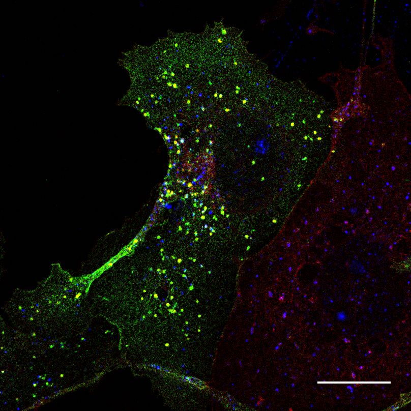
bioRxiv preprint first posted online Apr. 6, 2019; doi: http://dx.doi.org/10.1101/601575. The copyright holder for this preprint (which
       was not peer-reviewed) is the author/funder, who has granted bioRxiv a license to display the preprint in perpetuity.
                                      All rights reserved. No reuse allowed without permission.

                        that Par3 regulates CNTNAP2 spatial localization to endocytic compartments.

                        1. Introduction

                            CNTNAP2 is a prominent disease susceptibility gene associated with Autism
                        Spectrum Disorder (ASD), Intellectual Disability (ID), Schizophrenia (SCZ),
                        and epilepsy, and is causative for Cortical Dysplasia Focal Epilepsy (CDFE)
                        syndrome (Rodenas-Cuadrado et al., 2014). Similarly, Cntnap2 knockout mice,
                        zebrafish, and rats have recapitulated several disease endophenotypes, including
                        impaired social interaction, communication, and seizures (Penagarikano et al.,
                        2011; Hoffman et al., 2016; Thomas et al., 2017). CNTNAP2 encodes a type
                        I transmembrane cell adhesion molecule that has a wide range of functions,
                        including clustering potassium channels, migration, and dendritic stabilization
                        (Rodenas-Cuadrado et al., 2014). Moreover, multiple reports have shown that
                        CNTNAP2, as well as close family members, are more prominently expressed
                        in interneurons and preferentially affect interneuronal function (Mo et al., 2015;
                        Karayannis et al., 2014; Gao et al., 2018). Indeed, interneuron dysfunction may
                        be key factor for behavioral endophenotypes in Cntnap2 null mice (Jurgensen
                        and Castillo, 2015; Bridi et al., 2017; Vogt et al., 2014); however, the cellular
                        and molecular mechanisms remain poorly understood.
                            Polarity is an organizing process that is essential for cellular development,
                        allowing cells to form asymmetrical specializations critical for cellular differen-
                        tiation (Goldstein and Macara, 2007). The partitioning defective (Par) proteins
                        participate in an evolutionarily-conserved complex which initiates many of these
                        essential processes through the interaction of its three members – Partition-
                        defective protein 3 (Par3, a.k.a. PARD3), Partitioning-defective 6 (Par6), and
                        atypical protein kinase C (aPKC) – and downstream guanine nucleotide ex-
                        change factors (Johansson et al., 2000; Lin et al., 2000). Through this interplay,
                        Par3 assists in regulation of multiple polarity events including apical-basal es-
                        tablishment in epithelial cells, axon specification in neurons, asymmetric mitotic
                        spindle positioning during cell division, and astrocyte migration (Mertens et al.,

                                                                          2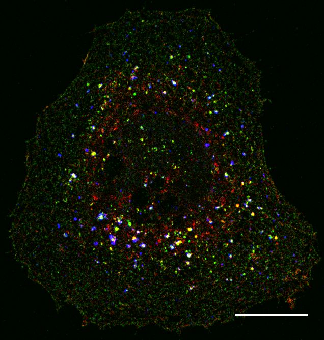
bioRxiv preprint first posted online Apr. 6, 2019; doi: http://dx.doi.org/10.1101/601575. The copyright holder for this preprint (which
       was not peer-reviewed) is the author/funder, who has granted bioRxiv a license to display the preprint in perpetuity.
                                      All rights reserved. No reuse allowed without permission.

                        2006; Macara, 2004). Conversely, Par3 dysfunction leads to loss of polar organi-
                        zation, abnormal GTPase signaling, and carcinogenesis/metastasis in some cell
                        types (McCaffrey et al., 2012).
                            Other than its canonical role with Par6 and aPKC, Par3 also has Par
                        complex-independent functions. For example, Par3 can spatially localize the
                        Rac-GEF TIAM1 to the membrane, a process important for events requiring
                        cytoskeletal rearrangement, such as spinogenesis (Duman et al., 2013; Zhang
                        and Macara, 2006), migration (Narayanan et al., 2013), and tight junction for-
                        mation (Chen and Macara, 2005; Narayanan et al., 2013). Par3 also is involved
                        in mammalian cell survival and phosphoinositide signaling through membra-
                        nous interactions with exocyst docking proteins (Ahmed and Macara, 2017)
                        and PTEN respectively (Feng et al., 2008). Previous studies have also impli-
                        cated cell adhesion molecules, such as JAMs, Ephrin receptors, and GPCRs, as
                        critical upstream effectors for recruitment of Par3 (Itoh et al., 2001; Lin et al.,
                        1999; Duman et al., 2013). Thus, Par3, in both canonical and non-canonical
                        roles, serves as a central recruitment scaffold for a multitude of interactors,
                        many of which likely remain unknown.
                            Here, using a yeast two-hybrid screen of CNTNAP2’s C-terminus, we discov-
                        ered Par3 is a novel CNTNAP2 interaction candidate. We validated this interac-
                        tion by co-immunoprecipitating Cntnap2 and Par3 from mouse brain. Through
                        domain mapping in HEK293T cells, we demonstrated this association to be de-
                        pendent on PDZ binding. Immunocytochemical analysis in COS7 cells revealed
                        both molecules in association within abnormally large clathrin-dependent endo-
                        cytic vesicles, while live imaging showed that this association altered CNTNAP2
                        mobility. Finally, overexpressed Par3 formed abnormally large vesicular struc-
                        tures in primary cultured neurons, some of which were enriched in endogenous
                        CNTNAP2. Taken together, we characterized Par3 as a novel CNTNAP2 inter-
                        action partner, which may contribute to a deeper fundamental understanding
                        of CNTNAP2’s molecular functions and associated biological pathways that
                        contribute to neurodevelopmental disorders.

                                                                          3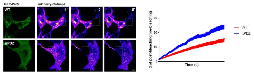
bioRxiv preprint first posted online Apr. 6, 2019; doi: http://dx.doi.org/10.1101/601575. The copyright holder for this preprint (which
       was not peer-reviewed) is the author/funder, who has granted bioRxiv a license to display the preprint in perpetuity.
                                      All rights reserved. No reuse allowed without permission.

                        2. Materials and Methods

                        Antibodies and Plasmids. A detailed antibody and primer list is presented in
                        the Supplementary Information.
                            pEGFP-N2 plasmid was purchased from Clontech (Mountain View, CA,
                        USA). pEBFP2-N1 was purchased from Addgene (Cambridge, MA, USA; #54595).
                        FLAG-CNTNAP2 was generated by subcloning human CNTNAP2 cDNA (gift
                        from Dr. Elior Peles, Weizmann Institute of Science, Israel) into the pEGFP-N2
                        vector (restriction sites BamHI and NotI, removing EGFP) using the Infusion
                        cloning system from Clontech. The FLAG sequence was then inserted down-
                        stream of the signaling peptide (amino acids 1-27) using the same technique.
                        mCherry-CNTNAP2 was constructed similarly, with mCherry being inserted
                        after the signal peptide. CNTNAP2 truncations were created by deleting the
                        C-terminus, 4.1-binding site, or PDZ-binding site.
                            Myc-Par3 was created by subcloning human myc-Par3 from the pK-myc-
                        Par3b plasmid (Addgene #19388) into the peGFP-N2 vector as described above
                        using BamHI and NotI restriction sites. Par3 truncation mutants were created
                        by deleting the first (amino acids 271-359), second (amino acids 461-546), third
                        (amino acids 590-677), or all PDZ domains (∆PDZall ; amino acids 271-677).
                        GFP-Par3 and GFP-Par3∆PDZall were generated by inserting EGFP into myc-
                        Par3 between the BglII and BamHI restriction sites.

                        Neuronal Culture and Transfections. High density (300,000 cells/cm2) cortical
                        neuron cultures were prepared from Sprague-Dawley rat E18 embryos. Cortical
                        neurons were transfected using Lipofectamine 2000 (Thermo Fisher Scientific,
                        Waltham, MA, USA) following the manufacturer’s recommendations. Neurons
                        were maintained in feeding media for 3 days post-transfection. Any visual signs
                        of poor neuronal health meant the exclusion of the cell from quantification.
                        COS7 or HEK293T cells (ATCC, Manassas, VA, USA; lines authenticated be-
                        fore shipment) were transfected using Lipofectamine 2000. Experiments were
                        performed 48h post-transfection.

                                                                          4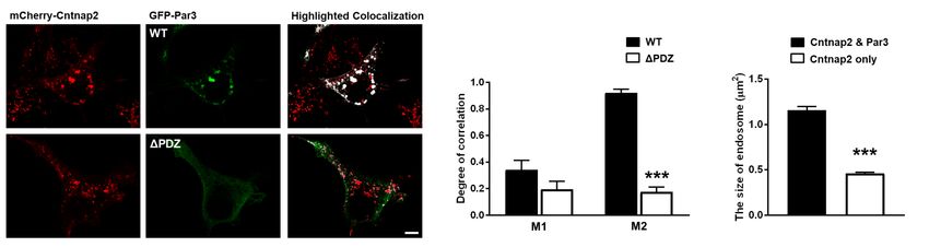
bioRxiv preprint first posted online Apr. 6, 2019; doi: http://dx.doi.org/10.1101/601575. The copyright holder for this preprint (which
       was not peer-reviewed) is the author/funder, who has granted bioRxiv a license to display the preprint in perpetuity.
                                      All rights reserved. No reuse allowed without permission.

                        Immunocytochemistry. Endogenous Par3 was detected as described (Zhang and
                        Macara, 2006). Briefly, neurons were fixed in 4% formaldehyde-sucrose-PBS
                        for 15 min at room temperature (RT), followed by permeabilization with 0.2%
                        Triton-X-100 for 5 minutes at RT, blocking in 10% bovine serum albumin (BSA)
                        for 1h at RT, and incubation of primary antibody in 3% BSA overnight at 4o C.
                        Secondary antibodies were applied in 3% BSA 1h at RT. For detection of all
                        other targets, ICC was performed as described (Gao et al., 2018). Coverslips
                        were mounted using ProLong Gold.

                        Immunoprecipitation. Mouse cortex or HEK293T cells were homogenized in
                        immunoprecipitation buffer (50 mM Tris pH 7.4, 150 mM NaCl, 0.5% Triton
                        X-100), with protease inhibitor cocktail (Roche, Basel, Switzerland) and sol-
                        ubilized for 1h at 4◦ C. Solubilized material was centrifuged at 20,000 g for 10
                        minutes at 4◦ C and the supernatant was precleared with protein A/G sepharose
                        beads (Thermo Fisher Scientific) for 30 minutes. Proteins were then immunopre-
                        cipitated with 3 µg of antibody overnight at 4◦ C, followed by a 1 hour incubation
                        with protein A/G sepharose beads the following day. Beads were then washed
                        3 times with IP buffer before adding 2x Laemmli buffer (Biorad, Hercules, CA,
                        USA). Samples were analyzed by SDS-PAGE and Western blotting.

                        Fractionation. Subcellular fractionation was performed as previously described
                        (Nakagawa et al. 2005). Briefly, cortices from 6-week old mice were homoge-
                        nized in cold sucrose buffer (20 mM HEPES pH 7.4, 320 mM sucrose, 5 mM
                        EDTA) with protease inhibitor cocktail (Roche). Homogenates were centrifuged
                        at 3,000 g for 20 min at 4◦ C speed to pellet nuclei . Supernatant (S1) was then
                        centrifuged at 38,000 g for 30 min at 4◦ C to obtain a crude membrane pellet
                        (P2). P2 was re-suspended in potassium iodide buffer (20 mM HEPES pH 7.4, 1
                        M KI, 5 mM EDTA) to remove membrane-associated proteins (S3). Membranes
                        were again collected by centrifugation (38,000 g for 20 min at 4◦ C). Membranes
                        were washed (20 mM HEPES pH 7.4, 5 mM EDTA) and pelleted once more (S4)
                        before solubilizing in CHAPS buffer (20 mM HEPES pH 7.4, 100 mM NaCl, 5
                        mM EDTA, 1% CHAPS) supplemented with protease inhibitors for 2 hours at

                                                                          5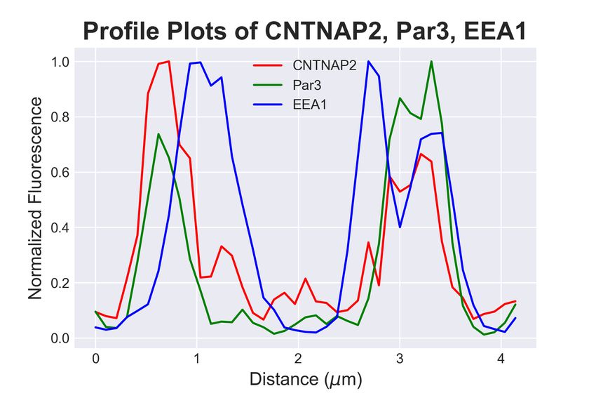
bioRxiv preprint first posted online Apr. 6, 2019; doi: http://dx.doi.org/10.1101/601575. The copyright holder for this preprint (which
       was not peer-reviewed) is the author/funder, who has granted bioRxiv a license to display the preprint in perpetuity.
                                      All rights reserved. No reuse allowed without permission.

                        4◦ C. Solubilized membranes were clarified by centrifugation at 100,000 g for 30
                        min at 4◦ C (S5). The final CHAPS-insoluble pellet was re-suspended in SDS
                        buffer (50mM TRIS pH 7.4, 150 mM NaCl, 1% SDS) supplemented with pro-
                        tease inhibitors, solubilized at 37◦ C for 20 mins and clarified by centrifugation
                        (S6).

                        Yeast Two-Hybrid Screening. Y2H screening was performed using the Match-
                        maker Gold System (Clontech). Briefly, the C-terminal region of CNTNAP2
                        (amino acids 1284-1331) was cloned into the bait vector (pGBKT7) and trans-
                        formed into yeast. Bait-positive yeast were then mated with yeast containing
                        cDNA (pGADT7) from a mouse brain cDNA library. Mated yeast were then
                        plated onto double amino acid (-Leu/-Trp) dropout, Aureobasidin A positive
                        (A), and X-α-Gal positive (X) plates (DDO/X/A), a low stringency medium
                        that selects for mated yeast containing both bait and prey vectors. DDO/X/A
                        colonies that were blue, indicative of a possible interaction between bait-prey,
                        were streaked onto quadruple amino acid (-Leu/-Trp/-His/-Ade) dropout, Aureobasidin-
                        A positive (A), and X-α-Gal positive (X) plates (QDO/X/A), which require a
                        genuine bait-prey interaction for growth. The blue colonies from QDO/X/A
                        plates were isolated and plasmids sequenced.

                        Confocal Microscopy. Confocal images of neurons were acquired using a Nikon
                        C2 confocal microscope using a 60X oil immersion objective with numerical
                        aperture (NA) = 1.4 with 0.4 µm z-stacks. All images were acquired in the
                        linear range of fluorescence intensity and a single plane image from the stack
                        was selected for analysis. For antibody validation, z-projections were used. The
                        acquisition parameters were kept the same for all conditions and images were
                        analyzed using Fiji software.

                        Colocalization analysis. Colocalization highlighter images and Manders’ colo-
                        calization coefficients were determined in ImageJ after thresholding. Total im-
                        munofluorescence intensity of region of interest (ROI) was measured automat-
                        ically. For the size of endosome quantification, a threshold was applied to the

                                                                          6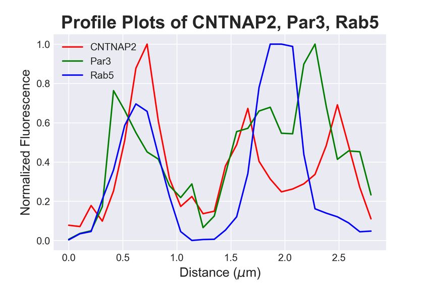
bioRxiv preprint first posted online Apr. 6, 2019; doi: http://dx.doi.org/10.1101/601575. The copyright holder for this preprint (which
       was not peer-reviewed) is the author/funder, who has granted bioRxiv a license to display the preprint in perpetuity.
                                      All rights reserved. No reuse allowed without permission.

                        maximum projection images to include all detectable endosomes. The endosome
                        restricted to objects with areas greater than 0.08 µm2, were manually detected,
                        and the endosome area was measured. For object-based binary classification of
                        colocalization, see Trafficking Marker Analysis.

                        Trafficking Marker Analysis. COS7 cells were transfected with mCherry-CNTNAP2,
                        Myc-Par3, and Blue Fluorescent Protein (BFP). Cells were fixed with standard
                        immunocytochemical methods (see Immunocytochemistry section) and stained
                        with respective antibodies (see Antibodies and Plasmids). Images were acquired
                        on a Nikon C2 scanning confocal microscope using a 60X objective (NA = 1.4)
                        at Nyquist sampling frequency. Cells were imaged after determining suitable
                        expression of Par3, CNTNAP2, and BFP.
                            ROIs were drawn on single planes encompassing whole cell cytoplasm and
                        excluding nuclei using BFP cell-fill as a guide. Images were processed and an-
                        alyzed using Python 3.6 with the scikit-image package (van der Walt et al.,
                        2014). Briefly, images were preprocessed using a 3x3 median filter and 20-
                        pixel disk white tophat transformation to reduce noise and background, respec-
                        tively. Thresholding was performed using the Triangle algorithm(Zack et al.
                        1977) for each color channel and converted to binary images. Small objects
                        (¡12 pixels2) were excluded from analysis. Puncta were segmented into objects
                        using a Watershed algorithm. Colocalization analysis was performed as follows:
                        For two-channel colocalization of Par3 and CNTNAP2, an object in the first
                        channel was considered colocalized with the second channel if >50% of its in-
                        cluded pixels were also above threshold in channel 2. For triple colocalization of
                        CNTNAP2+ /Par3+ puncta with trafficking markers, the intersection of these
                        channels was then segmented as described above. These objects were then used
                        as channel 1 for colocalization with markers (channel 2). See Supplemental
                        Figure 2 for illustration.

                        Code Availability. Python code for trafficking marker analysis is available upon
                        request.

                                                                          7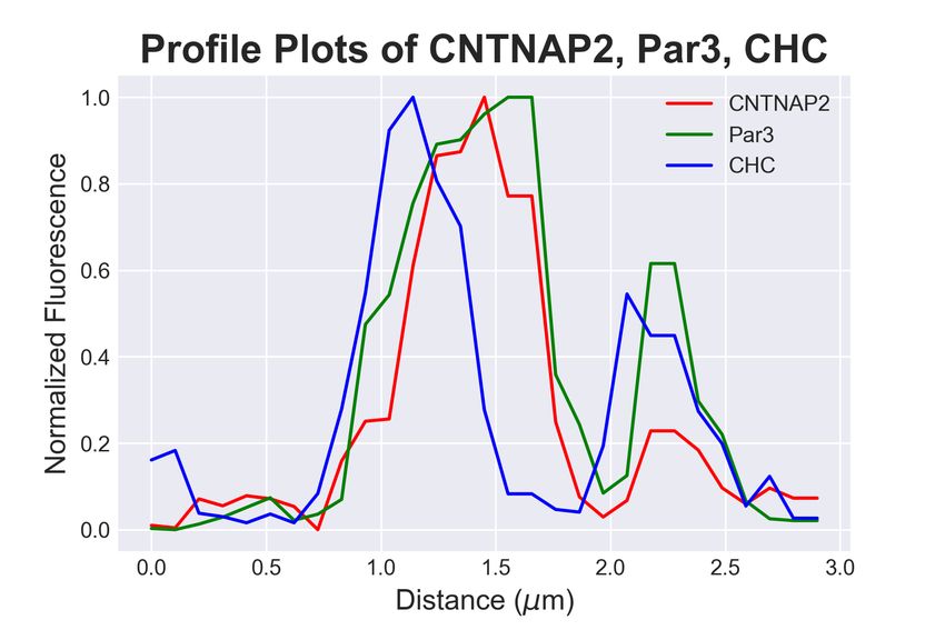
bioRxiv preprint first posted online Apr. 6, 2019; doi: http://dx.doi.org/10.1101/601575. The copyright holder for this preprint (which
       was not peer-reviewed) is the author/funder, who has granted bioRxiv a license to display the preprint in perpetuity.
                                      All rights reserved. No reuse allowed without permission.

                        Live Imaging and Analysis. COS7 cells for FRAP experiments were grown on
                        glass bottom culture dishes from MatTek (Ashland, MA, USA) and transfected
                        with mCherry-CNTNAP2 and GFP-Par3 or GFP-Par3∆PDZall . Cells were im-
                        aged with a confocal microscope (Nikon) in a CO2 incubator stage. Images were
                        taken with 256x256 pixel resolution every 1s for 300s after bleaching. 100% laser
                        power pulses of 1ms for 1 min were used to bleach GFP-Par3. Fluorescence in-
                        tensity of bleached GFP-Par3 was measured and normalized to the fluorescence
                        before bleaching with the same area. Recovery data points were then fitted to
                        a one-phase association exponential in GraphPad Prism (Version 7).

                        Blinding and Statistical Analysis. Data from cell line and primary neuron stud-
                        ies were obtained and analyzed under blinded conditions (coverslip identity hid-
                        den, cells selected randomly, filenames randomized). Cells of visibly poor health
                        were excluded from quantification. Sample sizes for cell line studies were be-
                        tween 10-30 cells. For primary neuronal studies, over 30-40 neurons were imaged
                        and the best representative neuron was used for display.
                            All statistical tests were performed with GraphPad Prism (Version 7) or in
                        Python using the SciPy library. Before analysis, data were first tested using
                        D’Agostino’s Omnibus Normality Test and Pearson correlation in order to de-
                        termine whether parametric or nonparametric tests were to be used. Post-hoc
                        tests were always used in multiple comparison analysis. P values < 0.05 were
                        considered significant.

                        3. Results
                        3.1. A yeast two-hybrid screen reveals Par3 as a novel CNTNAP2 interaction
                             partner
                            We performed a yeast two-hybrid screen of a mouse brain cDNA library with
                        the entire CNTNAP2 intracellular domain (ICD) as bait (amino acids 1284-
                        1331) to find intracellular interactors of CNTNAP2 (Figure 1a). Out of three
                        million candidates screened, we identified 41 unique clones (Gao et al., 2018),
                        with Par3 being one of the most intriguing, given its role as a spatial organizer
                        (Zhang and Macara, 2006; Duman et al., 2013; Ahmed and Macara, 2017). We

                                                                          8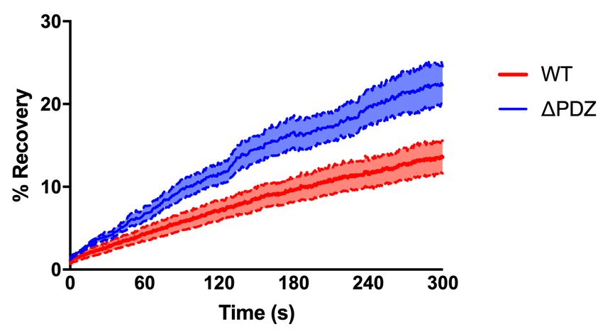
bioRxiv preprint first posted online Apr. 6, 2019; doi: http://dx.doi.org/10.1101/601575. The copyright holder for this preprint (which
was not peer-reviewed) is the author/funder, who has granted bioRxiv a license to display the preprint in perpetuity.
All rights reserved. No reuse allowed without permission.
a. Discoidin LamG LamG EGF FBG-like
bait b.
LamG EGF LamG 4.1 PDZ Input Cntnap2 IP IgG
CNTNAP2
150 kD Cntnap2
Δ4.1ΔPDZ
ΔCT
prey
150 kD Par3
CR1 PDZ1 PDZ2 PDZ3 aPKCBD
PAR3 50 kD IgG
ΔPDZ1 ΔPDZ2 ΔPDZ3
ΔPDZ_all
Input IP (FLAG)
c. + myc-Par3 + myc-Par3
myc-Par3 FLAG-WT FLAG-ΔPDZ FLAG-Δ4.1 FLAG-ΔCT myc-Par3 FLAG-WT FLAG-ΔPDZ FLAG-Δ4.1 FLAG-ΔCT
150 kD FLAG 150 kD FLAG
180 kD Myc 180 kD Myc
IgG
50 kD
Input IP (myc)
d. + FLAG-CNTNAP2 + FLAG-CNTNAP2
FLAG- FLAG-
CNTNAP2 myc-Par3 ΔPDZ1 ΔPDZ2 ΔPDZ3 ΔPDZ_all CNTNAP2 myc-Par3 ΔPDZ1 ΔPDZ2 ΔPDZ3 ΔPDZ_all
180 kD
Myc 180 kD Myc
150 kD
CNTNAP2 150 kD CNTNAP2
50 kD
IgG
Figure 1
Figure 1: Par3 binds with CNTNAP2 through PDZ interaction. (a) Cartoon showing the
domain topology of CNTNAP2 and Par3, with black lines representing the bait/prey regions
derived from the yeast-two hybrid screen and red lines indicating the truncated mutants used
for experimentation. (b) Representative western blot showing Par3 co-immunoprecipitation
with CNTNAP2 from mouse cortex homogenates (representative image from 2 independent
experiments). (c) Western blot showing co-immunoprecipitation of Par3 coexpressed with
various CNTNAP2 truncations to demonstrate this interaction is specific to CNTNAP2’s
PDZ-binding domain (representative image from 3 independent experiments) (d) Western blot
showing co-immunoprecipitation of CNTNAP2 with various Par3 truncations to demonstrate
each of Par3’s three PDZ domains can bind to CNTNAP2 (representative image from 2
independent experiments).
confirmed the this positive result by demonstrating yeast growth on high strin-
gency plates only when expressing both bait/prey plasmids, indicating a direct
physical interaction (data not shown). We then validated that CNTNAP2 and
Par3 are in the same protein complex in vivo by co-immunoprecipitation exper-
iments from mouse cortex homogenates (Figure 1b). To determine the protein
domains involved in their interaction, we co-expressed various CNTNAP2 trun-
cation mutants (Figure 1a; red lines) with Par3 in HEK293T cells and performed
co-immunoprecipitations. We found that the C-terminal PDZ-binding motif of
CNTNAP2 is necessary for interaction with Par3, as its deletion prevented Par3
9
bioRxiv preprint first posted online Apr. 6, 2019; doi: http://dx.doi.org/10.1101/601575. The copyright holder for this preprint (which
       was not peer-reviewed) is the author/funder, who has granted bioRxiv a license to display the preprint in perpetuity.
                                      All rights reserved. No reuse allowed without permission.

                           a.                                                                                                       b.
                                                 20 x mouse cortex
                                                                                                                                                  P1   S1   S2   S3   S5   S6
                                                                                                                                         150 kD                                 Par3

                                                                                                                                         150 kD                                 Cntnap2

                                                  sucrose buffer, 3000g
                                pellet (P1) + supernatant (S1)                             P1: myelin, meninges,
                                                                                           cell nuclei                                   100 kD                                 GluA1
                                                        sucrose buffer, 34’000g

                                                pellet (P2) + supernatant (S2)              crude membrane pellet (P2)
                                                                                                                                          95 kD                                 PSD95
                                                         potassium iodide (KI) buffer , 34’000g

                                                pellet (P3) + supernatant (S3)              S3: membrane-associated proteins              50 kD                                 β-Tubulin
                                                         wash buffer, 34’000g

                                                pellet (P4) + supernatant (S4)              Discard S4: wash to remove KI

                                                          1 % CHAPS for 2h, 100’000g

                                                pellet (P5) + supernatant (S5)              S5: Solubilized membrane protein fraction

                                                          1 % SDS for 30min

                                                supernatant (S6)                             S6: CHAPS insoluble material (PSD)

                        Figure 2: CNTNAP2 and Par3 are localized to membranes in mouse brain. (a) Schematic
                        showing the subcellular fractionation protocol. (b) Immunoblots of subcellular fractionations
                        from adult mouse forebrain probed with CNTNAP2 and Par3. Purple and red boxes highlight
                        fractions of interest. GluA1, PSD95, and β-tubulin served as compartment-specific markers.

                        pull-down (Figure 1c). Conversely, deletion of any one or all of Par3’s three PDZ
                        domains (Figure 1a; red lines) resulted in a dramatic reduction of CNTNAP2
                                                                                              Figure 2

                        co-immunoprecipitation efficiency (Figure 1d). From these results, we conclude
                        that CNTNAP2’s C-terminal PDZ-binding domain can interact with multiple
                        Par3 PDZ domains, a Par3 binding motif previously observed with Par6 (Ren-
                        schler et al., 2018). We decided to use a Par3 mutant lacking all PDZ sites
                        (Par3∆PDZall ) for subsequent studies. These experiments demonstrate that
                        Par3 is a novel CNTNAP2 interactor that utilizes PDZ binding for physical
                        association.

                        3.2. Subcellular compartmentalization of Par3 and CNTNAP2

                            We proceeded to characterize the spatial properties of both proteins by
                        performing an in-depth subcellular fractionation procedure using salt extrac-
                        tion and detergent solubilization (Figure 2). To confirm fractionated compart-
                        ments, we probed the fractions with GluA1, PSD95, and beta-tubulin anti-
                        bodies. GluA1 (synaptic membrane protein) was highly enriched in the crude
                        membrane (S5) and synaptic (S6) fractions while PSD95 (synaptic cytosolic
                        protein) was only enriched in the S6 fraction; beta-tubulin (non-synaptic cy-
                        tosolic protein) was absent from either S5 or S6 fractions (Figure 2b, bottom).

                                                                                                                                  10
bioRxiv preprint first posted online Apr. 6, 2019; doi: http://dx.doi.org/10.1101/601575. The copyright holder for this preprint (which
was not peer-reviewed) is the author/funder, who has granted bioRxiv a license to display the preprint in perpetuity.
All rights reserved. No reuse allowed without permission.
a. b.
mCherry Myc DAPI Merge
mCherry-
CNTNAP2
alone
myc-Par3
alone
mCherry- c. d.
CNTNAP2 +
myc-Par3
mCherry-
CNTNAP2ΔPDZ
+ myc-Par3
mCherry-
CNTNAP2 + myc-
Par3ΔPDZall
2525 um
Figure 3: Co-expressed CNTNAP2 and Par3 associate with large vesicles in COS7 cells.
(a) Representative confocal images of COS7 cells expressing mCherry-CNTNAP2/myc-Par3
alone, together, or with various truncation mutants shown to affect protein-protein interac-
tions. Arrows point to the abnormally large mCherry-CNTNAP2 + myc-Par3 aggregates Figure 3
induced by co-expression of the two (scale bar = 25 µm) (representative image from 2 in-
dependent experiments). (b) Selective representative images showing co-localization (white)
between mCherry-CNTNAP2 + GFP-Par3 (WT) or mCherry-CNTNAP2 GFP-Par3∆PDZall
(∆PDZ) (scale bar = 10 µm). (c) Quantification of Mander’s colocalization (WT: 9 cells,
∆PDZ: 10 cells from 1 independent experiment) by unpaired two-tailed t-test (d) Measure-
ment of the size of mCherry-CNTNAP2 vesicles alone or when GFP-Par3 is co-expressed (n
= 9 cells for both conditions from 1 independent experiment) by unpaired two-tailed t-test.
Values are means ± SEM. *** P < 0.001.
We found both Par3 and CNTNAP2 in the S6 fraction, consistent with pre-
vious publications (Chen et al., 2015; Oiso et al., 2009; Gao et al., 2018; Lin
et al., 2000). Moreover, Par3 was also highly enriched in the crude membrane-
associated (S3) fraction but almost nonexistent in S5, while Cntnap2 was most
abundant in the S5 fraction, as expected. Together, these data suggests Par3
is a cytosolic protein, which can transiently associate with membrane proteins
like CNTNAP2.
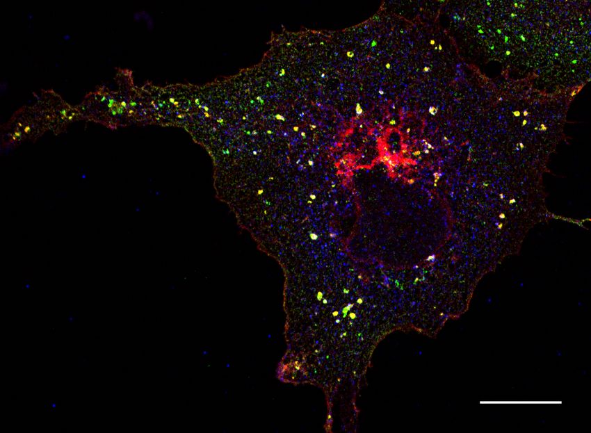
3.3. Par3 redistributes CNTNAP2 in COS7 cells
We next used COS7 cells to ascertain localization patterns for both molecules.
When CNTNAP2 was expressed alone, we noted distribution patterns at the
cell periphery, perinuclear region, and within small cytoplasmic vesicles (Figure
11bioRxiv preprint first posted online Apr. 6, 2019; doi: http://dx.doi.org/10.1101/601575. The copyright holder for this preprint (which
was not peer-reviewed) is the author/funder, who has granted bioRxiv a license to display the preprint in perpetuity.
All rights reserved. No reuse allowed without permission.
3a; mCherry-CNTNAP2 alone). Par3, on the other hand, displayed mostly
a diffuse cytoplasmic localization pattern (Figure 3a; myc-Par3 alone) (Lin
et al., 2000). Upon co-expression, respective expression patterns changed dras-
tically, where both molecules were found mostly within large punctate cytoplas-
mic structures (arrows, Figure 3a; mCherry-CNTNAP2 + myc-Par3). More-
over, CNTNAP2 puncta size increased significantly upon Par3 co-expression
(mCherry-CNTNAP2 + myc-Par3: 1.16 ± 0.04µm2 vs. mCherry-CNTNAP2
alone: 0.46 ± 0.014µm2 ; Figure 3d). These patterns were abolished with re-
placement of CNTNAP2 or Par3 with CNTNAP2∆PDZ or Par3∆PDZall mu-
tants, respectively (Figure 3a; mCherry-CNTNAP2∆PDZ + myc-Par3 and
mCherry-CNTNAP2 + myc-Par3∆PDZall ). Quantitative Manders colocaliza-
tion analysis confirmed these observations (Figure 3b-c; mCherry-CNTNAP2 +
myc-Par3 M2: 0.921 ± 0.02 vs. mCherry-CNTNAP2 + myc-Par3∆PDZall M2:
0.176 ± 0.03). Our data therefore suggests a possible role of Par3 in regulating
CNTNAP2 subcellular localization.
3.4. CNTNAP2 and Par3 co-associate with clathrin-mediated endocytic vesicles
Because the structures containing both CNTNAP2 and Par3 resembled traf-
ficking compartments, we next determined the identity of CNTNAP2+ /Par3+
structures by co-staining exogenously-expressed proteins in COS7 cells with en-
dogenous trafficking markers. Markers for endocytosis (Caveolin-1, Clathrin
heavy chain [CHC]), very early endosomes (APPL1), early endosomes (EEA1,
Rab5), and the Golgi-secretory complex (GOPC, Syntaxin-6 [Stx6]) were exam-
ined. Association of CNTNAP2+ /Par3+ puncta with these markers was mea-
sured using a novel object-based algorithm (Supplementary Figure 1a, also see
Materials and Methods). We found CNTNAP2+ /Par3+ vesicles to have a high
degree of co-localization with EEA1, Rab5, and CHC (Figure 4a); line scans and
quantitative co-localization analysis confirm synchronous peaks of fluorescence
and statistically significant overlap between all CNTNAP2+ /Par3+ and each of
these three markers (Figure 4b-d). On the other hand, the remaining markers
showed divergent fluorescent intensity peaks and low overlap values (Supplemen-
12bioRxiv preprint first posted online Apr. 6, 2019; doi: http://dx.doi.org/10.1101/601575. The copyright holder for this preprint (which
was not peer-reviewed) is the author/funder, who has granted bioRxiv a license to display the preprint in perpetuity.
All rights reserved. No reuse allowed without permission.
a. b. c. Fraction of Par3+/CNTNAP2+ Puncta
CHC EEA1
EEA1 Rab5 Rab5 0.8 Colocalized with Marker
Colocalized Fraction
0.6
0.4
0.2
0.0
1
C
L1
1
5
PC
6
CNTNAP2
-
A
ab
x
H
lin
St
PP
EE
O
C
R
eo
G
A
av
C
Ve ndo
En
TG athw
Ea
ry so
E
d
P
N/ ay
oc
rly
Ea me
Se
y to
En
rly
cre
sis
do
to
so
ry
d.
me
Tukey’s Multiple Comparison Test: Significant Results
Par3 Mean Diff. 95% CI of Diff. Summary Adjusted P
Value
Caveolin-1 vs. CHC -0.1725 -0.3255 to -0.01948 * 0.0167
Caveolin-1 vs. Rab5 -0.1884 -0.3307 to -0.04618 ** 0.0024
Caveolin-1 vs. EEA1 -0.3267 -0.4814 to -0.172 ****bioRxiv preprint first posted online Apr. 6, 2019; doi: http://dx.doi.org/10.1101/601575. The copyright holder for this preprint (which
was not peer-reviewed) is the author/funder, who has granted bioRxiv a license to display the preprint in perpetuity.
All rights reserved. No reuse allowed without permission.
a. b.
mCherry-CNTNAP2 FRAP Recovery
***
Figure 5: Par3 stabilizes CNTNAP2 within intracellular vesicles. (a) FRAP and time-lapse
imaging (5 minutes) of live COS7 cells co-expressing mCherry-CNTNAP2 + GFP-Par3 (WT)
or mCherry-CNTNAP2 + GFP-Par3∆PDZall (∆PDZ) (scale bar = 5 µm). Green channel
represents expression patterns of GFP-Par3 or GFP-Par3∆PDZall . Pseudo-color channel
represents mCherry-CNTNAP2 before (-1’), during (0), or after (5’) FRAP bleaching in either
condition. (b) Quantification of post-FRAP recovery in either condition by two-way ANOVA
(WT: n = 8 cells, ∆PDZ: n = 10 cells from 1 independent experiment). Values are means
±SEM. *** P < 0.001. WT τ =426.2s, ∆PDZall τ =361.3s
co-expressed with CNTNAP2 while Par3∆PDZall retains a diffuse cytoplasmic
Figure 5
staining (Figure 5a).
Time-lapse imaging of overexpressed mCherry-CNTNAP2 and GFP-Par3 in
COS7 cells revealed a mixed population of large and immobile CNTNAP2+ /Par3+
vesicles intermixed with smaller, more mobile ones (Supplemental Movie 1).
Conversely, the immobile CNTNAP2+ /Par3+ structures were abolished when
GFP-Par3 was replaced with the Par3∆PDZall mutant (Supplemental Movie
2). Using FRAP, we found CNTNAP2 mobility was increased when coexpressed
with the Par3∆PDZall mutant compared to WT. Five minutes following bleach-
ing, mCherry-CNTNAP2 fluorescence recovered to twice the extent when coex-
pressed with GFP-Par3∆PDZall compared to WT (Figure 5a-b). Additionally,
the rate of recovery was nearly 20% faster with the Par3∆PDZall mutant (WT:
τ = 426.2s, ∆PDZall : τ = 361.3s). These data suggest that Par3 restricts
CNTNAP2 mobility in a manner dependent on the PDZ domains of Par3.
3.6. Par3 affects the localization of CNTNAP2 in primary neuronal dendrites
Having shown that Par3 can influence CNTNAP2’s localization in COS7
cells, we set out to understand this relationship in cultured rat neurons. Be-
14bioRxiv preprint first posted online Apr. 6, 2019; doi: http://dx.doi.org/10.1101/601575. The copyright holder for this preprint (which
was not peer-reviewed) is the author/funder, who has granted bioRxiv a license to display the preprint in perpetuity.
All rights reserved. No reuse allowed without permission.
a. BFP only BFP + myc-Par3 BFP + myc-Par3ΔPDZ_all
BFP
myc
CNTNAP2 (endogenous)
Merge 5 um
Figure 6: Overexpressed Par3 clusters endogenous CNTNAP2 in primary inhibitory neurons.
(a) Representative images (from at least 3 independent experiments) of cultured primary
inhibitory neurons transfected with blue fluorescent protein (BFP), alone or together with
myc-Par3 or myc-Par3∆PDZall (scale bar = 5 µm).
cause our previous study and others show CNTNAP2 to be more highly ex-
pressed in interneurons (Gao and Penzes, 2015; Mo et al., 2015; Karayannis
Figure 6
et al., 2014), we focused our analysis on inhibitory neurons. We overexpressed
Par3 in interneurons and studied its expression patterns as well as that of en-
dogenous CNTNAP2. We confirmed the specificity of our interneuronal marker
by co-staining with GABA (Supplementary Figure 2a). In line with our obser-
vations in cell lines, Par3 was largely diffuse in the cytoplasm but also formed
large vesicular-like clusters (Figure 6a), consistent with endogenous staining
patterns (Supplementary Figure 2b) (Zhang and Macara, 2006). On the other
hand, overexpression of the Par3∆PDZall mutant produced mostly a cytoplas-
mic pattern, suggesting the PDZ interaction is critical for clustering (Figure
6a). To ensure that our observations are robust, we confirmed the specificity of
the CNTNAP2 antibody by staining CNTNAP2−/− neurons (Supplementary
Figure 3). Finally, as in COS7 cells, we occasionally found large aggregates of
endogenous CNTNAP2 that clustered strongly with overexpressed Par3, but not
Par3∆PDZall (Figure 6a). Altogether, our data indicate that Par3 can direct
localization of CNTNAP2 to punctate structures in neurons.
15bioRxiv preprint first posted online Apr. 6, 2019; doi: http://dx.doi.org/10.1101/601575. The copyright holder for this preprint (which
was not peer-reviewed) is the author/funder, who has granted bioRxiv a license to display the preprint in perpetuity.
All rights reserved. No reuse allowed without permission.
4. Discussion
Par3 is part of a conserved signaling complex required for inducing polarity
events critical for cellular function and survival, such as migration, apical-basal
polarity establishment, and axon specification (Mertens et al., 2006). Par3, with
its multi-domain scaffold partners, serves as a central recruiter of downstream
signaling molecules in these processes. Our work expands on our knowledge of
this function by characterizing the existence of a previously unknown interaction
with CNTNAP2 identified by unbiased yeast two-hybrid screening.
CNTNAP2 has multi-faceted roles in neuronal migration, development, and
maintenance; Par3 may serve as a spatial regulator for these functions (Zhang
and Macara, 2006; Ahmed and Macara, 2017). In support of this model, we show
that Par3 clusters CNTNAP2 into large vesicles via C-terminal PDZ binding
in heterologous cells. These CNTNAP2+ /Par3+ vesicles appear to be compo-
nents of the clathrin-dependent endosomal trafficking pathway, within which
Par3 stabilizes and spatially restricts CNTNAP2. Translating to neurons, it is
possible that relative levels of Par3 at a local scale within a neuron may influ-
ence CNTNAP2’s function (Ruch et al., 2017). For example, Par3 within spines
may influence local CNTNAP2 levels and function in that area (Zhang and
Macara, 2006; Duman et al., 2013), leading to alterations in synaptic density,
dynamics, or signaling (Gdalyahu et al., 2015; Varea et al., 2015). Addition-
ally, we observed CNTNAP2 and Par3 colocalized structures in dendrites of
inhibitory neurons (Figure 6). These structures were fairly sparse within single
cells but also suggest an spatially regulated assembly model. Importantly, these
colocalizing structures were absent when Par3 was replaced with the ∆PDZall
mutant.
Mechanistically, we found multiple Par3 PDZ domains are necessary for
proper CNTNAP2 binding. While this may be the result of artificial steric dis-
ruption, it is possible that such architecture exists to hold multiple CNTNAP2
molecules together within a tight spatial confine (Renschler et al., 2018). This
could explaining the abnormally large size of CNTNAP2+ /Par3+ vesicles upon
16bioRxiv preprint first posted online Apr. 6, 2019; doi: http://dx.doi.org/10.1101/601575. The copyright holder for this preprint (which
was not peer-reviewed) is the author/funder, who has granted bioRxiv a license to display the preprint in perpetuity.
All rights reserved. No reuse allowed without permission.
overexpression and suggesting the possibility of a network assembly (Hayashi
et al., 2009). This configuration may also be necessary to offset the naturally
weak CNTNAP2-Par3 interaction, as seen by our fractionation results (Figure
2).
Yet, how exactly does Par3 mechanistically regulate CNTNAP2? The an-
swer is difficult to determine since CNTNAP2 has multiple regions critical to
trafficking. It has been established that threonine 1292 on CNTNAP2’s in-
tracellular domain regulates endocytosis through protein kinase C (Bel et al.,
2009) and that proper surface targeting of family member neurexin1 requires its
PDZ-binding motif (Fairless et al., 2008). Furthermore, specific sites on CNT-
NAP2’s extracellular region are also critical for proper secretory and axonal traf-
ficking (Pinatel et al., 2017; Falivelli et al., 2012). Our immunocytochemistry
experiments link Par3-CNTNAP2 complexes with clathrin-coated endosomes,
suggesting that Par3 may, via PDZ binding, be another component critical for
CNTNAP2 internalization from the surface membrane. These questions will be
addressed in future studies.
Both CNTNAP2 and Par3 play important roles in several steps of cortex
development, including cell proliferation, migration, myelination, cell adhesion
and synapse development (Bultje et al., 2009; Liu et al., 2018; Tep et al., 2012;
Famulski et al., 2010; Varea et al., 2015; Penagarikano et al., 2011; Scott et al.,
2017). While defects in all of these processes have been implicated in neurode-
velopmental disorders, further work is needed to determine the specific processes
where their interaction plays a key role. Future work should focus on the molec-
ular pathways dependent on this newly identified CNTNAP2-Par3 interaction,
and to elucidate the specific processes in which this partnership operates.
Ethics Statement
All experiments were approved by the Institutional Animal Care and Use
Committee of Northwestern University, protocol number IS0005992.
17bioRxiv preprint first posted online Apr. 6, 2019; doi: http://dx.doi.org/10.1101/601575. The copyright holder for this preprint (which
was not peer-reviewed) is the author/funder, who has granted bioRxiv a license to display the preprint in perpetuity.
All rights reserved. No reuse allowed without permission.
Author Contributions
R.G. led the project. R.G. and C.P.P. performed all confocal imaging exper-
iments. R.G., M.D.M., and M.P.F. performed biochemistry experiments. S.Y.
performed live imaging experiments. P.P. supervised the project. R.G., C.P.P.
and P.P. wrote the manuscript.
Acknowledgments
This work was supported by grants NS100785 and MH097216 from the NIH-
NIMH to P.P and F30MH096457 to R.G.
Conflict of Interest
The authors declare no competing financial conflict of interests.
References
Ahmed, S. M., Macara, I. G., Mar. 2017. The Par3 polarity protein is an exo-
cyst receptor essential for mammary cell survival. Nature Communications 8,
14867.
Bel, C., Oguievetskaia, K., Pitaval, C., Goutebroze, L., Faivre-Sarrailh, C.,
Sep. 2009. Axonal targeting of Caspr2 in hippocampal neurons via selective
somatodendritic endocytosis. J Cell Sci 122 (Pt 18), 3403–13.
Bridi, M. S., Park, S. M., Huang, S., Sep. 2017. Developmental Disrup-
tion of GABAAR-Meditated Inhibition in Cntnap2 KO Mice. eNeuro 4 (5),
ENEURO.0162–17.2017.
Bultje, R. S., Castaneda-Castellanos, D. R., Jan, L. Y., Jan, Y. N., Kriegstein,
A. R., Shi, S. H., Jul. 2009. Mammalian Par3 regulates progenitor cell asym-
metric division via notch signaling in the developing neocortex. Neuron 63 (2),
189–202.
18bioRxiv preprint first posted online Apr. 6, 2019; doi: http://dx.doi.org/10.1101/601575. The copyright holder for this preprint (which
was not peer-reviewed) is the author/funder, who has granted bioRxiv a license to display the preprint in perpetuity.
All rights reserved. No reuse allowed without permission.
Chen, N., Koopmans, F., Gordon, A., Paliukhovich, I., Klaassen, R. V., van der
Schors, R. C., Peles, E., Verhage, M., Smit, A. B., Li, K. W., Jul. 2015. In-
teraction proteomics of canonical Caspr2 (CNTNAP2) reveals the presence of
two Caspr2 isoforms with overlapping interactomes. Biochimica et Biophysica
Acta (BBA) - Proteins and Proteomics 1854 (7), 827–833.
Chen, X., Macara, I. G., Mar. 2005. Par-3 controls tight junction assembly
through the Rac exchange factor Tiam1. Nat. Cell Biol. 7 (3), 262–269.
Duman, J. G., Tzeng, C. P., Tu, Y. K., Munjal, T., Schwechter, B., Ho, T. S.,
Tolias, K. F., Apr. 2013. The adhesion-GPCR BAI1 regulates synaptogen-
esis by controlling the recruitment of the Par3/Tiam1 polarity complex to
synaptic sites. J Neurosci 33 (16), 6964–78.
Fairless, R., Masius, H., Rohlmann, A., Heupel, K., Ahmad, M., Reissner,
C., Dresbach, T., Missler, M., Nov. 2008. Polarized targeting of neurexins
to synapses is regulated by their C-terminal sequences. J Neurosci 28 (48),
12969–81.
Falivelli, G., De Jaco, A., Favaloro, F. L., Kim, H., Wilson, J., Dubi, N., Ellis-
man, M. H., Abrahams, B. S., Taylor, P., Comoletti, D., Nov. 2012. Inherited
genetic variants in autism-related CNTNAP2 show perturbed trafficking and
ATF6 activation. Hum Mol Genet 21 (21), 4761–73.
Famulski, J. K., Trivedi, N., Howell, D., Yang, Y., Tong, Y., Gilbertson, R.,
Solecki, D. J., Dec. 2010. Siah regulation of Pard3A controls neuronal cell
adhesion during germinal zone exit. Science 330 (6012), 1834–8.
Feng, W., Wu, H., Chan, L.-N., Zhang, M., Aug. 2008. Par-3-mediated junc-
tional localization of the lipid phosphatase PTEN is required for cell polarity
establishment. J. Biol. Chem. 283 (34), 23440–23449.
Gao, R., Penzes, P., 2015. Common mechanisms of excitatory and inhibitory
imbalance in schizophrenia and autism spectrum disorders. Curr Mol Med
15 (2), 146–67.
19bioRxiv preprint first posted online Apr. 6, 2019; doi: http://dx.doi.org/10.1101/601575. The copyright holder for this preprint (which
was not peer-reviewed) is the author/funder, who has granted bioRxiv a license to display the preprint in perpetuity.
All rights reserved. No reuse allowed without permission.
Gao, R., Piguel, N. H., Melendez-Zaidi, A. E., Martin-de-Saavedra, M. D.,
Yoon, S., Forrest, M. P., Myczek, K., Zhang, G., Russell, T. A., Csernansky,
J. G., Surmeier, D. J., Penzes, P., Apr. 2018. CNTNAP2 stabilizes interneuron
dendritic arbors through CASK. Mol Psychiatry.
Gdalyahu, A., Lazaro, M., Penagarikano, O., Golshani, P., Trachtenberg, J. T.,
Geschwind, D. H., 2015. The Autism Related Protein Contactin-Associated
Protein-Like 2 (CNTNAP2) Stabilizes New Spines: An In Vivo Mouse Study.
PLoS One 10 (5), e0125633.
Goldstein, B., Macara, I. G., Nov. 2007. The PAR proteins: Fundamental play-
ers in animal cell polarization. Dev Cell 13 (5), 609–22.
Hayashi, M. K., Tang, C., Verpelli, C., Narayanan, R., Stearns, M. H., Xu,
R. M., Li, H., Sala, C., Hayashi, Y., Apr. 2009. The postsynaptic density
proteins Homer and Shank form a polymeric network structure. Cell 137 (1),
159–71.
Hoffman, E. J., Turner, K. J., Fernandez, J. M., Cifuentes, D., Ghosh, M., Ijaz,
S., Jain, R. A., Kubo, F., Bill, B. R., Baier, H., Granato, M., Barresi, M. J.,
Wilson, S. W., Rihel, J., State, M. W., Giraldez, A. J., Feb. 2016. Estrogens
Suppress a Behavioral Phenotype in Zebrafish Mutants of the Autism Risk
Gene, CNTNAP2. Neuron 89 (4), 725–33.
Itoh, M., Sasaki, H., Furuse, M., Ozaki, H., Kita, T., Tsukita, S., Aug. 2001.
Junctional adhesion molecule (JAM) binds to PAR-3: A possible mechanism
for the recruitment of PAR-3 to tight junctions. J. Cell Biol. 154 (3), 491–497.
Johansson, A., Driessens, M., Aspenstrom, P., Sep. 2000. The mammalian ho-
mologue of the Caenorhabditis elegans polarity protein PAR-6 is a binding
partner for the Rho GTPases Cdc42 and Rac1. J Cell Sci 113 ( Pt 18), 3267–
75.
Jurgensen, S., Castillo, P. E., Oct. 2015. Selective Dysregulation of Hippocam-
20bioRxiv preprint first posted online Apr. 6, 2019; doi: http://dx.doi.org/10.1101/601575. The copyright holder for this preprint (which
was not peer-reviewed) is the author/funder, who has granted bioRxiv a license to display the preprint in perpetuity.
All rights reserved. No reuse allowed without permission.
pal Inhibition in the Mouse Lacking Autism Candidate Gene CNTNAP2. J
Neurosci 35 (43), 14681–7.
Karayannis, T., Au, E., Patel, J. C., Kruglikov, I., Markx, S., Delorme, R.,
Heron, D., Salomon, D., Glessner, J., Restituito, S., Gordon, A., Rodriguez-
Murillo, L., Roy, N. C., Gogos, J. A., Rudy, B., Rice, M. E., Karayiorgou, M.,
Hakonarson, H., Keren, B., Huguet, G., Bourgeron, T., Hoeffer, C., Tsien,
R. W., Peles, E., Fishell, G., Jul. 2014. Cntnap4 differentially contributes
to GABAergic and dopaminergic synaptic transmission. Nature 511 (7508),
236–40.
Lin, D., Edwards, A. S., Fawcett, J. P., Mbamalu, G., Scott, J. D., Pawson, T.,
Aug. 2000. A mammalian PAR-3-PAR-6 complex implicated in Cdc42/Rac1
and aPKC signalling and cell polarity. Nat Cell Biol 2 (8), 540–7.
Lin, D., Gish, G. D., Songyang, Z., Pawson, T., Feb. 1999. The carboxyl ter-
minus of B class ephrins constitutes a PDZ domain binding motif. J. Biol.
Chem. 274 (6), 3726–3733.
Liu, W. A., Chen, S., Li, Z., Lee, C. H., Mirzaa, G., Dobyns, W. B., Ross, M. E.,
Zhang, J., Shi, S. H., Jun. 2018. PARD3 dysfunction in conjunction with dy-
namic HIPPO signaling drives cortical enlargement with massive heterotopia.
Genes Dev 32 (11-12), 763–780.
Macara, I. G., Mar. 2004. Parsing the polarity code. Nat Rev Mol Cell Biol
5 (3), 220–31.
McCaffrey, L. M., Montalbano, J., Mihai, C., Macara, I. G., Nov. 2012. Loss
of the Par3 polarity protein promotes breast tumorigenesis and metastasis.
Cancer Cell 22 (5), 601–614.
Mertens, A. E., Pegtel, D. M., Collard, J. G., Jun. 2006. Tiam1 takes PARt in
cell polarity. Trends Cell Biol 16 (6), 308–16.
Mo, A., Mukamel, E. A., Davis, F. P., Luo, C., Henry, G. L., Picard, S., Urich,
M. A., Nery, J. R., Sejnowski, T. J., Lister, R., Eddy, S. R., Ecker, J. R.,
21bioRxiv preprint first posted online Apr. 6, 2019; doi: http://dx.doi.org/10.1101/601575. The copyright holder for this preprint (which
was not peer-reviewed) is the author/funder, who has granted bioRxiv a license to display the preprint in perpetuity.
All rights reserved. No reuse allowed without permission.
Nathans, J., Jun. 2015. Epigenomic Signatures of Neuronal Diversity in the
Mammalian Brain. Neuron 86 (6), 1369–84.
Narayanan, A. S., Reyes, S. B., Um, K., McCarty, J. H., Tolias, K. F., Dec.
2013. The Rac-GAP Bcr is a novel regulator of the Par complex that controls
cell polarity. Mol Biol Cell 24 (24), 3857–68.
Oiso, S., Takeda, Y., Futagawa, T., Miura, T., Kuchiiwa, S., Nishida, K.,
Ikeda, R., Kariyazono, H., Watanabe, K., Yamada, K., Apr. 2009. Contactin-
associated protein (Caspr) 2 interacts with carboxypeptidase E in the CNS.
J. Neurochem. 109 (1), 158–167.
Penagarikano, O., Abrahams, B. S., Herman, E. I., Winden, K. D., Gdalyahu,
A., Dong, H., Sonnenblick, L. I., Gruver, R., Almajano, J., Bragin, A., Gol-
shani, P., Trachtenberg, J. T., Peles, E., Geschwind, D. H., Sep. 2011. Absence
of CNTNAP2 leads to epilepsy, neuronal migration abnormalities, and core
autism-related deficits. Cell 147 (1), 235–46.
Pinatel, D., Hivert, B., Saint-Martin, M., Noraz, N., Savvaki, M., Karagogeos,
D., Faivre-Sarrailh, C., Jul. 2017. The Kv1-associated molecules TAG-1 and
Caspr2 are selectively targeted to the axon initial segment in hippocampal
neurons. J Cell Sci 130 (13), 2209–2220.
Renschler, F. A., Bruekner, S. R., Salomon, P. L., Mukherjee, A., Kullmann, L.,
Schutz-Stoffregen, M. C., Henzler, C., Pawson, T., Krahn, M. P., Wiesner,
S., Feb. 2018. Structural basis for the interaction between the cell polarity
proteins Par3 and Par6. Sci Signal 11 (517).
Rodenas-Cuadrado, P., Ho, J., Vernes, S. C., Feb. 2014. Shining a light on CNT-
NAP2: Complex functions to complex disorders. Eur J Hum Genet 22 (2),
171–8.
Ruch, T. R., Bryant, D. M., Mostov, K. E., Engel, J. N., Jan. 2017. Par3
integrates Tiam1 and phosphatidylinositol 3-kinase signaling to change apical
membrane identity. Mol Biol Cell 28 (2), 252–260.
22bioRxiv preprint first posted online Apr. 6, 2019; doi: http://dx.doi.org/10.1101/601575. The copyright holder for this preprint (which
was not peer-reviewed) is the author/funder, who has granted bioRxiv a license to display the preprint in perpetuity.
All rights reserved. No reuse allowed without permission.
Scott, R., Sanchez-Aguilera, A., van Elst, K., Lim, L., Dehorter, N., Bae, S. E.,
Bartolini, G., Peles, E., Kas, M. J. H., Bruining, H., Marin, O., Dec. 2017.
Loss of Cntnap2 Causes Axonal Excitability Deficits, Developmental Delay
in Cortical Myelination, and Abnormal Stereotyped Motor Behavior. Cereb
Cortex 29 (2), 586–597.
Tep, C., Kim, M. L., Opincariu, L. I., Limpert, A. S., Chan, J. R., Appel,
B., Carter, B. D., Yoon, S. O., Jan. 2012. Brain-derived neurotrophic factor
(BDNF) induces polarized signaling of small GTPase (Rac1) protein at the
onset of Schwann cell myelination through partitioning-defective 3 (Par3)
protein. J Biol Chem 287 (2), 1600–8.
Thomas, A. M., Schwartz, M. D., Saxe, M. D., Kilduff, T. S., Jan. 2017. Cntnap2
Knockout Rats and Mice Exhibit Epileptiform Activity and Abnormal Sleep-
Wake Physiology. Sleep 40 (1).
van der Walt, S., Schönberger, J. L., Nunez-Iglesias, J., Boulogne, F., Warner,
J. D., Yager, N., Gouillart, E., Yu, T., scikit-image contributors, 2014. Scikit-
image: Image processing in Python. PeerJ 2, e453.
Varea, O., Martin-de-Saavedra, M. D., Kopeikina, K. J., Schürmann, B., Flem-
ing, H. J., Fawcett-Patel, J. M., Bach, A., Jang, S., Peles, E., Kim, E., Penzes,
P., May 2015. Synaptic abnormalities and cytoplasmic glutamate receptor
aggregates in contactin associated protein-like 2/Caspr2 knockout neurons.
Proc. Natl. Acad. Sci. U.S.A. 112 (19), 6176–6181.
Vogt, D., Hunt, R. F., Mandal, S., Sandberg, M., Silberberg, S. N., Nagasawa,
T., Yang, Z., Baraban, S. C., Rubenstein, J. L. R., Apr. 2014. Lhx6 directly
regulates Arx and CXCR7 to determine cortical interneuron fate and laminar
position. Neuron 82 (2), 350–364.
Zhang, H., Macara, I. G., Mar. 2006. The polarity protein PAR-3 and TIAM1
cooperate in dendritic spine morphogenesis. Nat Cell Biol 8 (3), 227–37.
23You can also read