Evolution of DNA Replication Origin Specification and Gene Silencing Mechanisms
←
→
Page content transcription
If your browser does not render page correctly, please read the page content below
bioRxiv preprint doi: https://doi.org/10.1101/2020.07.04.187286. this version posted July 4, 2020. The copyright holder for this preprint (which
 was not certified by peer review) is the author/funder. It is made available under a CC-BY-NC 4.0 International license.

 Evolution of DNA Replication Origin Specification and Gene Silencing Mechanisms

 Hu, Y.1,2, Tareen, A.3, Sheu, Y-J.1, Ireland, W. T.5, Speck, C.6, Li, H.7, Joshua-Tor, L. 1,4, Kinney, J. B.3
 and Stillman, B.1*

 Author Affiliations:

 1. Cold Spring Harbor Laboratory, 1 Bungtown Road, Cold Spring Harbor, NY 11724, USA.
 2. Program in Molecular and Cell Biology, Stony Brook University, Stony Brook, NY 11794, USA.
 3. Simons Center for Quantitative Biology, Cold Spring Harbor Laboratory, Cold Spring Harbor,
 New York 11724, USA
 4. W. M. Keck Structural Biology Laboratory, Howard Hughes Medical Institute, Cold Spring
 Harbor, NY, 11724, USA.
 5. Department of Physics, California Institute of Technology, Pasadena, California, USA
 6. DNA Replication Group, Institute of Clinical Sciences, Faculty of Medicine, Imperial College
 London, London W12 0NN, United Kingdom
 7. Structural Biology Program, Van Andel Institute, Grand Rapids, MI 49503, USA

 * Corresponding Author stillman@cshl.edu

 Abstract
 DNA replication in eukaryotic cells initiates from chromosomal locations, called replication origins,
 that bind the Origin Recognition Complex (ORC) prior to S phase. Origin establishment is guided
 by well-defined DNA sequence motifs in Saccharomyces cerevisiae and some other budding yeasts,
 but most eukaryotes lack sequence-specific origins. At present, the mechanistic and evolutionary
 reasons for this difference are unclear. A 3.9 Å structure of S. cerevisiae ORC-Cdc6-Cdt1-Mcm2-7
 (OCCM) bound to origin DNA revealed, among other things, that a loop within Orc2 inserts into a
 DNA minor groove and an a-helix within Orc4 inserts into a DNA major groove1. We show that
 this Orc4 a-helix mediates the sequence-specificity of origins in S. cerevisiae. Specifically, mutations
 were identified within this a-helix that alter the sequence-dependent activity of individual origins
 as well as change global genomic origin firing patterns. This was accomplished using a massively
 parallel origin selection assay analyzed using a custom mutual-information-based modeling
 approach and a separate analysis of whole-genome replication profiling and statistics. Interestingly,
 the sequence specificity of DNA replication initiation, as mediated by the Orc4 a-helix, has evolved
 in close conjunction with the gain of ORC-Sir4-mediated gene silencing and the loss of RNA
 interference.

 Main
 In the budding yeast S. cerevisiae, replication origins are specified by DNA sequence motifs that comprise
 an essential A element (about 11nt in length) and multiple B elements2. Such sequences enable the
 replication of extrachromosomal plasmids and are thus termed autonomously replicating sequences
 (ARSs)3. By contrast, replication origins are sequence non-specific in plants and animals, in the fission
 yeast Schizosaccharomyces pombe, and even in many other budding yeasts and fungi4. Nevertheless, the
 proteins involved in the initiation of DNA replication are highly conserved. In eukaryotes, the six subunit
 ORC complex (comprising Orc1-6) assembles on DNA prior to S-phase5. ORC then recruits Cell Division

 1
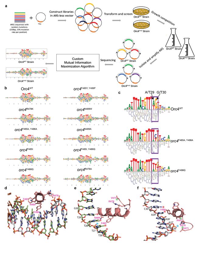
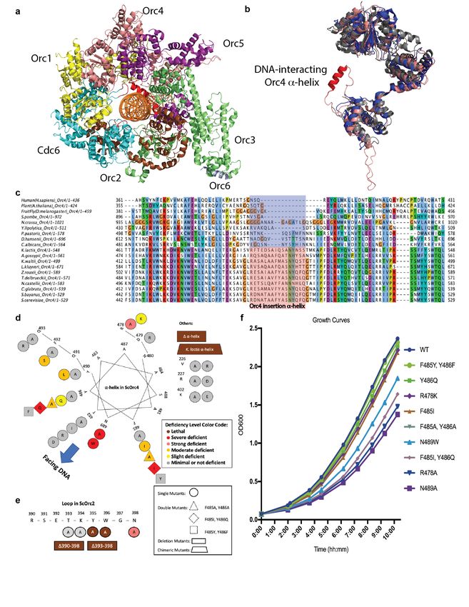
bioRxiv preprint doi: https://doi.org/10.1101/2020.07.04.187286. this version posted July 4, 2020. The copyright holder for this preprint (which
 was not certified by peer review) is the author/funder. It is made available under a CC-BY-NC 4.0 International license.

 Cycle 6 (Cdc6), chromatin licensing and DNA replication factor 1 (Cdt1), and the replication helicase
 subunits Mcm2-7 to form a pre-Replicative Complex (pre-RC)6. In prior work, a structure of a pre-RC
 assembly intermediate containing the S. cerevisiae ORC-Cdc6-Cdt1-Mcm2-7 (OCCM) bound to origin
 DNA (ARS1) was determined at ~3.9 Å by cryo-electron microscopy1. This structure revealed multiple
 OCCM-DNA interactions, including an Orc4 a-helix inserted into the DNA major groove and an Orc2
 loop inserted into the minor groove (Fig. 1a). These interactions were subsequently confirmed by a higher
 resolution ORC-DNA structure7. We note that a lysine-rich region of Orc1 interacts with DNA in this
 latter structure but not in the OCCM, suggesting considerable plasticity in origin recognition during pre-
 RC assembly.
 Interestingly, the Orc4 a-helix and Orc2 loop have evolved in a manner that parallels the evolution of
 origin sequence specificity. Sequence alignments suggest that these features have been acquired in a sub-
 group of Saccharomyces-related budding yeasts, but are absent in all other eukaryotes including other
 budding yeasts, other fungi (including S. pombe), plants, and animals (Fig. 1c, Extended Data Fig. 1a).
 High resolution structures of Human8 and Drosophila9 ORC show the lack of the Orc4 a-helix and Orc2
 loop (Fig. 1b, Extended Data Fig. 1b). The Orc4 a-helix and Orc2 loop are present but diverged in some
 other budding yeasts, such as Kluyveromyces lactis, which has sequence-specific origins that exhibit a
 DNA sequence motif that differs from the S. cerevisiae motif.
 These observations suggest that the Orc4 a-helix and/or Orc2 loop might play key roles in origin sequence
 specificity. To investigate which specific residues might be involved, 32 individual Orc4 a-helix mutants
 and 7 Orc2 loop mutants were examined using plasmid shuffle assay (Fig. 1d-e, Extended Data Fig. 2, 3;
 see Methods). The Orc2 loop mutants were either lethal, had strong defects, or had little effect (Fig. 1e,
 Extended Data Fig. 3). Deletion of the Orc4 a-helix or its replacement with the 13-amino acid K. lactis
 Orc4-a-helix were lethal. Other Orc4 a-helix mutants led to different levels of growth deficiency (Fig.
 1d, Extended Data Fig. 2). Based on the growth deficiency phenotype, nine viable Orc4 mutants were
 chosen for further detailed analysis. To perform the genetics in a complete manner, two conservative
 mutations (orc4F485Y, Y486F and orc4R478K) were chosen for comparison. The wild type and Orc4 mutants
 were tagged at the amino terminus (NTAP-tag) and integrated into the genome as the sole Orc4 subunit
 that formed a functional ORC (Extended Data Fig. 4). Some strains with the integrated version of the
 mutant Orc4 (strains G, Extended Data Fig. 6) proliferated far better compared to strains that relied on a
 mutant Orc4 subunit that was expressed from a single origin minichromosome (strains P, Extended Data
 Fig. 6). Five of these mutants were created to investigate the FY residues at positions 485-486, which have
 evolved to IQ in K. lactis. Specifically, orc4Y486Q, orc4F485I, and orc4F485I, Y486Q were used to study the
 effects of these evolutionary changes, while orc4F485A, Y486A and orc4F485Y, Y486F (a conservative swap of
 aromatic amino acids) were used to investigate these residues more generally. The other four mutants,
 orc4R478A, orc4R478K, orc4N489A, and orc4N489W, were chosen to investigate the roles of R478 and N489, two
 conserved residues at opposite ends of the a-helix that we predicted to mediate both protein-protein
 contacts and contacts with DNA backbone phosphates. Some of these Orc4 mutants exhibited slower
 growth rates (Fig. 1f) and slowed passage through S phase and mitosis (Extended Data Fig. 5). To better
 understand the effects of these mutations on origin activity and specificity, we performed two
 complementary, but independent deep-sequencing-based assays: massively parallel origin mutagenesis on
 plasmids and genome-wide DNA replication profiling.

 Massively parallel origin mutagenesis. To quantify the sequence-dependent activity of ORC at specific
 origins of interest, we performed a massively parallel origin selection assay (MPOS assay) on two different
 origins in wild type and nine yeast strains harboring the Orc4 variants. 150 base-pairs of either ARS1 (also

 2
bioRxiv preprint doi: https://doi.org/10.1101/2020.07.04.187286. this version posted July 4, 2020. The copyright holder for this preprint (which
 was not certified by peer review) is the author/funder. It is made available under a CC-BY-NC 4.0 International license.

 known as ARS416) or ARS317 DNA were synthesized at a 15% per-nucleotide substitution rate and cloned
 into plasmids that carried a selective marker10,11 (Fig. 2a). These two plasmid libraries were then separately
 transfected into the ten strains of yeast described above, and the mutated ARSs that remained after multiple
 cell divisions were sequenced. A custom motif inference algorithm, based on mutual information
 maximization, was then applied to these sequence data and used to infer quantitative motifs describing
 origin activity in each strain. This algorithm proved to be essential for the correct analysis of the data (see
 below).

 Some mutants, such as orc4F485Y, Y486F and orc4R478K, yielded motifs very similar to WT (Fig. 2b for ARS1,
 Extended Data Fig. 7a for ARS317). Other strains, such as the orc4N489A, orc4N489W, orc4R478A and orc4F485I,
 Y486Q
 retained a far less diverse set of mutant ARSs and yielded noisier motifs that, nevertheless, remained
 relatively similar to the WT ARS consensus sequence (Fig. 2b for ARS1, Extended Data Fig. 7a for
 ARS317). However, two Orc4 a-helix mutants exhibited robust changes to their ARS motifs: in both the
 orc4F485A, Y486A and orc4Y486Q mutants, and for both the ARS1 and ARS317 experiments (Fig. 2b and c,
 Extended Data Fig. 7), two dinucleotides present in the WT consensus sequence motif at position 29-30
 changed. In the ARS1 experiments, motif A/T G/T has been switched to T/A C/T in the orc4F485A, Y486A
 strain and switched to T/A T/G in the orc4Y486Q strain (Fig. 2c). Substantial changes were observed at the
 same position in the ARS317 MPOS assay (Extended Data Fig. 7b). A Principal Component Analysis
 (PCA) of the motifs inferred from multiple biological replicates confirmed that reproducible changes in
 motifs indeed resulted from the mutations in question (Extended Data Fig. 8a). A quantitative analysis of
 the mutual-information-based motif inference method (IM) compared to the standard enrichment ratio
 calculation (EM), showed that the new mutual-information-based motif inference method was essential
 for resolving these mutation-dependent changes in origin specificity (Extended Data Fig. 8).

 The structural basis for origin sequence specification. Observations using the ~3Å high-resolution
 structure7 of Orc4 a-helix on DNA (Fig. 2d, Supplementary Video 1) can further rationalize the mutation-
 dependent change in origin specificity. We suggest that Y486 interacts with the DNA base C/A30 (Fig.
 2f), which is on the complementary strand from the A/T G/T dinucleotide whose readout is altered in the
 orc4F485A, Y486A and orc4Y486Q mutants (Figure 2c, purple box) in a face-to-edge T-type p interaction, as
 often seen in protein-DNA interfaces12. In addition, F485 sits against a hydrophobic stretch comprised of
 the methyl groups emanation from a run of T’s, T26-29 (Fig. 2e).

 At each end of the Orc4 insertion a-helix are amino acids that we suggest provide affinity for ORC to
 DNA as well as help position this sequence-reading a-helix correctly in the major groove. R478 interacts
 with A487 on the a-helix as well as the adjacent V475 and could have an alternative conformation
 whereby contacts DNA phosphate (Fig. 2e and f). Even a conservative amino acid substitution R478K
 was slightly deficient (Fig. 1f and Extended Data Table 1) and had a subtle change in the genome wide
 origin firing pattern (Extended Data Fig. 11d and Fig. 3c) indicating that there is an additional role for the
 R478 side group that the charge and length of the lysine side group cannot fully replace the arginine at
 this location. Likewise, N489 that sits at the opposite end of the a-helix would be in range of a DNA
 phosphate contact and even a DNA base contact with T28 upon minor adjustments of the model that are
 well within the EM density7 (Fig. 2e). Mutation of either amino acid had the largest, non-lethal effect (Fig.
 1) and thus they could play key roles in both positioning the a-helix in the major groove and contributing
 to affinity of ORC to DNA.

 3
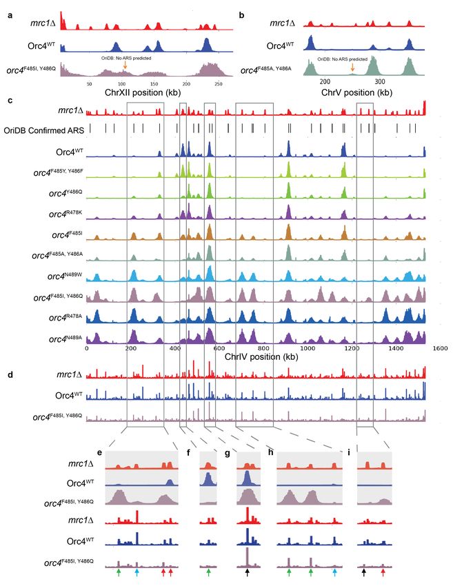
bioRxiv preprint doi: https://doi.org/10.1101/2020.07.04.187286. this version posted July 4, 2020. The copyright holder for this preprint (which
 was not certified by peer review) is the author/funder. It is made available under a CC-BY-NC 4.0 International license.

 Genome-wide replication origin profiling. To investigate the mutation-dependent origin usage changes
 in natural genomic replication origin profiling, cells were arrested in G1 phase and released into S phase
 in the presence of hydroxyurea (HU). HU treatment restricts (via checkpoint signaling) origin firing to
 those origins that normally become active in early S phase, and prevents the activation of origins that
 normally fire later. The addition of 5-Ethynyl-2'-deoxyuridine (EdU), followed by purification of EdU-
 labeled DNA and high-throughput DNA sequencing, was then used to map the locations and activities of
 early origins throughout the genome (Extended Data Fig. 9 and Fig. 3c). Mrc1 is a replication fork
 associated protein and mediator of intra-S phase checkpoint signaling. All origins fired, as expected, when
 the MRC1 gene was deleted. We observed more origins firing than have been confirmed in oriDB13. This
 Orc4WT mrc1∆ profile thus reveals a maximal set of possible origins against which to compare the
 replication profiles of NTAP tagged Orc4 integrated strains. As expected, only a subset of the maximal
 set of possible origins fired in wild type (WT) cells containing the NTAP-tagged Orc4WT protein. The
 genome-wide replication origin profiles were reproducible in biological replicates (Extended Data Fig. 9b
 for WT and mrc1∆; Fig. 3c and Extended Data Fig. 9c for all mutant Orc4 strains).

 Only two completely “de novo” replication origin locations were found in genomic origin firing profiles
 of all nine orc4 mutant strains, which were neither predicted to be ARS locations in OriDB nor exist in
 the Orc4WT mrc1∆ profile. One was found in orc4F485I, Y486Q (Fig. 3a) and another was found in orc4F485A,
 Y486A
 strain (Fig. 3b). Indeed, we didn’t expect to see a dramatic change in de novo origin locations because
 the mutations we made are only single or double point-mutations and would not be expected to create
 tremendous amount of “de novo” replication origin locations. However, extensive changes in the Orc4 a-
 helix, such as orc4Kla-helix or orc4∆a-helix, were lethal and therefore could not be assessed for “de novo”
 origins.

 Despite the very few “de novo” replication origin locations, genomic the origin firing pattern changed
 considerably in some strains (Extended Data Fig. 11b-j). There were numerous origins that are active in
 both mrc1∆ and the wild-type strain (aka. early origins) but were specifically repressed in the orc4 mutant
 strains (Fig. 3f and Extended Data Fig. 11a-j, orange arrow direction). At the same time, there were
 numerous origins that were inactive in the wild-type strain but were activated in the orc4 mutant strains
 (Fig. 3e and h, green arrows and Extended Data Fig. 11a-j, green arrow direction). The activation or
 repression pattern is mutant dependent. The chromosome IV profiles for the nine orc4 mutant strains was
 used as an example chromosome to show more details (Fig. 3c, Extended Data Fig. 9c). While some
 origins, either those that normally fire early or late, did not change (Fig. 3c, g and i, black arrows), other
 origins changed their firing pattern in Orc4 mutant strains. For example, an active origin in WT and most
 of the mutant strains was not active in the orc4F485I, Y486Q and orc4F485A, Y486A strains (Fig. 3c and f, green
 arrow). In contrast, many origins that normally do not fire in HU in WT became active in multiple mutants
 (Fig. 3c, e and h, green arrows). The firing pattern was mutant dependent. Interestingly, two origins on
 Chr. IV were active only in the orc4F485I, Y486Q mutant strain, in which the IQ are the amino acids that exist
 in K. lactis (Fig. 3c, e and i, red arrows). When, however, the conservative orc4F485Y, Y486F double mutant
 was analyzed, the firing pattern was like WT.

 There are chromatin and chromosome location context factors that were suggested in previous studies to
 play roles in controlling the origin timing14,15. Our genome-wide statistics result supports this idea. Origin
 firing peak heights generally do not correlate with how well they match the ACS motifs (Extended Data
 Fig. 12, section A). Only when the origin sequence recognition is reduced/disturbed, such as in the orc4
 F485 and Y486 mutants, the origin locations that originally can have high origin activities are then

 4
bioRxiv preprint doi: https://doi.org/10.1101/2020.07.04.187286. this version posted July 4, 2020. The copyright holder for this preprint (which
 was not certified by peer review) is the author/funder. It is made available under a CC-BY-NC 4.0 International license.

 severely affected (Extended Data Fig. 12, section B). The orc4 R478 and N489 mutations had a slower
 growth phenotype and this is likely due to reduced origin licensing, consistent with the proposal that the
 Orc4 a-helix that is not positioned correctly due to amino acid changes at each end of the a-helix, making
 it more difficult to stably position within the DNA major groove. As a consequence, poor DNA interaction
 would lead to overall much fewer origins that become active (Extended Data Fig. 11 q-t support this idea)
 resulting in larger replicon size (i.e., greater inter-origin distance) and a slower doubling time (Fig. 1f).
 These mutants would be forced to use late origins under HU to survive. This is also consistent with the
 observation that the peak widths on average for these slow growing mutants was larger than WT because
 the smaller number of origins utilized would not be restrained by rate limiting replication factors, which
 are known to exist 16,17. We suggest that the mutations at Orc4 R478 and N489 would not cause sequence
 specificity changes but instead general inefficient binding to the origins. Indeed, the correlation between
 the origin firing peak heights from genome-wide origin data and the sequence motif matching quality
 scores from the MPOS ARS selection assays did not correlate well in the Orc4 R478 and N489 mutant
 strains compared to those strains (e.g., orc4F484I, Y486Q and orc4F485A, F486A) that change DNA sequence
 specificity (Extended Data Fig. 12, section C). We suggest that mutations that affect the affinity and
 sequence specificity of ORC for origin DNA, coupled with the known influence of chromatin context on
 origin timing14,15, combine to dramatically change which origins are utilized in the genome.

 The recruitment of MCM2-7 to replication origins was analyzed by Mcm2 chromatin-
 immunoprecipitation (ChIP) in G1 phase (Fig. 3d, Extended Data Fig. 10b). The ChIP-Mcm2 results were
 reproducible in biological replicates (Extended Data Fig. 10a) and correspond well to the replication origin
 profiles (Fig. 3), although it is known that Mcm2-7 can move on chromosomes once loaded 18. A
 particularly interesting case is the origin switch in the orc4F485I, Y486Q mutant strain (Fig. 3d and e, two red
 arrows at right), where the Mcm2 binding also switched, albeit not completely. Another interesting case
 is the inactive late origins in HU in WT that have lower or no ChIP-Mcm2 signal in orc4F485I, Y486Q mutant
 strain are not active origins in the mutant strain (Fig. 3e and h, blue arrows). Combined, these results
 suggest that the Orc4 a-helix is a major determinant of origin utilization in the genome.

 The DNA sequence statistical analysis for genome-wide origin firing pattern changes. We examined
 the DNA sequences predicted to be ACS from OriDB13,37 under each EdU peak and performed a genome-
 wide statistical analysis. The result shows that, specifically in the Y486 mutant strains (orc4F485I, Y486Q,
 orc4F485A, Y486A and orc4Y486Q), the origin firing peak heights were significantly reduced when the
 dinucleotide sequence “AG” at the position corresponding to the ARS consensus sequence (ACS) 29-30
 nucleotides in the motifs that we determined from MPOS assay (Fig. 2c, Fig. 4a, c-e). Hereafter, these
 positions in the genomic ACS were defined as “position 29-30” for easier reference. The Orc4WT strain,
 together with the rest of the mutant strains, have relatively equal origin firing peak height regardless of
 the origin dinucleotide sequence at position 29-30 (Fig. 4a-b, Extended Data Fig. 13). This, both the
 MPOS assay data (Fig. 2c) and the analysis of the ACS in the origins used in the genome, show that Orc4
 F485 and Y486, especially Y486, are essential for recognizing origins with the “AG” dinucleotide
 sequence at position 29-30 These mutants effectively reduce the chances of utilizing origins with the “AG”
 sequence. These data show that the ORC4 a-helix defines the sequence specificity and hence location of
 active origins in the genome.

 Co-evolution of DNA Replication Origin Specification and Gene Silencing Mechanisms. The data
 using both whole genome analysis and the MPOS assays demonstrate that the ORC4 a-helix contribute
 to selection of origin sequences in the yeast genome, as predicted by the structure of the OCCM1 and

 5
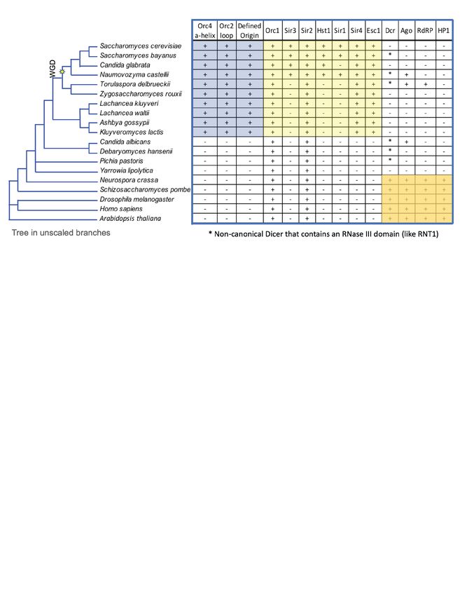
bioRxiv preprint doi: https://doi.org/10.1101/2020.07.04.187286. this version posted July 4, 2020. The copyright holder for this preprint (which
 was not certified by peer review) is the author/funder. It is made available under a CC-BY-NC 4.0 International license.

 ORC7 on origin DNA. The conservation of the a-helix and loop is restricted to a small clade of
 Saccharomyces-related species and where it has been determined, corresponds to the origins of DNA
 replication having a demonstrable consensus sequence (Raguraman, M.K and Liachko, I. in Kaplan19)
 (Fig. 5).

 It is known that in some budding yeasts, such as S. cerevisiae and K. lactis, ORC, functioning with Silent
 Information Regulator (SIR) proteins, is also required in transcriptional gene silencing of mating type loci,
 rDNA and telomeres20-22. Evolutionally, Sir2 and Sir4 preceded the acquisition of Sir1 and Sir3 (which is
 related to Orc1) in the ORC-Sir4-mediated transcriptional gene silencing pathway. In K. lactis, Sir4 binds
 directly to Orc1 but in S. cerevisiae, Sir1 binds to Orc1 and Sir4 binds to Sir3 that arose from Orc1 as a
 result of whole genome duplication22 (WGD, Fig. 5). In both species, Sir2 is required, as its histone
 deacetylase activity is essential for the gene silencing function. Interestingly, Sir4 binds to Esc1 which is
 located in the nuclear envelope, tethering the silent loci to the nuclear periphery23. Of relevance here is
 that Sir4 is related in structure to nuclear lamins, which are present in most eukaryotes but are absent in
 yeast24.

 We observed a very interesting co-evolution of origin sequence specification (Fig. 5, first three columns)
 and gene silencing (Fig. 5, remaining columns). The acquisition of sequence specific origins, the Orc4 a-
 helix and the Orc2 loop (Fig. 5, blue shadow) correlated precisely with the acquisition of ORC-Sir4-
 mediated transcriptional gene silencing (Fig. 5, yellow shadow).

 Dicer is an RNase III family member and a key mediator in the RNA interference (RNAi) pathway, which
 has been shown to control gene silencing by transcriptional and post-transcriptional mechanisms25.
 However, Dicer, but not other components of the RNAi pathway, has an RNAi-independent role in S.
 pombe in the termination of transcription at replication stress sites26. This may contribute to alleviation of
 R-loop mediated conflicts between DNA replication and transcription, particularly in repeated sequences
 and heterochromatin. The vast majority of eukaryotes that lack sequence-specific origins, including plants,
 animals and the majority of fungi including yeast have vast repeated sequences and heterochromatin and
 thus need RNAi25 (Fig. 5, orange shadow) or Dicer’s RNAi-independent role in maintaining genome
 stability, particularly if origin locations are stochastic, as has been shown in S. pombe27.

 Budding yeasts that lack sequence-specific origins, such as the pathogenic yeast Candida albicans and
 the industrial yeast Yarrowia lipolytica that can metabolize unusual hydrocarbons, have lost, or are in the
 process of losing RNAi28,29. Some retain a non-canonical Dicer (Dcr*) that has an RNase III domain and
 has been shown in C. albicans to exhibit RNAi to silence transposable elements and sub-telomeric
 repeated sequences28. They lack both the Orc4 a-helix and the Orc2 loop and do not have demonstratable
 sequence-specific origins. In this context, Y. lipolytica is an interesting case since it has lost Dicer and
 Argonaute (Ago) and lacks ORC-Sir4 silencing and sequence-specific origins. Y. lipolytica has dispersed
 rDNA gene clusters that are sub-telomeric and has a relatively low gene density (one gene per 3.3kb), far
 lower than the gene density found in S. cerevisiae (one gene per 2kb)30. It also uses Tay1, a TRF-like
 protein for telomeric and sub-telomeric gene silencing, which is more similar to the human shelterin
 complex mechanism31. Moreover, it is a heterothallic yeast, which does not switch its mating type and
 thus lacks silent mating type loci30. We suggest and that Y. lipolytica may be a useful specifies to
 investigate origin location and sequence specificity and we are studying replication patterning in this
 species.

 6
bioRxiv preprint doi: https://doi.org/10.1101/2020.07.04.187286. this version posted July 4, 2020. The copyright holder for this preprint (which
 was not certified by peer review) is the author/funder. It is made available under a CC-BY-NC 4.0 International license.

 The budding yeasts that have acquired sequence specific origins and ORC-Sir4-mediated gene silencing
 system are likely to have lost RNAi completely, albeit some retained the non-canonical Dicer (Dcr*). One
 budding yeast, T. delbrueckii, has ORC-Sir4 silencing and has retained Dcr* and Ago, but the latter are
 not involved in transcriptional gene silencing29. As species lost RNAi with a concomitant reduction in
 repeated sequences in the genome, including centromere associated repeated sequences, we suggest that
 in the Saccharomyces-related, ORC-Sir4 containing budding yeast that ORC evolved to bind DNA in a
 sequence specific manner, providing a mechanism to locate origins of DNA replication in intergenic
 regions6. Such a location would help maintain genome stability by reducing the possibility of conflicts
 between DNA replication and transcription, including the formation of R-loops32. The remaining repeated
 sequences in these species, such as the silent mating type loci in homothallic yeast, have evolved to be
 protected from loss by recombination and be transcriptionally silenced by an ORC-Sir4 dependent
 recruitment of the histone deacetylase Sir2. It is possible that in Y. lipolytica, Sir2 binds directly to ORC
 and thus bypasses the requirement for the other SIR proteins.

 In S. pombe, Orc4 has an AT-hook DNA binding domain at its amino-terminus that localizes initiation of
 DNA replication to AT-rich sequences in the genome33 even though replication origin utilization
 throughout the genome is known to be stochastic27. We found similar sequences are present in Orc4 in
 many fungi, including Neurospora crassa. Since the AT-hook sequences and the Orc4 a-helix and Orc2
 loop are absent in other fungi, animals and plants, they must have an alternative mechanism of specifying
 origin location, a topic of major interest.

 ORC is involved in maintenance of heterochromatin in Drosophila and Human, via an interaction between
 ORC1 and the heterochromatin protein HP134,35. Furthermore, ORC in Human cells is also involved in
 repression of transcription of the CCNE1 gene encoding Cyclin E via interactions with the histone
 methyltransferase SUV39H1 and the Retinoblastoma tumor suppressor protein (Rb)36. Thus ORC-
 dependent gene silencing may exist outside of species that have acquired Sir4.

 Main References
 1. Yuan, Z. et al. Structural basis of Mcm2-7 replicative helicase loading by ORC-Cdc6 and Cdt1.
 Nat Struct Mol Biol 24, 316–324 (2017).
 2. Marahrens, Y. & Stillman, B. A yeast chromosomal origin of DNA replication defined by
 multiple functional elements. Science 255, 817–823 (1992).
 3. Stinchcomb, D. T., Struhl, K. & Davis, R. W. Isolation and characterisation of a yeast
 chromosomal replicator. Nature 282, 39–43 (1979).
 4. Prioleau, M.-N. & MacAlpine, D. M. DNA replication origins-where do we begin? Genes Dev
 30, 1683–1697 (2016).
 5. Bell, S. P. & Stillman, B. ATP-dependent recognition of eukaryotic origins of DNA replication
 by a multiprotein complex. Nature 357, 128–134 (1992).
 6. Bell, S. P. & Labib, K. Chromosome Duplication in Saccharomyces cerevisiae. Genetics 203,
 1027–1067 (2016).
 7. Li, N. et al. Structure of the origin recognition complex bound to DNA replication origin. Nature
 355, 1–22 (2018).
 8. Tocilj, A. et al. Structure of the active form of human origin recognition complex and its ATPase
 motor module. eLife 6, 1822 (2017).
 9. Bleichert, F. & Berger, J. M. Crystal structure of the eukaryotic origin recognition complex.
 Nature 519, 321–326 (2015).

 7
bioRxiv preprint doi: https://doi.org/10.1101/2020.07.04.187286. this version posted July 4, 2020. The copyright holder for this preprint (which
 was not certified by peer review) is the author/funder. It is made available under a CC-BY-NC 4.0 International license.

 10. Liachko, I., Youngblood, R. A., Keich, U. & Dunham, M. J. High-resolution mapping,
 characterization, and optimization of autonomously replicating sequences in yeast. Genome Res
 23, 698–704 (2013).
 11. Hoggard, T. et al. High Throughput Analyses of Budding Yeast ARSs Reveal New DNA
 Elements Capable of Conferring Centromere-Independent Plasmid Propagation. G3 (Bethesda) 6,
 993–1012 (2016).
 12. Wilson, K. A., Kellie, J. L. & Wetmore, S. D. DNA-protein π-interactions in nature: abundance,
 structure, composition and strength of contacts between aromatic amino acids and DNA
 nucleobases or deoxyribose sugar. Nucleic Acids Res 42, 6726–6741 (2014).
 13. Siow, C. C., Nieduszynska, S. R., Müller, C. A. & Nieduszynski, C. A. OriDB, the DNA
 replication origin database updated and extended. Nucleic Acids Res 40, D682–6 (2012).
 14. Stevenson, J. B. & Gottschling, D. E. Telomeric chromatin modulates replication timing near
 chromosome ends. Genes Dev 13, 146–151 (1999).
 15. Soriano, I., Morafraile, E. C., Vázquez, E., Antequera, F. & Segurado, M. Different nucleosomal
 architectures at early and late replicating origins in Saccharomyces cerevisiae. BMC Genomics
 15, 791 (2014).
 16. Tanaka, S., Nakato, R., Katou, Y., Shirahige, K. & Araki, H. Origin Association of Sld3, Sld7,and
 Cdc45 Proteins Is a Key Stepfor Determination of Origin-Firing Timing. Curr Biol 21, 2055–
 2063 (2011).
 17. Lynch, K. L., Alvino, G. M., Kwan, E. X., Brewer, B. J. & Raghuraman, M. K. The effects of
 manipulating levels of replication initiation factors on origin firing efficiency in yeast. PLoS
 Genet 15, e1008430 (2019).
 18. Gros, J. et al. Post-licensing Specification of Eukaryotic Replication Origins by Facilitated
 Mcm2-7 Sliding along DNA. Mol cell 60, 797–807 (2015).
 19. Kaplan, D. L. The Initiation of DNA Replication in Eukaryotes. (Springer, 2016).
 doi:10.1007/978-3-319-24696-3
 20. Bell, S. P., Kobayashi, R. & Stillman, B. Yeast origin recognition complex functions in
 transcription silencing and DNA replication. Science 262, 1844–1849 (1993).
 21. Fox, C. A., Loo, S. & Dillin, A. The origin recognition complex has essential functions in
 transcriptional silencing and chromosomal replication. Genes Dev 9, 911–924 (1995).
 22. Hickman, M. A. & Rusche, L. N. Transcriptional silencing functions of the yeast protein
 Orc1/Sir3 subfunctionalized after gene duplication. Proceedings of the National Academy of
 Sciences 107, 19384–19389 (2010).
 23. Grunstein, M. & Gasser, S. M. Epigenetics in Saccharomyces cerevisiae. Cold Spring Harbor
 Perspectives in Biology 5, a017491 (2013).
 24. Diffley, J. F. & Stillman, B. Transcriptional silencing and lamins. Nature 342, 24–24 (1989).
 25. Martienssen, R. & Moazed, D. RNAi and heterochromatin assembly. Cold Spring Harbor … 7,
 a019323 (2015).
 26. Castel, S. E. et al. Dicer promotes transcription termination at sites of replication stress to
 maintain genome stability. Cell 159, 572–583 (2014).
 27. Dynamics of DNA replication in a eukaryotic cell. Proceedings of the National Academy of
 Sciences 116, 4973–4982 (2019).
 28. Drinnenberg, I. A. et al. RNAi in budding yeast. Science 326, 544–550 (2009).
 29. Ellahi, A. & Rine, J. Evolution and Functional Trajectory of Sir1 in Gene Silencing. Mol Cell
 Biol 36, 1164–1179 (2016).
 30. Yarrowia lipolytica. Yeast 29, 409–418 (2012).

 8
bioRxiv preprint doi: https://doi.org/10.1101/2020.07.04.187286. this version posted July 4, 2020. The copyright holder for this preprint (which
 was not certified by peer review) is the author/funder. It is made available under a CC-BY-NC 4.0 International license.

 31. Kramara, J. et al. Tay1 protein, a novel telomere binding factor from Yarrowia lipolytica. Journal
 of Biological Chemistry 285, 38078–38092 (2010).
 32. Hamperl, S., Bocek, M. J., Saldivar, J. C., Swigut, T. & Cimprich, K. A. Transcription-
 Replication Conflict Orientation Modulates R-Loop Levels and Activates Distinct DNA Damage
 Responses. Cell 170, 774–786 (2017).
 33. Chuang, R. Y. & Kelly, T. J. The fission yeast homologue of Orc4p binds to replication origin
 DNA via multiple AT-hooks. Proc Natl Acad Sci USA 96, 2656–2661 (1999).
 34. Pak, D. T. et al. Association of the origin recognition complex with heterochromatin and HP1 in
 higher eukaryotes. Cell 91, 311–323 (1997).
 35. Prasanth, S. G., Shen, Z., Prasanth, K. V. & Stillman, B. Human origin recognition complex is
 essential for HP1 binding to chromatin and heterochromatin organization. Proc Natl Acad Sci
 USA 107, 15093–15098 (2010).
 36. Hossain, M. & Stillman, B. Opposing roles for DNA replication initiator proteins ORC1 and
 CDC6 in control of Cyclin E gene transcription. eLife 5, 10.7554–eLife.12785 (2016).
 37. Nieduszynski, C. A., Knox, Y. & Donaldson, A. D. Genome-wide identification of replication
 origins in yeast by comparative genomics. Genes Dev 20, 1874–1879 (2006).

 Figure and Table Legends
 Fig. 1 | DNA interacting Orc4 a-helix and Orc2 loop are essential. a, Top-view of ORC-Cdc6 structure
 encircling an origin DNA with Orc1-6 and Cdc6 indicated. Recolored from previous cryo-EM work1
 OCCM structure (PDB code 5udb) and Orc4 a-helix and Orc2 loop that interact with DNA are colored in
 red. b, Orc4 structure superposition among Human Orc4 in blue (from PDB code 5uj7), Drosophila Orc4
 in grey (from PDB code 4xgc) and S. cerevisiae Orc4 in salmon (from PDB code 5udb). Orc4 a-helix that
 interacts with DNA is colored in red. c, Multiple sequence alignment of Orc4 among representing
 eukaryotic species as indicated. Orc4 a-helix region indicated with species that don’t have sequence
 specific origins shadowed in blue and species that sequence specific origins exist shadowed in pink. d-e,
 Maps of mutant viability phenotype from plasmid shuffle assay: Orc4 a-helix in a helical wheel (d) and
 Orc2 loop in connected line (e). Mutant deficiency phenotypes (Extended Data Fig. 2 and 3) are
 summarized in color codes as indicated. Amino acids indicated in one-letter abbreviation. Different mutant
 types are indicated with different shapes. f, Growth curves of NTAP-Orc4 integrated strains (see Methods)
 in YPD with initiation OD600 at 0.05 at 30˚C to measure OD600 at different time points.

 Fig. 2 | Selected origin sequence changes following a Massively Parallel Origin Selection (MPOS)
 assay. a, Schematic diagram of MPOS assay. b, ARS motifs for Orc4-integrated variants at A and B1
 elements generated using an ARS1 (ARS416) variant library. See methods for how motifs are graphically
 rendered. c, Magnified view of the A element region in b from Orc4WT, orc4F485A, Y486A, orc4Y486Q strains.
 Dark purple rectangles indicate the major changes at positions 29-30 in the Orc4 mutant strains. d, Top-
 view of Orc4 a-helix insertion from ORC-DNA structure at 3Å (PDB code 5zr1) positioned in the DNA
 major groove. F485, Y486, N489 and R478 interact with DNA in base-specific (specificity) and base-
 nonspecific (affinity) manner. e-f, same as in d, but view in different angles. Red asterisks denote the
 base-specific interactions between amino acid and DNA base. Blue asterisks denote the base-nonspecific
 interaction between amino acid and DNA phosphate backbone. Green asterisks denote the interaction
 between amino acids. Prime symbols denote bases on the opposite strand. Bases numbering denotes the
 positions in logo (see b). e shows the hydrophobic interaction between F485 and T-rich region T26-T29,
 base-specific interaction between N489 and T28, base-nonspecific interaction between N489 and
 phosphate backbone of T28, and R478 interaction with V475. f shows the aromatic edge-face interaction

 9
bioRxiv preprint doi: https://doi.org/10.1101/2020.07.04.187286. this version posted July 4, 2020. The copyright holder for this preprint (which
 was not certified by peer review) is the author/funder. It is made available under a CC-BY-NC 4.0 International license.

 between Y486 and A29’on the opposite strand, hydrophobic interaction between Y486 and C30’, base-
 nonspecific interaction between R478 and phosphate backbone of A27’, and R478 interaction with A487
 and V475.

 Fig. 3 | Orc4 a-helix mutants change the pattern of origin firing and MCM binding. Genome-wide
 origin firing profile, a-factor blocked and released into S phase in 200mM hydroxyurea (HU) for 90 mins.
 a, the completely new origin location ChrXII: 95,827-117,318 in orc4F485I, Y486Q strain. b, shows the
 completely new origin location ChrV: 248,069-251,910 in orc4F485A, Y486A strain. c, Whole genome
 replication profiles. Chromosome IV (ChrIV) is shown as a representation. Strains are in the order of
 shorter to longer doubling time (Extended Data Table 3) from top to bottom. d-i, ChIP profile of MCM
 (anti-Mcm2) of NTAP-Orc4 integrated strains at ChrIV in G1 phase and it’s comparison to replication
 origins profile. orc4F485I, Y486Q strain is used as an example of NTAP-Orc4 mutant strains to compare with
 Orc4WT strain in whole-chromosome view (d) and zoom-in views (e-i). Green arrows indicate the example
 locations of origins with firing pattern changes in the orc4F485I, Y486Q strain. At these locations, similar
 changes were also observed in other mutants (see c). Black arrows indicate the example locations of
 origins with firing pattern that remained the same in orc4F485I, Y486Q strain. At these locations, origins with
 firing pattern also remained the same in other mutants (see c). Red arrows indicate the example locations
 of origin firing pattern changes are unique in the orc4F485I, Y486Q strain but not in other mutant strains (see
 c). Blue arrows indicate the examples of inactive late origins in HU in WT that have lower or no ChIP-
 Mcm2 signal in orc4F485I, Y486Q mutant strain and are not active origins in the mutant strain.

 Fig. 4 | Genomic origin firing pattern changes are sequence specific. DNA sequences under each origin
 replication peaks that are predicted to match the ARS consensus sequence (ACS) were obtained from
 OriDB13,37. Genome-wide statistical analysis was performed to check the dependence of origin firing peak
 height on dinucleotide identity at position 29-30 that correspond to logo derived from the MPOS data (Fig.
 2c). P-values that correspond to a one-way ANOVA test. Asterisks denotation: *p
bioRxiv preprint doi: https://doi.org/10.1101/2020.07.04.187286. this version posted July 4, 2020. The copyright holder for this preprint (which
 was not certified by peer review) is the author/funder. It is made available under a CC-BY-NC 4.0 International license.

 Methods
 Yeast genetic methods and strain construction
 Yeast strains generated in this study (described in Supplementary Methods Table 1) were derived from
 W303-1a (MATa ade2-1 can1-100 his3-11,15 leu2-3,112 trp1-1 ura3-1).

 The YB51 (orc4∆::TRP1 + pORC4/URA3) strain was used for plasmid shuffle assay (see Methods,
 Plasmid shuffle assay). A PCR-based gene deletion strategy was used for disrupting endogenous Orc4
 with TRP1 and a URA3-containing plasmid (pRS416) carrying wildtype ORC4 gene is used as
 complement.

 The Orc4 site-directed mutation constructs-containing plasmids were used for plasmid shuffle assay (see
 Methods, Plasmid shuffle assay). Based on a CEN-based LEU2-containing plasmid constructs (pRS415)
 carrying wildtype ORC4 gene, Orc4 site-directed mutation constructs were created using PCR
 mutagenesis strategy, confirmed by DNA sequencing (see Methods, Plasmid shuffle assay).

 The NTAP-Orc4 integrated yeast strains were used for phenotype characterization assays, including the
 genome-wide DNA replication origin profile analyses, chromatin-immunoprecipitation and massively
 parallel origin mutagenesis and selection assay. NTAP-Orc4 integrated strains were derived from YB1588
 (MATa orc4∆::TRP1 bar1∆::TRP1 LEU2::BrdU-Inc + pORC4/URA3), which is a meiotic product of a
 diploid strain obtained by crossing YB51 (MATa orc4∆::TRP1 + pORC4/URA3) and YB1549
 (MATa bar1Δ::TRP1 LEU2::BrdU-Inc). YB1549 was derived from YS2251 (MATa bar1Δ::TRP1) by
 inserting a BrdU-INC cassette38 with LEU2 to facilitate EdU incorporation.

 NTAP-Orc4 construct was used for NTAP-Orc4 integrated strains construction, which was generated
 using a PCR based strategy with tag coming from pBS176139 (purchased from Euroscarf). The construct
 is inserted into his3 locus of YB1588 using CRISPR/Cas9 system40. Then, the plasmid containing Cas9
 gene was dropped off by non-selective culture and tested for loss of plasmid marker. Subsequently, the
 plasmid carrying wildtype Orc4 gene was dropped off by counter selecting on 5 fluoroorotic acid 5-FOA
 plates for loss of URA3. The loss of pORC4/URA3 were confirmed by PCR and sequencing in combination
 with phenotypic assessment.

 Plasmid shuffle assay
 The Orc4 a-helix mutants were screened for function in vivo using plasmid shuffle assay similar to
 previously described41. The Orc4 site-directed mutation constructs-containing plasmids with LEU2
 marker were transformed into YB51 (orc4∆::TRP1 + pORC4/URA3) and selected on SC-Leu-Ura plates.
 The transformants were isolated, grown in YPD overnight, and spotted onto 5-FOA plates with 10-fold
 serial dilutions starting from 1.5x10⁷ cells to select for loss of URA3 plasmid carrying the wild-type Orc4.
 As control, the same dilutions were spotted on YPD plates. YPD or 5-FOA plates were cultured under
 30˚C, or 25˚C or 37˚C to test their cold or temperature sensitivity.

 Cell extract preparation, immunoprecipitation, immunoblot analysis and antibodies
 Whole cell extraction from NTAP-Orc4 integrated strains (see Methods, Yeast genetic methods and strain
 construction) was prepared as previously described41. Cell extracts were analyzed for protein
 concentrations. Immunoprecipitation procedures were performed by mixing ∼1.6 mg of total proteins and
 30 µl of the IgG Sepharose 6 Fast Flow beads (GE Healthcare, Cat# 17-0969-01) at 4°C for 2h and
 precipitating the NTAP tagged Orc4. The beads were washed extensively with EBX buffer (recipe same

 11
bioRxiv preprint doi: https://doi.org/10.1101/2020.07.04.187286. this version posted July 4, 2020. The copyright holder for this preprint (which
 was not certified by peer review) is the author/funder. It is made available under a CC-BY-NC 4.0 International license.

 as previously published41) and boiled in 30ul loading sample buffer (cite cold spring harbor recipe online).
 Proteins from immunoprecipitation (IP) and cell extract (as IP input) were fractionated by SDS-10%
 PAGE and transferred to nitrocellulose membrane. Immunoblot analysis was performed using antibodies
 against Orc4 (SB12) used at 1:2000 dilution and Orc1 (SB13) used at 1:1000 dilution and TBS with 0.05%
 Tween 20 was used for preparing blocking and washing solutions.

 Cell growth, block, synchronization and flow cytometry analysis
 Exponentially growing yeast cells (∼107 cells/mL) in YPD were synchronized in G1 with 25 ng/mL of α-
 factor (bar1Δ strains are used in this study) for 3h at 30°C. To release from G1 arrest, cells were collected
 by filtration and promptly washed twice on the filter using one culture volume of H2O and then
 resuspended into YPD medium. 1ml of cells was collected at different time points by adding sodium azide
 to final concentration at 0.1%. Cells are quickly centrifuged, resuspended with 400ul H2O and fixed by
 adding 1ml 100% ethanol and rotate overnight at 4˚C. Cells then are quickly centrifuged, washed one
 time with H2O, resuspended in 250ul RNaseA (Sigma-Aldrich) solution (2mg/ml), incubated for 4h in a
 37˚C shaker and then sonicated using a Tekmar Sonic Disruptor with 630-0418 Tapered Microtip for 2
 cycles of pulse for 1 second “ON”, 1 second “OFF” at amplitude setting 22-25%. Proteinase K (Sigma-
 Aldrich) solution was added to final concentration at 1mg/ml and incubate for 1h in 50˚C in Eppendorf
 Thermomixer R Mixer, 1.5ml Block with speed at 750rpm. Cells were then quickly centrifuged, resuspend
 in 50mM Tris PH7.5 with SYBR green I (Thermo Fisher) diluted at 1:10,000 ratio and filtered through
 strainer cap tubes (Corning™ Falcon™ Test Tube with Cell Strainer Snap Cap). BD LSRFortessa Dual
 Special Order System instrument and BD FACSDiva Software Version 8.0.1 Firmware Version 1.4 (BD
 LSRFortessa) were used to collect the data by measuring SYBR green signal. Same number of yeast cells
 data (30,000 events per run) were collected for each sample. FlowJo Version 10.6.1 was used to analyze
 the data and no gating strategy used.

 Massively parallel origin selection (MPOS) assay
 Both the ARS1 (ARS416) and HMR-E (ARS317) libraries used ARS sequences 150bp in length and
 synthesized with a 15% mutation rate at each position. Variant ARSs were cloned in bulk into a HIS3-
 containing plasmid. The libraries were then transformed into NTAP-Orc4 integrated yeast strains (see
 Methods, Yeast genetic methods and strain construction). The transformed cells were plated on SC-his
 plate, grew in 30˚C incubator, washed off from plates when saturated, inoculated into SC-his medium and
 shook at 30˚C shaker till it reached saturation to harvest. ARS-containing plasmid DNA was isolated and
 PCR-amplified and ligated with custom inline barcodes (Supplementary Method Table 3), quantified,
 pooled, and submitted for sequencing. Computational analyses of MPOS assay data are described below
 (see Methods, Computational analyses of MPOS data). DNA sequencing data were submitted to the
 Sequence Read Archive database (see Methods, Data and code availability).

 Computational analyses of MPOS data
 Processing of MPOS data. Illumina reads from the MPOS experiments were analyze using custom
 Python scripts. The output of this pipeline was, for each library or selected sample, a list of variant ARS
 sequences with each sequence assigned a corresponding read count. These lists were used as input to both
 the ER and IM motif modeling algorithms described below

 Matrix models for ARS motifs. Our motif modeling effort aimed to predict the activity of a variant ARS
 based on its DNA sequence. Specifically, we sought a mathematical function ( ) that quantifies the

 12
bioRxiv preprint doi: https://doi.org/10.1101/2020.07.04.187286. this version posted July 4, 2020. The copyright holder for this preprint (which
 was not certified by peer review) is the author/funder. It is made available under a CC-BY-NC 4.0 International license.

 activity of an arbitrary input ARS DNA sequence . We assumed this function could be represented by a
 matrix model42, i.e.,

 ( ) = ( ( !" !"
 ! "

 where = , , , indexes the four DNA bases, = 1, 2, … , indexes nucleotide positions, the
 sequence = { !" } is represented by a 4 × matrix of indicator variables ( !" = 1 if base occurs at
 position ; !" = 0 otherwise), and = { !" } is a 4 × matrix of model parameters that must be inferred
 from data. All inferred motifs were limited to sequences of length = 50 encompasing both the A and
 B1 elements of the assayed ARSs. To facilitate the comparison of motifs to one another, both visually and
 #
 through PCA analysis, motif parameters were centered and rescaled via the transformation !" → !" /
 # $ #&
 where !" = !" − % ∑!! !! " and = ?∑! ∑" !" .

 Sequence logos. Sequence logos were generated by Logomaker 43. In these representations of motif
 parameters, the value of !" is represented by the height of character at position (or negative that height
 if the character is drawn below the x-axis).

 Principal component analysis (PCA). The PCAs shown in Extended Data Figure S10 were performed
 on inferred motifs as follows. The motif parameters were first centered and normalized as described
 above. Each parameter matrix was then unrolled into a 4 × 1 vector, where = 50. Standard PCA
 analysis was then performed on different motif subsets, as shown in panel a.

 Motif inference. The inference of motif parameters was performed using a second-generation version of
 the MPAthic software package44. MPAthic enables motif inference using either enrichment ratios (ER) or
 information maximization (IM).

 ER inference is the standard way of computing sequence motifs from massively parallel selection
 experiments45. Here, parameter values '( are given by
 ER
 !")*+*,-*.
 !" = log& +/01213 ,
 !"
 +/01213
 where !" is the fraction of sequences in the initial ARS library that have base at position , and
 )*+*,-*.
 !" is defined similarly for selected ARS sequences. These fractions were computed using a
 pseudocount of 1.

 IM inference seeks to identify parameters 45 that maximize the mutual information between the
 predicted activity of an assayed ARS sequence and the sample that sequence was observed in. Specifically,
 one aims to maximize

 ( |sample)
 ( ) = ( (sample) I ( |sample)log&
 ( )
 )267+*)

 where “sample” indicates either the library sample or the selected sample, and ( |sample) is the
 distribution of activities assigned to the sequences in that sample by a motif with parameters . For a given

 13
bioRxiv preprint doi: https://doi.org/10.1101/2020.07.04.187286. this version posted July 4, 2020. The copyright holder for this preprint (which was not certified by peer review) is the author/funder. It is made available under a CC-BY-NC 4.0 International license. choice of , the distribution ( |sample) was computed as in Kinney et al. 46: the activities in both samples combined were sorted, replaced by their ranks, and binned into 1000 equipopulated bins; for each sample, the distribution of sequence counts across bins was then smoothed using Gaussian kernel having a standard deviation of 20 bins. The marginal probability was subsequently computed as ( ) = ∑)267+*) (sample) ( |sample). The optimal values 45 were identified using a Metropolis Monte Carlo simulation in which each was assigned relative probability of 289(;) , where is the number of read counts in both the library sample and selected samples combined. Each Monte Carlo run was initiated at random parameter values then carried out for 25,000 steps. Each reported motif resulted from averaging together the end-points of five independent Monte Carlo runs. This IM inference strategy closely followed the one described previously46. The present work is the first to show that, as predicted from previous theoretical arguments47, IM motif inference removes systematic experiment-to-experiment variation that confounds ER motif inference. Genome-wide replication origin profile analysis Isolation and preparation of DNA for genome-wide replication origin profile analysis is similar to previously described48. Yeast cells were synchronized in G1 with α-factor and were released into medium containing 0.2 mg/mL pronase E, 0.2 M HU, and 0.5 mM EdU. Cells were collected by centrifugation at 90 mins after release into S phase. Genomic DNA was extracted and fragmented. EdU-genomic DNA was then biotinylated using the Click reaction and purified using Streptavidin T1 magnetic beads (Invitrogen). Libraries for Illumina sequencing were constructed using TruSeq ChIP Library Preparation Kit (Illumina). Computational analyses of sequencing data are described below (see Methods, Computational analyses of replication origin profile and ChIP-seq data). DNA sequencing data were submitted to the Sequence Read Archive database (see Methods, Data and code availability). Chromatin immunoprecipitation The ChIP-seq for Orc1 and Mcm2 were performed as described49 with modification. About 109 synchronized yeast cells were fixed with 1% formaldehyde for 15 min at room temperature (RT), then quenched with 130 mM glycine for 5 min at RT, harvested by centrifugation, washed twice with TBS (50 mM Tris.HCl pH 7.6, 150 mM NaCl), and flash frozen. Cell pellets were resuspended in 600 µl lysis buffer (50 mM HEPES-KOH pH 7.5, 150 mM NaCl, 1 mM EDTA, 1% Triton X-100, 0.1% Na- Deoxycholate, 0.1% SDS, 1 mM PMSF, protease inhibitor tablet (Roche)), and disrupted by bead beating using multi-tube vortex (Baxter Scientific Products SP Multi-Tube Vortexer S8215-1) for 12-15 × 30s at maximum setting. Cell extracts were collected and sonicated using Bioruptor (UCD-200, Diagenode) for 38 cycles of pulse for 30 seconds “ON”, 30 seconds “OFF” at amplitude setting High (H). The extract was centrifuged for 5 min at 14,000 rpm. The soluble chromatin was used for IP. Antibody against Mcm2 (mcm228) was preincubated with washed Dynabeads Protein A/G. For each immunoprecipitation, 80 μl antibody-coupled beads was added to soluble chromatin. Samples were incubated overnight at 4°C with rotation, after which the beads were collected on magnetic stands, and washed 3 times with 1 ml lysis buffer and once with 1 ml TE, and eluted with 250 μl preheated buffer (50 mM Tris.HCl pH 8.0, 10 mM EDTA, 1% SDS) at 65°C for 15 min. Immunoprecipitated samples were incubated overnight at 65°C to reverse crosslink, and treated with 50 μg RNase A at 37°C for 1 hr. 5μl proteinase K (Roche) was added and incubation was continued at 55°C for 1 hr. Samples were purified using MinElute PCR purification kit (Qiagen). Libraries for Illumina sequencing were constructed using TruSeq ChIP Library Preparation Kit (Illumina). Computational analyses of sequencing data are described below (see Methods, Computational analyses of replication origin profile and ChIP-seq data). 14
bioRxiv preprint doi: https://doi.org/10.1101/2020.07.04.187286. this version posted July 4, 2020. The copyright holder for this preprint (which
 was not certified by peer review) is the author/funder. It is made available under a CC-BY-NC 4.0 International license.

 Computational analyses of replication origin profile and ChIP-seq data
 Illumina reads from the genome-wide replication origin profiling and ChIP-seq experiments were mapped
 to the S. cerevisiae S288C genome using BWA, after which pileup files were created using SAMtools
 (http://www.htslib.org). Pileup counts were then smoothed via convolution with a uniform kernel of width
 5000 bp (for replication origin profiles) or 300 bp (for ChIP-seq). To normalize the profiles relative to one
 another, we computed the number of reads bounding 99.5% of positions within each profile and divided
 the entire profile by this number.

 Data availability
 Raw Illumina reads are available at [SRA ACCESSION NUMBER].

 Code availability
 Processed data files, analysis scripts, and scripts used for figure generation are available at [GITHUB
 URL].

 Note for reviewers: We will provide all code used for data analysis in Github as open source software
 upon acceptance. It will be freely available on Github. SRA accession number will be publicly available
 upon revision of the paper. In the meantime, all raw data, processed data and custom codes have currently
 been uploaded onto an unstructured repository:
 MPOS assay data link: labshare.cshl.edu/shares/stillmanlab/www-data/yixinhu/MPOS/
 Origin firing EdU profile data link: labshare.cshl.edu/shares/stillmanlab/www-data/yixinhu/EdU/
 ChIP-seq data link: labshare.cshl.edu/shares/stillmanlab/www-data/yixinhu/ChIP-seq/
 Custom codes link: labshare.cshl.edu/shares/stillmanlab/www-data/yixinhu/Code/

 Method References / Additional References
 38. Viggiani, C. J. & Aparicio, O. M. New vectors for simplified construction of BrdU-Incorporating
 strains of Saccharomyces cerevisiae. Yeast 23, 1045–1051 (2006).
 39. Puig, O. et al. The tandem affinity purification (TAP) method: a general procedure of protein
 complex purification. Methods 24, 218–229 (2001).
 40. Anand, R., Memisoglu, G. & Haber, J. Cas9-mediated gene editing in Saccharomyces cerevisiae.
 (2017). doi:10.1038/protex.2017.021a
 41. Sheu, Y.-J. & Stillman, B. The Dbf4-Cdc7 kinase promotes S phase by alleviating an inhibitory
 activity in Mcm4. Nature 463, 113–117 (2010).
 42. Kinney, J. B. & McCandlish, D. M. Massively Parallel Assays and Quantitative Sequence-
 Function Relationships. Annu Rev Genomics Hum Genet 20, 99–127 (2019).
 43. Tareen, A. & Kinney, J. B. Logomaker: beautiful sequence logos in Python. Bioinformatics 36,
 2272–2274 (2020).
 44. Ireland, W. T. & Kinney, J. B. MPAthic: Quantitative Modeling of Sequence-Function
 Relationships for massively parallel assays. bioRxiv 17, doi:10.1101–054676 (2016).
 45. Stormo, G. D. Modeling the specificity of protein-DNA interactions. Quant Biol 1, 115–130
 (2013).
 46. Kinney, J. B., Murugan, A., Callan, C. G. & Cox, E. C. Using deep sequencing to characterize the
 biophysical mechanism of a transcriptional regulatory sequence. Proceedings of the National
 Academy of Sciences 107, 9158–9163 (2010).

 15
You can also read