Protein interface redesign facilitates the transformation of nanocage building blocks to 1D and 2D nanomaterials - Nature
←
→
Page content transcription
If your browser does not render page correctly, please read the page content below
ARTICLE
                  https://doi.org/10.1038/s41467-021-25199-x                 OPEN

                  Protein interface redesign facilitates the
                  transformation of nanocage building blocks
                  to 1D and 2D nanomaterials
                  Xiaorong Zhang1, Yu Liu1, Bowen Zheng1, Jiachen Zang1, Chenyan Lv1, Tuo Zhang                                              1 ✉,   Hongfei Wang2 &
                  Guanghua Zhao 1 ✉
1234567890():,;

                  Although various artificial protein nanoarchitectures have been constructed, controlling the
                  transformation between different protein assemblies has largely been unexplored. Here, we
                  describe an approach to realize the self-assembly transformation of dimeric building blocks
                  by adjusting their geometric arrangement. Thermotoga maritima ferritin (TmFtn) naturally
                  occurs as a dimer; twelve of these dimers interact with each other in a head-to-side manner
                  to generate 24-meric hollow protein nanocage in the presence of Ca2+ or PEG. By tuning two
                  contiguous dimeric proteins to interact in a fully or partially side-by-side fashion through
                  protein interface redesign, we can render the self-assembly transformation of such dimeric
                  building blocks from the protein nanocage to filament, nanorod and nanoribbon in response
                  to multiple external stimuli. We show similar dimeric protein building blocks can generate
                  three kinds of protein materials in a manner that highly resembles natural pentamer building
                  blocks from viral capsids that form different protein assemblies.

                  1 Collegeof Food Science & Nutritional Engineering, China Agricultural University, Beijing Key Laboratory of Functional Food from Plant Resources, Beijing
                  100083, China. 2 Key Laboratory of Chemical Biology and Molecular Engineering of Education Ministry, Key Laboratory of Energy Conversion and Storage
                  Materials of Shanxi Province, Institute of Molecular Science, Shanxi University, Taiyuan, China. ✉email: zhangtuo@cau.edu.cn; gzhao@cau.edu.cn

                  NATURE COMMUNICATIONS | (2021)12:4849 | https://doi.org/10.1038/s41467-021-25199-x | www.nature.com/naturecommunications                                1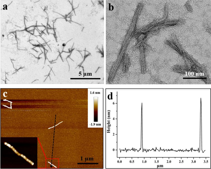
ARTICLE                                                                  NATURE COMMUNICATIONS | https://doi.org/10.1038/s41467-021-25199-x

S
      hape transformation phenomena are ubiquitous in nature.                 side manner by protein interface redesign, the self-assembly of
      Many living organisms by shape transformation perform                   these similar dimeric protein molecules are able to transform
      shape-to-function activities in response to the external                from the inherent hollow protein nanocage into nanoribbon in
environment1. For instance, Amoeba proteus undergoes multi-                   the presence of PEG (Fig. 1c). The dimensions of these filaments,
directional shape transformation to form pseudopod for naviga-                nanorods, and nanoribbons collectively span between nanometer
tion and rapid path alteration. These phenomena have triggered                and micrometer scales (50 nm to 4.0 µm). In-depth character-
tremendous interest in mimicking the structure–property rela-                 ization by X-ray crystallography, transmission electron micro-
tionship of living systems. At a molecular level, shape-shifting              scopy (TEM), and atomic force microscopy (AFM) confirmed
related to DNA2, RNA3, peptides4, and small molecules5 has been               that this protein interface redesign approach can regulate the
reported in recent years owing to their relatively simple and                 transformation between hollow protein nanocage and 1D and 2D
controllable structure. In viral capsids, a single protein fold can be        nanomaterials. This approach opens up an avenue for con-
evolved to form multiple oligomeric states with different                     structing 1D or 2D nanoarchitectures with the building blocks of
symmetries6, but to construct smart protein architectures artifi-              hollow protein nanocage as starting materials.
cially whose structure and shape transformation could be
modulated by external stimuli remains challenging.
                                                                              Results
   Proteins, as Nature’s most versatile building blocks, are mainly
                                                                              Natural dimeric TmFtn assembly into hollow protein nano-
responsible for the complexity of living organisms7. During
                                                                              cages. The four helix bundle structure, which is widely distributed
evolution, proteins have acquired self-assembly properties to
                                                                              in Nature, has been utilized as building blocks to construct a
construct a variety of large, complex, and symmetric architectures
                                                                              number of proteins and enzymes to perform a considerably wide
such as one-dimensional (1D) actin filaments8, two-dimensional
                                                                              range of functions, such as iron storage by ferritin26, DNA pro-
(2D) bacterial surface layers (S-layers)9, and three-dimensional
                                                                              tection by Dps protein27, copper storage by Csp142, electron
(3D) light-harvesting protein complexes of phycobilisomes10,
                                                                              transfer by cytochrome cb56243, ribonucleotide reduced into
thereby endowing their hosts with plenty of functions. It is well
                                                                              deoxynucleotide by R2 subunit of ribonucleotide reductase44,
known that protein–protein interactions (PPIs) at protein inter-
                                                                              methane oxidized into methanol by methane monooxygenase45,
faces are the chief contributors to construct the diversified protein
                                                                              and so on. Four helix bundles are also attractive for synthetic
nanostructures11–13. Following Nature’s inspiration to assemble
                                                                              chemists because its interfaces are dominated by side chain and
protein building blocks into exquisite nanostructures, various
                                                                              side chain interactions, which can be more tunable than β-
self-assembly strategies, such as symmetry-directed design14–17,
                                                                              strands. This study focuses on TmFtn, a naturally dimeric protein
metal coordination7,18–20, host–guest interactions21,22, and the
                                                                              consisting of two antiparallel four helix bundles, as shown in
use of bifunctional ligands23,24, have been applied to construct
                                                                              Supplementary Fig. 1. In contrast, most known ferritins are
1D, 2D, and 3D hierarchical protein nanostructures. Among
                                                                              usually composed of 24 identical or similar subunits that
these various protein nanostructures, natural protein nanocages
                                                                              assemble into a shell-like structure. Interestingly, the dimeric
represent a class of versatile nanomaterials that fulfill a wide
                                                                              TmFtn can convert into 24-meric protein nanocage in the pre-
range of functions, such as CO2 fixation by carboxysomes25, iron
                                                                              sence of divalent metal ions such as Mg(II) and Ca(II)46,47.
metabolism by ferritins26, DNA protection by Dps27, and nucleic
                                                                              Consistent with these recent findings, our results show that, in
acid storage and transport by viral capsids28. By taking advantage
                                                                              solution, purified TmFtn molecules (Supplementary Fig. 2) exist
of their well-defined architectures, isolated interiors, and high
                                                                              as dimers and can self-assemble into 24-meric hollow protein
biocompatibility, scientists have subverted the above natural
                                                                              nanocage in the presence of Ca2+, and addition of EDTA causes
functions of the protein nanocages and explored them as nano-
                                                                              the formed protein nanocage disassembly back into its dimeric
containers for encapsulation and delivery of bioactive cargo
                                                                              form (Supplementary Fig. 3a), indicative of a reversible process of
molecules29, as bio-templates for preparation of various
                                                                              protein assembly (Supplementary Fig. 3b).
nanomaterials30, and as reaction centers for multienzyme
                                                                                 The crystal structure of 24-meric Tmftn determined at a
catalysis31. So far, different strategies such as de novo design32–34,
                                                                              resolution of 2.2 Å (Supplementary Tables 1 and 2) shows that
fusion protein14,35,36, directed evolution37–39, and key interface
                                                                              one calcium ion located nearby C3–C4 interface is coordinated
redesign40,41 have been built to create a variety of artificial hollow
                                                                              with two acidic residues (Glu51 and Glu132) and three water
protein nanocages that rival the size, property, and functionality
                                                                              molecules (Supplementary Fig. 3c, d). Careful analyses of the
of their natural analogs. Despite these advances, rendering PPIs
                                                                              crystal structure revealed that a group of acidic amino acid
controllable to facilitate the transformation of the building blocks
                                                                              residues are lined along with the C3–C4 interface, producing
from protein nanocages into 1D or high-order nanomaterials in
                                                                              electrostatic repulsion along with this interface in the absence of
the laboratory has yet to be explored.
                                                                              calcium ions. This might be an important reason why TmFtn
   Herein we introduce a protein interface redesign approach that
                                                                              molecules naturally exist as protein dimers in solution in the
could be used for the self-assembly transformation of dimeric
                                                                              absence of metal ions. In contrast, calcium ions near the C3–C4
building blocks from hollow protein nanocage to filament,
                                                                              interface can essentially eliminate such electrostatic repulsion
nanorod, and nanoribbon. The basic building block of this
                                                                              through their interaction with the acidic residues, strengthening
approach is a naturally occurring dimeric protein—Thermotoga
                                                                              the stability of C3–C4 interface, finally producing the 24-meric
maritima ferritin (TmFtn), which tends to assemble into 24-
                                                                              protein nanocage. Besides, each protein nanocage is composed of
meric hollow protein nanocage induced by calcium ions (Fig. 1a).
                                                                              12 protein dimers, and any of the 2 adjacent protein dimers
The crystal structure analyses revealed that the assembly of two
                                                                              interact with each other in a head-to-side manner (Supplemen-
adjacent dimeric protein molecules in a head-to-side manner is
                                                                              tary Fig. 3b, d). Based on these findings, it is reasonable to believe
responsible for the formation of such a shell-like structure
                                                                              that calcium ions facilitate the interaction of these protein dimers
(Fig. 1a). In contrast, upon tuning such head-to-side state to a
                                                                              in a head-to-side manner, resulting in the formation of hollow
fully side-by-side manner by protein interface redesign, similar
                                                                              protein nanocage.
dimeric protein molecules self-assemble into filaments or
nanorods in the presence of calcium ions or polyethylene glycol
(PEG), respectively (Fig. 1b). Differently, when we adjusted the              Engineering dimeric TmFtn for assembly into filaments and
two adjacent dimeric proteins to interact in a partially side-by-             nanorods. The above head-to-side interaction manner is

2                 NATURE COMMUNICATIONS | (2021)12:4849 | https://doi.org/10.1038/s41467-021-25199-x | www.nature.com/naturecommunications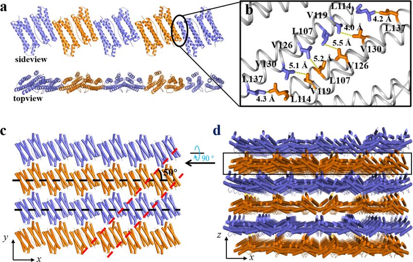
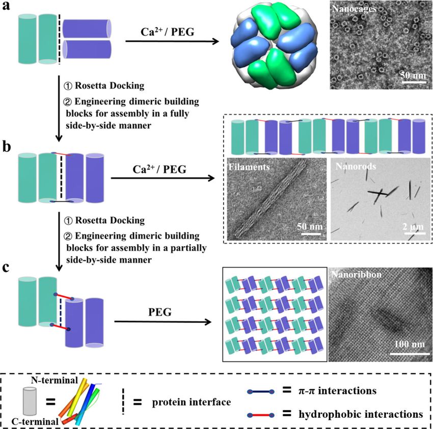
NATURE COMMUNICATIONS | https://doi.org/10.1038/s41467-021-25199-x                                                                     ARTICLE

Fig. 1 Schematic representation of the protein interface redesign approach used to control the self-assembly transformation of dimeric building blocks
between different types of nanomaterials. a The naturally occurring dimeric Thermotoga maritima ferritin (TmFtn) molecules interact with each other in a
head-to-side manner to form 24-meric protein nanocages in the presence of calcium ions or PEG. b, c Upon Rosetta docking and designing new PPIs
between adjacent dimeric building blocks in a fully or partially side-by-side manner, the assembly of similar dimeric building blocks can be transformed
from the above formed hollow protein nanocages into nanofilaments or nanorods or nanoribbons in the presence of calcium ions or PEG.

ubiquitous in nearly all known ferritins from animal, plant, to                 concentration of NaCl from 0 to 500 mM; Supplementary Fig. 7).
bacteria. Based on this interesting phenomenon, we envisioned                   This might be because the designed interaction forces are not
that if this head-to-side interaction manner between two adjacent               strong enough to drive protein dimers assembly in a side-by-side
dimeric TmFtn was adjusted to a fully side-by-side manner by                    manner in solution. This situation is, at least partly, derived from
protein interface redesign, the 24-meric TmFtn nanocages would                  the fact that a group of acidic amino acid residues from C-helix
transform into 1D protein nanomaterials. Therefore, we describe                 and D-helix occur between the designed subunits interface
a three-step computational assisted method for realizing this idea:             (Supplementary Fig. 8), reducing the designed attraction, thereby
(1) two adjacent dimers were manually placed in a fully side-by-                impeding adjacent FLAL molecule assembly in the designed side-
side manner as shown in Supplementary Fig. 4a, (2) RosettaDock                  by-side manner.
algorithm48 was carried out to optimize such fully side-by-side                    Inspired by the fact that Ca2+ has the ability to induce natural
interactions into a more complementary protein–protein inter-                   TmFtn dimer assembly into 24-meric protein nanocage through
face, and (3) building interactions at the designed interface to                its interaction with acidic residues (such interaction not only
drive self-assembly. After completing the first two steps, predicted             eliminates electrostatic repulsion from acidic residues but also
surface residues include sites 114 and 147 and sites 114’ and 147’              reinforces the attraction between dimeric building blocks;
that lie across the designed interface from each other as appro-                Supplementary Fig. 3c), we deemed that calcium ions might also
priate locations for installing the interactions (Supplementary                 have the ability to facilitate FLAL molecules to assemble side-by-
Fig. 4b, c). We made a mutant named FLAL where Asn147 was                       side because many acidic amino acids are lined along with the
replaced by aromatic phenylalanine (Phe) residue to create π–π                  designed interface. To confirm this idea, we examined the FLAL
interactions, while Glu112, Glu113, and Lys114 were replaced by                 self-assembly behavior in solution concerning Ca2+ and FLAL
three hydrophobic residues Leu, Ala, and Leu to decrease the                    concentrations. Upon screening the concentration of Ca2+ and
electrostatic repulsion and increase the hydrophobic interactions               FLAL (Supplementary Table 3), we found that the optimal
between two adjacent dimers at the same time (Supplementary                     condition for the formation of filaments is [FLAL] = 48.0 μM and
Fig. 5). Subsequently, this mutant was purified to homogeneity as                [Ca2+] = 80.0 mM, which is close to the Ca2+ concentration (50
characterized by sodium dodecyl sulfate (SDS) and native poly-                  mM) used for the above nanocage assembly47. TEM under the
acrylamide gel electrophoresis (PAGE; Supplementary Fig. 6).                    above conditions showed that FLAL molecules self-assemble into
However, similar to wild-type (wt) TmFtn, the mutant FLAL also                  two kinds of protein arrays: cuboid-like superlattices and
occurs as a dimer rather than liner arrangement in solution under               filaments, as shown in Supplementary Fig. 9a. Since FLAL
different experimental conditions (pH value from 6.0 to 10.0; the               concentration used here is significantly lower than that needs

NATURE COMMUNICATIONS | (2021)12:4849 | https://doi.org/10.1038/s41467-021-25199-x | www.nature.com/naturecommunications                              3
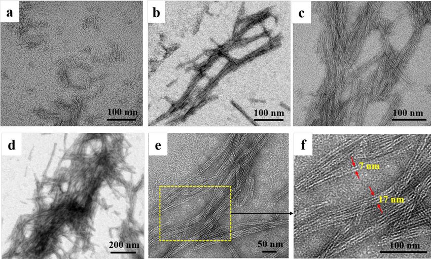
ARTICLE                                                                   NATURE COMMUNICATIONS | https://doi.org/10.1038/s41467-021-25199-x

during conventional crystallization procedures (at least 144.0 μM              obtained qualified single crystals suitable for X-ray diffraction.
or 6.0 mg/mL), the formation of these two kinds of protein arrays              The crystal structure was solved at a resolution of 2.1 Å
corresponds to a protein self-assembly process in solution.                    (Supplementary Tables 1 and 2). The packing pattern of FLAL
Enlargement of the small cuboid-like assembly revealed that 12                 molecules in the crystal is pronouncedly different from wt
dimeric FLAL molecules first assemble into 24-meric protein                     TmFtn. The side view of the crystal structure revealed that FLAL
nanocages, which further assemble into well-organized 3D                       molecules arrange in a repeating, side-by-side fashion to form
superlattices (Supplementary Fig. 9b–d) as we previously                       filaments (Fig. 3a). In contrast, native TmFtn exhibits a different
reported16,17. This result is not surprising for two reasons: (a)              packing pattern in its crystal where 12 TmFtn dimers assemble
the above genetic modification for native dimeric TmFtn is only                 into a 24-mer protein cage (Supplementary Fig. 3). Such a
involved in amino acid residues nearby the C3 and C4 interfaces                difference in protein assembly between TmFtn and mutant FLAL
of the metal-mediated 24-meric protein nanocage rather than its                agrees with our design. The crystal structure shows that the width
C3–C4 interfaces, thus hardly affecting its inherent assembly                  of the filament is about 5 nm from the side view and about 2 nm
property; (b) substitution of Asn147 with Phe in native TmFtn                  from the top view, these findings being in accordance with the
can also have the possibility to make two adjacent protein                     above TEM observation showing that the width of filaments is
nanocages join together along the fourfold channels through π–π                about 2 nm (Fig. 2).
interactions as reported recently16, resulting in the formation of                Further crystal analyses reveal that two adjacent FLAL
such 3D protein superlattices. In contrast, enlargement of the                 molecules have opposite orientations, forming another interface
linear-shaped species revealed the generation of filaments by                   along their C and D helixes. In the structure with resolutions that
FLAL mutant molecules (Supplementary Fig. 9e). Differently, wt                 permit detailed analysis of side-chain configurations, Phe147 and
TmFtn dimers can only assemble into discrete protein nanocages                 Leu114 side chains at the designed interface adopt suitable
under identical experimental conditions (Supplementary Fig. 10).               conformations to generate π–π stacking interactions and hydro-
These findings demonstrate that protein interface redesign in                   phobic interactions, respectively (Fig. 3b, c), again approving our
conjunction with appropriate solution conditions can realize the               design at an atomic level. Besides the designed noncovalent
self-assembly transformation of dimeric building blocks from                   interactions, Leu112 and Val119 also produce hydrophobic
protein nanocage into 1D nanomaterials.                                        interactions (Fig. 3d), which is unexpected. Additionally, one
   To visualize the morphology of the formed filaments in detail, they          calcium ion occurs nearby the designed protein interface, which
were observed by TEM in different visual fields. With excess Ca2+               binds to Glu108, Asp127, Asn105, and one water molecule
addition, filaments were formed rapidly within 30 min (Fig. 2a). As             (Fig. 3e). We believe that the cooperation of these noncovalent
time goes on, the filaments become longer and more numerous                     interactions and metal coordination along the inter-building-
(Fig. 2a–d), suggesting that the filament formation is thought to               block interfaces promotes the formation of the filaments. It
proceed through growth events. The length of these filaments grew               should be noted that only the bounded calcium ion with obvious
up to tens of nanometers with the most extended filament up to 1.3              densities can be identified in the crystal structure, but other
µm (Supplementary Fig. 11), and finally the formed filaments were                interactions with low occupancy were not counted. In the crystal
tangled together (Fig. 2d). In addition, the filaments have a tendency          structure, the formed filaments further arrange in the vertical
to stack together, and thus individual filament is hardly observed.             direction to create 2D protein assemblies (Supplementary
Usually, two or four filaments are stacked together, and these                  Fig. 12a). Besides, these filaments are parallel displaced from
filament assemblies exhibit widths on the order of 7 or 17 nm,                  the side view (Supplementary Fig. 12b), and two adjacent
respectively, with the pitch of two filaments approximately 3 nm                filaments are connected by weak interactions in the crystal
(Fig. 2e, f). The width of the individual filament is about 2 nm, which         structure. For example, Lys10 and Glu164 from two contiguous
is in good agreement with the thickness of FLAL dimer                          filaments are in close proximity, leading to the generation of
(Supplementary Fig. 1).                                                        electrostatic attraction (Supplementary Fig. 12c). However, TEM
   To obtain detailed structural information on the FLAL                       analyses revealed that the M1 mutant where Lys10 and Glu164
filament, we tried to crystallize this protein and eventually                   were mutated into Gly exhibits a similar assembly behavior to the

Fig. 2 Characterization of the filaments constructed by 48.0 µM FLAL and 80 mM Ca2+. a–d TEM images of the filaments at different time points (0.5, 1,
12, and 24 h). e High-magnification transmission electron microscopic (HRTEM) images of the filaments. f Real map of the inverted FFT of e.

4                  NATURE COMMUNICATIONS | (2021)12:4849 | https://doi.org/10.1038/s41467-021-25199-x | www.nature.com/naturecommunications
NATURE COMMUNICATIONS | https://doi.org/10.1038/s41467-021-25199-x ARTICLE
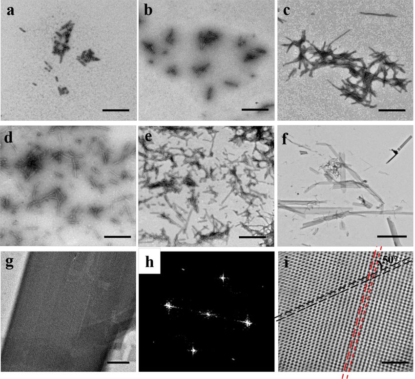
Fig. 4 TEM analyses of the nanorods constructed by 12.0 µM FLAL and
30% PEG1500. a Low-magnification TEM view of the formed nanorods.
b High-magnification view of a. The yellow dotted line indicated the
assembled direction of FLAL molecules. c Real map of the inverted FFT from
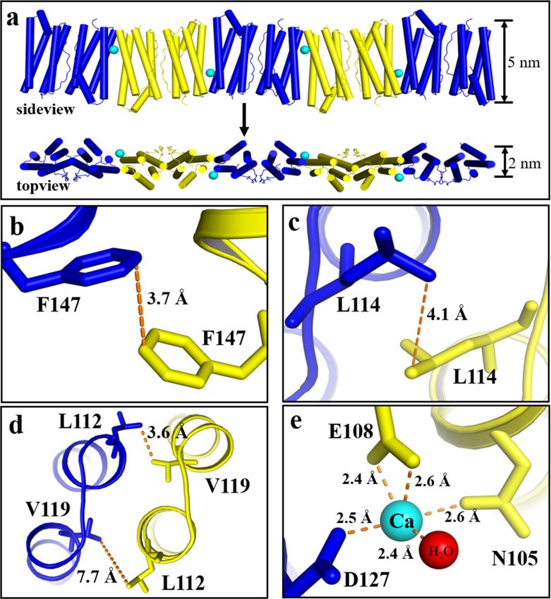
Fig. 3 Structural basis of the 1D filaments in atomic detail. a Side and top b. Inset: Fast Fourier transform image. d Structural diagram based on the
views of the FLAL filament in the crystal structure where the Ca2+ is shown reconstruction of c.
as cyan sphere. b–e Close-up views of the interfacial interactions between
two adjacent dimers in the 1D filament, including π–π stacking interaction resolution (Supplementary Tables 1 and 2). As expected, the
(b), hydrophobic interactions (c, d), and metal coordination (e). designed interface is responsible for driving self-assembly, but no
electron densities of the PEG were observed (Supplementary Fig. 16),
FLAL molecule (Supplementary Fig. 13), suggesting that other most likely due to the high flexibility of PEG and its weak ability to
kinds of non-covalent interactions might be also responsible bind to protein50. The crystal structure proved remarkably similar to
for the formation of the observed 1D filament. The detailed the design model: protein molecules connect through designed π
stacking mechanism between adjacent filaments needs further stacking and hydrophobic interactions in a side-by-side repeating
investigation. pattern with adjacent FLAL molecules antiparallel with each other
One overarching goal of synthetic biomimetic nanomaterials is (Supplementary Fig. 16a, e). Thus, the crystal structure is in good
to construct hierarchical assemblies that are able to respond to agreement with the above results observed by TEM (Fig. 4b, c),
various external stimuli so that the fabricated assemblies can be confirming the side-by-side interaction manner of dimeric FLAL
controlled. Therefore, besides calcium ions, we also investigated molecules.
the effect of PEG on the assembly behavior of FLAL molecules Further analyses of the crystal structure showed that the
because PEG is a hydrophilic nonionic polymer widely used in assembly process of FLAL molecules could be summarized as
many biochemical and pharmaceutical applications due to its follows: first, FLAL molecules pack along the x-axis in such a way
mild action on the biological activity of cell components49,50. mentioned above to form 1D arrays (Supplementary Fig. 16a),
After trying different molecular weights (PEG1000, PEG1500, and such assembly pattern was also observed by TEM shown in
and PEG3000) and concentrations (10, 20, and 30%) of PEG, we Fig. 4b; second, just like the stacking of filaments of FLAL
finally obtained nanorods formed in the presence of 30% molecules in the presence of Ca2+, these 1D arrays stack along
PEG1500. As shown in detail in Fig. 4a, nanorods of several the y-axis through electrostatic interaction to form 2D arrays
micrometers in length (up to 4 μm) can be visualized through (Supplementary Fig. 16b), which is likewise visualized by TEM
TEM. Magnification of the assembled components showed that (Fig. 4b, c); finally, the formed 2D arrays further arrange in the
FLAL molecules are well organized and arranged in a linear vertical direction to create 3D protein assemblies (Supplementary
manner as shown by the yellow dotted line (Fig. 4b). Fast Fourier Fig. 16c, d), and the weak interaction involved in this step mainly
transform (FFT) based on the TEM image presents a clear view of comes from their E helix (Supplementary Fig. 16f). Taken
the assembled arrangement (Fig. 4c). The corresponding together, all these findings demonstrate that our protein interface
assembly pattern of the FLAL molecules observed under TEM redesign approach yields assembly of dimeric FLAL protein
is shown in Fig. 4d. Further analysis by AFM with an intelligent building blocks into the filaments or nanorods induced by Ca2+
mode revealed that the height of the nanorod is 220 ± 5 nm or PEG rather than the inherent Ca2+-mediated 24-meric protein
(Supplementary Fig. 14). In contrast, wt TmFtn dimers only shell-like assembly.
assemble into discrete protein nanocages under identical experi-
mental conditions (Supplementary Fig. 15). These results again Engineering dimeric TmFtn for assembly into nanoribbons.
demonstrate that the designed driving force enables dimeric The above results demonstrated that tuning the action mode of
FLAL molecules to self-assemble into nanorods. two adjacent protein dimers from the head-to-side manner to the
To determine the structural basis of such nanorods, we set out to fully side-by-side manner facilitates the self-assembly transfor-
grow single crystals large enough for X-ray diffraction analyses in mation of the dimeric building blocks from hollow protein
the presence of PEG1500. We solved the crystal structure at 2.6 Å nanocage into nanofilaments and nanorods. Bioinspired by the
NATURE COMMUNICATIONS | (2021)12:4849 | https://doi.org/10.1038/s41467-021-25199-x | www.nature.com/naturecommunications 5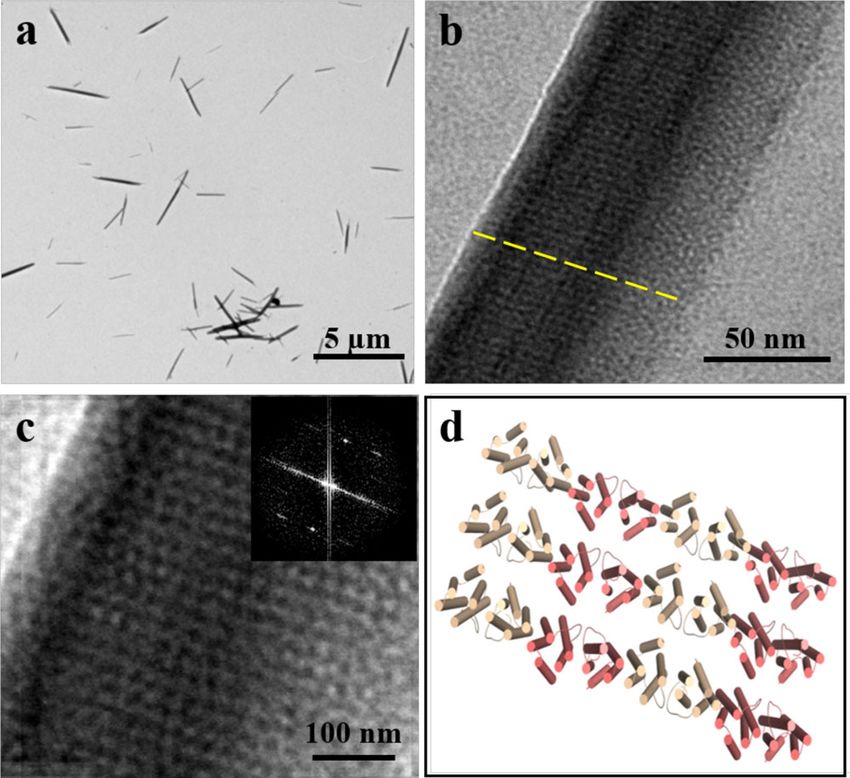
ARTICLE                                                                 NATURE COMMUNICATIONS | https://doi.org/10.1038/s41467-021-25199-x

structure of α-keratin protofilaments formed from two staggered               between two adjacent FLAL-L molecules are mediated entirely
rows of head-to-tail coiled coils51, we wonder what if we rear-              through hydrophobic interactions, as the contact region of
range two adjacent protein dimers in a staggered pattern, namely,            adjacent FLAL-L molecules is rich in hydrophobic amino acid
a partially side-by-side pattern. To answer this question, we use            residues (Fig. 7b). The engineered Leu114 and Leu137 side chains
the method mentioned above for protein docking but do not need               cooperated with the original V119, L107, V126, and V130 side
to preset the dimers to search for their possible arrangement. To            chains provide the bulk of the hydrophobic core, whereas the
this end, we simulated the orientations of two TmFtn dimers                  engineered Leu112, Ala113, and Phe147 are not involved in such
through global docking using RosettaDock48. One thousand                     interactions. The crystal structure provides direct evidence to
independent docking trajectories were carried out, and by com-               confirm the partially side-by-side manner of FLAL-L molecules.
bining energy simulation with a visual inspection, a model with              The above formed filaments coalesce in parallel along the y axes
dimers in a partially side-by-side manner was selected to assist             through hydrogen bonds (Supplementary Fig. 20b, c), leading to
interface redesign (Supplementary Fig. 17a). We found that sev-              the generation of a protein layer (Fig. 7c), corresponding to the
eral hydrophobic residues, including Leu107, Val119, Val126, and             ribbons observed by TEM (Fig. 6). The arrangement of the
Val130, are distributed along with the designed interface (Sup-              protein layer in the crystal perfectly matches the pattern seen in
plementary Fig. 17b). To effectively utilize these hydrophobic               the nanoribbons observed by TEM (Fig. 6f–i). Based on the
residues, we plan to design hydrophobic interactions as the                  observation that the thickness of each protein layer is about 2 nm
driving forces to construct such partially side-by-side protein              as suggested by the crystal structure (Fig. 7c) and the height of the
assembly. Based on the fact that FLAL mutant contains three                  nanoribbons detected by AFM (Supplementary Fig. 19) is around
more hydrophobic amino acid residues located on its outer sur-               ~10 nm, it is reasonable to believe that the observed nanoribbons
face than wt TmFtn, therefore, to build stronger hydrophobic                 are of five layers. In crystals, the formed protein layers further
interactions at this designed interface (Supplementary Fig. 17c),            arrange along the z axes to form 3D protein frameworks (Fig. 7d)
we chose FLAL mutant instead of wt TmFtn as starting materials               through π–π interactions and electrostatic interactions (Supple-
for further engineering.                                                     mentary Fig. 20d–f). All these results demonstrate that the
   To enable two dimers to interact with each other in a partially           hydrophobic interactions between two contiguous dimeric
side-by-side fashion, we made another mutant named FLAL-L                    protein-building blocks in a partially side-by-side fashion carried
where Arg137 in FLAL was replaced with Leu (Supplementary                    out by protein interface redesign facilitate the transformation of
Fig. 5) to increase the area of the hydrophobic patch as shown in            the dimeric building blocks from 24-meric nanocages into
Supplementary Fig. 17d. To gain insight into the assembly                    nanoribbons through filaments.
behavior of FLAL-L mutant, we purified it to homogeneity as
suggested by SDS-PAGE and native-PAGE (Supplementary
Fig. 18). We next investigated the assembly of FLAL-L molecules              Discussion
in the presence of PEG in solution. Upon screening the                       Particle polymorphism is a popular phenomenon in virus capsids,
concentration and molecular weight of PEG, we found that                     which possess inherent switches that allow efficient disassembly
FLAL-L molecules can likewise assemble into filaments in the                  and reassembly in response to solution conditions. For example,
presence of 15 or 20% PEG1500, as shown in Supplementary                     an icosahedral plant virus, cowpea chlorotic mottle virus, can
Table 4. The filaments with different sizes ranging from 100 nm               convert into a tubular nanostructure, which needs double-
to several micrometers can be visualized (Fig. 5a, b). Enlargement           stranded DNA to act as a template and take advantage of the
of the assembly revealed that the FLAL-L molecules are well                  nonspecific binding of capsid proteins and DNA52,53. Simian
organized in a linear fashion (Fig. 5b). To obtain more insights             virus 40 (SV40) represents another interesting example of shape
into the structural information, the formed filaments were further            transformation between protein nanostructures with different
characterized by AFM with an intelligent mode. As shown in                   dimensions. The SV40 capsid is mainly composed of 72 penta-
Fig. 5c, d, the height of the array is 6 ± 0.5 nm, which is                  mers of VP1. Electron microscopic observations revealed that, at
comparable with the height of FLAL-L molecules (∼5 nm) in the                pH 5.0, long and tubular structures are formed, whereas at high
crystal (Supplementary Fig. 1). To gain deep insight into the                salt concentrations, small (20 nm) particles predominate54.
above process, we investigated the formation of the filaments as a            However, how to transform the assembly of the building blocks
function of time. Short filaments were rapidly formed at 5 min.               from natural protein cages into 1D or 2D architecture by a
With an increase in time, the length and number of the formed                simple, effective method in the laboratory has yet to be explored.
filaments increased gradually (Fig. 6a–e). After 15 days, except for          The challenge lies in the fact that protein nanocages occurs as
the filament, nanoribbon-like nanomaterials appeared (Fig. 6f). A             discrete molecules in solution while filament/nanorod/ribbon
high-magnification TEM view (Fig. 6g) seized the periodic                     created in this study closely resemble 1D and 2D polymers and
parallel arrangement of protein arrays, and the real map from                that the intermolecular interface and assembly geometry for the
invert FFT (Fig. 6h) gave an excellent view on the structure of the          construction of hollow protein nanocage and filament/nanorod/
nanoribbon, which reveals that two kinds of lines interlaced to              ribbon nanomaterials are entirely different. We demonstrate that
form such structure (Fig. 6i). Further examination of the                    spatially directed assembly of protein-building blocks might be a
nanoribbons using AFM revealed that the height of the                        solution to solve such problems, which will reduce the compu-
nanoribbon is nearly 10 nm (Supplementary Fig. 19).                          tational workload and lower the barriers to protein assembly
   To shed light on structural information on the above filaments             design. To confirm this idea, we have built here a protein inter-
and nanoribbons, FLAL-L was also crystallized by vapor                       face redesign approach that is able to manipulate the directed
diffusion, and eventually, qualified single crystals suitable for X-          assembly of protein-building blocks, resulting in the self-assembly
ray diffraction were obtained (Supplementary Fig. 20a). We                   transformation of dimeric building blocks from hollow protein
solved the crystal structure at the resolution of 2.3 Å (Supple-             nanocage to 1D and 2D protein arrays with nanoscale and
mentary Tables 1 and 2). The crystal structure analyses revealed             microscale long-range order.
that FLAL-L molecules are perfectly aligned with each other in a                In pursuit of our goal to transform the dimeric building blocks
partially side-by-side manner to form filaments (Fig. 7a), the top            from hollow protein nanocages into 1D and 2D nanostructure,
view of which is in good agreement with the filament observed                 external stimuli were introduced to exert control over PPIs
under TEM (Fig. 5a, b). As expected, intermolecular associations             for dimeric FLAL or FLAL-L assembly. Researchers have

6                NATURE COMMUNICATIONS | (2021)12:4849 | https://doi.org/10.1038/s41467-021-25199-x | www.nature.com/naturecommunications
NATURE COMMUNICATIONS | https://doi.org/10.1038/s41467-021-25199-x                                                                  ARTICLE

Fig. 5 Characterization of the 1D filaments constructed by 12.0 µM FLAL-L and 15% PEG1500. a Low-magnification TEM view of FLAL-L filaments.
b Enlargement of FLAL-L filaments. c Atomic force microscopic (AFM) micrographs of the 1D filaments. d Height distributions measured along the black
arrow in c.

demonstrated that the self-assembly of a monomeric four helix                   of actin filaments and S-layers in terms of their dimensions and
bundle protein can be directed in all three dimensions through                  structural uniformity. Thus, our reported construction of 1D and
the inherent chemically tunable and modular nature of metal                     2D protein arrays from the dimeric protein as building blocks
coordination7. In this study, the transformation of dimeric                     provides a model to study the assembly mechanism of natural
building blocks into different protein nanomaterials including                  protein architectures.
hollow protein nanocage and filament/nanorod/nanoribbon                             Controlling self-assembly is critical to the advancement of
mainly depends on the redesigned PPIs. These PPIs are largely                   nanotechnology. Nowadays, scientists are able to accurately
mediated by the designed weak, noncovalent interactions,                        control the protein self-assembly behaviors to construct various
although external stimulation is required to trigger the assembly               supramolecular structures through the rational design of PPIs.
of the designed protein dimers.                                                 Though a variety of intricate protein nanostructures such as
   It is noteworthy that there is only one amino acid difference in             hollow protein cages, filaments/tubules, nanosheets, and 3D
amino acid sequence between FLAL and FLAL-L molecules, but                      crystalline frameworks have been created18–24,36–41, rendering
their assembly behavior is markedly different from each other.                  directed assembly of protein-building blocks into the custom-
FLAL molecules self-assemble into nanorods, whereas FLAL-L                      tailored nanoarchitectures remains challenging. Our reported
has the ability to assemble into the nanoribbons. The significant                protein engineering approach could tune the inherent head-to-
difference in self-assembly behavior between these two dimeric                  side interaction manner of two adjacent dimeric protein-building
protein molecules reflects the importance of amino acid residue                  blocks to the fully or partially side-by-side manner by redesigning
Arg137 in FLAL, suggesting that Arg137 could act as a switch to                 protein interfaces, yielding directed assembly of the building
control the conversion of nanorods to nanoribbons. In all cases,                blocks, thereby facilitating the self-assembly transformation of the
the crystal structures reveal that the backbones in the side-by-side            dimeric building blocks from hollow protein nanocage into 1D or
interaction manner were designed with high accuracy compared                    2D nanomaterials. PPIs at the designed protein interfaces are
to the computationally designed model (Supplementary Fig. 21).                  mainly contributed from the cooperation of the hydrophobic
The occurrence of 1D and 2D protein arrays with the dimeric                     interactions, aromatic π–π interactions, and external stimulation,
protein as building blocks recalls among natural proteins the case              which effectively stabilize the designed protein interface. The

NATURE COMMUNICATIONS | (2021)12:4849 | https://doi.org/10.1038/s41467-021-25199-x | www.nature.com/naturecommunications                          7
ARTICLE                                                                         NATURE COMMUNICATIONS | https://doi.org/10.1038/s41467-021-25199-x

Fig. 6 Kinetics of the formation of 2D nanoribbon. a–f TEM views of the formation of FLAL-L assemblies as a function of time (5 min, 30 min, 1 h, 12 h,
24 h, and 15 days, respectively) in the presence of 15% PEG1500. g High-magnification view of the 2D nanoribbon formed at 15 days. h Fast Fourier
transform (FFT) of g. i Real map from invert FFT of h. The black and red dotted lines indicate the arrangement of the 2D FLAL-L assembly. a–e Scale bars
represent 500 nm, while scale bars represent 2 µm in f, and 100 nm in g, i, respectively.

above protein engineering approach that focuses on adjustment                       TmFtn cDNA was performed with the fast site-directed mutagenesis kit (TIANGEN
for the geometric arrangement of protein-building blocks is                         Biotech Co., Ltd.). Polymerase chain reaction amplification was carried out using the
                                                                                    pET-3a plasmid with the TmFtn gene as a template, and primers for mutagenesis are
conceptually and operationally simple. It is interesting that the
                                                                                    listed in Supplementary Table 5. Plasmid sequences were verified by DNA
head-to-side interaction between two contiguous dimeric protein-                    sequencing. TmFtn, as well as mutants, were purified as follows. First, the plasmids
building blocks occurs not only in all known ferritins but also in                  corresponding to FLAL and FLAL-L were transformed into BL21 (DE3) Escherichia
other cage-like proteins such as Dps, where six dimeric protein-                    coli cells, respectively, and then cultured at 37 °C in 1 L of LB media containing
building blocks also assemble into a shell-like structure27.                        100 µg/mL ampicillin. After the cell density reached an absorbance of 0.6 at 600 nm,
                                                                                    protein expression was likewise induced with 200 µM IPTG for 10 h at 37 °C. Cells
Therefore, our engineering approach should, in principle, be                        were harvested by centrifugation (8609 × g) and the precipitate was re-suspended in
applicable to some other protein architectures. This would pro-                     50 mM Tris–HCl (pH 8.0), followed by sonication. The supernatant was collected
duce a variety of protein nanomaterials with different geometries.                  from lysed cell samples and subjected to heating at 90 °C for 10 min. Then thermal-
Furthermore, the filaments obtained in this study could provide a                    treated supernatant was collected after centrifugation at 8609 × g for 30 min and
multiple enzyme port scaffold to convene variant enzymes align                      passed through a membrane filter. Finally, the protein solution was applied to an
                                                                                    ion-exchange column (DEAE Sepharose Fast Flow, GE Healthcare), followed by
into a supramolecular enzyme system with unexplored                                 gradient elution with 0–1.0 M NaCl. The purified protein was then dialyzed against
property55.                                                                         50 mM Tris–HCl (pH 8.0) at 4 °C to exclude NaCl from the solution, and protein
                                                                                    concentrations were determined according to the Lowry method with bovine serum
                                                                                    albumin as standard. Protein purity was confirmed by SDS–PAGE. The molecular
Methods                                                                             weight of TmFtn subunit was estimated to be approximately 20 kDa, which is
Protein preparation. The gene encoding TmFtn was synthesized by Synbio              consistent with our electrophoretic band as shown in Supplementary Fig. 2b, and the
Technologies, which has been inserted into the plasmid pET-3a. Mutagenesis of the   molecular weight of the dimer is approximately 40 kDa.

8                    NATURE COMMUNICATIONS | (2021)12:4849 | https://doi.org/10.1038/s41467-021-25199-x | www.nature.com/naturecommunications
NATURE COMMUNICATIONS | https://doi.org/10.1038/s41467-021-25199-x ARTICLE
Fig. 7 The crystal structure and arrangement of FLAL-L mutant. a Side and top views of the 1D FLAL-L filament in the crystal structure. b Close-up view of
the hydrophobic interactions between two adjacent FLAL-L molecules. c The 1D filaments coalesce in parallel along the y axes to form 2D protein layer,
corresponding to the 2D ribbons observed by TEM. The dashed lines in c correspond to the lines shown in Fig. 6i. d From the top view, FLAL-L molecules
assemble into 3D protein frameworks.
Preparation of nanocages, filaments, nanorods, and nanoribbons. After pur- Crystallization, data collection, and structure determination. Purified proteins
ification, TmFtn molecules remain as a dimer in the absence of metal ions. For were concentrated to 6 mg/mL in a buffer consisting of 20 mM Tris–HCl at pH 8.0, and
protein nanocages, 50 mM Tris–HCl (pH 8.0) was used as buffer and 50 mM CaCl2 crystals were obtained using the hanging drop vapor diffusion method under different
was required to induce dimeric TmFtn self-assembly. Filaments were prepared by conditions, which are shown in Supplementary Table 1. X-ray diffraction data were
adding a certain amount of CaCl2 to FLAL solution (48 µM) at room temperature. collected at Shanghai Synchrotron Radiation Facility (SSRF; BL17U and BL19U) with
After being stirred for several minutes, the resulting mixture was incubated at 4 °C merging and scaling by the HKL-3000 software57. Data processing statistics are shown
overnight. To optimize the assembly conditions, different concentrations of CaCl2 in Supplementary Table 2. The structures were determined by molecular replacement
were tested over a range of 0–100 mM. Similarly, nanorods were prepared by using the Molrep program in CCP4 using the structure of TmFtn (PDB code 1VLG) as
adding a certain amount of PEG1500 to FLAL solution (12 µM) at room tem- a search model. Structure refinement was conducted using the Refmac5 program and
perature. After gently shaking for a few minutes, the resulting mixture was incu- PHENIX software58. The structure was rebuilt using COOT59, which made the model
bated at 4 °C overnight. Nanoribbons were prepared by mixing FLAL-L solution manually adjusted. All figures of the resulting structures were produced using the
(12 µM) with 30% PEG1500, and then the reaction mixtures were incubated for PyMOL60 program and UCSF61 Chimera package.
15 days. The concentrations and polymerization degree of PEG used were
optimized.
Data availability
The atomic coordinates and structure factors in this study have been deposited in the
Docking simulations. One thousand independent docking trajectories were car- Protein Data Bank under the accession PDB IDs: 7DYA, 7DY8, 7DY9, and 7DYB. Source
ried out using RosettaDock48. The two TmFtn or FLAL subunits that form the data are provided with this paper.
designed interface were used as the starting structure for the docking simulations.
One of the monomers was randomly spun along the axis connecting the centers of
mass of both partners and the same monomer was also allowed to search a space of Received: 28 January 2021; Accepted: 28 July 2021;
up to 3 Å normal to that axis, 8 Å in the plane perpendicular to the axis, and with
up to an 8˚ tilt from the axis and an 8˚ additional spin around the axis.
Polyacrylamide gel electrophoresis. The purity and molecular weight of protein
samples were estimated by PAGE. Gel electrophoresis under denaturing conditions References
was carried out using a 15% polyacrylamide–SDS gel as reported by Laemmli56, 1. Lay, C. L., Lee, M. R., Lee, H. K., Phang, I. Y. & Ling, X. Y. Transformative
and samples need to be heated in a water bath for 5 min. For native PAGE, a
two-dimensional array configurations by geometrical shape-shifting protein
4–20% polyacrylamide gradient gel was used and run at 5 mA for 10 h at 4 °C. Gels
microstructures. ACS Nano 10, 9708–9717 (2015).
were stained with Coomassie brilliant blue R250.
2. Rothemund, Paul & Folding, W. K. DNA to create nanoscale shapes and
patterns. Nature 440, 297–302 (2006).
High-resolution gel filtration chromatography analyses. High-resolution gel 3. Delebecque, C. J., Lindner, A. B., Silver, P. A. & Aldaye, F. A. Organization of
filtration chromatography analyses were performed using an ÄKTA pure system intracellular reactions with rationally designed RNA assemblies. Science 333,
coupled to a Superdex 200 Increase column (GE Healthcare) in buffer (50 mM Tris, 470–474 (2011).
100 mM NaCl, pH = 8.0) with a flow rate of 0.5 mL/min. 4. Aggeli, A. et al. Responsive gels formed by the spontaneous self-assembly of
peptides into polymeric β-sheet tapes. Nature 386, 259–262 (1997).
5. Waller, P. J. et al. Chemical conversion of linkages in covalent organic
TEM imaging. Protein samples (10 μL) were deposited on carbon-coated copper frameworks. J. Am. Chem. Soc. 48, 15519–15522 (2016).
grids and excess solution was removed with filter paper after a 2-min incubation. 6. Zhao, X., Fox, J. M., Olson, N. H., Baker, T. S. & Young, M. J. In vitro assembly
Then protein samples were stained using 2% uranyl acetate for 5 min. Transmis- of cowpea chlorotic mottle virus from coat protein expressed in Escherichia coli
sion electron micrographs were obtained at 80 kV through a Hitachi H-7650 and in vitro-transcribed viral cDNA. Virology 207, 486–494 (1995).
transmission electron microscope. 7. Brodin, J. D. et al. Metal-directed, chemically tunable assembly of one-, two- and
three-dimensional crystalline protein arrays. Nat. Chem. 4, 375–382 (2012).
AFM measurements. For AFM sample preparation, protein samples (10 μL) were 8. Reisler, E. & Egelman, E. H. Actin structure and function: what we still do not
pipetted on freshly cleaved mica (Beijing Zhongxingbairui Technology Co., Ltd.) understand. J. Biol. Chem. 282, 36133–36137 (2007).
and dried at room temperature. The AFM images were collected using a Nanoman 9. Chung, S., Shin, S. H., Bertozzi, C. R. & De Yoreo, J. J. Self-catalyzed growth of
VS (Bruker) with tapping mode at a resolution of 256 lines per image and a scan S layers via an amorphous-to-crystalline transition limited by folding kinetics.
rate of 1 Hz. The AFM images were processed using NanoScope Analysis. Proc. Natl Acad. Sci. USA 107, 16536–16541 (2010).
NATURE COMMUNICATIONS | (2021)12:4849 | https://doi.org/10.1038/s41467-021-25199-x | www.nature.com/naturecommunications 9ARTICLE NATURE COMMUNICATIONS | https://doi.org/10.1038/s41467-021-25199-x
10. Ma, J. et al. Structural basis of energy transfer in Porphyridium purpureum 40. Zhang, S. et al. “Silent” amino acid residues at key subunit interfaces regulate
phycobilisome. Nature 579, 146–151 (2020). the geometry of protein nanocages. ACS Nano 10, 10382–10388 (2016).
11. Kortemme, T. & Baker, D. Computational design of protein-protein 41. Zhang, S. et al. Conversion of the native 24-mer ferritin nanocage into its non-
interactions. Curr. Opin. Chem. Biol. 8, 91–97 (2004). native 16-mer analogue by insertion of extra amino acid residues. Angew.
12. Schreiber, G. & Fleishman, S. J. Computational design of protein–protein Chem. Int. Ed. 55, 16064–16070 (2016).
interactions. Curr. Opin. Chem. Biol. 23, 903–910 (2013). 42. Vita, N. et al. A four-helix bundle stores copper for methane oxidation. Nature
13. Jones, S. & Thornton, J. M. Principles of protein-protein interactions. Proc. 525, 140–143 (2015).
Natl Acad. Sci. USA 93, 13–20 (1996). 43. Wittung-Stafshede, P., Lee, J. C., Winkler, J. R. & Gray, H. B. Cytochrome
14. Padilla, J. E., Colovos, C. & Yeates, T. O. Nanohedra: using symmetry to b562 folding triggered by electron transfer: approaching the speed limit for
design self assembling protein cages, layers, crystals, and filaments. Proc. Natl formation of a four-helix-bundle protein. Proc. Natl Acad. Sci. USA 96,
Acad. Sci. USA 98, 2217–2221 (2001). 6587–6590 (1999).
15. Sinclair, J. C., Davies, K. M., Venien-Bryan, C. & Noble, M. E. Generation of 44. Kolberg, M., Strand, K. R., Graff, P. & Andersson, K. K. Structure, function,
protein lattices by fusing proteins with matching rotational symmetry. Nat. and mechanism of ribonucleotide reductases. Biochim. Biophys. Acta 1699,
Nanotechnol. 6, 558–562 (2011). 1–34 (2004).
16. Zhou, K. et al. On-axis alignment of protein nanocage assemblies from 2D to 45. Lee, S. J., Mccormick, M. S., Lippard, S. J. & Cho, U. Control of substrate
3D through the aromatic stacking interactions of amino acid residues. ACS access to the active site in methane monooxygenase. Nature 494, 380–384
Nano 12, 11323–11332 (2018). (2013).
17. Zheng, B., Zhou, K., Zhang, T., Lv, C. & Zhao, G. Designed two- and three- 46. Ceci, P., Forte, E., Di Cecca, G., Fornara, M. & Chiancone, E. The
dimensional protein nanocage networks driven by hydrophobic interactions characterization of Thermotoga maritima ferritin reveals an unusual subunit
contributed by amyloidogenic motifs. Nano Lett. 19, 4023–4028 (2019). dissociation behavior and efficient DNA protection from iron-mediated
18. Bailey, J. B., Zhang, L., Chiong, J. A., Ahn, S. & Tezcan, F. A. Synthetic oxidative stress. Extremophiles 15, 431–439 (2011).
modularity of protein-metal-organic frameworks. J. Am. Chem. Soc. 139, 47. Chakraborti, S. et al. Three-dimensional protein cage array capable of active
8160–8166 (2017). enzyme capture and artificial chaperone activity. Nano Lett. 19, 3918–3924
19. Gu, C. et al. Structural insight into binary protein metal-organic frameworks (2019).
with ferritin nanocages as linkers and nickel clusters as nodes. Chem. Eur. J. 48. Gray, J. J. et al. Protein-protein docking with simultaneous optimization of
26, 3016–3021 (2020). rigid-body displacement and side-chain conformations. J. Mol. Biol. 331,
20. Yang, M. & Song, W. J. Diverse protein assembly driven by metal and 281–299 (2003).
chelating amino acids with selectivity and tunability. Nat. Commun. 10, 5545 49. Wang, Y. & Annunziata, O. Comparison between protein-polyethylene glycol
(2019). (PEG) interactions and the effect of peg on protein-protein interactions using
21. Kitagishi, H. et al. Supramolecular hemoprotein linear assembly by successive the liquid-liquid phase transition. J. Phys. Chem. B. 111, 1222–1230 (2007).
interprotein heme-heme pocket interactions. J. Am. Chem. Soc. 129, 50. Cattani, G., Vogeley, L. & Crowley, P. B. Structure of a pegylated protein
10326–10327 (2007). reveals a highly porous double-helical assembly. Nat. Chem. 7, 823–828
22. Carlson, J. C. T. et al. Chemically controlled self-assembly of protein (2015).
nanorings. J. Am. Chem. Soc. 128, 7630–7638 (2006). 51. Mckittrick, J. et al. The structure, functions, and mechanical properties of
23. Yang, R. et al. 2D square arrays of protein nanocages through channel- keratin. JOM 64, 449–468 (2012).
directed electrostatic interactions with poly(α, L-lysine). Chem. Commun. 50, 52. Mukherjee, S., Pfeifer, C. M., Johnson, J. M., Liu, J. & Zlotnick, A. Redirecting
2879–2882 (2014). the coat protein of a spherical virus to assemble into tubular nanostructures. J.
24. Yang, G. et al. Highly ordered self-assembly of native proteins into 1D, 2D, Am. Chem. Soc. 128, 2538–2539 (2006).
and 3D structures modulated by the tether length of assembly-inducing 53. Xu, Y. et al. DNA-templated CMV viral capsid proteins assembly into
ligands. Angew. Chem. Int. Ed. 56, 10691–10695 (2017). nanotubes. Chem. Commun. 49–51 (2008).
25. Tanaka, S. et al. Atomic-level models of the bacterial carboxysome shell. 54. Kanesashi, S. et al. Simian virus 40 vp1 capsid protein forms polymorphic
Science 319, 1083–1086 (2008). assemblies in vitro. J. Gen. Virol. 84, 1899–1905 (2003).
26. Arosio, P., Ingrassia, R. & Cavadini, P. Ferritins: a family of molecules for iron 55. Hudalla, G. A. et al. Gradated assembly of multiple proteins into
storage, antioxidation and more. Biochim. Biophys. Acta 1790, 589–599 supramolecular nanomaterials. Nat. Mater. 13, 829–836 (2014).
(2009). 56. Laemmli, V. K. Determination of protein molecular weight in polyacrylamide
27. Grant, R., Filman, D., Finkel, S., Kolter, R. & Hogle, J. The crystal structure of gels. Nature 227, 680–685 (1970).
Dps, a ferritin homolog that binds and protects DNA. Nat. Struct. Biol. 5, 57. Otwinowski, Z. & Minor, W. Processing of X-ray diffraction data collected in
294–303 (1998). oscillation mode. Methods Enzymol. 276, 307 (1997).
28. Douglas, T. & Young, M. Viruses: making friends with old foes. Science 312, 58. Adams, P. D. et al. PHENIX: a comprehensive Python-based system for
873–875 (2006). macromolecular structure solution. Acta Crystallogr. D Biol. Crystallogr. 66,
29. Liang, M. et al. H-ferritin-nanocaged doxorubicin nanoparticles specifically 213–221 (2010).
target and kill tumors with a single-dose injection. Proc. Natl Acad. Sci. USA 59. Emsley, P., Lohkamp, B., Scott, W. G. & Cowtan, K. Features and development
111, 14900–14905 (2014). of Coot. Acta Crystallogr. D Biol. Crystallogr. 66, 486–501 (2010).
30. Wen, A. M. & Steinmetz, N. F. Design of virus-based nanomaterials for 60. DeLano, W. L. The PyMOL Molecular Graphics System (DeLano Scientific,
medicine, biotechnology, and energy. Chem. Soc. Rev. 45, 4074–4126 (2016). 2002).
31. Jordan, P. C. et al. Self-assembling biomolecular catalysts for hydrogen 61. Pettersen, E. F. et al. A visualization system for exploratory research and
production. Nat. Chem. 8, 179–185 (2016). analysis. J. Comput. Chem. 25, 1605–1612 (2004).
32. King, N. P. et al. Computational design of self-assembling protein
nanomaterials with atomic level accuracy. Science 336, 1171–1174 (2012).
33. King, N. P. et al. Accurate design of co-assembling multi-component protein Acknowledgements
nanomaterials. Nature 510, 103–108 (2014). This work was supported by the National Natural Science Foundation of China (Nos.
34. Hsia, Y. et al. Design of a hyperstable 60-subunit protein icosahedron. Nature 31972018 and 31730069). The Shanghai Synchrotron Radiation Facility (SSRF) is espe-
535, 136–139 (2016). cially acknowledged for beam time. We thank the staff from BL17U1/BL18U1/19U1
35. Lai, Y. T. et al. Structure of a designed protein nanocage that self-assembles beamline of the National Center for Protein Sciences Shanghai (NCPSS) at Shanghai
into a highly porous cube. Nat. Chem. 6, 1065–1071 (2014). Synchrotron Radiation Facility for assistance during data collection.
36. Cannon, K. A., Nguyen, V. N., Morgan, C. & Yeates, T. O. Design and
characterization of an icosahedral protein nanocage formed by a double-
fusion protein containing three distinct symmetry elements. ACS Synth. Biol.
9, 517–524 (2020).
Author contributions
G.Z. and T.Z. conceived and directed the project and wrote the paper. X.Z. designed and
37. Woersdoerfer, B., Woycechowsky, K. J. & Hilvert, D. Directed evolution of a
performed experiments, analyzed data, and co-wrote the paper. B.Z. performed experi-
protein container. Science 331, 589–592 (2011).
ments. Y.L. and H.W performed the crystal data collection. J.Z. and C.L. performed the
38. Worsdorfer, B., Pianowski, Z. & Hilvert, D. Efficient in vitro encapsulation of
experiments and co-wrote the paper.
protein cargo by an engineered protein container. J. Am. Chem. Soc. 134,
909–911 (2012).
39. Butterfield, G. L. et al. Evolution of a designed protein assembly encapsulating
its own RNA genome. Nature 552, 415–420 (2017). Competing interests
The authors declare no competing interests.
10 NATURE COMMUNICATIONS | (2021)12:4849 | https://doi.org/10.1038/s41467-021-25199-x | www.nature.com/naturecommunicationsNATURE COMMUNICATIONS | https://doi.org/10.1038/s41467-021-25199-x ARTICLE
Additional information Open Access This article is licensed under a Creative Commons
Supplementary information The online version contains supplementary material Attribution 4.0 International License, which permits use, sharing,
available at https://doi.org/10.1038/s41467-021-25199-x. adaptation, distribution and reproduction in any medium or format, as long as you give
appropriate credit to the original author(s) and the source, provide a link to the Creative
Correspondence and requests for materials should be addressed to T.Z. or G.Z. Commons license, and indicate if changes were made. The images or other third party
material in this article are included in the article’s Creative Commons license, unless
Peer review information Nature Communications thanks the anonymous reviewer(s) for
indicated otherwise in a credit line to the material. If material is not included in the
their contribution to the peer review of this work. Peer reviewer reports are available.
article’s Creative Commons license and your intended use is not permitted by statutory
regulation or exceeds the permitted use, you will need to obtain permission directly from
Reprints and permission information is available at http://www.nature.com/reprints
the copyright holder. To view a copy of this license, visit http://creativecommons.org/
licenses/by/4.0/.
Publisher’s note Springer Nature remains neutral with regard to jurisdictional claims in
published maps and institutional affiliations.
© The Author(s) 2021
NATURE COMMUNICATIONS | (2021)12:4849 | https://doi.org/10.1038/s41467-021-25199-x | www.nature.com/naturecommunications 11You can also read