Conserved strategies of RNA polymerase I hibernation and activation
←
→
Page content transcription
If your browser does not render page correctly, please read the page content below
ARTICLE
                  https://doi.org/10.1038/s41467-021-21031-8                 OPEN

                  Conserved strategies of RNA polymerase I
                  hibernation and activation
                  Florian B. Heiss          1,   Julia L. Daiß1, Philipp Becker1 & Christoph Engel                     1✉

                  RNA polymerase (Pol) I transcribes the ribosomal RNA precursor in all eukaryotes. The
                  mechanisms ‘activation by cleft contraction’ and ‘hibernation by dimerization’ are unique to
1234567890():,;

                  the regulation of this enzyme, but structure-function analysis is limited to baker’s yeast. To
                  understand whether regulation by such strategies is specific to this model organism or
                  conserved among species, we solve three cryo-EM structures of Pol I from Schizosacchar-
                  omyces pombe in different functional states. Comparative analysis of structural models
                  derived from high-resolution reconstructions shows that activation is accomplished by a
                  conserved contraction of the active center cleft. In contrast to current beliefs, we find that
                  dimerization of the S. pombe polymerase is also possible. This dimerization is achieved
                  independent of the ‘connector’ domain but relies on two previously undescribed interfaces.
                  Our analyses highlight the divergent nature of Pol I transcription systems from their coun-
                  terparts and suggest conservation of regulatory mechanisms among organisms.

                  1 Regensburg   Center for Biochemistry, University of Regensburg, Universitätsstraße 31, 93053 Regensburg, Germany. ✉email: christoph.engel@ur.de

                  NATURE COMMUNICATIONS | (2021)12:758 | https://doi.org/10.1038/s41467-021-21031-8 | www.nature.com/naturecommunications                             1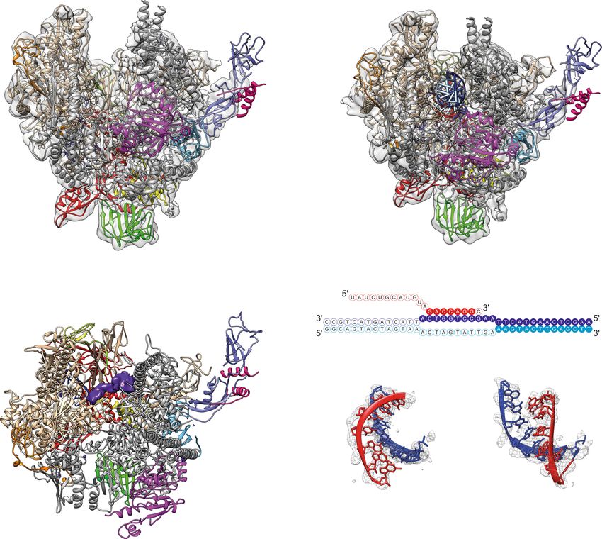
ARTICLE                                                                  NATURE COMMUNICATIONS | https://doi.org/10.1038/s41467-021-21031-8

T
        ranscription of the ribosomal RNA (rRNA) precursor by                 secondary structure elements for the entire polymerase and side
        RNA polymerase (Pol) I is a prerequisite for the bio-                 chain orientations in many regions (Fig. 1; Supplementary
        synthesis of ribosomes in all known eukaryotes1. To allow             Figs. 1–3). The architecture of Sc and Sp Pol I is similar, despite
transcription initiation in baker’s yeast Saccharomyces cerevisiae            poor overall sequence identity (Supplementary Fig. 4, Supple-
(Sc), monomeric Pol I is bound by the initiation factor Rrn3,                 mentary Table 2). Both, EC and monomeric Pol I, lack cryo-EM
enabling recruitment to the rDNA promoter via the hetero-                     density for the A49/A34.5 heterodimer, indicating that the sub-
trimeric core factor (CF), TBP and upstream activating factor                 complex is flexible or was lost during grid preparation (Fig. 1).
(UAF)2–4. Structures solved from Sc Pol I crystals revealed                   The presence of the A49/A34.5 subunit-complex in Pol I pre-
inactive polymerase dimers with widely expanded active center                 parations was confirmed by Coomassie−stained SDS-PAGE
clefts in three similar conformations5–7, matching biochemical                (Supplementary Fig. 1a) and mass-spectrometry analysis.
observations in extracts8, initial and recent Electron Microscopy                Differences between Sc and Sp Pol I include the lack of density
studies9,10. Such dimerization relies on a ‘connector’ domain at              for the Pol-I-specific helix α0 in subunit Rpb6 and insertions in the
the C-terminus of Pol I subunit A43. In detail, the ‘stalk’ subunit           ‘Dock’ and ‘Foot’ domains of Sp subunit A190. Helix α0 forms only
complex of one monomer is inserted into the ‘cleft’ of the other              in Sc Pol I and was presumed to strengthen stalk attachment
monomer from the upstream side. This allows formation of a                    compared to Pol II5,6. A divergent region (residues 549–559) and
connector α-helix in subunit A43 which attaches to the clamp                  an insertion (residues 591–600) in the ‘external 2’ domain of Sp
core helices of subunit A190 in the second monomer. Further-                  subunit A135 are adjacent to the lobe-region responsible for tight
more, a C-terminal connector β-hairpin traps the lid of subunit               association of the A49/34.5 sub-complex18 and may be responsible
A190, completely inactivating both polymerases5,6. Activation                 for its reduced affinity (compare Supplementary Figs. 3b and 4).
then requires Pol I monomerization, cleft contraction and stabi-              Poor stalk density is observed, indicating a high degree of flexibility,
lization of monomers by Rrn3. Further cleft contraction takes                 similar to human Pol II19 and potentially linked to a lack of Rpb6
place upon promoter melting and Pol I interaction with the                    α0 formation. Nevertheless, fitting of a stalk homology model
DNA/RNA hybrid11–13.                                                          created from the crystal structure of the Sc A14/A43 complex20 was
   Structures of monomeric states and of actively elongating Pol I            possible. In addition, local resolution is reduced in the jaw regions
from Sc were solved by single particle Electron Cryo Microscopy               and the tip of the clamp domain of subunit A190 in Sp Pol I EC
(cryo-EM)14,15. Furthermore, differential fluorescent allele tag-              and monomer reconstructions, also indicating a high degree of
ging demonstrated that Pol I dimerization is associated with                  flexibility (Supplementary Figs. 1 and 2). The C-terminal domain of
inactivation (hibernation) under certain starvation conditions in             subunit A12.2 is mostly flexible in ECs and monomers, although
an elegant in vivo approach16. In such experiments, deletion of               low-resolution density in the Sp EC (Supplementary Fig. 3a) may
the connector resulted in dimer disruption. To date, it is unclear            indicate a position outside subunit A135 as observed in some Sc
which mechanisms and structural features of Pol I regulation are              ECs21.
specific to Sc and which are conserved among organisms17.
   Here, we use single particle cryo-EM to solve the structure of
                                                                              Sp Pol I contains an expander element that is flexible in ECs. In
Pol I from Schizosaccharomyces pombe (Sp) in a monomeric (apo)
                                                                              ECs, we observe density for DNA and RNA, allowing us to build
and an actively elongating form at high resolution, showing that
                                                                              and refine a model for 25 template bases, 11 non-template bases
Pol I cleft contraction upon transcription activation is common to
                                                                              and 7 RNA bases (Fig. 1d). Interactions with Pol I, positioning in
both organisms. Strikingly, we also uncover that Pol I dimeriza-
                                                                              the cleft and relative orientation of the scaffold are similar to Sc
tion can take place independent of the A43 connector domain
                                                                              structures14,15,21. We do, however, observe a density that we
in vitro and solve the cryo-EM reconstruction of such a dimer.
                                                                              attribute to the ‘expander’ (‘DNA-mimicking’) element in Pol I
Our results allow discussing the evolutionary conservation of
                                                                              monomers (Fig. 1b). This density is located between the ‘pro-
structural features, hibernation and activation mechanisms.
                                                                              trusion’ domain of subunit A135 and the ‘clamp core’ region in
                                                                              subunit A190 on the upstream region of the active center cleft
Results                                                                       and appears to prevent unspecific DNA binding. Due to flexible
Preparation and cryo-EM of Schizosaccharomyces pombe Pol I.                   connections to the ‘jaw’ domain of subunit A190, we refrained
To study the structure of its Pol I, we generated an Sp strain                from modeling the exact residues of the section in the Sp
carrying a C-terminal flag-his tag on subunit AC40. Purification                monomer. Location of presumed expander density in the cleft is
using established protocols5 yielded pure Pol I that shows protein            comparable to that observed in Sc Pol I dimers (Supplementary
bands for all 14 subunits (Supplementary Fig. 1). Sp Pol I is active          Fig. 3d), while the element is flexible in Sc Pol I monomers.
in elongation and cleavage of an RNA primer from synthetic
constructs in vitro similar to Sc Pol I (Supplementary Fig. 2). To
                                                                              Sp Pol I dimerizes in vitro independent of the ‘connector’
determine the structure of a monomer, we stabilized Pol I by
                                                                              domain. Surprisingly, we observed many dimer-particles in the
crosslinking with BS3, followed by quenching and size exclusion
                                                                              monomer dataset (Supplementary Fig. 1). As described above, Pol
chromatography (Methods). In an independent experiment, we
                                                                              I subunit A43 does not contain a connector element in Sp and
established an Sp Pol I elongation complex (EC) in vitro similar
                                                                              therefore should be unable to form dimers, as inferred from
to its Sc counterpart15. Using three locked nucleic acid (LNA)
                                                                              connector deletions in Sc16. Previous studies described that Sc Pol
bases at the 3′ end of the RNA primer reduced sample hetero-
                                                                              I dimers and monomers are in equilibrium in solution. Buffer
geneity by preventing transcript cleavage via subunit A12.2
                                                                              conditions8 and concentration of highly purified Pol I5 can shift
(Methods). Two cryo-EM datasets were collected on a Titan Krios
                                                                              this equilibrium in vitro. To test whether dimerization is possible
Electron Microscope equipped with Falcon III direct electron
                                                                              in solution or may result from chemical crosslinking, we carried
detector: one from non-crosslinked, LNA-containing EC particles
                                                                              out analytical size exclusion chromatography (SEC) in different
and one from BS3-crosslinked Pol I, both following size exclusion
                                                                              buffers and in-solution dynamic light scattering (DLS) at different
chromatography (Supplementary Table 1).
                                                                              concentrations of Sp Pol I. A shift to earlier SEC elution volumes
                                                                              indicates an increased particle size under high-salt conditions
Pol I architecture is conserved but sub-complex occupancy                     (Fig. 2a). Negative stain EM analysis of SEC peak fractions
varies among organisms. The cryo-EM densities reveal                          revealed the presence of dimeric particles independent of the

2                 NATURE COMMUNICATIONS | (2021)12:758 | https://doi.org/10.1038/s41467-021-21031-8 | www.nature.com/naturecommunications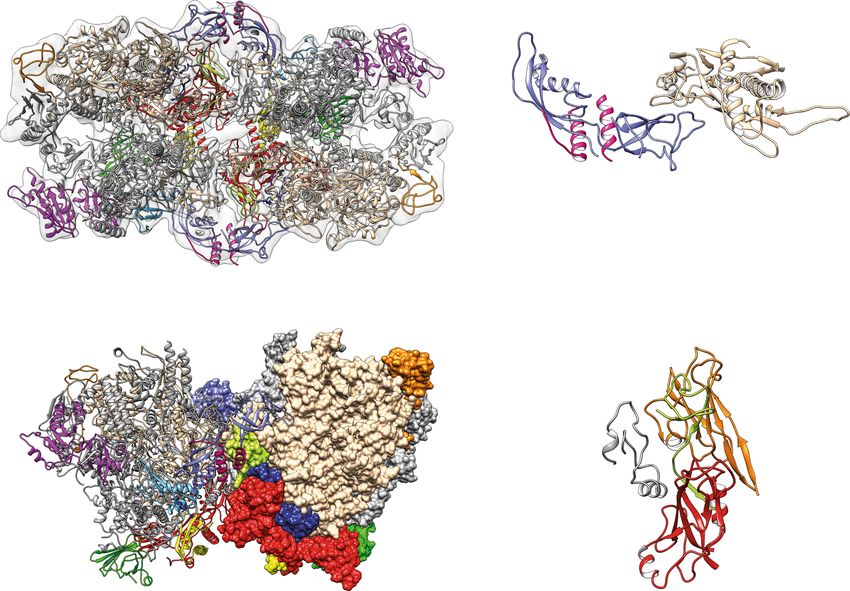
NATURE COMMUNICATIONS | https://doi.org/10.1038/s41467-021-21031-8 ARTICLE
a 1 91 361 485 514
580-587
597 670 842 1004 1066
∆1115 1159-1166
1189 1270 1344 1476 1520 1631 1689
Clamp Clamp head Dock Active Pore Funnel Cleft Foot Cleft Jaw Divergent Jaw Cleft Clamp
A190 core Clamp core site core
Expander
∆N-term 591-600 Anchor ∆1123
1 118 374 445 529 632 683 785 895 1053 1077 1174
Protrusion Lobe Fork External 2 Exter- Hybrid Wall Hybrid binding Clamp
A135 nal 1 binding
b Clamp Core c
A135 Helices Clamp Core
A43/A14 A135
Helices A43/A14
stalk
A12.2 stalk
A190 A12.2 A190
Rpb6 Rpb6
Rpb5
Rpb5
AC 40 AC 19 AC 19
template strand
Rpb8 non-template strand
Rpb8
Front view Front view
45° d
Sp ‘expander‘
Rpb12 A135 Wall
A135
Protrusion
DNA template strand
G
A
90°
C
A43/A14
stalk C
Rpb6
A
A190 G
A12.2 Product
G RNA
Front view Side view (A135)
A190 Jaw Rpb5
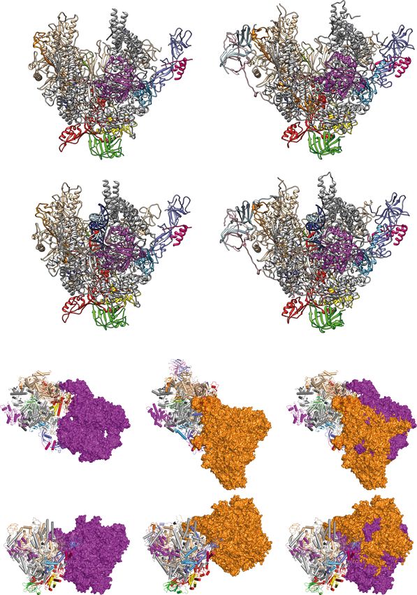
Fig. 1 Structure of monomeric and elongating S. pombe Pol I. a Sub-domain architecture of the Pol I subunits A190 and A135 in Sp. Insertion/Deletion of
more than five residues compared to Sc are highlighted. b Cryo-EM reconstruction of monomeric Sp Pol I. Transparent, unsharpened cryo-EM density
overlaid with ribbon model shows 12 subunits but lacks the A49/A34.5 subcomplex. The cleft is expanded and density (purple, space-filling) indicates the
location of the ‘expander’ element. c Cryo-EM reconstruction of an Sp Pol I Elongation complex. The cleft is contracted and the expander is displaced by the
hybrid. d Schematic representation of the artificial bubble construct used to establish an EC. Nucleotide bases included in the EC-density are highlighted. A
model of the template-DNA/product-RNA hybrid overlaid with sharpened EC density is shown, the 3′ nucleotide of the RNA is present but shows poor
density and was thus not modeled.
buffer (Supplementary Fig. 5). In a high-salt buffer, about 32% of Inactive Sp Pol I dimers arrange in a divergent architecture. A
identified particles (49% of Pol I molecules) appear to be in a three-dimensional reconstruction of Sp Pol I dimers at 4.5 Å
dimeric state as indicated by unsupervised 2D-classification. In resolution was obtained in C2 symmetry (Fig. 3; Supplementary
contrast, dimer-prevalence is below 2% of particles in low-salt Table 1). Unambiguous placement of two Sp Pol I monomers and
conditions. In line with this, DLS using the Prometheus Panta rigid body fitting of subdomains yielded a model of the Sp Pol I
technology (Nanotemper) indicates an increase of hydrodynamic dimer. Whereas establishment of Pol I dimers depends on stalk-
particle radius with increasing Sp Pol I concentration (Fig. 2b). insertion into the cleft of a second monomer in Sc, the Sp dimers
Taken together, we concluded that reversible, connector- form by interaction of the stalk with the protrusion domain of
independent dimerization of Sp Pol I is possible. To understand subunit A135 in the other monomer (Fig. 3). A second interface
the underlying molecular principles, we reconstructed the dimer forms between the ‘dock domain’ in subunit A190 of one
architecture from its cryo-EM density. monomer (including the Sp specific insertion of residues 580–587
NATURE COMMUNICATIONS | (2021)12:758 | https://doi.org/10.1038/s41467-021-21031-8 | www.nature.com/naturecommunications 3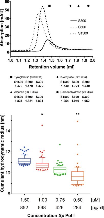
ARTICLE                                                                         NATURE COMMUNICATIONS | https://doi.org/10.1038/s41467-021-21031-8

    a                                                                               and the Pol I specific helix α12a) with the wall subdomain of
                                                                                    subunit A135, subunit AC40 and the common subunit Rpb12 of
                                                                                    the second monomer. These two interfaces allow a tight asso-
                                                                                    ciation via the establishment of contacts between both Pol I
                                                                                    upstream-faces independent of the connector element but uti-
                                                                                    lizing organism-specific regions. Low-resolution density on the
                                                                                    lobe of subunit A135 in both molecules in the dimers may
                                                                                    indicate the presence of A49/A34.5 (Supplementary Fig. 6). This
                                                                                    is in line with the suggestion that dissociation of this subcomplex
                                                                                    is independent of Pol I inactivation in Sp22.

                                                                                    Cleft contraction is common to active Pol I. Comparative
                                                                                    structural modeling reveals that, similar to Sc Pol I reconstruc-
                                                                                    tions, the cleft is extended in monomers and contracted in ECs
                                                                                    (Fig. 4; Supplementary Movie 1). Consequently, the ‘bridge’ helix
                                                                                    of subunit A190 is mostly disordered in monomers (poor density
                                                                                    despite higher overall resolution) and forms a well-structured
                                                                                    helix in ECs (Supplementary Fig. 3e). The ‘hinges’ identified by
                                                                                    comparing Pol II structures with Pol I dimers in crystals5,6,17 are
                                                                                    similar for Sp Pol I cleft modulation. Thus, polymerase activation
                                                                                    by contraction is conserved between Sc and Sp. Further cleft
                                                                                    expansion in Sc Pol I dimers likely results from mutual stalk-cleft
    b                                                                               insertion (Fig. 4b) and is not observed in Sp Pol I dimers (Fig. 4c;
                                                                                    Supplementary Movie 2). Despite this divergence, functional
                                                                                    importance is apparently conserved: Both molecules in a dimer
                                                                                    are likely unable to initiate transcription since binding of the
                                                                                    initiation factor Rrn3 to the stalk-dock region is blocked by the
                                                                                    neighboring monomer. This Rrn3-binding is necessary for pro-
                                                                                    moter recruitment in Sc10,16,23–25, and likely conserved in
                                                                                    Sp22,26,27.

                                                                                    Discussion
                                                                                    In this work, we described three single particle cryo-EM recon-
                                                                                    structions of Sp Pol I, representing the only structures of this
                                                                                    enzyme from an organism other than Sc to date. Availability of a
                                                                                    second in vitro transcription system allows cross-validation of
                                                                                    structural and functional investigations in future studies. Whereas
                                                                                    we find that the general architecture of Pol I is conserved,
                                                                                    structural details vary among organisms.
                                                                                       In contrast to Sc, density for the expander element was
                                                                                    observed in Sp Pol I monomers, hinting at an additional possi-
                                                                                    bility to prevent unspecific DNA-binding to Pol I monomers.
                                                                                    Absence of cryo-EM density for the A49/A34.5 heterodimer may
                                                                                    result from a loss of the subcomplex due to stress on the air-
Fig. 2 Sp Pol I can dimerize in vitro dependent on protein concentration            buffer-interface during freezing28, a flexible association in Sp, or
and buffer condition. a Analytical Size Exclusion Chromatography (SEC) of Sp        may indicate functional relevance. The A49/A34.5 subunit-
Pol I in buffers containing 1500 mM (S1500; dotted), 600 mM (S600; dashed)          complex plays an important role in Pol I initiation by stabiliz-
or 300 mM (S300; continuous) Potassium Acetate. The main peak (A280nm)              ing the non-template strand during promoterDNA melting and
elutes at higher apparent molecular weight in the high salt buffer. The average     may support promoter escape in Sc12,29. The subcomplex shows
elution peaks of reference proteins are indicated: black square – Tyroglobulin;     similarities to initiation factors TFIIF and TFIIE in the Pol II
Black diamond – ß-Amylase; Black triangle – Albumin; Black dot –                    transcription system, as suggested from homology to crystal
Carboanhydrase (exact retention volumes at the bottom). Compare Sup. Fig. 5         structures and native mass spectrometry analysis30. However, the
for negative-stain EM characterization of S300 and S1500 peak fractions and         A49/A34.5 subcomplex has also been described as important for
methods for details. b Dynamic Light Scattering (DLS) analysis of Sp Pol            Pol I elongation20,31,32 and is constantly attached to the Pol I core
I particles (S600 buffer) measured using the Panta technology (Methods).            throughout elongation in vivo33. Dissociation of the subcomplex
Each mark represents an individual measurement (n = 80 for each Pol I               from Sc Pol I was observed under specific experimental condi-
concentration). At higher Pol I concentration, the hydrodynamic radius of           tions in vitro20 or in ECs established using a nucleotide analo-
particles in solution increases, indicating a shift of equilibrium towards          gon21. Notably, the EC scaffold used in this study contains
dimerization. The boxes show the median (horizontal line inside box) and span       modified RNA which may lead to the loss of A49/A34.521, even
from the 25th to the 75th percentiles, the whiskers reach up to the 90th and        though this was not the case in an Sc Pol I EC structure deter-
down to the 10th percentile, measurements beyond these are shown as                 mined with the identical scaffold15. Taken together, it can be
individual points (* one outlier at 32.72 nm is not displayed; ** two outliers at   speculated that the A49/A34.5 subcomplex not only carries out
19.98 nm and 21.04 nm are not displayed).                                           multiple functions in Pol I initiation and elongation (as recently
                                                                                    reviewed34), but also that these roles may vary in importance

4                    NATURE COMMUNICATIONS | (2021)12:758 | https://doi.org/10.1038/s41467-021-21031-8 | www.nature.com/naturecommunications
NATURE COMMUNICATIONS | https://doi.org/10.1038/s41467-021-21031-8 ARTICLE
a A43*/A14* stalk Dimer Interface I
A135 Rpb5*
A12.2-NTD A190* Subunit A43
M1* N445*
Divergent loop
85*-102* H374*
Tip
domain P188*
R12
A135*
G59 Loop Protrusion
Subunit A14 D3 E126 - E132
N172 OB
domain
Rpb5
Rpb6 A12.2-NTD*
A190 A135*
A43/A14 stalk
Monomer A Monomer B Dimer Interface II
90°
A135* protrusion
A12.2-NTD*
A12.2-NTD Rpb12*
Rpb12* A135*
Insertion ‘wall‘ domain
loop
A190* K591
Rpb5 Y785*
N546
α12a
Rpb6 S895* AC 40*
A190 ‘dock‘ domain
AC 19
Rpb8 AC 40*
AC 40 AC 19*
Side view Rpb10*
b 1 59 84 126 147 174
S.pombe TIP domain OB domain OB domain
S.cerevisiae N-Terminus TIP domain OB domain OB domain Conncetor
1 32 92 103 128 172 Loop C1-C2 226 263 326
Tip loop
Fig. 3 The architecture of Sp Pol I dimers diverges from its Sc counterpart. a Cryo-EM reconstruction of dimeric Sp Pol I (transparent, unsharpened
density). Dimers form by interaction of two monomers via two interfaces (right) on their upstream faces. Subunits of monomer B (ribbon in top panel, space-
filling at the bottom) marked by asterisks. Interfaces are highlighted on the right and are largely composed of Sp-specific segments such as divergent loops in
the protrusion of subunit A135, the OB-fold of subunit A43 and the dock domain of subunit A190. Some additional density in the back-region of both
monomer clefts likely relates to the expander element. b Sub-domain architecture comparison of Pol I subunit A43 between Sp and Sc reveals the lack of a
connector and a divergent C1-C2 loop. This loop is located at the distal end of the stalk sub-complex and involved in dimer-formation (compare panel a) by
contacting the protrusion of the neighboring polymerase.
among organisms, even though functional complementation of Sc Apparently, dimerization of Pol I molecules is possible but
and Sp A49/A34.5 was previously shown22,35. involves divergent primary and secondary structure elements.
Comparison of Pol I cleft expansion states showed that tran- Dimers adopt a variable architecture, while their formation still
scription activation by active center cleft contraction is conserved results in a hibernating state. In vivo relevance of these findings
in Sc and Sp. This contraction distinguishes the Pol I transcription and the reason for the dynamic association of the A49/A34.5-
system from its Pol II/III counterparts2,36. Strikingly, well-defined related subunits are still under debate awaiting further investi-
dimers are present in Sp Pol I preparations that form independent gation. Nevertheless, we conclude that the principles underlying
of the A43 connector domain, which is required for dimerization Pol I regulation ‘activation by contraction’ and ‘hibernation by
in Sc16. Dimer prevalence in vitro is influenced by buffer condi- dimerization’ are conserved among organisms.
tions and Pol I concentration in a dynamic equilibrium in both
organisms and not an artefact of chemical crosslinking or crys- Methods
tallization. We find that different organism-specific regions are Construction of AC40-tagged S. pombe strain. A construct for genomic inser-
involved in formation of inactive dimers in both, Sp and Sc Pol I, tion of a 10xHis/Flag tag was ordered as plasmid (Gene Art). The construct was
occluding the binding site for Rrn3 in both organisms. Hence, amplified and genomically inserted into the haploid S. pombe strain 972h-: A 100
ml YPD culture was started at optical density (OD600) of 0.25 from an over night
conserved Rrn3-association23,37,38 can likely stabilize monomers culture at 30 °C. After 5–6 h, OD600 was at 1.0 and cells were harvested in 250 ml
as required for activation39. Regulated binding of Rrn340–43 then conical tubes (1361 g, 5 min). Cells were resuspended in 25 ml sterile water by
allows promoter recruitment and DNA-melting to take place29,44. vortexing and again centrifuged. Cells were resuspended in 1 ml of sterile 100 mM
NATURE COMMUNICATIONS | (2021)12:758 | https://doi.org/10.1038/s41467-021-21031-8 | www.nature.com/naturecommunications 5ARTICLE NATURE COMMUNICATIONS | https://doi.org/10.1038/s41467-021-21031-8
a Pol I Monomer and EC structures are similar in Sp and Sc
Sp monomer Sc monomer A190
A43/A14 A43/A14
A135 5M3M A12.2 A135 stalk
A190 stalk
A12.2
Rpb6 Rpb6
A49/A34.5
Rpb5 Rpb5
sub-complex
AC 40 AC 19
attached AC 19
AC 40
Front Rpb8
Rpb8
View
Sp EC Sc EC A43/A14
A43/A14 stalk
A135
A190 stalk
5M3F A12.2 A135 A190
A12.2
Rpb6 Rpb6
Rpb5 Rpb5
A49/A34.5
AC 40 AC 19 sub-complex AC 19
attached
Front Rpb8 Rpb8
AC 40
View
b Pol I dimer structures diverge between Sp and Sc
S. pombe Pol I Dimer S. cerevisiae Comparison
A135 A135 Pol I Dimer
A12.2 A12.2
Monomer B
Rpb5 Rpb5
A190 A190
A43/A14 stalk
A43/A14 stalk
Sp
Monomer A Sc
Monomer B
90° 90° 90°
c Active center cleft expansion is conserved between Sc and Sp Pol I but most pronounced in Sc
Organism Structure Downstream Cleft width [Å] Upstream Cleft width [Å]
Dimer 4C2M 42 39
Saccharomyces cerevisiae Monomer 5M3M 41 36
EC 5M3F 34 25
Dimer 41 35
Schizosaccharomyces pombe Monomer 40 35
EC 34 28
Fig. 4 Comparison of Sc and Sp Pol I structures. a Structural comparison of Pol I monomers (top) and ECs (bottom) in Sp (left) and Sc (right) displays the
similar architecture of both enzymes. Upon EC formation, both Pol I versions contract their active sites (Supplementary Movie 1). b Comparison of Sp and
Sc Pol I dimers. First monomer (cartoon tubes) overlaid via subunit A135. Second monomer (space-filling; Sp purple, Sc orange) is attached via upstream
face but globally shifted in location. Increased cleft expansion compared to monomers is observed in Sc but not in Sp. This is likely a consequence of stalk
insertion into the active center cleft and formation of the connector helix at the clamp core domain of the second monomer in Sc. Compare Supplemental
Movie 2. c Quantification of cleft widths in Sc and Sp Pol I structures. In Sc structures, upstream width measured between residues Arg 434 in subunit A135
and Val 418 in subunit A190, downstream between Gly 231 and Lys 1331 in subunit A190. In Sp structures, upstream width measured between the
corresponding residues Arg 409 in subunit A135 and Arg 425 in subunit A190, downstream between Lys 226 and Ser 1338 in subunit A190.
6 NATURE COMMUNICATIONS | (2021)12:758 | https://doi.org/10.1038/s41467-021-21031-8 | www.nature.com/naturecommunicationsNATURE COMMUNICATIONS | https://doi.org/10.1038/s41467-021-21031-8 ARTICLE
Li2Ac solution. The suspension was then transferred into a 1.5 ml reaction tube and DNA: 5′-CGCTCGACCTCG-3′; RNA: 5′-FAM-GACCAGGAC-3′) in transcrip-
centrifuged for 15 s (tabletop centrifuge, full speed). Supernatant was removed and tion buffer (20 mM HEPES pH 7.8, 60 mM (NH4)2SO4, 8 mM MgSO4, 10 µM
the pellet resuspended in 400 µl of fresh 100 mM Li2Ac. In parallel, 500 µl of ZnCl2, 10% (v/v) glycerol, 10 mM DTT) for 20 min at 20 °C. For RNA elongation,
salmon sperm DNA (2 mg/ml) were boiled at 95 °C for 5 min and quickly chilled NTPs (1.4 mM end concentration each) were added and the reaction was incubated
on ice. The cells were then split into 100 µl aliquots, pelleted and the supernatant for 30 min at 28 °C. To examine cleavage activity, the pre-incubated reaction with
removed. To a pellet, the following transformation mix was added in the following a twofold molar excess of Pol I compared to scaffold was incubated for 30 min at
order: (1) 240 µl sterile PEG3350 (50% w/v), (2) 36 µl 1 M Li2Ac, (3) 50 µl salmon 28 °C without the addition of NTPs. To stop the reaction an equal amount of 2x
sperm DNA (2 mg/ml), and (4) 34 µl PCR product of the insertion construct. RNA loading dye (8 M Urea, 2× TBE, 0.02% bromophenol blue, 0.02% xylene
Tubes were vigorously vortexed for more than 1 min and incubated at 30 °C for cyanol) was added and the sample was heated to 95 °C for 5 min. As control
30 min under shaking. Subsequently, reactions were transferred to 42 °C and 0.25 pmol of scaffold was treated identically, without the addition of polymerase
incubated under shaking for 25 min. Cells were then pelleted (tabletop centrifuge at and NTPs. 0.125 pmol of FAM-labeled RNA product (as well as a marker con-
6000 g for 15 s), the supernatant removed and cells were resuspended in 1 ml YPD taining 9 nt, 15 nt and 21 nt long FAM-labeled RNAs: 5′-FAM-GACCAGGAC-3′,
medium. Cells were transferred into 15 ml conical tubes and shaken at 30 °C for 5′-FAM-AACGGAGACCAGGAC-3′, 5′-FAM-UGUUCUUCUGGAAGUCCA
3 h. After centrifugation at 1361 g for 5 min, the pellet was resuspended in 500 µl GTT-3′) was separated by gel electrophoresis (20% polyacrylamide gel containing
sterile water and plated on YPD plates with Kanamycin/G418. The plates were 7 M Urea) and visualized with a Typhoon FLA9500 (GE Healthcare).
incubated at 30 °C for 3-4 days, single colonies picked and re-plated on fresh plates.
For verification of correct genomic insertion, the respective regions were amplified
Preparation of Pol I elongation complex. Synthetic DNA (IDT) and RNA
by PCR and sequenced.
(Qiagen) oligonucleotides were designed and assembled as described15, with the
scaffold sequence for the template DNA (5′-AAGCTCAAGTACTTAAGCCTGGT
Fermentation of S. pombe. S. pombe cells were plated on YPD plates and grown at CATTACTAGTACTGCC-3′), non-template DNA (5′-GGCAGTACTAGTAAAC
30 °C for 48–72 h. A preculture of 500 ml was started and grown over night in YPD TAGTATTGAAAGTACTTGAGCTT-3′), and RNA (5′-UAUCUGCAUGUAGAC
at 30 °C under shaking. Cells were inspected for contaminations via light micro- CAGGC-3′; for the underlined nucleotides a methylene bridge between the 2′-O
scopy and secondary cultures of 2 l each were inoculated at a starting OD600 of and the 4′-C of the ribose ring has been formed, thus creating a locked nucleic
0.3–0.5. After 10–12 h, cells were inspected visually and transferred into the 200 l acid, LNA). Annealing was achieved by equimolar mixing (40 µM), then
fermenter at a starting OD600 of 0.30–0.35. YPD medium was prepared in the heating to 95 °C, and gradually reducing the temperature to 20 °C over 90 min.
fermenter, but pH was not adjusted and was therefore at ~6.0 initially. The medium Pol I (1 mg/ml) was incubated with a 1.35-fold molar excess of pre-annealed
was autoclaved and Ampicillin and Tetracycline were added to final concentrations EC-scaffold for 30 min at room temperature.
of 100 µg/µl and 12.5 µg/µl, respectively. Antifoam reagent was added to reduce
foaming during the fermentation. The fermenter was operated at 22 Nl/min Crosslinking. Purified Pol I (Mono S Eluate at concentration 1.0–1.3 mg/ml) was
(normal litres per minute) air influx and with 250 rpm stirring at 30 °C. After incubated with BS3 (1 mM final concentration) for 30 min (30 °C, 300 rpm), the
11–13 h, an OD600 of 6.0 to 7.5 was reached and cells were harvested with a reaction was stopped by adding Asp-Lys (9 mM final; 25 °C, 300 rpm) for 20 min
continuous-flow centrifuge, resuspended in freezing buffer (150 mM HEPES followed by ammonium hydrogen carbonate (60 mM final; 25 °C; 300 rpm) for
pH 7.8, 60 mM MgCl2, 20% v/v glycerol, 5 mM DTT, 1 mM PMSF, 1 mM Ben- 20 min.
zamidine, 60 µM Leupeptin, 200 µM Pepstatin; 0.5 ml buffer for each g of cells) and
flash-frozen in liquid nitrogen for storage at −80 °C.
Cryo-EM grid preparation. The samples were centrifuged (4 °C; 21,130 g;
Eppendorf tabletop centrifuge) for 5 min, to remove aggregates, and the super-
Pol I purification. The protocol for the purification of Sc Pol I5,45 was slightly natant carefully transferred into a fresh tube. The sample was then applied to a
modified to be applicable for 10x His tagged S. pombe Pol I: Superose 6 Increase 3.2/300 column in Solo4 buffer (5 mM HEPES pH 7.8, 1 mM
Frozen fermenter pellets (=150 g cells in a total volume of 225 ml) were thawed MgCl2, 10 µM ZnCl2, 150 mM KCl, 5 mM DTT). The Pol I containing fraction was
and ammonium sulfate concentration adjusted to 400 mM. Cells were lysed after again centrifuged (4 °C; 21,130 g) for 5 min, and concentration was adjusted to
adding 3 ml PI (100x) and 200 ml glass beads (diameter 0.5 mm) by bead beating approximately 100 µg/ml. Four µl of sample was applied to a glow discharged (2x;
for 90 min (30 s mixing, 60 s break) under constant cooling. After cell lysis glass 0.4 mbar 15 mA; 100 s) R1.2/1.3 Cu #300 grid (Quantifoil) and plunge frozen in
beads were removed by filtering and washed with dilution buffer (100 mM HEPES liquid ethane (Vitrobot Mark IV, Thermo Fisher Scientific; 100 % humidity; 4 °C;
pH 7.8, 20 mM MgCl2, 400 mM (NH4)2SO4). The crude cell extract was then 5 s wait time; 5 s blotting time; blot force 12).
centrifuged (4 °C; 8,600 g; JLA 16.250) for 60 min to remove the cell debris. The
supernatant was afterwards ultracentrifuged (4 °C, 167,424 g; 45Ti rotor) for 90 Single-particle cryo-EM. Images were collected on a Titan Krios Electron
min. The top fat layer was carefully removed using a 25-ml pipette, the mid-layer Microscope (Thermo Fisher Scientific) at 300 keV. Movies of 40 frames were
was subsequently collected without disturbing the viscous bottom DNA-pellet. The acquired on a Falcon III direct electron detector at 75,000x magnification (pixel size
aspired mid-layer was dialysed overnight (16 h + ) at 4 °C against dialysis buffer 1.0635 Å). The movies were recorded in linear mode with a dose rate of ~19 e−/px/s
(50 mM KAc, 20 mM HEPES pH 7.8, 1 mM MCl2, 10 % v/v glycerol, 10 mM ß- and a total dose of around 86 e−/Å2. The defocus span from −1.4 µm to −2.4 µm
Mercaptoethanol, 1x PI (Benzamidine & PMSF)). The dialysed extract was alternating in 0.2 µm intervals with a total of four exposures per hole.
ultracentrifuged for 2 h (4 °C; 41,856 g; 45Ti rotor). The Pol I containing pellet was
resuspended and pellets pooled in Res/W1 buffer (1.5 M KAc, 20 mM HEPES pH
7.8, 1 mM MgCl2, 10 mM Imidazole, 10 % v/v glycerol, 10 mM ß-Mercaptoethanol, Data processing. The EC dataset was processed using the RELION 3.0 suite46
0.5 PI). After 2 h incubation on a rotating wheel (4 °C; 10 rpm) 4 ml equilibrated (Supplementary Fig. 2). Movie frames were aligned and dose weighted using
Ni-NTA beads were added to the suspension and further incubated for 4 h (4 °C, Relion’s own implementation of MotionCor and Contrast Transfer Function (CTF)
7 rpm). After incubation the suspension was decanted into gravity columns, the Pol parameters were estimated using GCTF. A total of 3,598 movies were chosen based
I binding Ni-NTA beads were subsequently washed with Res/W1 buffer (5 CV) and on accumulated motion, visual inspection and CTF values, astigmatism, defocus
W2 buffer (300 mM KAc, 20 mM HEPES pH 7.8, 1 mM MgCl2, 25 mM Imidazole, and maximal resolution. A set of 100 randomly picked micrographs throughout the
10 % v/v glycerol, 10 mM ß-Mercaptoethanol) (5 CV). Pol I was then eluted with dataset was chosen for reference-free auto-picking using the Laplacian-of-Gaussian
20 ml total volume of E200 buffer (300 mM KAc, 20 mM HEPES pH 7.8, 1 mM (LoG) routine and yielding 2,829 particles. Two-dimensional classification resulted
MgCl2, 200 mM Imidazole, 10 % v/v glycerol, 10 mM ß-Mercaptoethanol). in templates for reference-based auto-picking yielding 299,038 particles. Two-fold
The eluate was therefore ultracentrifuged (4 °C; 46,378 g; 45Ti rotor) for 20 min binned particles (128 pixel boxes) were subjected to reference-free 2D classification
and loaded on a MonoQ 10/100 column (GE Healthcare) equilibrated with 15% B (250 Å mask). Following removal of contaminants, a total of 156,493 unbinned
(Mono-Buffer A: 20 mM HEPES pH 7.8, 1 mM MgCl2, 10% v/v glycerol, 5 mM particles were selected and aligned in 3D using an initial model generated in
DTT; Mono-Buffer B: 2 M KAc, 20 mM HEPES pH 7.8, 1 mM MgCl2, 10% v/v RELION as reference. These particles then underwent CTF refinement, bayesian
glycerol, 5 mM DTT). Pol I was eluted with a linear gradient of 13 CVs from 0.3 M polishing, followed by another round of CTF refinement. Masked Auto-refinement
to 1.4 M KAc (elution at around 0.9 M KAc). Pol I containing fractions were resulted in a reconstruction at 3.89 Å overall resolution (0.143 FSC). Removal of
pooled and diluted 200 mM KAc with Buffer A and again centrifuged (4 °C; 16,696 particles showing increased flexibility of the Jaw and Clamp subdomains were
g; 45Ti rotor). Next, the sample was loaded on a MonoS 5/50 column (GE removed by 3D Classification, resulting in 61,954 particles that allow reconstruc-
Healthcare) equilibrated with 200 mM KAc. Pol I was eluted with a linear gradient tion of an Sp Pol I EC at 4.00 Å resolution.
from 0.2 M to 0.7 M KAc with a plateau of 5 CV at 0.35 M (elution at around 0.5 M The ‘monomer’ dataset was processed using the RELION 3.0 suite46 unless
KAc). The peak fractions were analyzed on a gel, pooled, concentrated (Amicon; stated otherwise (Supplementary Fig. 1). After importing pre-averaged movie
100 kDa Molecular weight cut-off), flash-frozen in liquid nitrogen, and stored at frames (sums) the Contrast Transfer Function (CTF) parameters were estimated
−80 °C. with the embedded Gctf program. Pre-processing was performed as described for
the EC dataset. LoG picking resulted in a total of 11,594 particles from which 1,142
were chosen for template-based auto-picking following 2D classification. Removal
RNA elongation and cleavage assays. Purified Sc or Sp Pol I (1, 0.5 or 0.25 pmol) of ~90% of initially picked particles can be attributed to contamination and damage
was pre-incubated with 0.25 pmol of pre-annealed minimal nucleic acid scaffold resulting from stress on the air-buffer interface. Initial auto-picking identified
(template DNA: 5′-CGAGGTCGAGCGTGTCCTGGTCTAG-3′, non-template 874,753 particles from 4,333 micrographs, of which ~50% were discarded as
NATURE COMMUNICATIONS | (2021)12:758 | https://doi.org/10.1038/s41467-021-21031-8 | www.nature.com/naturecommunications 7ARTICLE NATURE COMMUNICATIONS | https://doi.org/10.1038/s41467-021-21031-8
contamination based on 2D-Classification. A total of 477,791 particles were reference-free auto-picking using Laplacian-of-Gaussian (LoG) routine. These
subjected to 3D classification using PDB 5M3M as reference. This allowed the settings were then applied on all 90 images yielding 40,544 particles that were
removal of damaged particles and Pol I particles with highly flexible subdomains. applied to reference-free 2D classification (380 Å mask). After removal of junk, a
The remaining 79,313 particles were subjected to CTF-refinement and bayesian total of 20,054 particles were classified into 16 classes.
polishing as for EC particles. The final 3D-reconstruction of monomeric Sp Pol I at For the shown low-salt SEC peak fraction (S300), 129 micrographs were
a nominal resolution of 3.84 Å shows an even orientational distribution of particles analyzed. A set of 10 randomly chosen images was used to train and optimize the
and some flexibility in the peripheral jaw, clamp and stalk regions. reference-free auto-picking using Laplacian-of-Gaussian (LoG) routine. These
In initial 2D classifications, minor dimer-classes were noticed. Thus, auto- particles underwent selection based on 2D classification and 3D centering yielding
picked particles were re-extracted in larger boxes of 360 pixels and analyzed in a 3,532 particles that were subsequently used as a template for a reference-based
second, independent processing tree. From 510,315 Pol - like particles selected by auto-picking from all 129 images resulting in 36,733 particles. After 3D centering
2D classification, a class of 17,552 particles could be attributed to well-defined using the filtered density of PDB 5M3M as reference and removal of junk particles
dimers. Particles were centered on the interface between both Pols, re-extracted by 2D classification (380 Å mask), 24,462 particles remained. The outcome of a 2D
and another 450 poor particles removed based on 2D classification without classification into 16 classes was then compared to the high-salt particles
sampling. Final 3D auto refinement imposing C2 symmetry yielded a (Supplementary Fig. 5).
reconstruction of Sp Pol I dimers at an overall resolution of 4.5 Å.
Reporting summary. Further information on research design is available in the Nature
Model building. At nominal resolutions of 3.8–4.0 Å, we derive near-atomic Research Reporting Summary linked to this article.
models for most regions of Sp Pol I monomers and the EC. To commence model
interpretation, we constructed homology models of subunits A190, A135, AC40,
AC19, A43, A14 (ker1 in Sp) and A12.2 based on sequence comparison with their
Data availability
The cryo-EM density of Sp Pol I monomer, dimer and EC have been deposited in the
Sc homologs, alignment of actual and predicted secondary structures, and domain
searches using HHPRED47. To construct homology models, the MODELLER48 Electron Microscopy Data Bank under accession codes EMD-11840, EMD-11841 and
implementation of UCSF Chimera was used49. Structures of the general subunits EMD-11842, respectively. Coordinates of the Sp Pol I monomer, dimer and EC were
Rpb5, Rpb6, Rpb8, Rpb10 and Rpb12 were imported from the crystal structure of deposited with the Protein Data Bank under accession codes 7AOC, 7AOD and 7AOE,
Sp Pol II50. Subdomain boundaries were defined based on Sc homology (Fig. 1 and respectively. The data underlying Fig. 2 and unprocessed gel scans are provided in a
Supplementary Fig. 4). Subdomains were then rigid body fitted into EC densities separate Source Data File. Further material can be obtained from the corresponding
(which were first obtained) using COOT51. While many regions allowed accurate author upon reasonable request. Source data are provided with this paper.
fitting of sidechain orientations in the sharpened cryo-EM map, others suffered
from poor main-chain tracing. Hence, density-guided modeling was performed in Received: 3 June 2020; Accepted: 5 January 2021;
the clamp head, dock-insertion, foot-insertion and part of the jaw regions in
subunit A190, as well as the subunit A12.2 and the toe domain of subunit AC40.
Modeling of stalk-subunits A43 and A14 was limited to rigid body fitting of
trimmed homology model domains in the unsharpened cryo-EM density. As a final
step, real-space refinement was carried out using phenix.refine52. The Sp Pol I
monomer was built by placement of the EC model and adjustment of subdomains
in COOT, followed by manual inspection and real-space refinement using phenix. References
refine. The dimer model was constructed by placement of two monomers, rigid 1. Klinge, S. & Woolford, J. L. Ribosome assembly coming into focus. Nat. Rev.
body fitting of subdomains and refinement in phenix-refine using NCS restraints. Mol. cell Biol. 20, 116–131 (2019).
2. Engel, C., Neyer, S. & Cramer, P. Distinct mechanisms of transcription
initiation by RNA polymerases I and II. Annu. Rev. biophysics 47, 425–446
Concentration-dependent dimerization using dynamic light scattering. Frozen
(2018).
Sp Pol I was thawed and diluted (1.5 µM, 1.0 µM, 0.75 µM, 0.5 µM) in S600 buffer
3. Khatter, H., Vorländer, M. K. & Müller, C. W. RNA polymerase I and III:
(10 mM HEPES pH 7.8, 1 mM MgCl2, 0.01 mM ZnCl2, 5 mM DTT, 1.5 % (v/v)
similar yet unique. Curr. Opin. Struct. Biol. 47, 88–94 (2017).
glycerol, and 0.6 M KAc) to a final volume of 20 µl. Technical duplicates of 10 µl of
each Pol I concentration were loaded into glass capillaries (Prometheus NT.48 4. Moss, T., Langlois, F., Gagnon-Kugler, T. & Stefanovsky, V. A housekeeper
Series nanoDSF Grade Standard Capillaries) and each capillary mounted into a with power of attorney: the rRNA genes in ribosome biogenesis. Cell. Mol. life
Prometheus Panta (NanoTemper Technologies GmbH). Fourty consecutive DLS Sci. 64, 29–49 (2007).
measurements of each capillary were taken at 75 % LED power, 100% Laser power 5. Engel, C., Sainsbury, S., Cheung, A. C., Kostrewa, D. & Cramer, P. RNA
and 15 °C (total measurements per condition: n = 80). Calculation and visualiza- polymerase I structure and transcription regulation. Nature 502, 650–655
tion were carried out using GraphPad Prism version 8.0.1 for Windows, GraphPad (2013).
Software, La Jolla California USA, www.graphpad.com. The boxes extend from the 6. Fernández-Tornero, C. et al. Crystal structure of the 14-subunit RNA
25th to 75th percentiles53. The whiskers in Fig. 2b are drawn down to the 10th polymerase I. Nature 502, 644–649 (2013).
percentile and up to the 90th. Points below and above the whiskers are drawn as 7. Kostrewa, D., Kuhn, C.-D., Engel, C. & Cramer, P. An alternative RNA
individual points. polymerase I structure reveals a dimer hinge. Acta Crystallogr. Sect. D Biol.
Crystallogr. 71, 1850–1855 (2015).
8. Milkereit, P., Schultz, P. & Tschochner, H. Resolution of RNA polymerase I
Analytical size exclusion chromatography. A total of 50 µg of frozen Sp Pol I was into dimers and monomers and their function in transcription. Biol. Chem.
thawed and diluted to 2.93 µM with SEC buffer (5 mM HEPES pH 7.8, 1 mM 378, 1433–1443 (1997).
MgCl2, 10 μM ZnCl2, 5 mM DTT, and 1.5 M KAc (S1500), 600 mM (S600), or 9. Bischler, N. et al. Localization of the yeast RNA polymerase I-specific subunits.
300 mM (S300)) to a total volume of 30 µl. The sample was centrifuged (4 °C;
EMBO J. 21, 4136–4144 (2002).
21,130 g; Eppendorf tabletop centrifuge) for 5 min, to remove aggregates, and the
10. Pilsl, M. et al. Structure of the initiation-competent RNA polymerase I and its
supernatant carefully transferred into a fresh tube. The sample was then applied to
implication for transcription. Nat. Commun. 7, 12126 (2016).
a Superose 6 Increase 3.2/300 column (GE Healthcare; flow 0.035 ml/min; 50 µl
11. Engel, C. et al. Structural Basis of RNA Polymerase I Transcription Initiation.
fractions) in the respective buffer (S1500, S600, or S300). Peak fractions (Fig. 2)
were diluted and negatively stained. After each run, a 30 µl mixture of marker Cell 169, 120–131.e22 (2017).
proteins (Thyroglobulin (669 kDa), ß-Amylase (223 kDa), Albumin (66.5 kDa), 12. Han, Y. et al. Structural mechanism of ATP-independent transcription
Calmodulin (29 kDa) was applied to the column for calibration in each buffer. initiation by RNA polymerase I. eLife 6; https://doi.org/10.7554/eLife.27414
(2017).
13. Sadian, Y. et al. Structural insights into transcription initiation by yeast RNA
Negative staining, EM data collection and image processing. Analytical SEC polymerase I. EMBO J. 36, 2698–2709 (2017).
peak fractions were diluted to 20% (v/v) and 10% (v/v) in their respective buffers and 14. Tafur, L. et al. Molecular Structures of Transcribing RNA Polymerase I. Mol.
were centrifuged (4 °C; 21,130 g; Eppendorf tabletop centrifuge) for 5 min. Five µl of cell 64, 1135–1143 (2016).
the samples were then applied to 400-mesh copper grids (G2400C; Plano) with a self- 15. Neyer, S. et al. Structure of RNA polymerase I transcribing ribosomal DNA
made carbon film of ~7 nm thickness (self-made). After 30 s, grids were washed in genes. Nature; https://doi.org/10.1038/nature20561 (2016).
200 µl ddH2O for 30 s, and stained three times in 20 µl saturated uranyl formate 16. Torreira, E. et al. The dynamic assembly of distinct RNA polymerase I
solution (30 s). After each step, excess liquid was removed with a filter paper. Images complexes modulates rDNA transcription. eLife 6; https://doi.org/10.7554/
were collected on a JEOL 2100-F Transmission Electron Microscope operated at eLife.20832 (2017).
200 keV and equipped with TVIPS-F416 (4kx4k) CMOS-detector at 40,000x mag- 17. Fernández-Tornero, C. RNA polymerase I activation and hibernation: unique
nification (pixel size 2.7 Å) with alternating defocus (−2.5 to −4.5 µm). mechanisms for unique genes. Transcription 9, 248–254 (2018).
The images were processed using RELION 3.1 (see above). For the high-salt
18. Darrière, T. et al. Genetic analyses led to the discovery of a super-active
SEC peak fraction (S1500), a total of 90 out of 98 collected micrographs were
mutant of the RNA polymerase I. PLoS Genet. 15, e1008157 (2019).
analyzed. A set of 10 randomly chosen images was used to train and optimize the
8 NATURE COMMUNICATIONS | (2021)12:758 | https://doi.org/10.1038/s41467-021-21031-8 | www.nature.com/naturecommunicationsNATURE COMMUNICATIONS | https://doi.org/10.1038/s41467-021-21031-8 ARTICLE
19. Bernecky, C., Herzog, F., Baumeister, W., Plitzko, J. M. & Cramer, P. 47. Zimmermann, L. et al. A completely reimplemented MPI bioinformatics toolkit
Structure of transcribing mammalian RNA polymerase II. Nature 529, with a new HHpred server at its core. J. Mol. Biol. 430, 2237–2243 (2018).
551–554 (2016). 48. Webb, B. & Sali, A. Comparative protein structure modeling using
20. Kuhn, C.-D. et al. Functional architecture of RNA polymerase I. Cell 131, MODELLER. Curr. Protoc. Bioinformatics 54, 5.6.1–5.6.37 (2016).
1260–1272 (2007). 49. Pettersen, E. F. et al. UCSF Chimera–a visualization system for exploratory
21. Tafur, L. et al. The cryo-EM structure of a 12-subunit variant of RNA research and analysis. J. Comput. Chem. 25, 1605–1612 (2004).
polymerase I reveals dissociation of the A49-A34.5 heterodimer and 50. Spåhr, H., Calero, G., Bushnell, D. A. & Kornberg, R. D. Schizosacharomyces
rearrangement of subunit A12.2. eLife 8; https://doi.org/10.7554/eLife.43204 pombe RNA polymerase II at 3.6-A resolution. Proc. Natl. Acad. Sci. USA 106,
(2019). 9185–9190 (2009).
22. Nakagawa, K. et al. The fission yeast RPA51 is a functional homolog of the 51. Emsley, P. & Cowtan, K. Coot: model-building tools for molecular graphics.
budding yeast A49 subunit of RNA polymerase I and required for maximizing Acta Crystallogr. Sect. D. Biol. Crystallogr. 60, 2126–2132 (2004).
transcription of ribosomal DNA. Genes Genet. Syst. 78, 199–209 (2003). 52. Liebschner, D. et al. Macromolecular structure determination using X-rays,
23. Blattner, C. et al. Molecular basis of Rrn3-regulated RNA polymerase I neutrons and electrons: recent developments in Phenix. Acta Crystallogr. Sect.
initiation and cell growth. Genes Dev. 25, 2093–2105 (2011). D. Struct. Biol. 75, 861–877 (2019).
24. Engel, C., Plitzko, J. & Cramer, P. RNA polymerase I-Rrn3 complex at 4.8 Å 53. Hyndman, R. J. & Fan, Y. Sample quantiles in statistical packages. Am.
resolution. Nat. Commun. 7, 12129 (2016). Statistician 50, 361 (1996).
25. Peyroche, G. et al. The recruitment of RNA polymerase I on rDNA is
mediated by the interaction of the A43 subunit with Rrn3. EMBO J. 19,
5473–5482 (2000). Acknowledgements
26. Imazawa, Y., Hisatake, K., Nakagawa, K., Muramatsu, M. & Nogi, Y. The We thank Michael Pilsl, Herbert Tschochner, Achim Griesenbeck and Philipp Milkereit
fission yeast RPA21 subunit of RNA polymerase I: an evolutionarily conserved for help and discussion, and Mona Höcherl for technical assistance. We thank Astrid
subunit interacting with ribosomal DNA (rDNA) transcription factor Rrn3p Bruckmann for mass spectrometry, Gerhard Lehmann and Nobert Eichner for IT sup-
for recruitment to rDNA promoter. Genes Genet. Syst. 77, 147–157 (2002). port, Gernot Längst for assistance with DLS measurements, Ralph Witzgall for
27. Imazawa, Y. et al. The fission yeast protein Ker1p is an ortholog of RNA JEM2100F access, and Achilleas Frangakis and Utz Ermel for initial cryo-EM screening.
polymerase I subunit A14 in Saccharomyces cerevisiae and is required for Cryo-EM data were collected at the cryo-EM facility of the University of Würzburg with
stable association of Rrn3p and RPA21 in RNA polymerase I. J. Biol. Chem. support from Bettina Böttcher and Christian Kraft. This work was supported by
280, 11467–11474 (2005). Deutsche Forschungsgemeinschaft SFB 960 (TP-A8) and the Emmy-Noether Programm
28. Passmore, L. A. & Russo, C. J. Specimen preparation for high-resolution cryo- (DFG grant no. EN 1204/1-1 to CE).
EM. Methods Enzymol. 579, 51–86 (2016).
29. Sadian, Y. et al. Molecular insight into RNA polymerase I promoter Author contributions
recognition and promoter melting. Nat. Commun. 10, 5543 (2019). F.H. carried out Sp Pol I purification and characterization, prepared cryo-EM grids and
30. Geiger, S. R. et al. RNA polymerase I contains a TFIIF-related DNA-binding carried out sequence analysis. F.H. and C.E. processed cryo-EM data. J.D. carried out
subcomplex. Mol. cell 39, 583–594 (2010). functional elongation/cleavage assays. P.B. and C.E. built Sp Pol I models. C.E. designed
31. Viktorovskaya, O. V., Appling, F. D. & Schneider, D. A. Yeast transcription and supervised research, established strains and purification protocols and prepared the
elongation factor Spt5 associates with RNA polymerase I and RNA manuscript with input from all authors.
polymerase II directly. J. Biol. Chem. 286, 18825–18833 (2011).
32. Merkl, P. E. et al. RNA polymerase I (Pol I) passage through nucleosomes
depends on Pol I subunits binding its lobe structure. J. Biol. Chem. 295, Funding
4782–4795 (2020). Open Access funding enabled and organized by Projekt DEAL.
33. Beckouet, F. et al. Two RNA polymerase I subunits control the binding and
release of Rrn3 during transcription. Mol. Cell. Biol. 28, 1596–1605 (2008).
34. Knutson, B. A., McNamar, R. & Rothblum, L. I. Dynamics of the RNA
Competing interests
The authors declare no competing interests.
polymerase I TFIIF/TFIIE-like subcomplex: a mini-review. Biochem. Soc.
Trans. 48, 1917–1927 (2020).
35. Albert, B. et al. RNA polymerase I-specific subunits promote polymerase Additional information
clustering to enhance the rRNA gene transcription cycle. J. Cell Biol. 192, Supplementary information The online version contains supplementary material
277–293 (2011). available at https://doi.org/10.1038/s41467-021-21031-8.
36. Vannini, A. & Cramer, P. Conservation between the RNA polymerase I, II,
and III transcription initiation machineries. Mol. Cell 45, 439–446 (2012). Correspondence and requests for materials should be addressed to C.E.
37. Moorefield, B., Greene, E. A. & Reeder, R. H. RNA polymerase I transcription
factor Rrn3 is functionally conserved between yeast and human. Proc. Natl. Peer review information Nature Communications thanks the anonymous reviewer(s) for
Acad. Sci. USA 97, 4724–4729 (2000). their contribution to the peer review of this work. Peer reviewer reports are available.
38. Miller, G. et al. hRRN3 is essential in the SL1-mediated recruitment of RNA
Polymerase I to rRNA gene promoters. EMBO J. 20, 1373–1382 (2001). Reprints and permission information is available at http://www.nature.com/reprints
39. Milkereit, P. & Tschochner, H. A specialized form of RNA polymerase I,
essential for initiation and growth-dependent regulation of rRNA synthesis, is Publisher’s note Springer Nature remains neutral with regard to jurisdictional claims in
disrupted during transcription. EMBO J. 17, 3692–3703 (1998). published maps and institutional affiliations.
40. Mayer, C., Zhao, J., Yuan, X. & Grummt, I. mTOR-dependent activation of
the transcription factor TIF-IA links rRNA synthesis to nutrient availability.
Genes Dev. 18, 423–434 (2004).
Open Access This article is licensed under a Creative Commons
41. Bodem, J. et al. TIF-IA, the factor mediating growth-dependent control of
Attribution 4.0 International License, which permits use, sharing,
ribosomal RNA synthesis, is the mammalian homolog of yeast Rrn3p. EMBO
adaptation, distribution and reproduction in any medium or format, as long as you give
Rep. 1, 171–175 (2000).
appropriate credit to the original author(s) and the source, provide a link to the Creative
42. Cavanaugh, A. H. et al. Rrn3 phosphorylation is a regulatory checkpoint for
ribosome biogenesis. J. Biol. Chem. 277, 27423–27432 (2002). Commons license, and indicate if changes were made. The images or other third party
43. Zhao, J., Yuan, X., Frödin, M. & Grummt, I. ERK-dependent phosphorylation material in this article are included in the article’s Creative Commons license, unless
of the transcription initiation factor TIF-IA is required for RNA polymerase I indicated otherwise in a credit line to the material. If material is not included in the
transcription and cell growth. Mol. Cell 11, 405–413 (2003). article’s Creative Commons license and your intended use is not permitted by statutory
44. Pilsl, M. & Engel, C. Structural basis of RNA polymerase I pre-initiation regulation or exceeds the permitted use, you will need to obtain permission directly from
complex formation and promoter melting. Nat. Commun. 11, 1206 (2020). the copyright holder. To view a copy of this license, visit http://creativecommons.org/
45. Engel, C. Purification of crystallization-grade RNA polymerase I from S. licenses/by/4.0/.
cerevisiae. Methods Mol. Biol. 1455, 85–97 (2016).
46. Zivanov, J. et al. RELION-3: new tools for automated high-resolution cryo-EM
© The Author(s) 2021
structure determination. eLife 7, e42166 (2018).
NATURE COMMUNICATIONS | (2021)12:758 | https://doi.org/10.1038/s41467-021-21031-8 | www.nature.com/naturecommunications 9You can also read