Meta-analysis of neural systems underlying placebo analgesia from individual participant fMRI data - Nature
←
→
Page content transcription
If your browser does not render page correctly, please read the page content below
ARTICLE
                  https://doi.org/10.1038/s41467-021-21179-3                 OPEN

                  Meta-analysis of neural systems underlying
                  placebo analgesia from individual participant fMRI
                  data
                  Matthias Zunhammer                 1,   Tamás Spisák1, Tor D. Wager                 2 ✉,   Ulrike Bingel1 ✉ & The Placebo Imaging
                  Consortium*
1234567890():,;

                  The brain systems underlying placebo analgesia are insufficiently understood. Here we
                  performed a systematic, participant-level meta-analysis of experimental functional neuroi-
                  maging studies of evoked pain under stimulus-intensity-matched placebo and control con-
                  ditions, encompassing 603 healthy participants from 20 (out of 28 eligible) studies. We find
                  that placebo vs. control treatments induce small, widespread reductions in pain-related
                  activity, particularly in regions belonging to ventral attention (including mid-insula) and
                  somatomotor networks (including posterior insula). Behavioral placebo analgesia correlates
                  with reduced pain-related activity in these networks and the thalamus, habenula, mid-cin-
                  gulate, and supplementary motor area. Placebo-associated activity increases occur mainly in
                  frontoparietal regions, with high between-study heterogeneity. We conclude that placebo
                  treatments affect pain-related activity in multiple brain areas, which may reflect changes in
                  nociception and/or other affective and decision-making processes surrounding pain.
                  Between-study heterogeneity suggests that placebo analgesia is a multi-faceted phenomenon
                  involving multiple cerebral mechanisms that differ across studies.

                  1 Center for Translational Neuro- and Behavioral Sciences, Dept. of Neurology, University Hospital Essen, Essen, Germany. 2 Cognitive and Affective

                  Neuroscience Laboratory, Department of Psychological and Brain Sciences, Dartmouth College, Hanover, NH, USA. *A list of authors and their affiliations
                  appears at the end of the paper. ✉email: tor.d.wager@dartmouth.edu; ulrike.bingel@uk-essen.de

                  NATURE COMMUNICATIONS | (2021)12:1391 | https://doi.org/10.1038/s41467-021-21179-3 | www.nature.com/naturecommunications                            1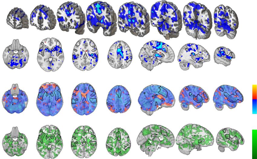
ARTICLE                                                                   NATURE COMMUNICATIONS | https://doi.org/10.1038/s41467-021-21179-3

P
        lacebo effects contribute substantially to treatment out-             and de-activations were found in regions typical for experimental
        comes in both medical research and clinical practice. A               pain (compare: Fig. 1b and ref. 13). The τ-statistic indicated
        better understanding of the underlying mechanisms is thus             considerable between-study heterogeneity in pain-related activity
important for optimizing drug development and clinical care1.                 throughout most of the brain (Supplementary Fig. 7), which was
Placebo analgesia is the most robust and best-studied type of                 expected given the large inter-study diversity regarding experi-
placebo effect1–3. A growing number of neuroimaging studies                   mental pain-induction, image acquisition, and image processing
elucidate the brain correlates of placebo analgesia. These studies,           (Supplementary Tables 2–5).
and meta-analyses of their findings, have provided evidence for
the involvement of brain regions linked to nociceptive processing,
                                                                              Voxel-wise results: effect of placebo treatment. In general,
including early pain-gating mechanisms, but also to decision-
                                                                              placebo treatment had a small (g < 0.2) effect on pain-related
making, cognitive appraisal, reward/motivation, emotional reg-
                                                                              brain activity, as compared to the matched control conditions
ulation4–7, and other forms of learning and social cognition8
                                                                              (Fig. 2a, b). Significant placebo-associated decreases were found
relevant for health behaviors.
                                                                              in the right insula, near the habenula and the splenium of the
   Nevertheless, the results of these studies vary substantially4, and
                                                                              corpus callosum, and in the cerebellum (p < 0.05, FWER cor-
the lack of large-sample assessments hampers the detection of small
                                                                              rected with pTFCE; Fig. 2a light blue, Supplementary Fig. 8,
to moderate effects9 and makes it difficult to identify precisely
                                                                              Supplementary Table 10). No areas showed placebo-related
which structures are consistently altered by placebo treatment.
                                                                              increases at the FWER-corrected threshold treating study as a
Previous meta-analyses have all relied on published coordinates of
                                                                              random effect.
activation peaks. These incomplete summaries of the full activation
                                                                                 Estimated between-study heterogeneity in voxel-level effect
maps provide only approximate information on replicability across
                                                                              sizes was low in the significantly de-activated regions (Fig. 2c,
studies and are susceptible to bias10. These limitations can be
                                                                              Supplementary Fig. 8, Supplementary Table 10). However, many
overcome by meta-analyses based on single-participant, whole-
                                                                              regions of the brain showing sub-threshold placebo-related
brain images, which are sometimes referred to as ‘mega-analyses’11.
                                                                              increases showed statistically significant τ-values, indicating
As meta-analyses on participant-level data are preferable in terms of
                                                                              between-study heterogeneity in effects (Fig. 2c). These included
statistical power and risk-of-bias11, a mega-analysis of placebo-
                                                                              multiple prefrontal cortical areas, perigenual anterior cingulate
induced brain activity can be expected to foster convergence in our
                                                                              cortex, intraparietal sulcus, precuneus, basal ganglia, and the left
understanding of placebo analgesia, to provide novel insights into
                                                                              middle insula. A brain-wide correlation analysis indicated that
the underlying neural mechanisms, and guide the development of
                                                                              placebo treatment effects were positively and significantly
predictive methods of individual placebo analgesia from neuroi-
                                                                              correlated across brain regions with τ-values (r = 0.191, 95% CI
maging data, which would be of crucial importance both from a
                                                                              [0.187, 0.196], p < 0.001, Supplementary Fig. 9), indicating that
clinical and drug development point of view. Here, we conducted a
                                                                              areas showing placebo-induced increases tended to have higher
systematic participant-level meta-analysis of 20 independent neu-
                                                                              levels of between-study heterogeneity. Thus, activation increases
roimaging studies on experimental placebo analgesia. Based on
                                                                              varied more substantially across studies than activation decreases.
whole-brain activation patterns in a total of N = 603 healthy par-
                                                                                 We therefore performed an exploratory fixed-study-effects
ticipants, we mapped the effects of placebo treatment on pain-
                                                                              analysis of placebo effects, which tests for effects within this set of
related brain activity and identified neural correlates of individual
                                                                              studies without the intent of generalizing to new, unobserved
differences in behavioral placebo analgesia.
                                                                              studies. In addition to decreases reported above, this analysis
                                                                              showed reduced activity in the middle cingulate cortex, the
Results                                                                       bilateral supplementary motor area (SMA), left fusiform cortex
Image quality. All included studies (N = 603 participants from                and cerebellum (Fig. 2d, light blue). The fixed-effects analysis
20 studies, see ref. 7 and Table 1) aimed at covering the whole               revealed significant placebo-induced activation in the anterior
brain down to the mid-pons/superior cerebellar level. Image                   intraparietal sulcus, precuneus, and dorsolateral prefrontal cortex
alignment to MNI-space was satisfactory for all studies, but                  (DLPFC) (Fig. 2d gold, Supplementary Table 11).
coverage was often incomplete near the boundaries of the brain                   To further follow up on potential sources of between-study
(see Supplementary Figs. 2 and 3), particularly in the inferior               heterogeneity, we explored the possibility of explaining hetero-
brainstem, cerebellum, and ventral prefrontal regions. These                  geneity through study-level experimental features, such as the
partially missing data are likely due to between-study differences            method of placebo induction. A preliminary comparison of
in field-of-view and/or signal dropout artifacts. For one study12,             placebo induction methods (conditioning and suggestions versus
only maps with white-matter regions masked out were available.                suggestions only) showed no significant differences in placebo-
   Outlier screening (see Supplementary Methods and Results)                  related brain activity after correction for multiple comparisons
indicated pain ratings that were too low for inclusion in six                 (Supplementary Fig. 10).
participants (responses  0.8); with the           corrected with pTFCE; Fig. 3a light blue; also see Supplementary
largest located in the insula, bilaterally (Fig. 1a, Supplementary            Fig. 11, Supplementary Table 12). The activity of contralateral
Fig. 6, Supplementary Table 9). In general, cerebral activations              (left) areas of the insula (z-score = 3.9, r = 0.17, p = 0.00005), the

2                 NATURE COMMUNICATIONS | (2021)12:1391 | https://doi.org/10.1038/s41467-021-21179-3 | www.nature.com/naturecommunications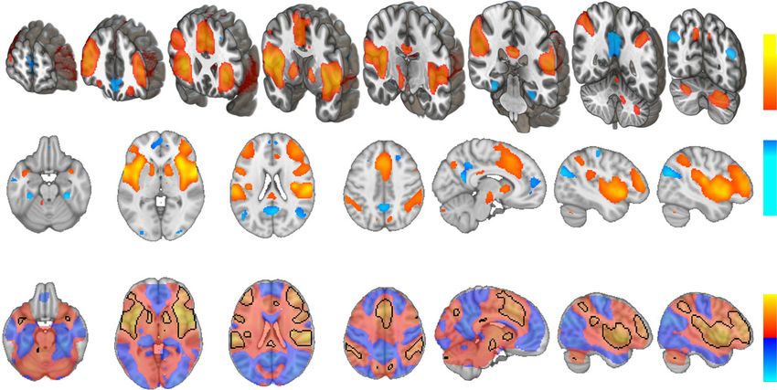
NATURE COMMUNICATIONS | https://doi.org/10.1038/s41467-021-21179-3 ARTICLE
Table 1 Studies included in the meta-analysis.
First author Year n Design Mean age (y) Sex (% Pain stimulus Placebo Treatment
male) induction
1 Atlas48 2012 21 within 25 48 heat sug IV-infusion
2 Bingel53 2006 19 within 24 79 laser sug + cond topical cream
3 Bingel70 2011 22 within 28 68 heat sug + cond IV-infusion
4 Choi54 2011 15 within 25 100 electrical sug + cond IV-infusion
5 Eippert56 2009 40 within 26 100 heat sug + cond topical cream
6 Ellingsen71 2013 28 within 26 68 heat sug nasal spray
7 Elsenbruch72 2012 36 within 26 42 distension sug IV-infusion
8 Freeman51 2015 24 within 27 50 heat sug + cond topical cream
9 Geuter55 2013 40 within 26 100 heat sug + cond topical cream
10 Kessner73 2013 39 between 26 51 heat cond topical cream
11 Kong49 2006 10c within 27 60 heat sug + cond sham acupuncture
12 Kong50 2009 12a within 26 42 heat sug + cond sham acupuncture
13 Lui74 2010 31 within 23 45 laser sug + cond sham TENS
14 Rütgen52 2015 102 between 25 31 electrical sug + cond pill
15 Schenk75 2014 32 within 26 53 cap + heat sug topical cream
16 Theysohn76 2014 30 within 35 50 distension sug IV-infusion
17 Wager12,A 2004 24 within NA NA electrical sug topical cream
18 Wager12,B 2004 23 within NA NA heat sug + cond topical cream
19 Wrobel57 2014 38 within 26 58 heat sug + cond topical cream
20 Zeidan43 2015 17a within 28 47 heat sug + cond topical cream
A Sub-study 1, B Sub-study 2, between between-group design, cap, capsaicin, cond conditioning, IV intravenous, L left, NA not available, R right, sug suggestions, TENS transcutaneous electrical nerve
stimulation, within within-subject design.
aPlacebo-treatment groups, only.
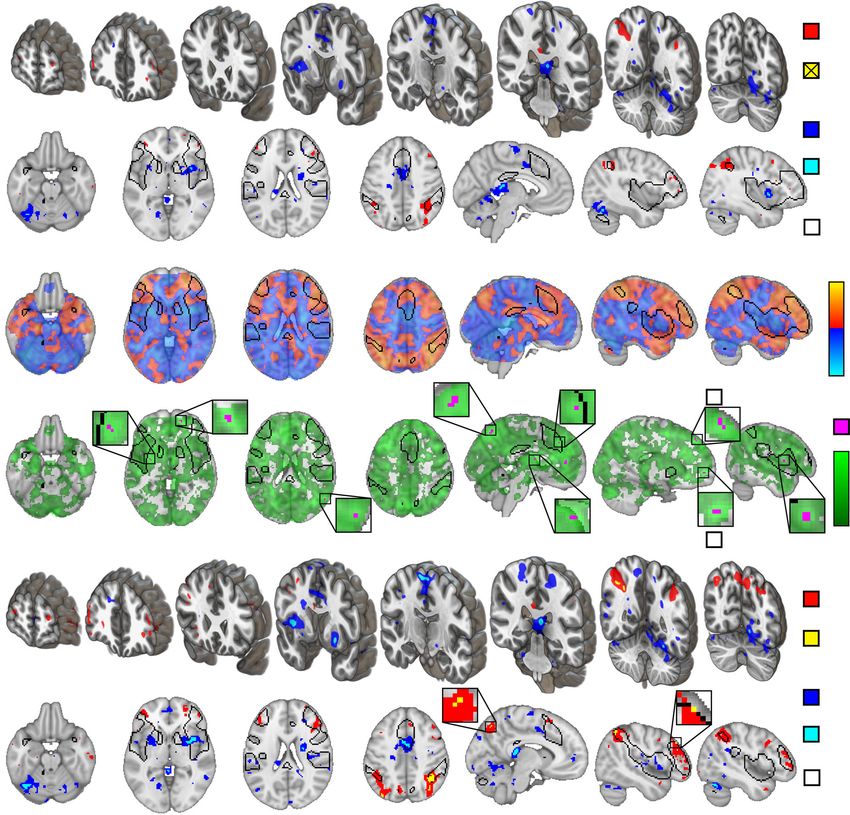
Fig. 1 Pain-related activity in experimental placebo imaging studies. a Statistically significant pain-responses (permutation test, controlled for FWER,
two-sided p < 0.05), and b whole-brain unthresholded standardized effect size g, of acute pain stimulation > baseline, pooled across placebo and control
conditions (FWER-corrected permutation test results are delineated as a back contour); range g: [−0.82, 1.68]; all: n = 543–603 individuals from 17 to 20
independent studies per voxel. Three-dimensional coronal slices are equidistantly distributed from y = 60 to −68 mm. Axial slices range equidistantly from
z = −22 to 42 mm. Custom coordinates for sagittal slices are displayed in mm. Source data (results as 3d-volumes) are provided at https://osf.io/n9mb3/.
secondary somatosensory cortex (z-score = 4.0, r = 0.21, p = analgesia and brain activity (Fig. 3b, Supplementary Fig. 11,
0.00003) was also negatively correlated with analgesia but was Supplementary Table 12), suggesting that correlations were driven
only significant without correcting for multiple comparisons by inter-individual differences rather than systematic differences
(Fig. 3a, blue). across studies. Across the brain, between-study heterogeneity did
Positive correlations between behavioral placebo analgesia and not reach FWER significance, but was largest in the basal ganglia,
brain activity, i.e. increasing brain activity with stronger placebo orbitofrontal and dorsolateral prefrontal cortices; see Fig. 3c).
response, did not reach statistical significance (p < 0.05, FWER Between-study heterogeneity was not spatially associated with
corrected, pTFCE). Without correcting for multiple comparisons, correlations across voxels (Supplementary Fig. 12); thus, the most
positive correlations (Fig. 3a, b, red) were observed near the heterogeneous regions were not those with the strongest effects.
subgenual cingulate cortex (z-score = 1.8, r = 0.14, puncorr = 0.036),
in the orbitofrontal cortex (z-score = 2.7, r = −0.17, puncorr = 0.003), Network- and region-based results: effects of painful stimula-
and the prefrontal pole (z-score = 2.6, r = −0.13, puncorr = 0.005). tion. Activation for painful stimulation compared to baseline
Levels of between-study heterogeneity were negligible in regions (averaged across placebo and control conditions) showed acti-
showing significant correlations between behavioral placebo vation of multiple expected cortical and subcortical regions
NATURE COMMUNICATIONS | (2021)12:1391 | https://doi.org/10.1038/s41467-021-21179-3 | www.nature.com/naturecommunications 3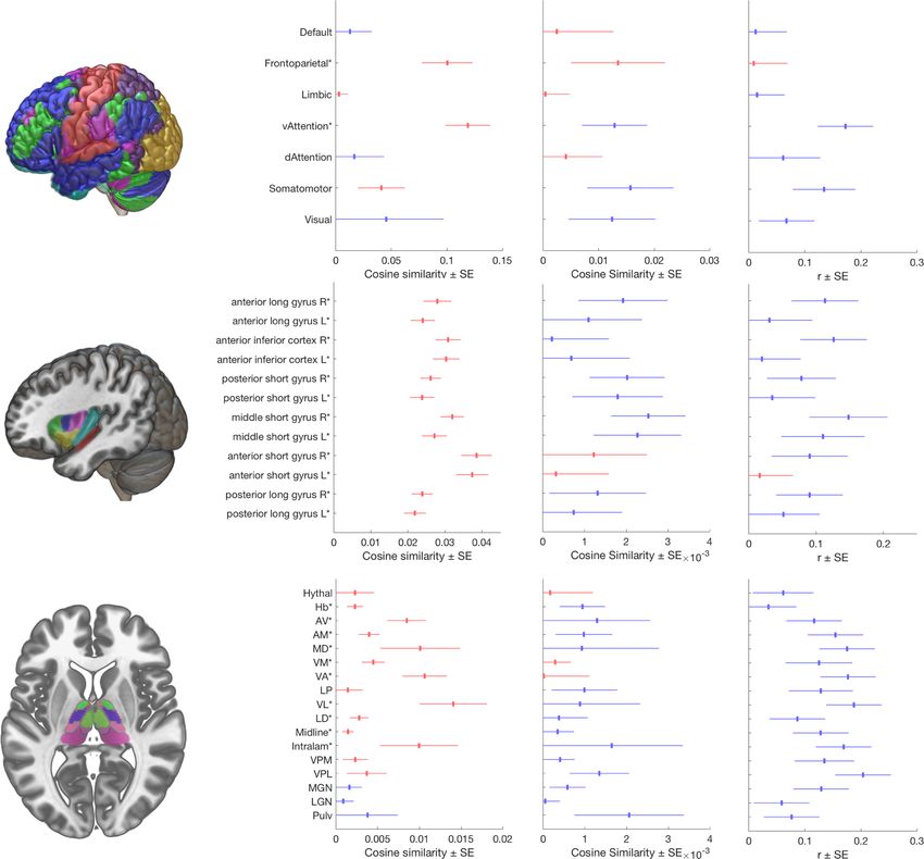
ARTICLE                                                                     NATURE COMMUNICATIONS | https://doi.org/10.1038/s41467-021-21179-3

Fig. 2 Placebo-induced changes in pain-related activity. a Areas of statistically significant placebo treatment effect, assuming random study-effect,
thresholded according to z-tests (uncorrected for multiple comparisons, two-sided p < 0.01, red and blue) and thresholded according to pTFCE-enhanced
permutation test (controlled for FWER, two-sided p < 0.05, light blue, activity increases did not reach statistical significance); b unthresholded standardized
effect size g of placebo treatment effect (range: [−0.19, 0.17]); c between-study heterogeneity τ (range: [0, 0.43]) with permutation test results
(controlled for FWER, one-sided, p < 0.05, green); τ is plotted as τ2 to emphasize regions of high heterogeneity. d significant placebo-effects assuming fixed
study-effect (range: [−0.22, 0.22]) thresholded according to z-tests (uncorrected for multiple comparisons, two-sided p < 0.01, red and blue) and
thresholded according to pTFCE-enhanced permutation test (controlled for FWER, two-sided p < 0.05, light blue and gold); all: n = 543 to 603 individuals
from 17 to 20 independent studies per voxel. b, c, and d are shown with a contour of FWER-corrected permutation test results for pain > baseline, as shown
in Fig. 1a. Small FWER-corrected clusters are zoomed in insets. Three-dimensional coronal slices are equidistantly distributed from y = 60 to −68 mm.
Axial slices range equidistantly from z = −22 to 42 mm. Custom coordinates for sagittal slices are displayed in mm and were chosen to highlight important
areas of activation. Source data (results as 3d-volumes) are provided at https://osf.io/n9mb3/.

(Fig. 4, Column 2). These included activation in the ventral                    Network- and region-based results: effect of placebo treatment.
attention network (which encompasses the insulae), the fronto-                  Network similarity analysis indicated that placebo treatment
parietal network14, and the somatomotor network. Positive                       reduced activity in the ventral attention and the somatomotor
associations were also found in all insular sub-regions and most                networks14; (Fig. 4, Column 3) which includes the mid-cingulate
thalamic nuclei, including the intralaminar nuclei targeted by                  cortex (localized particularly to area 24pr in ref. 16). In the insula,
ascending nociceptive pathways, the mediodorsal ‘limbic asso-                   placebo reduced activity in bilateral middle short gyrus and right
ciation’ nucleus, and the ventro-basal complex, including the                   posterior short gyrus, corresponding to the dorsal anterior/mid-
ventro-posterior lateral (VPL) nucleus15. A tendency towards                    insula, as well as a trend towards reduced activity in the right
negative associations was found for the lateral geniculate, medial              anterior long gyrus (posterior insula, contralateral to stimulation
geniculate, and pulvinar nuclei, which are known for their pre-                 in most studies). Thalamic nuclei showed tendencies towards
dominantly visual and auditory roles15.                                         placebo-induced decreases in areas strongly activated in pain. The

4                   NATURE COMMUNICATIONS | (2021)12:1391 | https://doi.org/10.1038/s41467-021-21179-3 | www.nature.com/naturecommunications
NATURE COMMUNICATIONS | https://doi.org/10.1038/s41467-021-21179-3                                                                          ARTICLE

Fig. 3 Correlations of behavioral placebo analgesia and changes in pain-related brain activity. a Whole-brain areas of statistically significant correlation
(Pearson’s r) between behavioral placebo analgesia (paincontrol − painplacebo) and placebo-related activity changes (painplacebo − paincontrol), thresholded
according to z-tests (uncorrected for multiple comparisons, two-sided p < 0.01, red and blue), and thresholded according to pTFCE-enhanced permutation
test (controlled for FWER, two-sided p < 0.05, light blue, increased correlations did not reach corrected statistical significance); b unthresholded Pearson’s
r, range: [−0.26; 0.17]; c between-study heterogeneity τ (range: [0, 0.32]) for the same relationship (permutation test controlled for FWER, one-sided p <
0.05, indicated no statistically significant voxels); τ is plotted as τ2 to emphasize regions of high heterogeneity. all: n = 384–460 individuals from 15 to 18
independent studies per voxel. a, b, and c are shown with a contour of FWER-corrected permutation test results for pain > baseline, as shown in Fig. 1a.
Correlations were computed across individual participants in the full sample, excluding between-group studies (where individual estimates of behavioral
placebo analgesia are not possible). On panels a and b, red-yellow and blue-light blue shades denote increased and decreased activity associated with
larger placebo analgesia, respectively. Three-dimensional coronal slices are equidistantly distributed from y = 60 to −68 mm. Axial slices range
equidistantly from z = −22 to 42 mm. Custom coordinates for sagittal slices are displayed in mm. Source data (results as 3d-volumes) are provided at
https://osf.io/n9mb3/.

strongest decreases were found in the VPL, a primary target of the               Discussion
spinothalamic tract, and the habenula. Observed placebo effects                  In this collaborative effort, we performed a comprehensive, large-
in other regions tended to be smaller.                                           scale (N = 603) participant-level voxel-based neuroimaging meta-
                                                                                 analysis of placebo analgesia, involving the majority of eligible
                                                                                 experimental neuroimaging studies. Our results provide a reliable,
Network- and region-based results: correlations with placebo                     aggregated view of the size, localization, significance, and het-
analgesia. As with the main effects of placebo vs. control, net-                 erogeneity of placebo-effects on pain-induced brain activity. In a
work similarity-based analysis of regions correlated with placebo                previous paper, we focused on the question of whether placebo
analgesia indicated that activity in the ventral attention and                   analgesia involves changes in the neurologic pain signature
somatomotor networks was negatively correlated with behavioral                   (NPS)17, a machine-learning based weighted, multi-voxel sum-
placebo responses (Fig. 4), i.e., strong placebo responders showed               mary metric (covering about 10% of the brain), that can be
larger deactivation with placebo treatment. Within the right                     interpreted as a neuromarker of nociceptive pain. This previous
insula, several regions tended towards negative correlations with                study revealed that behavioral placebo analgesia was associated
placebo responses, especially the anterior long gyrus (posterior                 with significant but small effects in the NPS, pointing to the
insula), middle short gyrus (dorsal anterior/mid insula), and                    relevance of other brain areas and networks. Accordingly, char-
anterior inferior cortex (ventral insula). In the thalamus, stronger             acterizing this potentially broader set of changes was the key
placebo analgesia was correlated with reductions in multiple                     focus of this voxel-wise whole-brain investigation (for a com-
thalamic regions, including all seven regions that responded                     parison with regions involved in the NPS, see Supplementary
strongly to pain in this sample (intralaminar, ventrolateral, ven-               Fig. 13). The present results corroborated previous findings of
tral anterior, ventromedial, mediodorsal, antero-medial, and                     increases in frontal-parietal regions and reductions in the insula.
anterio-ventral nuclear groups), and thalamic targets of the spi-                In addition, they revealed new effects systematically missed
nothalamic tract (ventro-posterior-lateral [VPL] and -medial                     in previous smaller-scale analyses, including reductions in
[VPM]).                                                                          the habenula, specific parts of the thalamus (particularly VPL, a

NATURE COMMUNICATIONS | (2021)12:1391 | https://doi.org/10.1038/s41467-021-21179-3 | www.nature.com/naturecommunications                                    5
ARTICLE NATURE COMMUNICATIONS | https://doi.org/10.1038/s41467-021-21179-3 Fig. 4 Similarity-based analysis of brain activity in functional cortical networks, insula, and thalamus. Column 1 (header): Depiction of atlases: Row 1: whole-brain cortical networks of functional connectivity14, Row 2: insular sub-regions66 Row 3: thalamic nuclei67. See Supplementary Fig. 14 for further details. Column 2: Mean (±SEM) cosine similarity (c) of pain-related activity, n = 603 from 20 independent studies; Column 3: Mean (±SEM) cosine similarity (c) of placebo-induced changes in pain-related activity (Column 2); all: n = 603 from 20 independent studies. Column 4: Correlation (Pearson’s r ± SEM) between behavioral placebo response and cosine similarity estimates of placebo-related activity; n = 460 from 18 independent studies. In Columns 2 and 3, red and blue colors denote increased and decreased pain-related activity, respectively. In Column 4 red and blue shades denote increased and decreased activity associated with larger placebo analgesia, respectively. Asterisks (*) denote significant differences from zero, according to two-sided t- tests (p < 0.05, uncorrected for multiple comparisons). Source data (results as 3d-volumes) are provided at https://osf.io/n9mb3/. Hythal Hypothalamus, Hb Habenular, AV anterior ventral, AM anterior medial, MD mediodorsal, VM ventral medial, VA ventral anterior, LP lateral posterior, VL ventral lateral, LD lateral dorsal, Intralam intralaminary, VPM ventral posterior medial, VPL ventral posterior lateral, MGN Medial Geniculate Nucleus, LGN Lateral Geniculate Nucleus, Pulv Pulvinar. nociceptive nucleus), and the cerebellum, promising new targets Our study-as-random-effect results provide strong evidence for in explaining placebo analgesia. placebo-associated reductions of pain-related activity in some Here, we discuss our results in correspondence to two key open brain areas linked to nociception and pain and indicate that these questions: (i) how strongly do placebo treatments affect the same are generalizable across experimental paradigms. We also provide systems involved in nociception and pain generation (as indicated strong evidence that the degree to which pain-related activity is by the contrast control > placebo); and (ii) which systems are reduced in these brain areas correlates with the magnitude of engaged by placebo treatments (as indicated by the contrast behavioral hypoalgesia across individuals. placebo > control) and may therefore reflect top-down mod- Specifically, the placebo-associated decrease and its correlation ulatory mechanisms. with behavioral pain ratings were most prominent in regions 6 NATURE COMMUNICATIONS | (2021)12:1391 | https://doi.org/10.1038/s41467-021-21179-3 | www.nature.com/naturecommunications
NATURE COMMUNICATIONS | https://doi.org/10.1038/s41467-021-21179-3                                                                   ARTICLE

located within the ventral attention and the somatomotor cortical                due to insufficient cerebellar coverage across studies. Here, the
networks, including the anterior insula and targets of the spi-                  dorsomedial cerebellum showed a profile of responses to painful
nothalamic tract18, including the thalamic VPL complex the                       events, reductions with placebo, and correlations with the mag-
posterior insula and, moreover, in the habenula (Figs. 2b and 3b;                nitude of placebo analgesia. Some cerebellar regions have been
Fig. 4, Column 3 and 4). Correlations were additionally pro-                     linked to pain, and others to other cognitive, affective, and motor
nounced in both lateral (VL and VPM/VPL) and medial (intra-                      processes28,29 and patients with cerebellar infarctions show
laminar and MD) thalamic nuclei (Fig. 4). These regions are                      reduced placebo analgesia30. Cerebellar reductions and correla-
targets of ascending nociceptive systems and, as expected, were                  tions are centered in vermis areas V and (to a lesser degree)
also strongly engaged during painful stimulation. In summary,                    II31–33, which are associated with somatomotor and limbic cor-
placebo-associated down-regulation seems to affect thalamocor-                   tical networks, respectively. Thus, the best interpretation of cer-
tical pathways related to nociception and pain13,19, particularly in             ebellar effects here is that they are related partly, but not
strong placebo responders. The relatively low between-study                      exclusively, to somatomotor networks and pain. Placebo
heterogeneity in these regions indicates that variability in                     hypoalgesia-related activity changes in the VL nucleus, a target of
placebo-related reductions is primarily a function of the indivi-                cerebellothalamic tract15, also suggest that fronto-cerebellar
dual responder rather than the paradigm used.                                    connectivity may pose a promising novel target for future in-
   These findings complement previous findings of small but                        depth studies on the mechanisms of placebo analgesia.
statistically significant placebo-induced reductions in the NPS7.                    In contrast to these placebo-related deactivations, some regions
In this previous study, NPS reductions also correlated with the                  displayed increased pain-related activity as an effect of placebo
magnitude of placebo analgesia. Here, findings of VPL reductions                  treatment, which is often interpreted as participating in the
with placebo and widespread correlations between analgesia and                   construction of top-down representations of context (including
correlations in broadly pain-related systems support the conclu-                 beliefs and expectations). These regions tend to be localized in the
sion that alterations in nociception and pain construction are an                frontoparietal network (FPN). These increases were statistically
important element of placebo analgesia. The small effect sizes                   significant only in the fixed effect analysis and involved the right
however indicate that nociceptive changes are unlikely to be a                   DLPFC (with subthreshold activation on the left side), as well as
complete explanation. The strengths of the previous NPS findings                  the bilateral intraparietal sulcus. While the fixed-effect analysis
were that the neuromarker was identified independently from the                   provides limited generalizability, this result is very much in line
present sample and validated in over 40 published cohorts to                     with previous neuroimaging studies highlighting the importance
date. However, limitations were that (1) the NPS may not per-                    of the DLPFC in initiating and maintaining the top-down effect
fectly characterize nociceptive processing in this sample, and                   of treatment expectation on nociceptive processing and pain. E.g.
some nociceptive pain-related effects may be missed; (2) it cannot               activity in the DLPFC precedes and scales with activity changes in
provide a broad view of effects across the brain, including areas                downstream pain modulatory areas and prevents the extinction of
like the habenula and many cortical areas; and (3) it tests a dis-               once learned placebo analgesia27. Moreover, transient inhibition
tributed pattern response and is not suited to identify placebo                  of the DLPFC using transcranial magnetic stimulation led to
effects in VPL (or any other region) in particular. The present                  reduced placebo analgesia34.
findings of effects in specific thalamic and other regions are                        The lack of FWER-corrected increases might be attributed to
complementary and important, in that they provide a region-level                 the reduced power in the study-as-random-effect analysis, which
inference about effects in neuroanatomically defined nociceptive                  is very conservative. However, the heterogeneity analysis indicates
pathways. Thus, overall, we believe that placebo effects affect the              that the between-study variance is significantly higher than
circuitry involved in pain generation to some degree, in a manner                expected in key fronto-parietal areas (Fig. 2c). This suggests that,
that varies across individuals more than across studies in the                   in contrast to the consistent placebo-induced decreases across
present dataset, but also includes other brain effects beyond                    studies, placebo-related increases in brain activity are more het-
nociception that may be important for the emotions, decision-                    erogenous across placebo induction techniques. For instance,
making, and behaviors surrounding pain.                                          significant heterogeneity in the DLPFC and perigenual ACC
   Placebo-related decreases were not restricted to pathways                     might reflect the differing engagement of descending pain reg-
associated with nociception. Brain regions traditionally linked to               ulatory mechanisms across studies, although these regions are
self-regulation and high-level action selection, particularly the                clearly not exclusively associated with pain modulation (e.g. see
SMA20–25 also showed reduced activity during placebo analgesia,                  ref. 35).
particularly in strong placebo responders (Figs. 2 and 3). Thus, it                 Between-study heterogeneity was statistically significant
is possible that some of these effects reflect shifts in motivation               throughout the frontal lobe (Fig. 2c), which may reflect inter-
and decision-making in the context of pain. These findings                        study variation in participants’ appraisal of the context and
extend previous meta-analyses, which all highlighted de-                         internal responses, e.g., expectations36. The degree to which
activations in the mid-cingulate, but not the SMA or pre-motor                   prefrontal systems are required for analgesia may vary. For
areas4–6. In addition to action planning and self-regulation, they               example, in ref. 37, placebo analgesia was predicted by fronto-
may be related to other cognitive operations related to evaluating               parietal activity in regions associated with emotion regulation but
pain under placebo treatment, including error monitoring, pre-                   not working memory. Emotion regulation, in particular reap-
diction errors, and sequence processing20,26.                                    praisal strategies involving self-generated positive contexts for
   One of the strongest effects was found in the left putamen,                   experiences, appears to involve fronto-parietal networks in
which de-activated with placebo in proportion to analgesia. This                 reducing negative affect. Other recent studies have also found
is in line with multiple studies reporting correlations between                  correlations between placebo analgesia and DLPFC con-
placebo analgesia and both fronto-parietal and limbic fronto-                    nectivity38,39 (e.g., with the nucleus accumbens40) and opioid
striatal pathways8 and might be related to the (prefrontal) sup-                 binding in prefrontal cortex41,42. By contrast, other studies have
pression of striatal prediction errors or other aversive circuits27.             found that mindfulness practice can reduce pain without fronto-
   Interestingly, each of the main analyses revealed prominent                   parietal activation or appreciable deactivation in spinothalamic
placebo-related reductions in cerebellar regions. While this in line             targets43. These strategies focus on acceptance without judgment
with some previous findings (e.g.12), cerebellar effects were not                 rather than active re-contextualization, which may be another
reported in previous meta-analyses of placebo analgesia, possibly                important component of placebo analgesia.

NATURE COMMUNICATIONS | (2021)12:1391 | https://doi.org/10.1038/s41467-021-21179-3 | www.nature.com/naturecommunications                           7
ARTICLE                                                                               NATURE COMMUNICATIONS | https://doi.org/10.1038/s41467-021-21179-3

   In sum, placebo analgesia may involve multiple alterations in                          Data acquisition. As previously described7, we performed a systematic literature
appraisal systems, reflecting multiple underlying mechanisms44.                            search to identify experimental functional magnetic resonance imaging (fMRI)
                                                                                          investigations of placebo analgesia (see Supplementary Fig. 1, Supplementary
Our results suggest that placebo effects are not restricted solely to                     Table 1, and ref. 7 for details). Criteria for study eligibility were: (a) published in
either sensory/nociceptive or cognitive/affective processes, but                          peer-reviewed journal in the English language; (b) original investigation; (c) human
likely involve a combination of mechanisms that may differ                                participants; (d) functional neuroimaging of the brain during evoked pain; (e) pain
depending on the paradigm and other individual factors.                                   delivered under matched placebo and control conditions. Definitions of placebo and
Understanding the neural and neurochemical pathways under-                                control conditions (see Supplementary Methods and Results) were identical to our
                                                                                          previous meta-analysis7. Investigators of eligible studies were contacted and invited
lying this variability will pave the way to systematically utilize/                       to share data. We collected single-participant, first-level, whole-brain standard-
modulate placebo responses in a context-, patient-, and disease-                          space summary images of pain response (statistical parametric maps) from the
specific manner. Fostering the therapeutic processes underlying                            original analyses, as published, as well as corresponding pain ratings, experimental
placebo effects in clinical settings promises to boost the efficacy                        design parameters, and demographic data (Supplementary Tables 2–5).
(and tolerability) of analgesic drug treatments. Likewise, con-
trolling and homogenizing placebo responses during drug                                   Outcome definition and comparisons. The main outcome was pain-related
development can enhance the assay sensitivity in clinical trials.                         change in fMRI signal (i.e., blood oxygen level-dependent signal, perfusion chan-
                                                                                          ges), i.e., the effect of painful stimulation compared to baseline, as estimated in the
Finally, biomarkers based on the types of brain alterations we                            original analyses (i.e., beta or contrast images). Based on this outcome, we per-
identify here, and reported in other studies38,45, may help to                            formed three comparisons: (i) main effect of pain vs baseline, averaging placebo
dissect placebo from analgesic drug responses in pre-clinical                             and control conditions; (ii) pain-related activity acquired under matched placebo
trials.                                                                                   and control conditions (placebo–control); and (iii), for studies that manipulated
                                                                                          placebo vs. control within-subject, correlations across individuals between the
   The present findings must be interpreted in the light of several                        effect of placebo treatment on brain activity and behavioral placebo analgesia (i.e.,
limitations. First, as our findings are based on experimental pla-                         [placebo–control] in pain ratings).
cebo interventions in healthy volunteers, they may not generalize                             Non-painful or mildly painful12,48–52 stimulus conditions were excluded. For
to clinical settings. Second, the present study covered a wide                            studies that involved left- and right-lateralized stimulation53, strong and weak
                                                                                          placebo conditions54,55, or early- and late heat-pain periods55–57 maps were
range of experimental placebo paradigms and conditions. This is                           averaged on subject level, as in the previous analysis7 (see Supplementary Table 6
favorable in terms of establishing the broad generalizability of                          for details). The main effect of pain vs baseline was averaged across placebo and
results, but it also means that findings have to generalize over                           control conditions (instead of just using no-placebo conditions) because for some
many sources of variation: Paradigm, population/sample, scan-                             studies48 only pooled estimates of the main effect of pain were available (see
                                                                                          Supplementary Methods and Results for details).
ner, and choice of analysis methods. Effect size estimates are thus
likely overly conservative compared with what may be possible as
analysis methods continue to become standardized and metho-                               Analysis. We applied the Cochrane risk of bias tool58 to assess the risk-of-bias of
                                                                                          included studies. (Supplementary Methods and Results and Supplementary
dological advances reduce inter-subject and inter-study varia-                            Table 7).
bility. Further, the fact that this meta-analysis was based on                                Images underwent systematic quality control, as described previously7 (see
participant-level statistical summary images from variety of dif-                         Supplementary Methods and Results for details). Voxels missing in >10% of
ferent pre-processing pipelines (s. Supplementary Table 5) likely                         participants (n > 60) or outside of the MNI152 brain-template (as implemented in
                                                                                          SPM1259, probability of brain tissue
NATURE COMMUNICATIONS | https://doi.org/10.1038/s41467-021-21179-3 ARTICLE
summarized using the GIV method, with statistics based on t-tests across studies, 16. Glasser, M. F. et al. A multi-modal parcellation of human cerebral cortex.
treating study as a random effect. No correction for multiple comparisons was Nature 536, 171–178 (2016).
performed for the atlas-based analyses due to their exploratory nature. Cosine 17. Wager, T. D. et al. An fMRI-based neurologic signature of physical pain. N.
similarity is equivalent to Pearson’s correlation except for mean-centering, so it Engl. J. Med. 368, 1388–1397 (2013).
remains sensitive to the overall level of activation across the brain and thus reflects 18. Brooks, J. & Tracey, I. From nociception to pain perception: imaging the
absolute normalized activity levels in the regions/networks tested rather than spinal and supraspinal pathways. J. Anat. 207, 19–33 (2005).
relative activity across regions. 19. Segerdahl, A. R., Mezue, M., Okell, T. W., Farrar, J. T. & Tracey, I. The dorsal
All analyses were performed with MATLAB 2016b, SPM12, the CANlab Core posterior insula subserves a fundamental role in human pain. Nat. Neurosci.
Tools neuroimaging analysis toolbox (https://github.com/canlab/CanlabCore), and 18, 499–500 (2015).
custom functions implementing the GIV method. Further analysis details are 20. Cona, G. & Semenza, C. Supplementary motor area as key structure for
provided in the Supplementary Methods and Results. domain-general sequence processing: a unified account. Neurosci. Biobehav.
MRIcroGL (v28.5.2017) was used to create illustrations of statistical parametric Rev. 72, 28–42 (2017).
maps. All neuroimages shown follow the neurological convention (left side 21. Woo, C.-W., Roy, M., Buhle, J. T. & Wager, T. D. Distinct brain systems
corresponds to left hemisphere in coronal- and axial-sections). Effect sizes are mediate the effects of nociceptive input and self-regulation on pain. PLoS Biol.
interpreted as small, moderate, and large according to the recommendations by 13, e1002036 (2015).
Cohen69. All result maps from this meta-analysis are available for download as 3d
22. Buhle, J. T. et al. Cognitive reappraisal of emotion: a meta-analysis of human
NIFTI images at https://osf.io/n9mb3/.
neuroimaging studies. Cereb. Cortex 24, 2981–2990 (2014).
23. Soon, C. S., Brass, M., Heinze, H.-J. & Haynes, J.-D. Unconscious
Reporting summary. Further information on research design is available in the Nature determinants of free decisions in the human brain. Nat. Neurosci. 11, 543–545
Research Reporting Summary linked to this article. (2008).
24. Stuss, D. T. & Alexander, M. P. Is there a dysexecutive syndrome? Philos.
Trans. R. Soc. B Biol. Sci. 362, 901–915 (2007).
Data availability 25. Lau, H. C. & Passingham, R. E. Unconscious activation of the cognitive control
Results as 3d-volumes are provided at https://osf.io/n9mb3/. Participant-level source data
system in the human prefrontal cortex. J. Neurosci. 27, 5805–5811 (2007).
are available from the authors upon reasonable request and with permission of the
26. Botvinick, M. M., Niv, Y. & Barto, A. C. Hierarchically organized behavior and
Placebo Imaging Consortium.
its neural foundations: a reinforcement learning perspective. Cognition 113,
262–280 (2009).
Code availability 27. Schenk, L. A., Sprenger, C., Onat, S., Colloca, L. & Büchel, C. Suppression of
The full analysis code is available at https://github.com/mzunhammer/ striatal prediction errors by the prefrontal cortex in placebo hypoalgesia. J.
PlaceboImagingMetaAnalysis. Neurosci. 37, 9715–9723 (2017).
28. Stoodley, C. J. & Schmahmann, J. D. Functional topography in the human
cerebellum: a meta-analysis of neuroimaging studies. Neuroimage 44, 489–501
Received: 19 January 2020; Accepted: 4 January 2021; (2009).
29. Baumann, O. et al. Consensus Paper: the role of the cerebellum in perceptual
processes. Cerebellum 14, 197–220 (2015).
30. Ruscheweyh, R. et al. Altered experimental pain perception after cerebellar
infarction. Pain 155, 1303–1312 (2014).
31. Diedrichsen, J., Balsters, J. H., Flavell, J., Cussans, E. & Ramnani, N. A
References probabilistic MR atlas of the human cerebellum. Neuroimage 46, 39–46
1. Enck, P., Bingel, U., Schedlowski, M. & Rief, W. The placebo response in (2009).
medicine: minimize, maximize or personalize? Nat. Rev. Drug Discov. 12, 32. Choi, E. Y., Yeo, B. T. T. & Buckner, R. L. The organization of the human
191–204 (2013). striatum estimated by intrinsic functional connectivity. J. Neurophysiol. 108,
2. Hróbjartsson, A. & Gøtzsche, P. C. Placebo Interventions For All Clinical 2242–2263 (2012).
Conditions (Review) Placebo Interventions For All Clinical Conditions. 33. Buckner, R. L. The cerebellum and cognitive function: 25 years of insight from
Cochrane Database Syst. Rev. https://doi.org/10.1002/14651858 (2010). anatomy and neuroimaging. Neuron 80, 807–815 (2013).
3. Vase, L., Petersen, G. L., Riley, J. L. & Price, D. D. Factors contributing to large 34. Krummenacher, P., Candia, V., Folkers, G., Schedlowski, M. & Schönbächler,
analgesic effects in placebo mechanism studies conducted between 2002 and G. Prefrontal cortex modulates placebo analgesia. Pain 148, 368–374 (2010).
2007. Pain 145, 36–44 (2009). 35. D’Souza, D. & D’Souza, H. Bilingual language control mechanisms in anterior
4. Atlas, L. Y. & Wager, T. D. A meta-analysis of brain mechanisms of placebo cingulate cortex and dorsolateral prefrontal cortex: a developmental
analgesia: consistent findings and unanswered questions. Handb. Exp. perspective. J. Neurosci. 36, 5434–5436 (2016).
Pharmacol. 225, 37–69 (2014). 36. Peciña, M., Stohler, C. S. & Zubieta, J.-K. Neurobiology of placebo effects:
5. Amanzio, M., Benedetti, F., Porro, C. A., Palermo, S. & Cauda, F. Activation expectations or learning? Soc. Cogn. Affect. Neurosci. 9, 1013–1021 (2014).
likelihood estimation meta-analysis of brain correlates of placebo analgesia in 37. Wager, T. D., Atlas, L. Y., Leotti, L. A. & Rilling, J. K. Predicting individual
human experimental pain. Hum. Brain Mapp. 34, 738–752 (2013). differences in placebo analgesia: contributions of brain activity during
6. Ashar, Y. K., Chang, L. J. & Wager, T. D. Brain mechanisms of the placebo effect: anticipation and pain experience. J. Neurosci. 31, 439–452 (2011).
an affective appraisal account. Annu. Rev. Clin. Psychol. 13, 73–98 (2017). 38. Tétreault, P. et al. Brain connectivity predicts placebo response across chronic
7. Zunhammer, M., Bingel, U. & Wager, T. D., Placebo Imaging Consortium. pain clinical trials. PLoS Biol. 14, 1–22 (2016).
Placebo effects on the neurologic pain signature: a meta-analysis of individual 39. Vachon-Presseau, E. et al. Brain and psychological determinants of placebo
participant functional magnetic resonance imaging data. JAMA Neurol. 75, pill response in chronic pain patients. Nat. Commun. 9, 3397 (2018).
1321–1330 (2018). 40. Hashmi, J. A. et al. Functional network architecture predicts psychologically
8. Wager, T. D. & Atlas, L. Y. The neuroscience of placebo effects: connecting mediated analgesia related to treatment in chronic knee pain patients. J.
context, learning and health. Nat. Rev. Neurosci. 16, 403–418 (2015). Neurosci. 34, 3924–3936 (2014).
9. Cremers, H. R., Wager, T. D. & Yarkoni, T. The relation between statistical 41. Peciña, M. & Zubieta, J. K. Molecular mechanisms of placebo responses in
power and inference in fMRI. PLoS ONE 12, 1–20 (2017). humans. placebo pain from bench to bedside 25–36, https://doi.org/10.1016/
10. Kober, H. & Wager, T. D. Meta-analysis of neuroimaging data. Wiley B978-0-12-397928-5.00004-0 (2013).
Interdiscip. Rev. Cogn. Sci. 1, 293–300 (2010). 42. Wager, T. D., Scott, D. J. & Zubieta, J.-K. Placebo effects on human mu-opioid
11. Salimi-Khorshidi, G., Smith, S. M., Keltner, J. R., Wager, T. D. & Nichols, T. E. activity during pain. Proc. Natl Acad. Sci. USA 104, 11056–11061 (2007).
Meta-analysis of neuroimaging data: a comparison of image-based and 43. Zeidan, F. et al. Mindfulness meditation-based pain relief employs different
coordinate-based pooling of studies. Neuroimage 45, 810–823 (2009). neural mechanisms than placebo and sham mindfulness meditation-induced
12. Wager, T. D. et al. Placebo-induced changes in FMRI in the anticipation and analgesia. J. Neurosci. 35, 15307–15325 (2015).
experience of pain. Science 303, 1162–1167 (2004). 44. Benedetti, F. Placebo and the new physiology of the doctor-patient
13. Duerden, E. G. & Albanese, M.-C. Localization of pain-related brain relationship. Physiol. Rev. 93, 1207–1246 (2013).
activation: a meta-analysis of neuroimaging data. Hum. Brain Mapp. 34, 45. Vachon-Presseau, E. et al. Corticolimbic anatomical characteristics
109–149 (2013). predetermine risk for chronic pain. Brain 139, 1958–1970 (2016).
14. Yeo, B. T. T. et al. The organization of the human cerebral cortex estimated by 46. Bowring, A., Maumet, C. & Nichols, T. E. Exploring the impact of analysis
intrinsic functional connectivity. J. Neurophysiol. 106, 1125–1165 (2011). software on task fMRI results. Hum. Brain Mapp. 40, 3362–3384 (2019).
15. Herrero, M. T., Barcia, C. & Navarro, J. M. Functional anatomy of thalamus 47. Brett, M., Johnsrude, I. S. & Owen, A. M. The problem of functional
and basal ganglia. Child’s Nerv. Syst. 18, 386–404 (2002). localization in the human brain. Nat. Rev. Neurosci. 3, 243–249 (2002).
NATURE COMMUNICATIONS | (2021)12:1391 | https://doi.org/10.1038/s41467-021-21179-3 | www.nature.com/naturecommunications 9ARTICLE NATURE COMMUNICATIONS | https://doi.org/10.1038/s41467-021-21179-3
48. Atlas, L. Y. et al. Dissociable influences of opiates and expectations on pain. J. 75. Schenk, L. A., Sprenger, C., Geuter, S. & Büchel, C. Expectation requires
Neurosci. 32, 8053–8064 (2012). treatment to boost pain relief: an fMRI study. Pain 155, 150–157 (2014).
49. Kong, J. et al. Brain activity associated with expectancy-enhanced placebo 76. Theysohn, N. et al. Are there sex differences in placebo analgesia during
analgesia as measured by functional magnetic resonance imaging. J. Neurosci. visceral pain processing? A fMRI study in healthy subjects.
26, 381–388 (2006). Neurogastroenterol. Motil. 26, 1743–1753 (2014).
50. Kong, J. et al. Expectancy and treatment interactions: a dissociation between
acupuncture analgesia and expectancy evoked placebo analgesia. Neuroimage
45, 940–949 (2009). Acknowledgements
51. Freeman, S. et al. Distinct neural representations of placebo and nocebo This research was funded by the Mercator Research Center Ruhr (MERCUR) and
effects. Neuroimage 112, 197–207 (2015). National Institutes of Health R01 MH076136 (T.D.W.) and the German Research
52. Rütgen, M. et al. Placebo analgesia and its opioidergic regulation suggest that Foundation, TRR 289 Treatment Expectation. Gefördert durch die Deutsche For-
empathy for pain is grounded in self pain. Proc. Natl Acad. Sci. USA 112, schungsgemeinschaft (DFG) – Projektnummer 422744262 – TRR 289.
E5638–E5646 (2015).
53. Bingel, U., Lorenz, J., Schoell, E., Weiller, C. & Büchel, C. Mechanisms of Author contributions
placebo analgesia: rACC recruitment of a subcortical antinociceptive network. U.B. and T.D.W. had full access to all the data in the study and take responsibility for the
Pain 120, 8–15 (2006). integrity of the data and the accuracy of the data analysis. Concept and design: M.Z.,
54. Choi, J. C. et al. Placebo effects on analgesia related to testosterone and U.B., and T.D.W. Acquisition of the data: All authors. Drafting of the manuscript: M.Z.,
premotor activation. Neuroreport 22, 419–423 (2011). U.B., and T.D.W. Critical revision of the manuscript for important intellectual content:
55. Geuter, S., Eippert, F., Hindi Attar, C. & Büchel, C. Cortical and subcortical All authors. Statistical analysis: M.Z., T.D.W., and T.S. Visualization of Results: T.S.,
responses to high and low effective placebo treatments. Neuroimage 67, T.D.W., and M.Z. Obtained funding: U.B. and T.D.W. Administrative, technical, or
227–236 (2013). material support: U.B. Supervision: U.B. and T.D.W.
56. Eippert, F. et al. Activation of the opioidergic descending pain control system
underlies placebo analgesia. Neuron 63, 533–543 (2009).
57. Wrobel, N., Wiech, K., Forkmann, K., Ritter, C. & Bingel, U. Haloperidol Funding
blocks dorsal striatum activity but not analgesia in a placebo paradigm. Cortex Open Access funding enabled and organized by Projekt DEAL.
57, 60–73 (2014).
58. Cochrane Handbook for Systematic Reviews of Interventions Version 5.1.0
[updated March 2011]. (The Cochrane Collaboration, 2011).
Competing interests
M.Z. is a full-time employee of Takeda Pharma; the present publication has been pre-
59. Ashburner, J. et al. SPM12 Manual The FIL Methods Group (and honorary
pared independently and outside of the employment; the employer is not involved in any
members). Functional Imaging Laboratory (Functional Imaging Laboratory,
of the subjects dealt within this publication and did not provide any form of support. The
Wellcome Trust Centre for Neuroimaging Institute of Neurology, UCL, 2014).
authors declare no competing interests.
60. Lakens, D. Calculating and reporting effect sizes to facilitate cumulative science: a
practical primer for t-tests and ANOVAs. Front. Psychol. 4, 1–12 (2013).
61. Borenstein, M., Hedges, L. V., Higgins, J. P. T. & Rothstein, H. R. In Introduction Additional information
to Meta-Analysis, https://doi.org/10.1002/9780470743386.ch6 (2009). Supplementary information The online version contains supplementary material
62. Deeks, J. J. & Higgins, J. P. Statistical algorithms in Review Manager 5 on available at https://doi.org/10.1038/s41467-021-21179-3.
behalf of the Statistical Methods Group of The Cochrane Collaboration.
Documentation 2010, (2010). Correspondence and requests for materials should be addressed to T.D.W. or U.B.
63. Nichols, T. E. & Holmes, A. P. Nonparametric permutation tests for
functional neuroimaging: a primer with examples. Hum. Brain Mapp. 15, Peer review information Nature Communications thanks Karin Jensen, Rebeccah Slater,
1–25 (2002). and the other, anonymous, reviewer(s) for their contribution to the peer review of this
64. Knijnenburg, T. A., Wessels, L. F. A., Reinders, M. J. T. & Shmulevich, I. Fewer work. Peer reviewer reports are available.
permutations, more accurate P-values. Bioinformatics 25, 161–168 (2009).
65. Spisák, T. et al. Probabilistic TFCE: a generalized combination of cluster size and Reprints and permission information is available at http://www.nature.com/reprints
voxel intensity to increase statistical power. Neuroimage 185, 12–26 (2019).
66. Faillenot, I., Heckemann, R. A., Frot, M. & Hammers, A. Macroanatomy and Publisher’s note Springer Nature remains neutral with regard to jurisdictional claims in
3D probabilistic atlas of the human insula. Neuroimage 150, 88–98 (2017). published maps and institutional affiliations.
67. Krauth, A. et al. A mean three-dimensional atlas of the human thalamus:
generation from multiple histological data. Neuroimage 49, 2053–2062 (2010).
68. Eickhoff, S. B., Constable, R. T. & Yeo, B. T. T. Topographic organization of
Open Access This article is licensed under a Creative Commons
the cerebral cortex and brain cartography. Neuroimage 170, 332–347 (2018).
Attribution 4.0 International License, which permits use, sharing,
69. Cohen, J. A power primer. Psychol. Bull. 112, 155–159 (1992).
adaptation, distribution and reproduction in any medium or format, as long as you give
70. Bingel, U. et al. The effect of treatment expectation on drug efficacy: imaging the
appropriate credit to the original author(s) and the source, provide a link to the Creative
analgesic benefit of the opioid remifentanil. Sci. Transl. Med. 3, 70ra14 (2011).
71. Ellingsen, D.-M. et al. Placebo improves pleasure and pain through opposite Commons license, and indicate if changes were made. The images or other third party
modulation of sensory processing. Proc. Natl Acad. Sci. USA 110, material in this article are included in the article’s Creative Commons license, unless
17993–17998 (2013). indicated otherwise in a credit line to the material. If material is not included in the
72. Elsenbruch, S. et al. Neural mechanisms mediating the effects of expectation in article’s Creative Commons license and your intended use is not permitted by statutory
visceral placebo analgesia: An fMRI study in healthy placebo responders and regulation or exceeds the permitted use, you will need to obtain permission directly from
nonresponders. Pain 153, 382–390 (2012). the copyright holder. To view a copy of this license, visit http://creativecommons.org/
73. Kessner, S., Wiech, K., Forkmann, K., Ploner, M. & Bingel, U. The effect of licenses/by/4.0/.
treatment history on therapeutic outcome: an experimental approach. JAMA
Intern. Med. 173, 1468–1469 (2013).
© The Author(s) 2021
74. Lui, F. et al. Neural bases of conditioned placebo analgesia. Pain 151, 816–824
(2010).
The Placebo Imaging Consortium
Lauren Atlas3,4,5, Fabrizio Benedetti6,7, Ulrike Bingel1 ✉, Christian Büchel8, Jae Chan Choi9,10, Luana Colloca11,
Davide Duzzi12, Falk Eippert13, Dan-Mikael Ellingsen14,15, Sigrid Elsenbruch16, Stephan Geuter17,
Ted J. Kaptchuk18, Simon S. Kessner19, Irving Kirsch18, Jian Kong20, Claus Lamm21, Siri Leknes22,23, Fausta Lui12,
10 NATURE COMMUNICATIONS | (2021)12:1391 | https://doi.org/10.1038/s41467-021-21179-3 | www.nature.com/naturecommunicationsNATURE COMMUNICATIONS | https://doi.org/10.1038/s41467-021-21179-3 ARTICLE Alexa Müllner-Huber21, Carlo A. Porro12, Markus Rütgen21, Lieven A. Schenk8, Julia Schmid24, Tamás Spisák1, Nina Theysohn25, Irene Tracey26, Tor D. Wager 2 ✉, Nathalie Wrobel27, Fadel Zeidan28 & Matthias Zunhammer 1 3 National Center for Complementary and Integrative Health, National Institutes of Health, Bethesda, MD, USA. 4National Institute on Drug Abuse, National Institutes of Health, Baltimore, MD, USA. 5National Institute of Mental Health, National Institutes of Health, Bethesda, MD, USA. 6 University of Turin, Turin, Italy. 7Plateau Rosà Labs, Plateau Rosà, Switzerland. 8Dept. of Systems Neuroscience, University Medical Center Hamburg-Eppendorf, Hamburg, Germany. 9Yonsei University, Wonju College of Medicine, Wonju, South Korea. 10Cham Brain Health Institute, Seoul, South Korea. 11University of Maryland, Baltimore, MD, USA. 12Dept. of Biomedical, Metabolic and Neural Sciences, University of Modena and Reggio Emilia, Modena, Italy. 13Max Planck Institute for Human Cognitive and Brain Sciences, Leipzig, Germany. 14Norwegian Center for Mental Disorders Research (NORMENT), Oslo University Hospital, Oslo, Norway. 15Dept. of Psychology, University of Oslo, Oslo, Norway. 16Dept. of Medical Psychology and Medical Sociology, Ruhr University Bochum, Bochum, Germany. 17Johns Hopkins University, Baltimore, MD, USA. 18Beth Israel Deaconess Medical, Harvard Medical School, Boston, MA, USA. 19Dept. of Neurology, University Medical Center Hamburg-Eppendorf, Hamburg, Germany. 20Massachusetts General Hospital, Harvard Medical School, Cambridge, MA, USA. 21Social, Cognitive and Affective Neuroscience Unit, Dept. of Cognition, Emotion, and Methods in Psychology, Faculty of Psychology, University of Vienna, Vienna, Austria. 22Dept. of Psychology, University of Oslo, Oslo, Norway. 23Dept. Diagnostic Physics, Oslo University Hospital, Oslo, Norway. 24Institute of Medical Psychology and Behavioral Immunobiology, University Hospital Essen, Essen, Germany. 25Insitute for Diagnostic and Interventional Radiology and Neuroradiology, University Hospital Essen, Essen, Germany. 26University of Oxford, Oxford, UK. 27Karolinska Institute, Solna, Sweden. 28Wake Forest School of Medicine, Winston-Salem, NC, USA. NATURE COMMUNICATIONS | (2021)12:1391 | https://doi.org/10.1038/s41467-021-21179-3 | www.nature.com/naturecommunications 11
You can also read