Lack of cathelicidin processing in Papillon-Lefèvre syndrome patients reveals essential role of LL-37 in periodontal homeostasis
←
→
Page content transcription
If your browser does not render page correctly, please read the page content below
Eick et al. Orphanet Journal of Rare Diseases 2014, 9:148
http://www.ojrd.com/content/9/1/148

 RESEARCH                                                                                                                                     Open Access

Lack of cathelicidin processing in Papillon-Lefèvre
syndrome patients reveals essential role of LL-37
in periodontal homeostasis
Sigrun Eick1*, Magdalena Puklo2, Karina Adamowicz2, Tomasz Kantyka2, Pieter Hiemstra3, Henning Stennicke4,
Arndt Guentsch5, Beate Schacher6, Peter Eickholz6 and Jan Potempa2,7

  Abstract
  Background: Loss-of-function point mutations in the cathepsin C gene are the underlying genetic event in
  patients with Papillon-Lefèvre syndrome (PLS). PLS neutrophils lack serine protease activity essential for cathelicidin
  LL-37 generation from hCAP18 precursor.
  Aim: We hypothesized that a local deficiency of LL-37 in the infected periodontium is mainly responsible for one of
  the clinical hallmark of PLS: severe periodontitis already in early childhood.
  Methods: To confirm this effect, we compared the level of neutrophil-derived enzymes and antimicrobial peptides
  in gingival crevicular fluid (GCF) and saliva from PLS, aggressive and chronic periodontitis patients.
  Results: Although neutrophil numbers in GCF were present at the same level in all periodontitis groups, LL-37 was
  totally absent in GCF from PLS patients despite the large amounts of its precursor, hCAP18. The absence of LL-37
  in PLS patients coincided with the deficiency of both cathepsin C and protease 3 activities. The presence of other
  neutrophilic anti-microbial peptides in GCF from PLS patients, such as alpha-defensins, were comparable to
  that found in chronic periodontitis. In PLS microbial analysis revealed a high prevalence of Aggregatibacter
  actinomycetemcomitans infection. Most strains were susceptible to killing by LL-37.
  Conclusions: Collectively, these findings imply that the lack of protease 3 activation by dysfunctional cathepsin
  C in PLS patients leads to the deficit of antimicrobial and immunomodulatory functions of LL-37 in the gingiva,
  allowing for infection with A. actinomycetemcomitans and the development of severe periodontal disease.
  Keywords: Papillon-Lefèvre syndrome, Periodontitis, Cathepsin C, Proteinase 3, Cathelicidin LL-37

Background                                                                          cathepsin C activity is essential for activation of neutrophil
Papillon-Lefèvre syndrome (PLS), a rare autosomal reces-                            elastase, cathepsin G, protease 3 and neutrophil serine
sive disease, with an incidence of 1–4 cases/million people                         protease 4 [6,7], PLS neutrophils show no or severely
is characterized by palmar and plantar hyperkeratosis and                           reduced activity of these four enzymes [8]. Nevertheless,
severe periodontitis affecting primary and permanent                                despite this deficiency, only rarely recurrent invasive bacter-
teeth leading to early loss of primary and permanent teeth                          ial infections, e.g. a pyogenic liver abscess are reported [1].
[1,2]. This periodontitis is classified as a periodontitis as a                        Periodontitis in general is characterised by inflammation
manifestation of systemic diseases associated with genetic                          of the supporting tissues surrounding the teeth and is one
disorders [3]. PLS is caused by lost-of-function mutations                          of the most prevalent inflammatory diseases in humans.
in the cathepsin C (CTSC) gene [4,5]. To date, more than                            The prevalence of severe periodontitis increases with age,
50 different mutations have been identified and since                               ranging from 1% in young individuals to about 30% in the
                                                                                    older population [9]. Disease is initiated by colonization of
* Correspondence: sigrun.eick@zmk.unibe.ch                                          certain bacterial species which may change via immuno-
1
 Department of Periodontology, Laboratory of Oral Microbiology, School of           modulation a symbiotic microbiota into a dysbiotic one,
Dental Medicine, University of Bern, Bern, Switzerland
Full list of author information is available at the end of the article
                                                                                    e.g. recently Porphyromonas gingivalis was postulated to

                                       © 2014 Eick et al.; licensee BioMed Central Ltd. This is an Open Access article distributed under the terms of the Creative
                                       Commons Attribution License (http://creativecommons.org/licenses/by/4.0), which permits unrestricted use, distribution, and
                                       reproduction in any medium, provided the original work is properly credited. The Creative Commons Public Domain
                                       Dedication waiver (http://creativecommons.org/publicdomain/zero/1.0/) applies to the data made available in this article,
                                       unless otherwise stated.
Eick et al. Orphanet Journal of Rare Diseases 2014, 9:148 Page 2 of 11 http://www.ojrd.com/content/9/1/148 be a “keystone” pathogen [10]. In aggressive periodontitis, Material and methods a form starting normally in adolescence, Aggregatibacter Subjects actinomycetemcomitans is highly prevalent, its virulence A total of 11 PLS patients (two females) were examined at was mainly equated to the production of a leukotoxin [11]. the Department of Periodontology, Center for Dental, Oral, Polymorphonuclear neutrophils (PMNs) play a major and Maxillofacial Medicine, Goethe-University Frankfurt/ role in immune defence against bacteria. Three serine Main. Fasting venous blood and saliva samples were proteases, proteinase 3 (PR3), neutrophil elastase (NE) collected from all 11 patients but gingival crevicular fluid and cathepsin G (CTSG), which are components of the only from eight individuals (two were edentulous, one sam- neutrophil azurophilic granules, participate in the intra- ple failed collection) (Additional file 1: Table S1) [19-24]. cellular killing of phagocytosed pathogens [6]. Further, Blood was collected from the antecubital fossa (lithium neutrophils are the most abundant source of the anti- heparin tube, Monovette, Sarstedt AG, Nümbrecht, microbial peptides α-defensins 1–4 (human neutrophil Germany) from PLS patients and PMNs were isolated peptides 1–4, HNPs 1–4) and hCAP18/LL-37. HNP1-4 using dextran sedimentation followed by hypotonic are synthesized as prepro forms and stored in a fully lysis of erythrocytes. Then cells were resuspended with processed, biologically active form in primary granules Hanks balanced salt solution (HBSS) to a density of in PMNs [12]. 3.3*106/mL. PMNs from three healthy subjects prepared The sole human cathelicidin (hCAP18/LL-37) is encoded in the same way were used as positive controls. by the CAMP gene, and encompasses two distinct domains. A cohort of patients attending the Center for Dental, The N-terminal, “cathelin-like” domain is structurally con- Oral and Maxillary Medicine at the University Hospital of served amongst vertebrates, which is in stark contrast to Jena was recruited for this study. The subjects included the highly diverse antimicrobial peptide constituting the seven patients with aggressive periodontitis (AP; mean age C-terminal domain [13]. The human cathelicidin is highly 30.9 years) and 12 with chronic periodontitis (CP; mean expressed in the myeloid-linage of bone marrow cells, and age 56.3 years). The patients were diagnosed according also in many types of epithelial cells [14]. In neutrophils to recommendation by the American Academy of Peri- hCAP18/LL-37 is stored in specific (secondary) granules odontology [3]. Severe periodontitis was diagnosed as as a biologically inactive precursor. During phagocytosis, an attachment loss of ≥5 mm at a minimum of five sites, or in otherwise stimulated neutrophils, bactericidal pep- in different quadrants, after receiving initial therapy. Nine tide LL-37 is released from hCAP18/LL-37 by limited periodontally healthy subjects were included as a control proteolysis, which is exerted by PR 3 [15]. group (con; mean age 32.2 years). Subjects were free of There has been reported a consistent association of systemic diseases, and had at least 20 teeth in occlusion. severe periodontitis with a number of syndromic diseases, Less than 35% of the patients were active smokers. especially with those involving neutrophil disorders such Individuals who had received systematic periodontal as deficient numbers of polymorphonuclear leukocytes treatment in the preceding year, those who had taken or aberrant neutrophil function [2]. E.g., patients with antibiotics within the previous 3 months, or those who Kostmann syndrome have a lack of bactericidal peptide were pregnant or nursing were excluded from this LL-37 [16], other severe congenital neutropenia types study. Clinical examinations included plaque index as a affect neutrophil elastase [17]. Recently it was shown measure for oral hygiene, bleeding on probing (BoP) as that exocytosed material of peripheral blood PMNs of a common used index associated with inflammation, the PLS patients contained abundant hCAP-18 but low probing depths and attachment loss at six sites per tooth. levels of LL-37 [18]. Furthermore, the plaque index had to be less than 0.35 to Here, we postulate that lack of functional cathepsin C in be selected for the study. PLS patients is associated with the absence of functional The study protocol was approved by the Ethics Com- LL-37 in the gingival region. We further hypothesize mittees of the Universities of Jena (#2030-05/07) and that the lack of LL-37 in periodontal tissue is a pivotal Frankfurt (#31/05), Germany. All participants gave their factor in the development of severe periodontitis in informed consent. PLS patients. Towards this aim, levels of hCAP18, LL- 37 and neutrophil defensins in gingival crevicular Sampling of saliva and crevicular fluid fluid, saliva and peripheral neutrophils of PLS patients From all subjects included in the study, saliva and gingival were quantified and correlated to LL-37 susceptibility of crevicular fluid (GCF) samples (only dentate individuals) clinical strains of the causative organism, Aggregatibacter were collected in the morning, 2–3 h after breakfast. actinomycetemcomitans. Cumulatively, our results strongly Whole saliva samples were collected using a sterile glass suggest that antimicrobial and immunomodulatory funnel into weighed 10 mL sterile polypropylene con- functions of LL-37 are essential for homeostasis of the tainers for 10 minutes. No oral stimulus was permitted for periodontium. 120 minutes prior to collection to exclude any influence
Eick et al. Orphanet Journal of Rare Diseases 2014, 9:148                                                         Page 3 of 11
http://www.ojrd.com/content/9/1/148

of mastication or foods. The seated patients collected             The NE activity was determined by measuring the rate of
the saliva over the period and pooled the saliva in the         release of p-nitroanilide (pNa) from N-methoxysuccinyl-
bottom of the mouth and drained to a collection tube            Ala-Ala-Pro-Val-p-nitroanilide (MeSuc-AAPV-pNA) used
when necessary.                                                 as substrate (Sigma, Munich, Germany). The assay was
  Crevicular washes were obtained using a previously            performed in total volume of 150 μL with 0.75 mM final
described method [25]. The sites to be sampled were             substrate concentration in 50 mM Tris–HCl, pH 7.5. The
isolated with cotton rolls and gently air-dried. A tip was      rate of pNA released was recorded at 405 nm using a
carefully inserted into the crevice at a level of approxi-      Spectromax 250 (Molecular Devices Corp., Sunnyvale,
mately 1 mm below the gingival margin. In each case,            USA) for 30 min.
seven sequential washes with 10 μL phosphate-buffered              PR3 activity was determined using Abz-GVADnVADYQ-
saline were performed using a micropipette. The GCF             Y(N02)-D (nV, norvaline) as a substrate at final concentra-
was obtained as a pooled sample from the deepest site           tion of 50 μM in 0.1 M Tris–HCl, 5 mM EDTA, 0.15 M
in each quadrant, and transferred into an Eppendorf             NaCl, 0.05% Tween-20, 5% dimethylforamide, pH 7.5.
tube. After this, samples were immediately frozen and           Substrate hydrolysis was measured as an increase of
kept at −20°C until analyzed.                                   fluorescence at λex = 320 nm and λem = 420 nm for 3 h
                                                                at 37°C using a Spectramax GEMINI XS.
Microbiota                                                         The activity of CTSC, NE and PR3 in GCF and saliva
DNA was extracted from 5 μL of GCF washing using                was calculated as a percentage of activities of individual
the Genomic Mini Kit (A&A Biotechnology, Gdynia,                proteases in lysates of healthy control neutrophils set
Poland) according to the manufacturer’s recommendations.        as 100%.
Real-time PCR for determining the counts of A. actinomy-
cetemcomitans, P. gingivalis, P. intermedia, T. forsythia and   Western blot of LL-37
T. denticola was carried out as described recently [26].        Semiquantitative Western blot analysis of LL-37 was
                                                                performed as described recently [27]. GCF and saliva
                                                                samples were diluted 4 times with sample buffer
ELISAs                                                          (0.125 mM Tris–HCl, 20% glycerol, 4% SDS), and resolved
MPO and α-defensin were detected in GCF and saliva              by SDS-PAGE (16% peptide gel (49.5%T/6%C)) using the
samples using Human MPO and Human HNP1–3 (neu-                  Tris-Tricine discontinuous buffer system [28]. LL-37
trophil defensins) ELISA test kits, respectively, according     was synthesized on an Applied Biosystems model 433A
to manufacturer’s protocol. Both kits were obtained from        synthesizer, and purified by preparative reversed-phase
HyCult Biotechnology (Uden, The Netherlands). Samples           high-performance liquid chromatography (HPLC) [29]. It
were diluted 10-100-fold (saliva), and 10,000-fold (GCF),       was used at a concentration of 20 ng/mL (6 ng/well =
in PBS and plasma dilution buffer, respectively, for MPO        4 μg/mL) as a standard. Electrophoresed gels were
and defensins determination.                                    electroblotted (Trans-Blot Semi-Dry; Bio-Rad) onto poly-
                                                                vinylidene difluoride (PVDF) membranes (Amersham-
Enzyme activities                                               Pharmacia Co., Uppsala, Sweden). Nonspecific binding
Enzyme activities of CTSC and the neutrophil serine pro-        sites on the membranes were blocked overnight in 5%
teases NE and PR3 were determined in GCF, saliva and            skimmed milk (Difco) and immunoblotted. The blots were
neutrophil lysates from control subjects and PLS patients.      probed with monoclonal mouse antibodies against LL-37
Cell lysates were obtained by mixing the neutrophil             (clone 1.1C12, 42) and goat anti-mouse IgG horse-raddish
suspension at a 1:1 ratio with 0.1% hexadecyltrimethyl          peroxidase conjugated antibodies (Sigma). Immunoreac-
ammonium bromide (CTAB) followed by incubation at               tive peptides were detected with ECL Plus (Amersham-
37°C for 15 min.                                                Pharmacia Co.) according to manufacturer’s protocol,
  The CTSC activity was assayed using H-glycyl-L-ar-            before membranes were exposed to X-ray films (Kodak,
ginine-7-amido-4-methylcoumarin (H-Gly-Arg-AMC)                 Rochester; NY; USA).
(Bachem, Weil, Germany) as a substrate at 500 μM
final concentration of 25 mM 2-(N-morpholino)etha-              Determination of antimicrobial activity against
nesulfonic acid (MES, Sigma, Munich, Germany),                  Aggregatibacter actinomycetemcomitans
50 mM NaCl, and 5 mM dithiothreitol (DTT) at pH 6.0.            Suspensions of several strains of A. actinomycetemcomitans
The enzymatic substrate turnover was monitored as the           (ATCC 33844 and six clinical isolates) were preincubated
increase of fluorescence (excitation and emission wave-         with different concentrations of LL-37 for 1 h at 37°C.
lengths at 380 nm and 460 nm, respectively) for 60 min          Strains incubated in the same conditions but without the
using a Spectramax GEMINI XS (Molecular Devices                 peptide constituted the control of survival (100% survival).
Corp., Sunnyvale, CA, USA).                                     After incubation bacteria were suspended and plated on
Eick et al. Orphanet Journal of Rare Diseases 2014, 9:148 Page 4 of 11
http://www.ojrd.com/content/9/1/148
blood agar plates. Colony forming units were counted after
4 days of cultivation in anaerobic atmosphere.
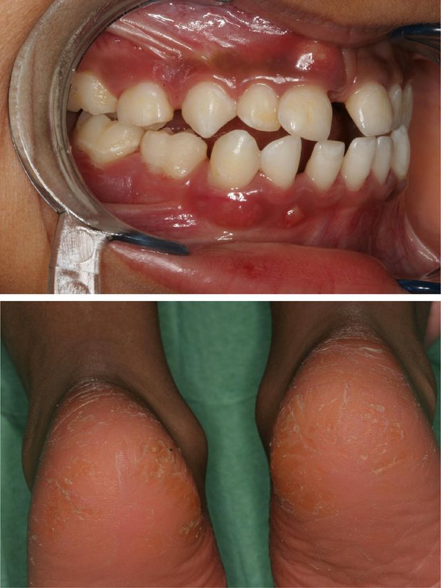
A)
Data analysis
All data were entered into the SPSS 21.0 (SPSS Inc,
Chicago, IL, USA) program, and were analyzed using
the Kruskal-Wallis test. Mann–Whitney U-test was
used for comparisons with PLS and periodontal healthy
subjects each. In GCF, correlations using Spearman test
were determined in PLS group and the subjects without
known genetic disorders. The level of significance was
set to p < 0.05.
Results
PLS patients
B)
The characteristics, including year of birth, ethnicity, gen-
der, phenotype, the presence of A. actinomycetemcomitans
in subgingival plaque before treatment and mutation
in the CTSC gene (nucleotide, exon, affected amino
acid residue) of the 11 PLS patients are summarized in
Additional file 1: Table S1). A. actinomycetemcomitans
was detected in all 9 microbiologically characterized
samples before periodontal treatment. Ten out of 11
patients exhibited typical skin lesions. Clinical view of
one of the patients is presented in Figure 1. In one
patient onset of periodontitis was late (at the age of 22) Figure 1 Papillon Lefèvre syndrome patient. Clinical view of a
Papillon-Lefèvre syndrome patient at 5 years age (#3, Table 1) with
but, nevertheless, led to edentulism (Additional file 1:
typical symptoms: severe periodontitis with a periodontal abscess at
Table S1). CTSC, NE and PR3 activity was measured in the upper central incisor 51 (A) and plantar hyperkeratosis (B).
peripheral blood neutrophils of PLS patients. The CTSC
activity was absent in neutrophils from nine patients while
in two patients it was detected at the low level (up to 10% fluids reflects neutrophil level [27]. Therefore we deter-
of the average activity of three matched healthy controls). mined MPO concentration to illustrate relative abundance
In none of neutrophil lysates from PLS patients the PR3 of neutrophils in our samples. In saliva of PLS patients the
activity was measurable. Low NE activity (at 2.6% of median concentration of MPO was determined at 864 pg/
controls) was detected only in one patient with the ml (range 116 – 2050 pg/ml), thus higher, although with-
highest CTSC activity (at 10% of control neutrophils). out statistical significance, than in other subject groups
For the purpose of this study GCF sampled from eight (CP: 747 pg/ml, range 522–790 pg/ml; AP: 591 pg/ml,
of the nine dentate patients has been analysed again for range 472–621 pg/ml, controls: 533 pg/ml, range 237–
bacteria present there. The results were compared to 836 pg/ml) (Figure 2A). However, after exclusion of the
simultaneously performed analysis of samples derived edentulous patients in the PLS group (median 909 pg/ml,
from patients with chronic and aggressive periodontitis, as range 496–2050 pg/ml), significantly higher concentration
well as healthy controls. Significantly, despite intensive of MPO (p < 0.05) was found in the PLS saliva than in the
treatment A. actinomycetemcomitans was still detected in saliva from other groups.
GCF from four out of the eight PLS patients at the similar In GCF, results were similar, the highest levels of MPO
prevalence as in GCF from non-treated aggressive were also measured in samples derived from PLS patients
periodontitis. On the other hand, the prevalence of P. (median 7.50 ng/μl, range 1.95-13.90 ng/μl), followed
gingivalis and T. denticola was much higher in chronic by chronic periodontitis patients, aggressive periodon-
periodontitis than in PLS patients and the aggressive titis, and periodontally healthy controls, respectively
form of the disease (Table 1). No periodontal pathogens (Figure 2B). Nevertheless, no significant difference was
were found in healthy controls. observed between any groups of samples.
MPO as an equivalent for neutrophil numbers α-Defensins
Myeloperoxidase (MPO) occurs in high amounts exclusively The levels of α-defensins in saliva of PLS patients (median
in neutrophils and its concentration in pathophysiological 0.33 μg/ml, range 0.00-6.58 μg/ml) were not strikinglyEick et al. Orphanet Journal of Rare Diseases 2014, 9:148 Page 5 of 11
http://www.ojrd.com/content/9/1/148
Table 1 Prevalence of the periodontopathogens Aggregatibacter actinomycetemcomitans, Porphyromonas gingivalis,
Tannerella forsythia, Treponema denticola and Prevotella intermedia in gingival crevicular fluid obtained from PLS
patients (at the time of the study), periodontitis patients as well as healthy controls
Species PLS (n = 8) Aggressive periodontititis Chronic periodontitis Periodontally healthy
(n = 7) (n = 12) subjects (n = 9)
A. actinomycetemcomitans 4 (50%) 4 (57%) 4 (33%) 0 (0%)
P. gingivalis 0 (0%) 4 (57%) 10 (83%) 0 (0%)
T. denticola 1 (13%) 3 (43%) 7 (58%) 0 (0%)
T. forsythia 3 (38%) 4 (57%) 11 (92%) 0 (0%)
P. intermedia 1 (13%) 2 (29%) 6 (50%) 0 (0%)
different from those in saliva of AP patients and controls patients and combined periodontitis patients (AP + CP)
(AP: median 0.25 μg/ml, range 0.00-0.5 μg/ml, and the levels of MPO correlated well with amount of
controls: median 0.45 μg/ml, range 0.00-6.6 μg/ml). In detected HNP1-3 (PLS: r = 0.742, p = 0.035; CP + AP:
contrast, chronic periodontitis patients possessed rela- r = 0.533, p = 0.004).
tively high concentration of HNP1-3 in saliva (median
3.62 μg/ml, range 0.12-19.76 μg/ml) (Figure 2C). hCAP18 and cathelicidin LL-37
In contrast to saliva, HNP1-3 were detected in GCF Except for edentulous PLS patients, the unprocessed
from PLS patients (median: 28.98 μg/μl, range: 0.14- cathelicidin hCAP18 was always detected in saliva.
103.94 μg/μl) in significantly higher concentration in hCAP18 was present in highest levels in PLS, followed
comparison with periodontally healthy controls (median: by chronic periodontitis patients and in low abundance
1.20 μg/μl, range: 0–29.19 μg/μl). Only moderate amounts in AP patients’ and healthy control individuals’ saliva
of HNP1-3 were detected in samples from aggressive (Figure 3A). This probably reflects the relatively high level
periodontitis (median: 15.29 μg/μl, range: 0.36-49.61 μg/μl). of neutrophils in saliva from PLS and chronic periodon-
In chronic periodontitis patients however, the levels of titis groups (Figure 1). The lack of hCAP18 in saliva of
HNP1-3 were high (median: 50.63 μg/μl, range: 0.40- edentulous PLS patients correlates with the near absence
116.17 μg/μl) (Figure 2D). Significantly, both in PLS of neutrophils as indicated by very low amount of MPO in
Figure 2 Levels of myeloperoxidase and α-defensins in saliva and gingival crevicular fluid. Levels of myeloperoxidase (MPO) (A and B)
and α-defensins (HNP1-3) (C and D) in saliva (A and C) and gingival crevicular fluid (B and D) obtained from Papillon-Lefèvre patients (PLS),
aggressive periodontitis patients (AP), chronic periodontitis patients (CP) and healthy controls (con). The results present median, and 25th and
75th percentiles.Eick et al. Orphanet Journal of Rare Diseases 2014, 9:148 Page 6 of 11 http://www.ojrd.com/content/9/1/148 Figure 3 Levels of unprocessed cathelicidin hCAP18 and mature LL-37 in saliva and gingival crevicular fluid. Levels of unprocessed cathelicidin hCAP18 and mature LL-37 in saliva (A) and gingival crevicular fluid (C) obtained from Papillon-Lefèvre patients (PLS), aggressive periodontitis patients (AP), chronic periodontitis patients (CP) and healthy controls (con) determined by western blot analysis. The results present median, and 25th and 75th percentiles. Panel B shows Western blot patterns of LL-37 immunoreactive proteins/peptides in GCF samples collected from PLS patients (#1 - #11) as well as representative samples of chronic periodontitis (CP) and aggressive periodontitis (AP) as well as from healthy controls (con). Arrows point to bands immunoreactive against anti-LL-37 antibody, representing intact hCAP18/LL-37, 11 kDa fragment of hCAP18/LL-37, and the LL-37 peptide. saliva from these individuals. This finding suggests that of hCAP18 in PLS patients was comparable to that of the gingival crevice around teeth is a major port of entry healthy subjects but lower than in GCF from AP and CP for neutrophils into the oral cavity. But using Western patients (Figure 3C). blot analysis with mAb anti-LL-37 we failed to detect Although the LL-37 peptide was found in all GCF mature LL-37 peptide in any saliva samples regardless samples from periodontally healthy controls, the level of disease status. of the peptide in CP patients was significantly higher High levels of hCAP18, comparable to that of CP, AP (Figure 3C). In the later group the amount of hCAP18 and healthy controls, were detected in five (out of eight) correlated with mature LL-37 (r = 0.523, p < 0.001) and the analysed samples of GCF from the PLS patients. In three load of P. gingivalis (r = 0.644, p < 0.001). In AP patients remaining samples barely visible amounts of hCAP18 LL-37 was detected at concentrations higher than in were found (Figure 3B). In general, the estimated amount controls but the difference was not significant. In strike
Eick et al. Orphanet Journal of Rare Diseases 2014, 9:148                                                                                 Page 7 of 11
http://www.ojrd.com/content/9/1/148

contrast we failed to detect the mature LL-37 peptide                         healthy subjects where the enzyme activity was detected
in any of GCF samples of PLS patients despite in part the                     in two out of nine analysed samples. In contrast, the NE
abundant presence of the precursor protein (Figure 3B).                       activity was high in chronic (CP versus PLS and controls:
Interestingly in this group the level of hCAP18 showed                        p < 0.001) and aggressive periodontitis patients (AP versus
a correlation to the load of A. actinomycetemcomitans                         PLS and controls: p = 0.005 and p = 0.006, respectively)
(r = 0.793, p = 0.019). Of note, the lack of LL-37 in these                   (Figure 4B).
samples was not due to proteolytic degradation of the                            The PR3 activity was detected in six out of the 11 PLS
peptide since the externally added peptide was intact in                      saliva samples (among them in two with the measurable
GCF samples even after 24 h incubation (data not shown).                      CTSC activity) but at much lower level than in other
                                                                              groups. With exception of one periodontally healthy
Activities of cathepsin C, neutrophil elastase and                            subject the PR3 activity was present in all other samples
proteinase 3                                                                  (Figure 4C). Significantly, no PR3 activities were detected
We failed to find any CTSC activity in nine out of 11                         in saliva of the edentulous PLS patients in keeping with
PLS saliva samples. In all other samples CTSC activity                        the low level of MPO. This further supports the conclu-
was detected but at highly variable levels. In patients                       sion that the junction or pocket epithelium around teeth
with CP and AP the CTSC activity was slightly higher,                         is the most important gate for neutrophils migration into
but not significantly, compared to periodontally healthy                      the otherwise healthy oral cavity.
controls (Figure 4A). The lack of the CTSC activity                              In GCF, the activity of CTSC varied considerably
coincided in PLS patients with very low levels of the NE                      between the groups (p < 0.001). In five samples of PLS
activity comparable to that in saliva from periodontally                      patients the CTSC activity was absent while in three other

 Figure 4 Activities of cathepsin C, neutrophil elastase and proteinase 3 in saliva and gingival crevicular fluid. Activities of cathepsin C
 (CTSC) (A and D), neutrophil elastase (NE) (B and D) and PR3 (C and E) in saliva (left panels) gingival crevicular fluid (right panels) obtained from
 Papillon-Lefèvre patients (PLS), aggressive periodontitis patients (AP), chronic periodontitis patients (CP) and healthy controls (con). The results
 present median, and 25th and 75th percentiles.
Eick et al. Orphanet Journal of Rare Diseases 2014, 9:148 Page 8 of 11
http://www.ojrd.com/content/9/1/148
samples it was present at very low levels (Figure 4D). This In oral fluids such as saliva and GCF from PLS subjects,
correlated with the lack of the NE (Figure 4E) and PR3 the CTSC activity was most absent or detected at very low
activities (Figure 4F). In general, the CTSC activity in GCF levels in comparison to the activity in other periodontitis
from PLS was far below that in periodontally healthy patients and even the periodontally healthy subjects.
subjects (p = 0.002). Conversely, in CP and AP groups the Deficiency of the CTSC activity in tested fluids was
CTSC activity was significantly higher than in healthy associated with the absence or severe reduction of activity
controls (AP 103-fold: p = 0.001; CP 66-fold: p = 0.004) of neutrophil serine proteases, such as NE and PR3. Thus,
(Figure 4D). the inflammatory response to pathogenic bacteria must be
disturbed in PLS patients since neutrophil serine proteases
Activity of LL-37 against Aggregatibacter activate gingival fibroblasts to produce inflammatory cyto-
actinomycetemcomitans strains kines [31] and are components of neutrophil extracellular
With the exception for one clinical isolate of serotype c, traps which trap and kill pathogens [32]. Further, reactive
which was resistant, all other tested strains were moder- oxygen species generation and MPO activity is not suffi-
ately susceptible to bactericidal activity of LL-37 (Table 2). cient to kill microbes and proteases are primarily respon-
The resistance does not seem to correlate with the strain sible for the destruction of phagocytosed bacteria [33]. This
serotype since three other strains of the same serotype antibacterial activity is exerted in different manners. For
were sensitive. example, NE cleaves outer membrane proteins in Gram
negative bacteria [34]. In keeping, it was shown that neutro-
Discussion phils from peripheral blood of PLS patients were incapable
PLS is caused by different mutations in the CTSC gene of neutralizing leukotoxin produced by A. actinomycetem-
resulting in typical and atypical pathological outcomes, comitans in the process dependent on serine proteases [18].
including those with isolated keratosis and periodontitis In stark contrast to saliva samples from PLS patients
[5,24]. Here 11 patients among them nine with typical half of which contain the low but detectable PR3 activity,
signs of PLS, including palmoplantar hyperkeratosis and no trace of the PR3 activity was detected in GCF. Interest-
severe early onset periodontitis already in the deciduous ingly, in four cases the PR3 activity was found in saliva
dentition in some patients, have been analysed. One totally deficient of the CTSC activity. This suggests a pos-
patient (#2) had periodontitis of the deciduous dentition, sible activation of PR3 by other proteases present in saliva.
but only suffered from mild skin symptoms. Another This is in keeping with results of a study using a leukae-
PLS patient (#7) suffered from typical skin symptoms, mia cell line which indicated that cathepsin C is not the
however, exhibited late onset of periodontitis (Additional sole enzyme involved in post-translational processing of
file 1: Table S1). In all cases we have shown that muta- PR3 [35]. The results implicated PR3 as a promising
tions affected the CTSC activity, never exceeded 10% of screening parameter for detection of periodontitis because
the average activity of systemically healthy subjects. the PR3 activity was significantly higher in every peri-
Furthermore, we confirmed the observation by Cagli odontitis patient without known genetic disorder than
et al. [30] that the expression of dysfunctional CTSC in any periodontally healthy control. This conclusion is
correlated with severely decreased activity of PR3, NE supported by the recent finding that the PR3-like activity
and cathepsin G in neutrophils. was increased in saliva of periodontitis patients and
correlated with severity of the disease [36]. The missing
activity in GCF of PLS patients can be an additional hint
for checking genetic disorders in selected periodontitis
Table 2 Minimal bactericidal concentrations (MBC) of
LL-37 against Aggregatibacter actinomycetemcomitans patients. PR3 seems to be an important player in the
strains process of inflammation since it activates oral epithelial
A. actinomycetemcomitans strain Origin MBC50 MBC90
cells to produce interleukin-8 and monocyte chemo-
(μg/ml) (μg/ml) attractant protein as well as to express intracellular
ATCC 33384 Laboratory strain 50 70 adhesion molecule (ICAM)-1 [37]. This might be in
J1 Clinical isolate 20 70
accordance to findings that IL-8 levels were lower in GCF
of PLS patients in comparison with controls [38]. In
J2 Clinical isolate 10 50
addition, chemotaxis of PMNs to IL-8 is diminished in
J3 Clinical isolate 20 50 PLS [39]. However in GCF of PLS patients, neutrophils
J7 Clinical isolate 10 50 are present in abundant numbers and this is associated
J11 Clinical isolate 70 >150 with high levels of α-defensins, other important neutrophil
J76 Clinical isolate 20 70 derived antimicrobial peptides. But the used method does
MBC50 and MBC90 indicate the minimal bactericidal concentration (MBC)
not allow distinguishing between the HNP1 precursor
values at which 50% and 90% of bacterial cells were killed, respectively. and functional HNPs. Considering the postulated role ofEick et al. Orphanet Journal of Rare Diseases 2014, 9:148 Page 9 of 11
http://www.ojrd.com/content/9/1/148
neutrophil serine protease in processing and activation of show a good to moderate sensitivity to LL-37 [46,47]
HNP-1 [40], the lack of their activity in PLS neutrophils which was confirmed by our clinical isolates.
may also cause the deficiency of active HNP1 in periodon- Additionally to the missing direct antimicrobial activity
tal lesions of PSL patients. a disturbance of other functions modulated by LL-37
Our results indicate that the lack of active neutrophil may be suggested in PLS patients. These include, direct
serine proteases and mature LL-37 is associated with chemotaxis of immune cells, induction of chemokines,
severity of periodontal disease in PLS patients. Recently regulation of chemokine receptor expression, inhibition
blood of PLS patients was analysed for LL-37 also using of the release of pro-inflammatory mediators, suppression
the WB method [18]. In contrast to our finding of the of neutrophil apoptosis, modification of dendritic cells
total absence of the peptide in GCF samples, a low differentiation and protection against inflammatory shock
amount of mature LL-37 and intermediate-size hCAP18- (reviewed in [48]). E.g., LL-37 neutralizes the lipopolysac-
derived fragments were detected in this study. We found charide activity of certain periodontopathogens among
intermediate hCAP18 fragments in chronic periodontitis them A. actinomycetemcomitans in human oral fibroblasts
associated with a high prevalence of P. gingivalis, T. [49]. Although stimulating in low concentrations proli-
forsythia and T. denticola [27], but never in PLS patients. feration of peripheral blood monocytes LL-37 inhibits
This discrepancy may arise from different sources of in vitro generation of osteoclasts from these cells [50].
analysed material: isolated neutrophils activated in vitro to These numerous immunomodulatory activities of LL-37
degranulate versus clinical samples in which neutrophils are apparently essential for maintaining homeostasis in
were in vivo exposed to bacteria. periodontal tissues by providing protective anti-microbial
All patients were initially colonized by A. actinomyce- responses without the excessive harmful inflammation.
temcomitans, after receiving an extensive periodontal
treatment still four of eight were tested positively for Conclusions
that species, however at low counts. This corroborates with In summary, the lack of functional cathepsin C impairs
a high prevalence of A. actinomycetemcomitans in PLS activation of neutrophil serine proteases in neutrophils
patients found in other studies [20,41]. Recently by using responding to bacterial challenge in periodontium of PLS.
a 16S rRNA-based microarray A. actinomycetemcomitans patients. One consequence is the loss of antimicrobial and
was found among the species in medium to high levels in immunomodulatory functions of LL-37 in the gingiva.
13 untreated PLS patients [42]. Our data strongly suggests This supports infection with A. actinomycetemcomitans
that the lack LL-37 is a condition selectively supporting and the development of severe periodontal disease.
growth of A. actinomycetemcomitans, the bacterium dir-
ectly linked to development and progression of aggressive Additional file
periodontitis. The importance of A. actinomycetemcomi-
tans in PLS patients is underlined by findings of high Additional file 1: Table S1. Characteristics (year of birth, ethnicity, sex,
IgG titers against that species [41,43] and the fact that a phenotype) of the 11 PLS patients including results of microbiological analysis
for detection of Aggregatibacter actinomycetemcomitans (A.a.) before treatment.
successful treatment of localized prepubertal periodontitis
in PLS correlates with eradication of A. actinomycetemco-
Competing interests
mitans [20,21]. The authors declare that they have no competing interests.
Interestingly, only in PLS patients the level of unpro-
cessed hCAP18 was correlated with the load of A. actino- Authors’ contributions
SE made the microbiological analysis and performed the statistical analysis.
mycetemcomitans. No such correlation between hCAP18 MP and TK measured the protease activities and the levels of antimicrobial
and any periodontal pathogen was found in other groups peptides in the samples. KA determined the antimicrobial activity of LL-37
clearly due to hCAP18 processing and LL-37 release. against the A. actinomycetemcomitans strains. PH and HS provided essential
reagents; AG recruited, diagnosed the periodontitis patients and periodontally
In these patients LL-37 in conjunction with α- and β-
healthy individuals and collected the samples. BS and PE recruited, diagnosed
defensins may prevent robust proliferation of A. acti- and treated the PLS patients and provided the respective samples. SE, AG, PE
nomycetemcomitans generating conditions favouring and JP participated in the study design and wrote the manuscript. All authors
read and approved the final manuscript.
the growth of other periodontal pathogens, including
P. gingivalis, T. forsythia and P. intermedia. Indeed, in Acknowledgements
periodontitis patients without known genetic disorders We are indebted to Dr. Ky-Anh Nguyen for fruitful discussion and critical reading
the load of P. gingivalis and the other proteolytic period- of the manuscript. This work was supported by the National Institutes of Health
(NIH/NIDCR) [R01 DE 022597], the European Commission [FP7-HEALTH-F3-2012-
ontopathogens is correlated with activity of CTSC, NE 306029 “TRIGGER”], Polish Ministry of Science and Higher Education (MNiSW)
and PR3 and the released α-defensins and mature LL-37. [2975/7.PR/13/2014/2], and National Science Center [Krakow, Poland, 2011/01/B/
Data regarding the susceptibility of A. actinomycetemco- NZ6/00268 and 2012/04/A/NZ1/00051]. The statistical support of Mr. Walter B.
Bürgin, Dipl. Biomed. Ing., is appreciated.
mitans to the various antibacterial peptides are rare, The authors declare no potential conflicts of interest with respect to the
strains were totally insensitive to HNP1-3 [44,45] and authorship and/or publication of this article.Eick et al. Orphanet Journal of Rare Diseases 2014, 9:148 Page 10 of 11
http://www.ojrd.com/content/9/1/148
Author details 17. Ye Y, Carlsson G, Wondimu B, Fahlen A, Karlsson-Sjoberg J, Andersson M,
1
Department of Periodontology, Laboratory of Oral Microbiology, School of Engstrand L, Yucel-Lindberg T, Modeer T, Putsep K: Mutations in the ELANE
Dental Medicine, University of Bern, Bern, Switzerland. 2Department of gene are associated with development of periodontitis in patients with
Microbiology, Faculty of Biochemistry, Biophysics and Biotechnology, severe congenital neutropenia. J Clin Immunol 2011, 31:936–945.
Jagiellonian University, Krakow 30-386, Poland. 3Department of Pulmonology, 18. de Haar SF, Hiemstra PS, van Steenbergen MT, Everts V, Beertsen W: Role of
Leiden University Medical Center, Leiden, The Netherlands. 4Haemostasis polymorphonuclear leukocyte-derived serine proteinases in defense
Biology, Novo Nordisk A/S, Maaloev, Denmark. 5Clinic of Prosthetic Dentistry, against Actinobacillus actinomycetemcomitans. Infect Immun 2006,
Center for Dentistry, Jena University Hospital, Jena, Germany. 6Department of 74:5284–5291.
Periodontology, Center for Dental, Oral, and Maxillofacial Medicine 19. Rudiger S, Petersilka G, Flemmig TF: Combined systemic and local
(Carolinum), Johann Wolfgang Goethe–University Frankfurt am Main, antimicrobial therapy of periodontal disease in Papillon-Lefevre
Frankfurt am Main, Germany. 7Center of Oral Health and Systemic Diseases, syndrome. A report of 4 cases. J Clin Periodontol 1999, 26:847–854.
University of Louisville School of Dentistry, Louisville, KY 40202, USA. 20. Schacher B, Baron F, Ludwig B, Valesky E, Noack B, Eickholz P: Periodontal
therapy in siblings with Papillon-Lefevre syndrome and tinea capitis: a
Received: 23 July 2014 Accepted: 11 September 2014 report of two cases. J Clin Periodontol 2006, 33:829–836.
21. Eickholz P, Kugel B, Pohl S, Naher H, Staehle HJ: Combined mechanical and
antibiotic periodontal therapy in a case of Papillon-Lefevre syndrome.
J Periodontol 2001, 72:542–549.
References 22. Hewitt C, Wu CL, Hattab FN, Amin W, Ghaffar KA, Toomes C, Sloan P, Read
1. Dalgic B, Bukulmez A, Sari S: Pyogenic liver abscess and peritonitis due to AP, James JA, Thakker NS: Coinheritance of two rare genodermatoses
Rhizopus oryzae in a child with Papillon-Lefevre syndrome. Eur J Pediatr (Papillon-Lefevre syndrome and oculocutaneous albinism type 1) in two
2011, 170:803–805. families: a genetic study. Br J Dermatol 2004, 151:1261–1265.
2. Hart TC, Atkinson JC: Mendelian forms of periodontitis. Periodontol 2000 23. Noack B, Gorgens H, Hoffmann T, Fanghanel J, Kocher T, Eickholz P,
2007, 45:95–112. Schackert HK: Novel mutations in the cathepsin C gene in patients with
3. Armitage GC: Development of a classification system for periodontal pre-pubertal aggressive periodontitis and Papillon-Lefevre syndrome.
diseases and conditions. Ann Periodontol 1999, 4:1–6. J Dent Res 2004, 83:368–370.
4. Pham CT, Ivanovich JL, Raptis SZ, Zehnbauer B, Ley TJ: Papillon-Lefevre 24. Noack B, Gorgens H, Schacher B, Puklo M, Eickholz P, Hoffmann T, Schackert
syndrome: correlating the molecular, cellular, and clinical consequences HK: Functional Cathepsin C mutations cause different Papillon-Lefevre
of cathepsin C/dipeptidyl peptidase I deficiency in humans. J Immunol syndrome phenotypes. J Clin Periodontol 2008, 35:311–316.
2004, 173:7277–7281. 25. Guentsch A, Kramesberger M, Sroka A, Pfister W, Potempa J, Eick S:
5. Toomes C, James J, Wood AJ, Wu CL, McCormick D, Lench N, Hewitt C, Comparison of gingival crevicular fluid sampling methods in patients
Moynihan L, Roberts E, Woods CG, Markham A, Wong M, Widmer R, Ghaffar with severe chronic periodontitis. J Periodontol 2011, 82:1051–1060.
KA, Pemberton M, Hussein IR, Temtamy SA, Davies R, Read AP, Sloan P, 26. Eick S, Straube A, Guentsch A, Pfister W, Jentsch H: Comparison of real-time
Dixon MJ, Thakker NS: Loss-of-function mutations in the cathepsin C polymerase chain reaction and DNA-strip technology in microbiological
gene result in periodontal disease and palmoplantar keratosis. Nat Genet evaluation of periodontitis treatment. Diagn Microbiol Infect Dis 2011,
1999, 23:421–424. 69:12–20.
6. Korkmaz B, Horwitz MS, Jenne DE, Gauthier F: Neutrophil elastase, 27. Puklo M, Guentsch A, Hiemstra PS, Eick S, Potempa J: Analysis of neutrophil-
proteinase 3, and cathepsin G as therapeutic targets in human diseases. derived antimicrobial peptides in gingival crevicular fluid suggests
Pharmacol Rev 2010, 62:726–759. importance of cathelicidin LL-37 in the innate immune response against
7. Perera NC, Wiesmuller KH, Larsen MT, Schacher B, Eickholz P, Borregaard N, periodontogenic bacteria. Oral Microbiol Immunol 2008, 23:328–335.
Jenne DE: NSP4 is stored in azurophil granules and released by activated 28. Schagger H, von Jagow G: Tricine-sodium dodecyl sulfate-polyacrylamide
neutrophils as active endoprotease with restricted specificity. J Immunol gel electrophoresis for the separation of proteins in the range from 1 to
2013, 191:2700–2707. 100 kDa. Anal Biochem 1987, 166:368–379.
8. Hewitt C, McCormick D, Linden G, Turk D, Stern I, Wallace I, Southern L, 29. Shafer WM, Pohl J, Onunka VC, Bangalore N, Travis J: Human lysosomal
Zhang L, Howard R, Bullon P, Wong M, Widmer R, Gaffar KA, Awawdeh L, cathepsin G and granzyme B share a functionally conserved broad
Briggs J, Yaghmai R, Jabs EW, Hoeger P, Bleck O, Rüdiger SG, Petersilka G, spectrum antibacterial peptide. J Biol Chem 1991, 266:112–116.
Battino M, Brett P, Hattab F, Al-Hamed M, Sloan P, Toomes C, Dixon M, 30. Cagli NA, Hakki SS, Dursun R, Toy H, Gokalp A, Ryu OH, Hart PS, Hart TC:
James J, Read AP, Thakker N: The role of cathepsin C in Papillon-Lefevre Clinical, genetic, and biochemical findings in two siblings with Papillon-
syndrome, prepubertal periodontitis, and aggressive periodontitis. Hum Lefevre Syndrome. J Periodontol 2005, 76:2322–2329.
Mutat 2004, 23:222–228. 31. Uehara A, Muramoto K, Takada H, Sugawara S: Neutrophil serine proteinases
9. Demmer RT, Papapanou PN: Epidemiologic patterns of chronic and activate human nonepithelial cells to produce inflammatory cytokines
aggressive periodontitis. Periodontol 2000 2010, 53:28–44. through protease-activated receptor 2. J Immunol 2003, 170:5690–5696.
10. Hajishengallis G, Darveau RP, Curtis MA: The keystone-pathogen hypothesis. 32. Wartha F, Beiter K, Normark S, Henriques-Normark B: Neutrophil extracellular
Nat Rev Microbiol 2012, 10:717–725. traps: casting the NET over pathogenesis. Curr Opin Microbiol 2007, 10:52–56.
11. Henderson B, Ward JM, Ready D: Aggregatibacter (Actinobacillus) 33. Reeves EP, Lu H, Jacobs HL, Messina CG, Bolsover S, Gabella G, Potma EO,
actinomycetemcomitans: a triple A* periodontopathogen? Periodontol Warley A, Roes J, Segal AW: Killing activity of neutrophils is mediated
2000 2010, 54:78–105. through activation of proteases by K + flux. Nature 2002, 416:291–297.
12. Lehrer RI, W L: alpha-Defensins in human innate immunity. Immunol Rev 34. Belaaouaj A: Neutrophil elastase-mediated killing of bacteria: lessons
2012, 245:84–112. from targeted mutagenesis. Microbes Infect 2002, 4:1259–1264.
13. Zaiou M, Nizet V, Gallo RL: Antimicrobial and protease inhibitory functions 35. Rao NV, Rao GV, Marshall BC, Hoidal JR: Biosynthesis and processing of
of the human cathelicidin (hCAP18/LL-37) prosequence. J Invest Dermatol proteinase 3 in U937 cells. Processing pathways are distinct from those
2003, 120:810–816. of cathepsin G. J Biol Chem 1996, 271:2972–2978.
14. Vandamme D, Landuyt B, Luyten W, Schoofs L: A comprehensive summary 36. Komine K, Kuroishi T, Ozawa A, Komine Y, Minami T, Shimauchi H, Sugawara
of LL-37, the factoctum human cathelicidin peptide. Cell Immunol 2012, S: Cleaved inflammatory lactoferrin peptides in parotid saliva of
280:22–35. periodontitis patients. Mol Immunol 2007, 44:1498–1508.
15. Sorensen OE, Follin P, Johnsen AH, Calafat J, Tjabringa GS, Hiemstra PS, 37. Uehara A, Sugawara S, Muramoto K, Takada H: Activation of human oral
Borregaard N: Human cathelicidin, hCAP-18, is processed to the epithelial cells by neutrophil proteinase 3 through protease-activated
antimicrobial peptide LL-37 by extracellular cleavage with proteinase 3. receptor-2. J Immunol 2002, 169:4594–4603.
Blood 2001, 97:3951–3959. 38. Ullbro C, Crossner CG, Nederfors T, Parhar R, Al Mohanna F, Meikle MC,
16. Putsep K, Carlsson G, Boman HG, Andersson M: Deficiency of antibacterial Reynolds JJ, Twetman S: Cytokines, matrix metalloproteinases and tissue
peptides in patients with morbus Kostmann: an observation study. inhibitor of metalloproteinases-1 in gingival crevicular fluid from patients
Lancet 2002, 360:1144–1149. with Papillon-Lefevre syndrome. Acta Odontol Scand 2004, 62:70–74.Eick et al. Orphanet Journal of Rare Diseases 2014, 9:148 Page 11 of 11
http://www.ojrd.com/content/9/1/148
39. Liu R, Cao C, Meng H, Tang Z: Leukocyte functions in 2 cases of
Papillon-Lefevre syndrome. J Clin Periodontol 2000, 27:69–73.
40. Tongaonkar P, Golji AE, Tran P, Ouellette AJ, Selsted ME: High fidelity
processing and activation of the human alpha-defensin HNP1 precursor
by neutrophil elastase and proteinase 3. PLoS One 2012, 7:e32469.
41. Clerehugh V, Drucker DB, Seymour GJ, Bird PS: Microbiological and
serological investigations of oral lesions in Papillon-Lefevre syndrome.
J Clin Pathol 1996, 49:255–257.
42. Albandar JM, Khattab R, Monem F, Barbuto SM, Paster BJ: The subgingival
microbiota of Papillon-Lefevre syndrome. J Periodontol 2012, 83:902–908.
43. Van Dyke TE, Taubman MA, Ebersole JL, Haffajee AD, Socransky SS, Smith DJ,
Genco RJ: The Papillon-Lefevre syndrome: neutrophil dysfunction with
severe periodontal disease. Clin Immunol Immunopathol 1984, 31:419–429.
44. Raj PA, Antonyraj KJ, Karunakaran T: Large-scale synthesis and functional
elements for the antimicrobial activity of defensins. Biochem J 2000,
347(Pt 3):633–641.
45. Miyasaki KT, Bodeau AL, Ganz T, Selsted ME, Lehrer RI: In vitro sensitivity of
oral, gram-negative, facultative bacteria to the bactericidal activity of
human neutrophil defensins. Infect Immun 1990, 58:3934–3940.
46. Tanaka D, Miyasaki KT, Lehrer RI: Sensitivity of Actinobacillus
actinomycetemcomitans and Capnocytophaga spp. to the bactericidal
action of LL-37: a cathelicidin found in human leukocytes and epithelium.
Oral Microbiol Immunol 2000, 15:226–231.
47. Ji S, Hyun J, Park E, Lee BL, Kim KK, Choi Y: Susceptibility of various oral
bacteria to antimicrobial peptides and to phagocytosis by neutrophils.
J Periodontal Res 2007, 42:410–419.
48. Choi KY, Chow LN, Mookherjee N: Cationic host defence peptides:
multifaceted role in immune modulation and inflammation. J Innate
Immun 2012, 4:361–370.
49. Suphasiriroj W, Mikami M, Shimomura H, Sato S: Specificity of
Antimicrobial Peptide LL-37 to Neutralize Periodontopathogenic
Lipopolysaccharide Activity in Human Oral Fibroblasts. J Periodontol 2013,
84:256–264.
50. Supanchart C, Thawanaphong S, Makeudom A, Bolscher JG, Nazmi K,
Kornak U, Krisanaprakornkit S: The antimicrobial peptide, LL-37, inhibits
in vitro osteoclastogenesis. J Dent Res 2012, 91:1071–1077.
doi:10.1186/s13023-014-0148-y
Cite this article as: Eick et al.: Lack of cathelicidin processing in Papillon-
Lefèvre syndrome patients reveals essential role of LL-37 in periodontal
homeostasis. Orphanet Journal of Rare Diseases 2014 9:148.
Submit your next manuscript to BioMed Central
and take full advantage of:
• Convenient online submission
• Thorough peer review
• No space constraints or color figure charges
• Immediate publication on acceptance
• Inclusion in PubMed, CAS, Scopus and Google Scholar
• Research which is freely available for redistribution
Submit your manuscript at
www.biomedcentral.com/submitYou can also read