Straumann Sla Scientific evidence firSt edition (2011) - Straumann Dental Implant System
←
→
Page content transcription
If your browser does not render page correctly, please read the page content below
Straumann ® SlA
 Scientific Evidence
  First Edition (2011)

   Straumann® Dental Implant System
The ITI (International Team for Implantology) is academic partner of Institut Straumann AG
in the areas of research and education.
Contents

2	Introduction

3   Study overview

4   Preclinical studies

12	Clinical studies

26 Additional studies

30	References

                          1
Introduction
Straumann® SLA – reliable and scientifically well documented
Roughened surfaces for dental implants have consistently demonstrated advantages over the older machined-
type titanium surfaces. Significantly greater success rates, enhanced bone-to-implant contact (BIC) and great-
er biomechanical and functional stability have all been demonstrated by rough surface implants compared
to those with smoother surfaces1. Straumann’s proprietary Sandblasted, Large-grit, Acid-etched (SLA®) surface
has become one of the best documented rough surfaces in implantology, having shown advantages over
other roughened implant surfaces1. In particular, preclinical analysis has shown the superiority of SLA® over
the previously most common rough surface, titanium plasma-sprayed (TPS), with better maintenance of verti-
cal bone height2 and significantly greater BIC3. Shear strength of the bone-to-implant interface has also been
shown to be greater with SLA® compared to TPS and machined surfaces4, and compared to a machined
and acid-etched surface5 in preclinical studies; a biomechanical comparison using implants of the same de-
sign showed that the better bone anchorage was a result of the surface topography rather than the implant
design.6
Sandblasted, Large-grit, Acid-etched (SLA®)
The extent of the bone-to-implant interface appears to increase with increasing surface roughness,7,8 but prob-
lems have been noted with surfaces that are very rough, suggesting that there may be an ‘optimum range’
of moderate surface roughness9. The SLA® surface is sandblasted with large-grit (250–500 μm), which results
in a peak-to-peak macro-roughness of approximately 20–40 μm, followed by micro-roughness of approxi-
mately 2–4 μm upon acid etching. Micro-rough surfaces increase the rate of cell spreading and the number
of cells attached to the surface, and increase the rate that cells produce factors regulating the differentiation
of bone-forming cells (osteoblasts) and reduce the activity of bone-destroying cells (osteoclasts).10,11
The clinical benefit
The advent of the SLA® surface, with its proven osseointegration properties, therefore revolutionized the con-
ventional timing of provisionalization, allowing it to be cut by half, from 12 weeks for the previous TPS surface
to just 6 weeks*12,13,14; subsequently, even earlier loading (e.g. early and immediate) have been shown to be
successful and predictable.14,15,16,17,18,19 The longevity of Straumann® Soft Tissue Level implants with the SLA®
surface has also been demonstrated by success in numerous long-term investigations (e.g. 5 years and
over).20,21,22,23,24 The substantial level of evidence for SLA® also acts as a proven foundation for the SLActive®
surface, which is based on the SLA® technology.
The SLA® surface is available and has proven to be successful on various Straumann implant designs, Nar-
row Neck implants,25 Wide Neck implants,26 Tapered Effect implants27 and short implants28. The SLA® sur-
face is also available on BL implant.
The 20 years of investigation and 10 years of clinical evidence for SLA®24 have provided a wealth of infor-
mation, and implant survival rates of > 98 % have consistently been observed.12,14,15,20,21,22,29,30 The following
information shows data from selected† key preclinical and clinical studies with Straumann’s SLA® -surfaced
implants, representing a range of indications, loading protocols and implant types. The excellent data dem-
onstrate the advantages and versatility of the SLA® surface.
* With good bone quality and adequate bone quantity and implants of Ø 4.1 mm or Ø 4.8 mm, over 8.0 mm lengths.
†
Please note that this Straumann Scientific Evidence document contains a selection of studies and makes no claim to be a complete study list. Articles were selected on
merit from the results of a literature search using combinations of the following key words: sand-blasted and grit-blasted, sand-blasted and particle blasted, tapered
effect, standard implant, standard plus, narrow neck implant, sand-blasted and large-grit and acid-etched, wide neck implant, ITI implant, sand-blasted and acid-etched,
Straumann, rough surfaces, blasted SLA®, surface topography, dental implant, surface chemistry, surface modification, surface roughness, acid-etching, titanium,
synOcta®, Locator®, IPS e.max®, preshaped abutment
2Study overview
Preclinical Studies
# Topic Authors Reference Page
1 Interface shear strength Wilke HJ et al. Clinical Implant Materials (Heimke G, Soltész U, Lee AJC, 04
eds). Advances in Biomaterials; 9, 1990: 309–314.
2 Surface characteristics on bone reactions Buser D et al. J Biomed Mater Res 1991;25(7):889–902. 05
3 Implant surface under loaded / Cochran DL et al. Clin Oral Implants Res 1996;7(3):240–252. 06
unloaded conditions
4 Histometric analysis SLA® Cochran DL et al. J Biomed Mater Res 1998;40(1):1–11. 07
5 Comparative Removal Torque Evaluation Buser D et al. Int J Oral Maxillofac Implants 1998;13(5):611–619. 08
6 Interface shear strength Buser D et al. J Biomed Mater Res 1999;45(2):75–83. 09
7 Immediate and early loading Quinlan P et al. Int J Oral Maxillofac Implants 2005;20(3):360–370. 10
8 Comparative study to investigate bone healing de Sanctis M et al. J Clin Periodontol 2009;36(8):705–711. 11
around 4 different implant systems
Clinical Studies
# Topic Authors Reference Page
9 Early healing (6 weeks) Cochran DL et al. Clin Oral Implants Res 2002;13(2):144–153. 12
10 Early loading in partially edentulous patients Bornstein MM et al. Int J Oral Maxillofac Implants 2003;18(5):659–666. 13
(6 weeks)
11 Early loading in posterior mandible Salvi GE et al. Clin Oral Implants Res 2004;15(2):142–149. 14
(2 or 6 weeks)
12 Early loading in edentulous posterior maxilla and Nordin T et al. Int J Oral Maxillofac Implants 2004;19(6):880–886. 15
mandible – 12-month follow-up
13 Immediate loading with 3-unit fixed Cornelini R et al. Int J Oral Maxilliofac Implants 2006;21(6):914–918. 16
partial dentures
14 5-year life table analysis on WN SLA® implants Bischof M et al. Clin Oral Implants Res 2006;17(5):512–520. 17
15 Clinical field trial with Soft Tissue Level Cochran D et al. J Periodontol 2007;78(56):974–982. 18
SLA® implants
16 Clinical performance of WN SLA® – 3-year Bornstein MM et al. Int J Oral Maxillofac Implants 2007;22(4):631–638. 19
follow-up
17 5-year follow-up RCT in the edentulous maxilla Fischer K et al. Clin Oral Implants Res 2008;19(5):433–441 20
18 Staged sinus floor elevation in partially edentulous Bornstein MM et al. Clin Oral Implants Res 2008;19(10):1034–1043. 21
patients
19 Kaplan-Meier curve for TPS and SLA® implants De Boever AL et al. Clin Oral Implants Res 2009 ;20(12) :1341–1350. 22
20 Mandibular overdentures supported by 2 implants: Cehreli MC et al. Clin Implant Dent Relat Res 2010;12(2):114–121. 23
a 5-year RCT
21 10-year data on Soft Tissue Level SLA® implants in Fischer K ITI World Symposium, Geneva, Switzerland, 24
the edentulous maxilla 15–17 Apr 2010.
31
The influence of various titanium surfaces on the interface shear strength
between implants and bone
Wilke HJ, Claes L, Steinemann S.
Clinical Implant Materials (Heimke G, Soltész U, Lee AJC, eds). Advances in Biomaterials; 9, 1990: 309–314.
Abstract: Mechanical effects of bone healing were evaluated for implants with various surface treatments. Maximum removal torque was
shown by the SLA® surface.
Introduction Results
Bone healing around titanium implants can be influenced by the tita- SLA® implants showed the maximum removal torque, and the maxi-
nium surface. This investigation therefore examined the mechanical mum removal torque increased over time for SLA® and TPS implants
effect of the bone healing response to various surface morphologies (Figure 1); in contrast, no significant changes in removal torque were
of titanium implants in terms of implantation time. observed for the remaining implants.
Methods
Titanium screws with six different surface treatments (electropolished;
sandblasted with fine grit/acid-pickled (HF/HNO3)/anodized; tita-
nium plasma-sprayed (TPS); sandblasted with medium grit/acid-pick-
led (HF/HNO3); sandblasted with large grit/acid-pickled (HF/
HNO3); sandblasted with large grit/acid-etched (HCl/H2SO4)
(SLA®) were placed in the tibiae of 10 sheep. Removal torque was
evaluated after 2, 9, 12, 24 and 52 weeks, and histological analy-
sis was performed after 9, 24 and 52 weeks.
Preclinical studies
8
SLA®
V
6 IV
III
II
SLA®
Nm
4
I III
2
V
IV
II
I
0
2 weeks 8 weeks 9 weeks 12 weeks 18 weeks 24 weeks 52 weeks
Figure 1: Removal torque (Nm) of titanium screws according to surface treatment and time in place.
I: electropolished
II: sandblasted with fine grit, acid pickled using HF/HNO3 and anodized
III: titanium plasma-sprayed
IV: sandblasted with medium grit and acid pickled using HF/HNO3
V: sandblasted with large grit and acid pickled using HF/HNO3
Conclusions
SLA®: sandblasted with large grit and acid etched using HCl/H2SO4
• The interface shear strength of implants can be increased
by altering the surface structure morphology.
• An optimal surface can potentially be developed for vari-
ous biomechanical situations, e.g. strong anchorage for
permanent implants, or weaker anchorage for tempo-
rary/removable implants.
4Influence of surface characteristics on bone integration of titanium implants. 2
A histomorphometric study in miniature pigs
Buser D, Schenk RK, Steinemann S, Fiorellini JP, Fox CH, Stich H.
J Biomed Mater Res 1991;25(7):889–902.
Abstract: Bone healing with various implant surface characteristics was evaluated. Rough surface promoted greater bone apposition, with an
additional effect shown by SLA®.
Introduction Results
Several techniques have been employed to improve anchorage of Histological analysis showed direct bone-to-implant contact for all
titanium implants in bone by surface modification or coatings. The implants, but with significant differences in the percentages obtained.
aim of this study was to investigate the influence of various surface The lowest bone-to-implant contact was observed with the HF/
characteristics on bone reactions in titanium implants. HNO3 implants (25.1 % ± 7.4 % and 21.6 % ± 9.3 %, respectively,
at 6 weeks), while the highest bone-to-implant contact was found
Methods with the SLA® and HA implants (57.7 % ± 9.5 % and 69.5 % ±
Hollow cylinder implants with six different surface treatments (elec- 6.5 %, respectively, at 6 weeks). The results showed increasing
tropolished (E); sandblasted with medium grit/acid-pickled (HF/ bone-to-implant contact with increasing surface roughness (Fig. 2).
HNO3) (SMP); sandblasted with large grit (SL); sandblasted with
large grit/acid-etched (HCl/H2SO4) (SLA®); titanium plasma-sprayed
(TPS); hydroxylapatite plasma-sprayed (HA)) (Fig. 1) were placed in
the metaphyses of the tibia and femur in six mini-pigs. Twelve im-
plants of each type were placed. Histomorphometric analysis was n.s.
80
performed after 3 and 6 weeks. 3 weeks
6 weeks
Preclinical studies
60
% Bone Contact
* * 40
20
0
E SMP SL TPS SLA® HA
* *
Figure 2: Mean direct bone-to-implant contact (%) of different implant surfaces
after 3 and 6 weeks; E = electro-polished; SMP = sandblasted with medium grit
(0.12–0.25 µm) and acid pickling with HF/HNO3; SL = sandblasted with large
grit (0.25–0.50 µm)
A narrow band of bone was often seen around SLA® and HA im-
plants as an extension of perpendicularly oriented bone trabeculae;
*
however, the HA coating consistently showed signs of resorption,
predominantly in areas without bone covering.
Conclusions
• Greater bone apposition was observed with rough surfac-
Figure 1: Scanning electron micrographs of different implant surfaces (bar is 20 es compared to polished or fine structured surfaces.
µm; original magnification x1700). (A) SMP, (B) SL, (C) SLA, (D) TPS, (E) HA • The SLA® surface showed an additional effect on bone
apposition compared to the sandblasted surface.
• The highest bone-to-implant contact was found with SLA®
and HA surfaces.
• The HA coating consistently showed signs of resorption.
* Reproduced with permission of John Wiley & Sons, Inc.
53
Evaluation of an endosseous titanium implant with a sandblasted and
acid-etched surface in the canine mandible: radiographic results
Cochran DL, Nummikoski PV, Higginbottom FL, Hermann JS, Makins SR, Buser D.
Clin Oral Implants Res 1996;7(3):240–252.
Abstract: SLA® and TPS implants were compared under loaded and unloaded conditions in vivo. SLA® implants showed advantages in early
healing and showed less crestal bone loss.
Introduction Results
Surface characteristics of titanium implants have a direct impact on Excellent tissue integration with ankylotic stability and no signs of
integration with hard and soft tissues, and radiography is the only peri-implant infection were observed for all implants, and the radiog-
non-invasive method to assess the bone reaction. The aim of this raphy showed no signs of continuous peri-implant radiolucency
study was to compare titanium implants with TPS and SLA® surfaces around the implants. Stability was maintained after loading, and no
in a dog mandible model under loaded and unloaded conditions complications were observed.
over 15 months.
Mean crestal bone loss at the preload and 3-month evaluations were
Methods 0.52 mm and 0.73 mm, respectively, for SLA® implants and 0.69 mm
Cylindrical implants with either a TPS or an SLA® surface (Fig. 1) were and 1.06 mm, respectively, for TPS implants. All crestal bone loss
placed in the mandibles of six dogs after 3 months of healing follow- occurred during the first 6 months after implant placement, 65 % be-
ing tooth extraction. A total of 69 implants were placed. In four dogs fore loading and 35 % in the first 3 months after loading, regardless
(groups B and C), impressions were taken 2 months after implant of implant surface. Bone loss was significantly less with the SLA® im-
placement for the fabrication of screw-retained gold crowns, and the plants at both preload and after 3 months of loading, and this was
implants (48 implants) were loaded 3 months after implant place- maintained during the 1-year follow-up period (Fig. 2).
ment. Radiographic analysis was performed for assessment of bone-
to-implant contact, by measuring the distance from the implant shoul-
Preclinical studies
der to the most coronal bone-to-implant contact, and bone density,
measured using computer-assisted densitometric image analysis (CA-
DIA). Measurements were taken 3 months after implant placement
SLA®
for the unloaded implants (group A, 2 dogs), 3 and 6 months after
1.4
implant placement in group B (2 dogs), and 3, 6, 9, 12 and TPS
15 months after implant placement (12 months of loading) in group 1.2 *
C (2 dogs).
1.0
% Bone Contact
0.8 *
* 0.6
0.4
0.2 *pBone response to unloaded and loaded titanium implants with a sandblasted 4
and acid-etched surface: a histometric study in the canine mandible
Cochran DL, Schenk RK, Lussi A, Higginbottom FL, Buser D.
J Biomed Mater Res 1998;40(1):1–11.
Abstract: SLA® and TPS implants were evaluated under loaded and unloaded conditions in vivo. Greater bone-to-implant contact was
observed with SLA® implants.
Introduction Results
Many investigations of titanium implants focus on the surface charac- All implants were stable after 3 months, with healthy peri-implant tis-
teristics, and BIC has been shown to be greater with an SLA® surface sues and no significant peri-implant inflammation. Bone-to-implant
compared to a TPS surface. This study was designed to evaluate the contact was significantly greater for the unloaded SLA® implants ver-
SLA® implant surface by histometric analysis in the canine mandible, sus the unloaded TPS implants after 3 months and after 12 months
compared to TPS surface implants. of loading, but there was no significant difference after 3 months of
loading (Fig. 2). No qualitative differences in bone tissue were ob-
Methods served between the SLA® and TPS implants.
Cylindrical implants with either a TPS or an SLA® surface were
placed in the mandibles of six dogs after 3 months of healing follow-
ing tooth extraction. A total of 69 implants were placed. Implants
were left unloaded in two dogs (Group A), while the implants in the
remaining four dogs were loaded with screw-retained gold crowns
after 3 months (Groups B and C). Histometric analysis of the bone- SLA®
to-implant contact was performed 3 months after implant placement TPS
100
in the unloaded group and 6 and 15 months after implant placement *p5
Removal torque values of titanium implants in the maxilla of miniature pigs
Buser D, Nydegger T, Hirt HP, Cochran DL, Nolte NP.
Int J Oral Maxillofac Implants 1998;13(5):611–619.
Abstract: Removal torque was evaluated for SLA® and Osseotite® implants in vivo over 12 weeks, and was found to be significantly higher
for SLA.
Introduction Results
After the introduction of SLA®, another acid-etched implant surface, Both surfaces showed similar features under scanning electron mi-
Osseotite®, was introduced onto the market, but there were no avail- croscopy (i.e. small micropits of 1–2 μm in diameter), although the
able data comparing this and other rough titanium surfaces. The aim Osseotite® surface appeared to exhibit a flatter profile. Average
of this investigation, therefore, was to compare an implant with this roughness (Ra) was greater for the SLA® surface (Ra = 2.0 μm versus
acid-etched surface (Osseotite®) with the sandblasted and acid- 1.3 μm). The mean removal torque was significantly higher for SLA®
etched surface (SLA®) by measurement of removal torque values. implants compared to Osseotite® implants at all time points (Fig. 3).
Methods
*
In the maxilla of each of nine adult miniature pigs, a total of 70 im-
plants were placed (six to eight implants per animal) at least 6
months after tooth extraction. Biomechanical testing was performed
on 54 of these implants by evaluation of removal torque after 4, 8
or 12 weeks. The implants had either a sandblasted and acid-etched
(SLA®) surface or a machined and acid-etched (Osseotite®) surface
(Fig. 1, Fig. 2). A commercially available standard hex device was
used for removal of the Osseotite® implants, while a specially manu-
Preclinical studies
factured adapter was used for removal of the SLA® implants. Since
the hex connection of the Osseotite® implants was rather short and
unable to withstand the shear stresses during removal torque testing,
the failure torque of these implants was also assessed. Torque rota- Figure 2: The two implants evaluated: the 8 mm long
tion curves were recorded for each test and the removal torque was SLA® implant without apical grooves (left) and the 10 mm
long Osseotite® implant with four apical grooves (right)
defined as the maximum torque on the curve.
SLA®
240 *
Osseotite ® *
210
*
180
150
Torque (Ncm)
120
*
90
60
30 * *pInterface shear strength of titanium implants with a sandblasted and 6
acid-etched surface: a biomechanical study in the maxilla of miniature pigs
Buser D, Nydegger T, Hirt HP, Cochran DL, Nolte NP.
J Biomed Mater Res 1999;45(2):75–83.
Abstract: Interface shear strength was evaluated for SLA®, TPS and machined implants in vivo. Removal torque was significantly higher for
SLA® and TPS, and was higher for SLA® versus TPS at 4 weeks.
Introduction 2.0
SLA® TPS M
The SLA® titanium surface has demonstrated enhanced bone apposi-
tion versus the TPS surface. This study was designed to evaluate the
interface shear strength of the SLA® surface in the maxilla of miniature 1.5
Removal torque (Nm)
pigs versus TPS and machined surfaces.
Methods 1.0
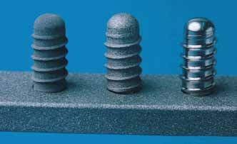
A total of 54 implants with an SLA®, TPS or machined surface (Fig. 1)
were placed in the maxillae (three implants on each side) of nine
adult miniature pigs at least 6 months after tooth extraction. The an- 0.5
terior-posterior position of each implant type was varied so that no
preference was given for any one implant type. Shear strength was
evaluated by removal torque testing after 4, 8 and 12 weeks; torque 0
4 8 12
rotation curves were recorded for each test and the removal torque Healing periods (weeks)
was defined as the maximum torque on the curve. Histologic and Figure 2: Removal torque values for SLA®, TPS and machined surface implants
histomorphometric analysis was also performed. after 4, 8 and 12 weeks
Preclinical studies
* anterior middle posterior
2.0
1.5
Removal torque (Nm)
1.0
0.5
Figure 1: The three tested implants, which had the same design but different 0
SLA® TPS M
surfaces: SLA® (left), TPS (center) and machined (right)
Figure 3: Removal torque values for SLA®, TPS and machined surface implants
according to implant position
Results
Mean removal torque for the machined surface implants varied from
0.13 to 0.26 Nm, while mean values for the rough surfaced implants Conclusions
were between 1.14 and 1.56 Nm. Mean removal torque was high- • Interface shear strength of titanium implants is significantly
er for the SLA® surface after 4 weeks compared to the TPS surface, influenced by the surface characteristics.
while mean values were similar for both surfaces at 8 and 12 weeks • SLA® and TPS surfaces showed significantly higher remov-
(Fig. 2). Implant position had a significant influence on removal al torque values than machined surface implants.
torque – lower removal torque values were found for more posterior • Mean removal torque was higher for SLA® compared to
locations (Fig. 3), which correlated significantly with decreasing TPS implants after 4 weeks.
bone density from anterior to posterior sites. Histological analysis
showed a separation between the bone and implant surface for ma-
chined surface implants. Bone trabeculae fractures were often ob-
served for TPS and SLA® implants, but the bone-to-implant interface
was maintained.
* R eproduced with permission of John Wiley & Sons, Inc.
97
Immediate and early loading of SLA® ITI single-tooth implants: an in vivo study.
Quinlan P, Nummikoski P, Schenk R, Cagna D, Mellonig J, Higginbottom F, Lang K, Buser D, Cochran D.
Int J Oral Maxillofac Implants 2005;20(3):360–370.
Abstract: SLA® implants were loaded immediately or early in vivo; no significant differences were found between the loading protocols.
Introduction Results
A healing time of 6 weeks has been suggested for SLA® surfaced All implants were well osseointegrated; implant survival was 100 %.
implants, indicating the potential for the implants to be loaded im- Clinically normal peri-implant tissues were observed, with no signs of
mediately or early. The aim of this study was to determine whether inflammation or suppuration. Changes in crestal bone height (com-
immediate or early loading of SLA® implants has any adverse effects bined mesial and distal aspects) for implants loaded after 2, 10 and
compared to conventional loading, as assessed by clinical, radio- 21 days and 3 months were 0.35 ± 0.18 mm, 0.15 ± 0.08 mm,
graphic and histological examination. 0.30 ± 0.08 mm and 0.02 ± 0.07 mm, respectively. The differ-
ences were not statistically significant, suggesting that loading time
Methods does not adversely affect the peri-implant crestal bone. There were
Mandibular premolars and first molars were removed from four dogs also no significant differences in primary or secondary BIC, total BIC
and a total of 48 SLA® surfaced implants (12 per dog) were placed (Fig. 1) or total connective tissue-to-implant contact. For bone mar-
4 months later. The implants were loaded with single screw-retained row-to-implant contact, the amount was marginally significantly great-
gold crowns 2, 10 or 21 days or 3 months after implant placement er for the implants placed 2 days before loading (19.16 ± 35 %)
and placed in functional occlusion. Block sections were obtained for compared to those placed 3 months before loading (13.98 ±
histologic examination and the changes in crestal bone height mesi- 2.02 %), but no other differences were observed.
ally and distally at each implant, and the change in bone density of
the coronal 3 mm of crestal bone, were recorded. Histological eval-
Preclinical studies
uation included primary, secondary and total BIC as well as bone
marrow-to-implant contact and connective tissue-to-implant contact.
Primary bone
100 Secondary bone
90
80
70
Bone-to-implant contact (%)
60
50
40
30
20
10
0
A B C D
Figure 1: Total BIC for implants placed 3 months (group A), 21 days (group B),
10 days (group C) and 2 days (group D) before loading
Conclusions
• No significant differences were observed between the
four loading protocols.
• Immediate and early loading of SLA® implants is therefore
possible with no adverse peri-implant tissue effects.
10Immediate implants at fresh extraction sockets: bone healing in four different 8
implant systems.
de Sanctis M, Vignoletti F, Discepoli N, Zucchelli G, Sanz M.
J Clin Periodontol 2009;36(8):705–711.
Abstract: Bone healing in extraction sockets was evaluated for SLA®, OsseoSpeed™, Osseotite® and SPI® Element implants. Healing
patterns were similar in all groups, but bone-to-implant contact was highest with SLA®.
Introduction Results
Implant placement immediately after tooth extraction has become a BIC proved to be highly variable, ranging from 39.84 % to 78.63 %
common procedure, but the outcomes may depend on implant de- for 3i implants (3i Osseotite® Certain straight miniplant), 35.78 % to
sign and/or surface. The aim of this study, therefore, was to investi- 76.58 % for Astra Tech implants (Astra Tech MicroThread
gate the differences in bone healing around four different implant OsseoSpeed™ Ø 3.5 mm), 50.13 % to 86.88 % for Thommen im-
systems placed in fresh extraction sockets in the dog, and to observe plants (Thommen SPI® ELEMENT Ø 3.5 mm) and 58.14 % to 83.40 %
the influence on modelling of the buccal plate. for Straumann implants (Straumann® Standard Plus Ø 3.3 mm); BIC
was therefore most consistent around Straumann implants. Mean BIC
Methods was also highest for Straumann implants (Fig. 2), although the differ-
Third and fourth premolars were removed in each of eight dogs and ence was not statistically significant. Marked bone remodelling of the
four different implant types were randomly placed in the distal extrac- buccal plate of approximately 2.5 mm was observed, regardless of
tion sockets: Thommen SPI® ELEMENT Ø 3.5 mm; Straumann® the implant system. Similar bone area percentages and amounts of
Standard Plus Ø 3.3 mm; Astra Tech MicroThread OsseoSpeed™ new bone formation were observed for all implant systems.
Ø 3.5 mm; 3i Osseotite® Certain straight miniplant (Fig. 1). Histo-
logic, histometric and histomorphometric analysis was performed af-
ter 6 weeks of healing to assess BIC, bone area, new bone forma-
tion and ridge alterations.
Preclinical studies
90.0
*
Bone to implant contact (%)
67.5
45.0
22.5
0
3i Astra Tech Straumann Thommen
Figure 1: The four implant systems evaluated: (from left to right) Thommen SPI ®
Figure 2: Mean BIC (± SD) for the four implant systems: 3i Osseotite® Certain;
ELEMENT; Straumann® Standard Plus; Astra Tech MicroThread OsseoSpeed™; 3i
Astra Tech MicroThread OsseoSpeed™; Straumann® Standard Plus; Thommen
Osseotite® Certain
SPI® ELEMENT
Conclusions
• Healing patterns for implants placed in fresh extraction
sockets after 6 weeks were similar for all four implant
systems.
• The highest BIC was observed with SLA® implants.
SPI® is a registered trademark of Thommen Medical AG, Switzerland.
OsseoSpeed™ is a trademark of Astra Tech AB, Sweden.
Osseotite® is a registered trademark of Biomet 3i, USA.
* Reproduced with permission of John Wiley & Sons, Inc.
119
The use of reduced healing times on ITI implants with a sandblasted and
acid-etched (SLA®) surface: early results from clinical trials with ITI SLA® im-
plants
Cochran DL, Buser D, ten Bruggenkate CM, Weingart D, Taylor TM, Bernard JP, Peters F, Simpson JP.
Clin Oral Implants Res 2002;13(2):144–153.
Abstract: SLA® implants were placed in 133 patients and restored after 6 weeks (12 weeks in class IV bone). The cumulative survival rate
after 2 years was 99.3 %.
Introduction Results
Since the SLA® surface has demonstrated advantages in earlier heal- There were 80 patients (198 implants), 21 patients (46 implants and
ing following implant placement, the aim of this investigation was to 32 patients (139 implants) in groups A, B and C, respectively. At the
determine whether SLA® implants could be predictably and safely time of publications, 110 patients with 326 implants had completed
restored after 6 weeks after placement in class I to III bone and after the 1-year follow-up evaluation, and 47 patients with 138 implants
12 weeks in class IV bone. had completed the 2-year recall. Three implants were lost before
abutment connection, giving a success rate of 99.3 %, for a mean
Methods implant loading time of 49 days. No implant failures or implant-relat-
This was a prospective multicenter trial to evaluate the success of ed adverse events were reported after abutment connection. The
abutment connection by descriptive analysis and implant success by cumulative implant success rate, as determined by life table analysis,
life table analysis. A total of 133 patients were recruited into the trial, was 99.1 % for both the 1-year and 2-year evaluations (Table 1).
and 383 implants were placed. Abutments were placed and torqued
to 35 Ncm 42–63 days after implant placement for implants in bone
class I, II and III, and after 84–105 days in bone class IV. The patient
groups were as follows: at least one tooth missing in the posterior
mandible and a fixed implant-to-implant restoration on at least two
implants (group A); at least one tooth missing in the posterior maxilla
and a fixed implant-to-implant restoration on at least two implants
clinical studies
(group B); at least 1 tooth missing in the posterior mandible and
either a fixed restoration on at least four implants or a removable
denture on at least four implants (group C). Implants were placed in
a non-submerged technique and restorations were either cement- or
screw-retained. Implant mobility, radiographic bone loss, gingival
health and plaque accumulation were assessed.
Interval No implants at No drop-outs No implants lost
Group (mo) start interval during intervals during interval CSR (%)
0–12 383 6 3 99.1
A, B, C
12–24 326 0 0 99.1
0–12 198 2 1 99.4
A
12–24 165 0 0 99.4
0–12 46 0 0 100
B
12–24 35 0 0 100
0–12 139 4 2 98.4
C
12–24 126 0 0 98.4
Table 1: Life table analyses
Conclusions
• Implants with an SLA® surface can be restored after 6
weeks with high levels of predictability and success.
• Very high implant success rates were observed after 1
and 2 years.
• The results confirm the concept of enhanced bone forma-
tion around SLA® surfaced implants and confirm the possi-
bility of reduced healing times prior to restoration.
12Early loading on nonsubmerged titanium implants with a sandblasted and 10
acid-etched (SLA®) surface: 3-year results of a prospective study in partially
edentulous patients
Bornstein MM, Lussi A, Schmid B, Belser UC, Buser D.
Int J Oral Maxillofac Implants 2003;18(5):659–666.
Abstract: Non-submerged SLA® implants were evaluated in 51 partially edentulous patients after 3 years; the success rate was 99 %.
Introduction Results
To evaluate the success rate of SLA® implants loaded after 6 weeks One implant failed to osseointegrate and became unstable during
of healing in posterior sites in partially edentulous patients, and to healing, so was consequently removed; A second implant was
determine whether the success rate is comparable to previous studies placed 3 months later and osseointegrated with no complications.
for implants with the TPS surface. One patient was lost to follow-up and considered a drop-out from
the study. The remaining 102 implants were successfully osseointe-
Methods grated after 3 years, resulting in a success rate of 99.03 %.
In 51 partially edentulous patients, 104 implants (89 mandibular, 15
maxillary) were placed in posterior sites in bone densities of class I, A local peri-implant infection as a result of excess cement was noted
II or III. Solid abutments were connected and torqued to 35 Ncm at two implants at the 3-month follow-up; the cement was removed
and the implants functionally loaded with cement-retained single and the infection successfully treated (Fig. 1). All other implants were
crowns (39 implants), splinted single crowns (43 implants) or fixed firmly osseointegrated with no signs of peri-implant infection or radio-
partial dentures (21 implants) after 6 weeks. Clinical and radiograph- lucency. Mean values for the clinical and radiographic examinations
ic examination was performed after 3, 12, 24 and 36 months – the are shown in Table 1. Mean PLI significantly decreased at 1, 2 and
parameters measured were modified plaque index (mPLI), modified 3 years compared to 3 months, and the mean decreases in mSBI
sulcus bleeding index (mSBI), probing depth (PD), distance from im- from 3 months were also significant. PD, DIM, CAL and DIB re-
plant shoulder to mucosal margin (DIM), clinical attachment level mained stable throughout the study period, while there was a ten-
(CAL), mobility assessed by Periotest (PTV), and distance from implant dency for decreasing PTV score.
clinical studies
shoulder to most coronal visible BIC (DIB).
*
Conclusions
• Early loading of SLA® implants after 6 weeks leads to
highly successful and predictable tissue integration in
partially edentulous patients.
• Successful tissue integration is maintained for up to 3
years of follow-up.
• An excellent 3-year success rate of 99.03 % was
observed.
Figure 1: The two implants with previous peri-implantitis
following successful treatment – at the 3-year examina-
tion both implants fulfilled the success criteria (Picture
courtesy of M Bornstein).
Time mPLI mSBI PD (mm) DIM (mm) CAL (mm) PTV DIB (mm)
3 mo 10.49 0.65 4.29 -1.12 3.18 -2.08 2.64
1y 0.28 0.49 4.47 -1.24 3.22 -3.57 2.76
2y 0.24 0.33 4.32 -1.12 3.19 -4.08 2.82
3y 0.28 0.26 4.23 -1.09 3.15 -3.17 2.72
Table 1: Mean values for clinical and radiographic parameters at 3, 12, 24 and 36 months
* Reproduced with permission of Quintessence Publishing Co Inc., Chicago.
1311
Early loading (2 or 6 weeks) of sandblasted and acid-etched (SLA®) ITI implants
in the posterior mandible. A 1-year randomized controlled clinical trial
Salvi GE, Gallini G, Lang NP.
Clin Oral Implants Res 2004;15(2):142–149.
Abstract: SLA® implants were placed in 27 patients and loaded after 2 or 6 weeks. No implants were lost after 1 year, showing that 2-week
loading does not jeopardize osseointegration.
Introduction Results
Implants placed in a two-stage surgical protocol are traditionally re- The implant survival rate after 1 year was 100 %. For three implants
stored after a non-loaded healing period of 3–6 months, but evi- (two in the test group and one in the control group), abutment rotation
dence suggests that the SLA® surface can promote faster osseointe- occurred at abutment connection, so the final impression was not
gration. The aim of this investigation, therefore, was to assess clinical taken and the abutments were successfully re-tightened to 35 Ncm
and radiographic outcomes of SLA® surfaced implants in the poste- 12 weeks later. There were no significant differences between the
rior mandible loaded after 2 or 6 weeks. test and control groups for any of the clinical or radiographic param-
eters measured (Table 1).
Methods
The study enrolled 27 patients with bilateral posterior mandibular
edentulous areas requiring prosthetic reconstruction. A total of 67
implants were placed, 31 in one side of the mandible (test) and 36
in the contralateral side (control). In the control group, abutment
placement was carried out after 35 days and single-tooth crowns
were placed after 42 ± 2 days. Abutments were placed after 7 days
in the test group and single-tooth crowns were placed after 14 ± 1
days. All abutments were torqued to 35 Ncm and all crowns were Conclusions
porcelain-fused-to-metal and were cemented. Clinical data (CAL, PD, • Loading of SLA® surfaced implants as early as 2 weeks
clinical studies
bleeding on probing (BOP), implant stability by Periotest (PTV) were does not jeopardize the process of osseointegration.
assessed 2, 6, 12, 24 and 52 weeks after implant placement; width • Proper handling of implant abutment rotation does not ad-
of keratinized mucosa (KM) was evaluated at implant placement and versely affect tissue integration or implant stability.
after 1 year. Distance from implant shoulder to most coronal BIC was • Clinical and radiographic outcomes are similar for im-
evaluated radiographically at baseline and after 6 weeks and 1 plants in the posterior mandible loaded after 2 weeks as
year to assess bone loss (BL). for those loaded after 6 weeks.
Test sites Control sites Statistic
(N=31) (N=36)
PPD (mm±SD) 2.6±0.5 2.7±0.5 ns
CAL (mm±SD) 3.1±0.4 3.2±0.5 ns
BOP (%) 9.7 8.3 ns
Width of KM (mm±SD) 1.8±0.4 1.9±0.5 ns
PTV (±SD) -1.4±0.9 -1.6±0.8 ns
BL (mm±SD) 0.57±0.49 0.72±0.5 ns
Table 1: Clinical and radiographic parameters at test and control sites at the
12-month follow-up evaluation
14A 3-arm study of early loading of rough-surface implants in the completely 12
edentulous maxilla and in the edentulous posterior maxilla and mandible:
results after 1 year of loading
Nordin T, Nilsson R, Frykholm A, Hallman M.
Int J Oral Maxillofac Implants 2004;19(6):880–886.
Abstract: A total of 234 SLA® implants were placed in patients with an edentulous maxilla or mandible. Cumulative survival after 1 year was
99.1 %, with favourable results for both immediate and early loading.
Introduction Results
Previous studies have indicated that early loading of SLA® implants Two of the 234 implants were lost, giving an overall cumulative sur-
may be feasible. The aim of this study was to investigate early load- vival rate of 99.1 %; survival rates for groups A, B and C were
ing of SLA® implants in the edentulous maxilla and posterior edentu- 99.2 %, 98.3 % and 100 %, respectively. Implant placement torque
lous maxilla and mandible using a larger patient population than varied from 29 to 35 Ncm among the groups, and no significant
previously studied. differences were observed between placement torque for implants
(Fig. 1). The mean reduction in marginal bone level from baseline to
Methods implant placement was -0.75 ± 0.13 mm (range 0 to 3.5 mm); the
The study enrolled 54 patients in three groups: patients with a com- mean changes in marginal bone level were -0.63 mm, -1.03 mm
pletely edentulous maxilla (20 patients, group A); patients with an and -0.96 mm for groups A, B and C, respectively. No significant
edentulous posterior left and/or right maxilla (19 patients, group B); differences were found between the groups or between implants
and, patients with an edentulous posterior left and/or right mandible placed in extractions sockets and those placed in healed bone.
(15 patients, group C). Patients each received from 2 to 7 implants
– 234 implants were placed (122 implants in group A, 59 in group
B and 53 in group C); 58 of the implants were placed immediately
after tooth extraction. Straumann® synOcta abutments were mounted
prior to flap suture and impressions were taken immediately after sur-
gery. Placement torque was measured at all implant sites at the time
clinical studies
the abutments were mounted. Sixty screw-retained prosthetic restora-
tions were used to restore 2–12 teeth and were loaded after a mean
period of 9 days (range 4 to 22 days). Intraoral radiographs were Conclusions
obtained at baseline and after 1 year of functional loading to evalu- • Favorable results were obtained for immediate and early
ate change in marginal bone level, measured as the change in dis- loading of SLA® implants in this patient population.
tance from the implant shoulder to the first bone apposition. • Early loading with SLA® implants can have predictable
outcomes for the rehabilitation of the edentulous maxilla
and edentulous posterior maxilla and mandible.
Immediate Healed
43
36
Torque (Ncm)
30
23
17
10
A B C
Group
Figure 1: Implant placement torque for implants placed in extraction sockets and
in healed bone for the three groups (A – edentulous maxilla; B – edentulous
posterior left and/or right maxilla; C – edentulous posterior left and/or right
mandible)
1513
Immediate loading of implants with 3-unit fixed partial dentures: a 12-month
clinical study
Cornelini R, Cangini F, Covani U, Barone A, Buser D.
Int J Oral Maxillofac Implants 2006;21(6):914–918.
Abstract: SLA® implants were immediately loaded with 3-unit fixed partial dentures in 20 patients. The survival rate after 1 year was 97.5 %.
Introduction Results
Studies have shown that shortened treatment times (e.g. loading after One implant was removed 2 months after placement due to acute
6 weeks) with SLA® implants can be predictable and have advan- infection at the implant site – a new implant was placed, which was
tages for both patients and clinician. The aim of this investigation subsequently successful. The remaining 39 implants were successful,
was to evaluate implant success after 12 months for immediately giving a 1-year success rate of 97.5 %. No significant differences
loaded SLA® implants in the posterior mandible supporting 3-unit were observed between implant placement and 12 months for DIB,
fixed partial dentures (FPDs). KM or ISQ (Table 1). No technical complications (e.g. screw loosen-
ing, resin fracture, pain during chewing) were observed.
Methods
Twenty patients with missing mandibular molars or premolars were
enrolled, and a total of 40 implants were placed. Copings were
connected to the implants, wound closure was achieved and impres-
sions were taken. Healing caps were then placed and temporary
screw-retained acrylic resin 3-unit FPDs in functional occlusion were
connected to the implants within 24 hours, i.e. immediate loading.
Definitive abutments and metal-ceramic restorations were placed af-
ter 6 months. Implant stability (ISQ), width of keratinized mucosa
(KM) and distance from implant should to point of first BIC (DIB) were
clinical studies
measured at implant placement and after 12 months. Implants were
included in the study only when the primary stability exceeded an
ISQ measurement of 62.
Baseline 12 months
Mean SD Mean SD P-value
DIB 2.4 0.4 2.5 0.4 ns
Mesial 2.6 0.6 3.1 0.6 ns
Distal
Width of keratinized mucosa
Midbuccal 2.0 0.5 2.0 0.5 ns
Middlingual 2.4 0.5 2.2 0.6 ns
ISQ 72.0 5.7 74.5 7.3 ns
Table 1: Changes in clinical and radiographic parameters between baseline (implant placement) and 12-month follow-up
Conclusions
• Immediate functional loading of SLA® implants resulted in
an excellent survival rate.
• The placement of SLA® implants in the mandible and im-
mediate loading with 3-unit FPDs has been shown to be a
successful procedure in this study.
16A five-year life table analysis on wide neck ITI implants with prosthetic 14
evaluation and radiographic analysis: results from a private practice
Bischof M, Nedir R, Abi Najm S, Szmukler-Moncler S, Samson J.
Clin Oral Implants Res 2006;17(5):512–520.
Abstract: A 5-year life table analysis was performed for 263 wide neck SLA® implants in 212 patients. The cumulative survival rate after
5 years was 97.9 % and the mean bone loss after 2 years was comparable to standard SLA® implants.
Introduction Results
Implants with diameters in the range of 3.75 to 4.1 mm have been The 1- and 2-year implant survival rates, based on 259 implants and
extensively used and investigated, but conflicting clinical data have 174 implants, respectively, were 98.8 % and 97.7 %; the 5-year cu-
been reported for implants with wider diameters (e.g. 5–6 mm). The mulative survival rate was 97.89 %.Two early failures occurred before
aim of this investigation was to report on the follow-up of SLA® Wide loading, and three late failures occurred after loading. A total of 157
Neck implants (4.8 mm) in a 5-year life table analysis, including single crowns and 80 FPDs were placed; the majority of prosthetic
prosthetic outcomes. restorations were cement-retained. Over 5 years, 93 % of the single
crowns and 95 % of FPDs were free from complications. Porcelain
Methods fracture was noted for 11 prostheses supported by 11 implants in 11
Over a 5-year period, 263 SLA® Wide Neck implants were placed patients; all of these were cement-retained. Of these, five were major
in 212 patients; 61.2 % were placed in the maxilla and 38.8 % in fractures, all of which occurred on single crowns, and six were minor
the mandible. The majority (97 %) were placed in the molar region, fractures, four with single crowns and two with FPDs. After 2 years,
with only 3 % in the premolar region. The mean implant lengths used mean crestal bone loss mesially and distally was 0.71 ± 0.62 mm
were 8.9 mm and 9.7 mm in the maxilla and mandible, respectively. and 0.60 ± 0.64 mm, respectively. Crestal bone loss > 1 mm was
Single crowns were used to replace single missing molars, and observed at 21.3 % of mesial and distal sides, and only 2.5 %
bridges (two splinted crowns, FPDs with pontics and/or unit exten- showed crestal bone loss > 2 mm (Table 1).
sions) were used in larger edentulous spaces. The width of the ves-
tibular and buccal lamellae was ≥ 1 mm in 89 % of cases (234 im-
clinical studies
plant sites), while 9.1 % (24 implant sites) had one lamellae < 1 mm
and 1.9 % (five implant sites) had both lamellae < 1 mm. Sinus per-
foration of 1–2 mm was tolerated and occurred with 52 % of the
maxillary implants placed. Simultaneous bone augmentation was
performed at 37 sites, 28 of which were vertical and 9 of which
were lateral. Slightly detectable mobility was observed for 20 im-
plants (7.6 %); the remaining 92.4 % were stable.
Conclusions
• Wide Neck SLA® implants supporting single crowns and
FPDs in molar regions are highly predictable.
• Mean bone loss after 2 years was comparable to stan-
dard SLA® implants.
CBL Mesial side Distal side Sum
• The safe and predictable use of Wide Neck implants sim-
Gain (mm) plified implant treatment.
0–1 6 (6 %) 11 (10.8 %) 17 (8.4 %) • Very few prosthetic complications were noted.
Loss (mm)
0–0.5 33 (33 %) 41 (40.2 %) 74 (36.6 %)
0.5–1 30 (30 %) 21 (40.6 %) 51 (25.2 %)
1–1.5 22 (22 %) 21 (40.6 %) 43 (21.3 %)
1.5–2 6 (6 %) 6 (5.9 %) 12 (5.9 %)
>2 3 (3 %) 2 (2.0 %) 5 (2.5 %)
Sum 100 (100 %) 102 (100 %) 202 (100 %)
Table 1: Crestal bone loss distribution after 2 years
1715
Clinical field trial examining an implant with a sand-blasted, acid-etched
surface
Cochran D, Oates T, Morton D, Jones A, Buser D, Peters F.
J Periodontol 2007;78(6):974–982.
Abstract: A clinical field trial was performed with 990 implants in 509 patients. Survival and success after 5 years were 99.3 % and 97.4 %,
respectively, comparable to those achieved in formal clinical trials.
Introduction Results
Traditionally, dental implants required 3 to 6 months of undisturbed A total of 509 patients with 990 implants (270 in the maxilla and
healing before the prosthetic restoration could take place. Formal 720 in the mandible) were treated according to the protocol. There
clinical trials have demonstrated that implants with the SLA® surface were four early implant failures (i.e. prior to or at abutment connec-
can be successfully restored after 6 weeks. The aim of this study, tion), and another implant failed 3 months after abutment placement;
therefore, was to evaluate the use of SLA® surfaced implants and a of these five, one was still in function after 3 years and was rated as
reduced healing time (6–8 weeks) in a large number of patients successful by the investigator. One late implant failure occurred after
treated in predominantly private practice settings. 49 months of function. The cumulative survival rates after 3 and
5 years were 99.56 % and 99.26 %, respectively, while the corre-
Methods sponding success rates were 99.12 % and 97.38 % (Table 1). Com-
The study was designed to include over 500 patients and over plications noted included gingivitis in two patients (four and two
800 implants. The patients were to be treated no differently to any implants),l recurrent peri-implant infection in two patients (one implant
other implant patients in the private practice setting. Requirements for each) and pain/discomfort in one patient (one implant). Successfully
treatment included good general health and sufficient bone at the treated complications included implant rotation on abutment place-
implant site (crest width > 6 mm and height > 10 mm). The implants ment (12 patients/15 implants), pain/discomfort on abutment place-
were placed in a non-submerged technique and had abutments ment (22 patients/34 implants), gingival recession (one patient), par-
placed and restored in full occlusion by 63 days following place- esthesia of the lower lip (two patients) and gingival hyperplasia (one
ment. Follow-up examinations were performed after 3 months and patient).
clinical studies
annually thereafter. A total of 86 investigators worldwide placed
1,406 implants in 706 patients; 27 patients (79 implants) were ex-
cluded because six investigators did not return documentation, and
170 patients (337 implants) were excluded due to implant restoration
after > 63 days (Fig. 1).
Implant survival Implant success
Interval Imlants at Failures Cumulative Failures Cumulative
(months) baseline survival rate success rate
(%) (%)
0–12 990 4 99.56 4 99.56
12–24 809 0 99.56 1 99.42
1'406 implants 24–36 686 0 99.56 2 99.12
706 patients 36–48 603 0 99.56 3 98.56
92 investigators 48–60 462 1 99.26 4 97.38
Compliance with study protocol? 60–72 202 0 99.26 0 97.38
> 72 11 0 99.26 0 97.38
Yes No
1'327 implants 79 implants Table 1: Cumulative implant survival and success rates – life table analysis
679 patients 27 patients
86 investigators 6 investigators
Conclusions
Implants restored within 63 days? • SLA® implants can be successfully restored after 6–8
Yes No weeks in a private practice setting.
• Very high survival and success rates were documented
990 implants 337 implants (99.26 % and 97.38 % after 5 years).
509 patients 170 patients • Cumulative survival rates were comparable to those from
formal clinical trials with stricter inclusion criteria.
Per protocol set Off protocol • The high numbers of investigators and patients suggested
that the characteristics of the SLA® surface are such that
critical aspects of the healing process are accounted for.
Figure 1: Study plan
18Clinical performance of wide-body implants with a sandblasted and 16
acid-etched (SLA®) surface: results of a 3-year follow-up study in a referral clinic
Bornstein MM, Harnisch H, Lussi A, Buser D.
Int J Oral Maxillofac Implants 2007;22(4):631–638.
Abstract: A total of 116 patients received SLA® wide body implants in a specialist clinic. Survival and success rates of 99.3 % were achieved
after 3 years.
Introduction Results
The aim of this investigation was to evaluate the clinical performance One implant in the left maxilla became unstable during the healing
of SLA® wide-body implants with a regular or wide neck in a referral period due to a peri-implant infection and was removed; no signs of
clinic to assess whether the success rates were similar to those peri-implant infection or mobility were observed for the remaining
achieved under strict, well-defined conditions. 150 implants. Six patients with 11 implants did not attend the
36-month examination and were therefore lost to follow-up. Clinical
Methods and radiographic analysis therefore included 109 patients with
This was a prospective investigation of patients referred by their pri- 139 implants, all of which were successfully integrated. The 3-year
vate dentist to a specialist clinic for implant therapy. The study en- survival and success rate was 99.3 %. All 139 implants showed fa-
rolled 116 partially edentulous patients, including smokers and those vorable clinical and radiographic findings – mean mPI and mSBI
with defects requiring bone augmentation. A total of 151 wide-body were 0.26 and 0.6, respectively. Mean PD and CAL were 3.87 mm
implants with either a regular neck or wide neck configuration were and 2.79 mm, respectively. Mean DIM at 3 years was -1.13 mm,
placed (75 in distal extension situations, 56 in single-tooth gaps and and the mean DIB increased from 2.52 mm at implant insertion to
20 in extended edentulous spaces). Prosthetic rehabilitation took 2.85 mm after 3 years; the difference was significant, but progres-
place after 6–8 weeks for implants placed without bone augmenta- sive bone loss was not detected at any implant over the 3-year
tion and after 10–14 weeks for implants with local bone augmenta- evaluation period (Fig. 1). Frequency analysis showed that most im-
tion. Single crowns were used for 95 implants, 29 were restored with plants had a DIB between -0.5 and +0.5 mm (Fig. 2), corresponding
splinted single crowns and 20 were used as abutments for implant- to < 0.2 mm bone gain or loss per year.
clinical studies
supported FPDs. After 36 months, clinical and radiographic follow-
up was performed, and the following parameters were evaluated:
modified plaque index (mPI); modified sulcus bleeding index (mSBI);
probing depth (PD); distance from implant shoulder to mucosal mar-
gin (DIM); clinical attachment level (CAL); mobility, as measured by
Periotest (PTV); and, distance from implant shoulder to first visible BIC. < 1.0 mm
+0.5 mm to
1.0 mm
0 mm to
+0.5 mm
0 mm to
-0.5 mm
-0.5 mm to
* -1.0 mm
-1.0 mm
0 10 20 30 40 50 60 70 80
Implants
Figure 2: Frequency analysis of bone gain or loss around 134 implants using
ΔDIB values
Conclusions
• Wide body SLA® implants with regular or wide neck con-
figurations achieved successful integration and high pre-
dictability in patients referred for implant therapy.
• Favorable clinical and radiographic results were ob-
served.
• The 3-year survival and success rates was 99.3 %.
• The implant survival compares well with that from clinical
Figure 1: 36-month peri-apical radiograph showing normal bone structures
studies with standard diameter SLA® implants in selected
around implants, with no signs of peri-implant radiolucencies
patient populations.
* Reproduced with permission of Quintessence Publishing Co Inc., Chicago.
1917
Five-year results from a randomized, controlled trial on early and delayed
loading of implant supporting full-arch prosthesis in the edentulous maxilla
Fischer K, Stenberg T, Hedin M, Sennerby L.
Clin Oral Implants Res 2008;19(5):433–441.
Abstract: SLA® implants were placed in the edentulous maxillae of 24 patients and loaded with full-arch prostheses either early or conventionally.
The 5-year cumulative survival rate was 95.1 %, indicating that early loading in the maxilla is a viable treatment option.
Introduction Results
Studies with SLA® implants have shown stable radiographic bone During the 5-year study period, seven implants were lost (five in the
levels, but few studies have presented long-term radiographic data. test group and two in the control group); three were early failures
The aim of this investigation, therefore, was to compare the clinical (before loading) and four were late failures. In addition, one patient
outcome and stability of early and delayed loaded one-stage im- with six implants did not attend the 5-year follow-up evaluation. The
plants with the SLA® surface in the edentulous maxilla. 5-year cumulative survival rate was 95.1 % (94.7 % and 95.7 % in
the test and control groups, respectively). There were no significant
Methods differences between the groups for RFA values. Plaque and bleeding
The study included 24 patients with totally edentulous maxillae re- on probing were greater in the control patients, and more control
quiring implant treatment. Each patient received five or six implants implants showed probing depths > 3 mm. Mean marginal bone
(diameter 4.1 mm, length 8–12 mm). The patients were assigned to level after 5 years was 2.9 ± 1.1 mm in the test group and 3.7 ±
either early implant loading (16 patients with 95 implants loaded 1.2 mm in the control group (Fig. 1); mean bone loss was 0.8 ±
after 9–18 days) or delayed loading (eight patients with 47 implants 1.2 mm and 0.3 ± 1.0 mm for test and control implants, respectively
loaded after 2.5–5.1 months). Prostheses were fabricated from cast – the difference was significant. It was noted, however, that the ear-
titanium alloy with acrylic resin crowns. Clinical and radiographic ly loaded implants were generally placed deeper into the bone. The
evaluations (sulcus bleeding index, plaque index, probing depth, numbers of patients with > 2 mm bone loss was similar in both
distance from implant shoulder to crestal bone level) were performed groups, but more patients in the test group showed > 3 mm bone
after 1, 3 and 5 years of loading; the prostheses were removed on loss. Resin-related technical complications were also more prevalent
clinical studies
each occasion. Implant stability was also recorded in mesial-distal in the test group (18 versus 12 in the control group), but implant, abut-
and buccal-palatal directions for each implant using resonance fre- ment, abutment screw or assembly screw fractures did not occur in
quency analysis (RFA). either group.
Conclusions
4.5
Control Test • Survival rates for early and delayed loaded SLA® implants
in the edentulous maxilla were not significantly different
4.0
after 5 years.
• Minimal bone resorption indicated favorable long-term
3.5 marginal bone response to the SLA® surface.
Bone level (mm)
• Early loading of SLA® implants to support full-arch prosthe-
3.0 ses in the edentulous maxilla is therefore a viable treat-
ment option.
2.5 • Technical complications were mainly resin-related, which
may be improved by the use of a lingual gold onlay.
2.0
1.5
Baseline 1-year 2-year 3-year 5-year
Figure 1: Marginal bone levels in relation to the upper part of the implant
shoulder for test and control implants from baseline to 5 years
20You can also read