COVID-19 - MORE THAN RESPIRATORY DISEASE: A GASTROENTEROLOGIST'S PERSPECTIVE
←
→
Page content transcription
If your browser does not render page correctly, please read the page content below

JOURNAL OF PHYSIOLOGY AND PHARMACOLOGY 2020, 71, 2
                                                www.jpp.krakow.pl | DOI: 10.26402/jpp.2020.2.02

Review article

                                  P.C. KONTUREK1, I.A. HARSCH1, M.F. NEURATH2, Y. ZOPF2

                             COVID-19 - MORE THAN RESPIRATORY DISEASE:
                               A GASTROENTEROLOGIST’S PERSPECTIVE

       1
        Department of Internal Medicine II, Thuringia-Clinic Saalfeld, Teaching Hospital of the University of Jena, Germany
                                  2
                                    Department of Medicine 1, Erlangen-Nuremberg, Germany

              Severe acute respiratory syndrome-coronavirus-2 (SARS-CoV2) outbreak is the most dramatic event since World War
              II. Originating as a cluster of unexplained cases of pneumonia, it turned out that this viral disease termed COVID-19 is
              not only a respiratory infection, but a systemic disease associated with a number of extrapulmonary complications. One
              of the medical disciplines that is strongly affected by this viral infection is gastroenterology. COVID-19 causes in some
              patients typical symptoms of enteritis such as diarrhea or abdominal pain. There is also evidence that this infection may
              lead to liver and pancreatic injury. Since the SARS-CoV2 virus was detected in stool, a fecal-oral route of transmission
              is possible. Moreover, viral receptor angiotensin converting enzyme 2 (ACE2) is highly expressed in the gastrointestinal
              tract and enables the invasion of the gastrointestinal epithelium as demonstrated in vitro and in vivo. COVID-19
              pandemic has an impact on the daily practice and the workflows in endoscopy leading to a dramatic decrease of
              screening and surveillance procedures. COVID-19 impacts the therapy of patients with inflammatory bowel disease
              (IBD), particularly those using high doses of corticosteroids, immunosuppressive agents and biologics. Patients with
              preexisting liver disease, especially metabolic associated liver fatty disease (MALFD) with fibrosis or liver cirrhosis,
              are at high risk for severe COVID-19. As long as no active vaccine against SARS-CoV2 is available, gastroenterologists
              have to be aware of these problems that affect their daily routine practice.

    K e y w o r d s : SARS-CoV2, pandemic, COVID-19, gastrointestinal manifestations, anti-inflammatory therapy, gut microbiota,
                      endoscopy, inflammatory bowel disease, liver disease

                       INTRODUCTION                                    dramatically and the true numbers are undercounted (4). The recent
                                                                       updated evidence indicates that almost 4,923,969 persons were
    In December 2019 in Wuhan (China), the first cases of              infected and 320,790 patients died from this infection. SARS-
“mysterious” pneumonia have been reported by health                    CoV2 infection represents the most dramatic human and health
authorities. A novel beta-coronavirus named severe acute               crisis since the World War II and places unprecedented strain on the
respiratory syndrome (SARS)-CoV2 was identified as the cause           health care systems worldwide. For control of the pandemic,
of the disease termed COVID-19. The SARS-CoV2 infection                different strategies have been developed such as the “Chinese
spread rapidly from China to almost all countries worldwide            approach” (very strict isolation of cities and individuals); flatten the
leading to the greatest pandemic outbreak since the Spanish flu        curve (Germany; involving different levels of isolation, social
in 1918. The World Health Organization (WHO) declared                  distancing, using face masks in the public and widespread virus
COVID-19 as a global pandemic on March 11, 2020 (1).                   testing), and herd immunity (Sweden) (3).
    SARS-CoV2 as a member of the beta-coronaviruses is an
enveloped single stranded positive sense RNA virus with an
average diameter of 60 – 149 nm. SARS-CoV2 shares up to 80%               CLINICAL SYMPTOMS OF SARS-CoV2 INFECTION
sequence homology with severe acute respiratory syndrome
coronavirus (SARS-CoV) and 50% with Middle East respiratory                Human-to-human transmission occurs mainly through the
syndrome coronavirus (MERS-CoV), respectively (2). All these           respiratory tract, by droplets, respiratory secretions, and direct
three viruses originated in bats as a natural reservoir and jumped     contact. The incubation period for the virus is between two and 14
to humans via different intermediate mammal hosts such as              days. The spectrum of SARS-CoV2-induced clinical
pangolin, civet and camel, respectively (3) (Fig. 1).                  manifestations ranges from asymptomatic cases through symptoms
    The development of the pandemic occurred in three stages:          of common cold (headache, dry cough, fatigue, fever), progressing
viral transmission from the intermediate reservoir to the humans;      in some patients to interstitial pneumonia complicated by acute
human-to-human transmission, and pandemic outbreak. The                respiratory distress syndrome with a high mortality rate (5) (Fig.
number of infections and death toll are daily increasing               2). The number of asymptomatic cases is significant, ranging
2
BAT
CIVET CAT CAMEL PANGOLIN
MERS
SARS CoV SARS-CoV2
Fig. 1. Possible transmission of
beta-coronaviruses, SARS CoV,
MERS and SARS-CoV2 from
bats to humans via intermediate
hosts.
Vomiting nausea 1 – 12 %
Anorexia
Hepatitis 14 – 53 % 30 – 40 %
Systemic
Digestive inflammation
IL-6
symptoms only Direct mucosal damage
via ACE2 TNF-aa
3 – 23 %
receptors GI-tract
t
Virus shedding
in stool
~ 50 % Diarrhea
2 – 50 %
Fig. 2. Gastrointestinal and hepatic manifestations of SARS-CoV2 infection in humans.
between 20% and 50%, making the tracing of this disease very and 26% of the patients complained of fever, cough and
difficult (6). Moreover, SARS is already infectious before first gastrointestinal (GI) symptoms, respectively (8). The mortality
symptoms appear (7). Therefore, the asymptomatic carriage of the rate ranged from 1 – 10% due to the development of fatal
virus could act as an important source of ongoing transmission. pneumonia. Especially older people and patients with
COVID-19 is a systemic disease; many infected patients show comorbidities such as hypertension, diabetes mellitus and
extra-pulmonary symptoms including diarrhea, vomiting, smell coronary heart disease have an increased risk for fatal clinical
and taste dysfunction. In a recent publication by Zhou et al. outcome (9). The disease may lead to a number of complications
including 254 patients with SARS-CoV-2 infection, 83%, 38,6% such as acute respiratory distress syndrome (ARDS), arrhythmia,
3

shock, acute kidney injury, skin manifestations, coagulation            on symptomatic and respiratory support. Recently, different
disorder (particularly pulmonary embolism), neurological                antiviral therapeutic approaches have been investigated in
complications, acute cardiovascular problems, liver dysfunction         several studies. The potential therapeutics target different steps
or secondary infections (10-15) (Fig. 3).                               of the viral infection including 1) interfering with angiotensin
    Compared to adults, children appear to be less affected by          converting enzyme 2 (ACE2) receptor (hydroxychloroquine and
COVID-19. In the pediatric population, asymptomatic cases or a          chloroquine), 2) premature termination of RNA transcription
mild course of the disease are much more common (16). However,          (remdesvir), 3) blockade of protein processing (lopinavir and
a concurrent development of Kawasaki disease in children with           ritonavir) and blocking of the “cytokine storm” (interleukin-1
COVID-19 disease has been described recently (17). These                (IL-1) receptor antagonist - anakinra, IL-6 receptor antagonist
reports shed a new light on the pathogenesis of COVID-19 disease        toclizumab) (21). For the time being, a final assessment of these
among children showing a higher risk for autoimmune diseases.           therapies is not possible, however, remdesvir shows promising
                                                                        results in the context of shortening the period of mechanical
                                                                        ventilation and of stay at the intensive care unit (ICU) (22).
                  DIAGNOSIS OF COVID-19                                     Another promising therapeutic approach is the use of plasma
                                                                        gained from convalescent carriers of SARS-CoV-2 containing
     Currently, diagnosis of COVID-19 involves the molecular            neutralizing antibodies that led to the clinical improvement of
detection of SARS-CoV2 by real-time PCR of viral RNA obtained           patients critically ill with COVID-19 (23).
from the oropharynx or nasopharynx (18). In computerized                    Since no vaccine therapy exists so far, a number of off-label
tomography, the SARS-CoV2-induced interstitial pneumonia                medications have been tested in different observational studies
shows up as ground glass opacity and patchy infiltrates in the chest.   to reduce the severity of COVID-19 infection. Some potential
However, it is difficult to differentiate between pneumonia caused      candidates for the adjuvant therapy of COVID-19 include
by COVID-19 and other pneumonia on chest CT alone (19). The             melatonin, famotidine, dipyridamole, cholesterol-lowering
definitive diagnosis should be based on a positive medical history      fenofibrate and benzofibrate and finally sildenafil, the
of the patient (direct contact to SARS positive person), positive RT-   phosphodiesterase-5 inhibitor (PDE-5) (24).
PCR test of nasopharyngeal and oropharyngeal swabs and typical              Especially interesting candidates are melatonin and
CT findings in the lungs.                                               famotidine that are used in gastroenterology. Melatonin shows
     The seroconversion takes place on about the 4th day after          potent antioxidant and anti-inflammatory activities that may
infection with a consecutive rise of IgM and IgG antibodies that        reduce the overproduction of proinflammatory cytokines
may be detected by an enzyme-linked immunosorbent assay                 (“cytokine storm”) observed during COVID-19. Especially
(ELISA) based on the recombinant nucleocapsid protein of                elderly persons would benefit from this therapy, as the
SARS-CoV2 (20).                                                         endogenous melatonin production is age-dependent and
                                                                        significantly diminished in older individuals (25).
                                                                            Famotidine is a histamine H2 blocker approved for the
                     COVID-19 THERAPY                                   treatment of the gastroesophageal reflux disease. Recent data
                                                                        demonstrated that this gastric acid suppressant has powerful
    Due to the lack of an effective antiviral therapy, particularly     effects on the immune system, reducing the inflammation and
an effective vaccine, the treatment of COVID-19 focuses so far          pathological clotting. A recent retrospective trial performed by

                                                           Pneumonia
                                                             ARDS                                         Coagulation
                   Skin
                                                                                                           disorders
               manifestations

          Acute kidney                                              COVID-19
             injury

                                                                                                        Cardiovascular
               Enteritis                                                                                complications
            acute abdomen                                                                          (acute myocardial infarction,
                                                                                                     myocarditis, heart failure
                                                                                                       risk, arrhythmia risk)
                             Mental                                     Neurologic complications
                             illness                  Hepatitis          (headache, ataxia, seizures,
                                                                          cerebrovascular problems,
                                                                                encephalitis)

Fig. 3. Different clinical complications of SARS-CoV2 infection.
4
Freedberg et al. revealed that the use of famotidine was demonstrated the immunofluorescent staining for ACE2 and
associated with a reduced risk for death or intubation. viral nucleocapsid in the gastric, duodenal and rectum mucosa
Interestingly, the proton pump inhibitors that are also powerful (34). These observations indicate that the fecal-oral transmission
gastric suppressants did not show such beneficial effects (26). could represent an additional route for the viral spread.
The explanation for these phenomena is the fact that famotidine,
as histamine-2 receptor antagonist, inhibits an essential step for
the viral replication due to the inhibition of 3-chymotrypsin-like COVID-19 AND ENDOSCOPY
protease (3CLpro) (27).
SARS-CoV2 outbreak has a dramatic influence on the daily
practice of GI endoscopy. Patients scheduled for an endoscopic
SARS-CoV2 AND GASTROENTEROLOGICAL procedure need to be stratified individually according to risk of
MANIFESTATIONS COVID-19 (low, intermediate and high risk). Low risk patients
undergoing the endoscopic procedure show no symptoms of
SARS-CoV2 outbreak exerts a very relevant impact on COVID-19 (cough, fever, shortness of breath), no contact to
gastroenterology. Many patients with SARS-CoV2 infection SARS-CoV2 positive person(s) and no stay in high-risk areas for
exhibit GI and hepatobiliary manifestations. In different studies, COVID-19 within the last 14 days. Patients at intermediate risk
the prevalence of GI manifestations ranges between 11.3% and may show symptoms (cough), but no medical history for contact
79.1% (Fig. 4 and 5). The most common symptoms include with SARS-CoV2 positive persons or stay in a high-risk area.
anorexia, nausea, vomiting, diarrhea, and abdominal pain (28, Patients with no symptoms, but medical history of contact with
29). Interestingly, the time from onset of GI symptoms to a SARS-CoV2 positive person or stay in a high-risk area are also
hospital presentation is delayed compared to respiratory at intermediate risk. Finally, high-risk patients include patients
symptoms (9 versus 7.3 days). As a consequence, such patients with at least one typical COVID-19 symptom (cough,
can be relocated mistakenly to the gastroenterological instead of breathlessness, fever) and contact to SARS-CoV2 positive
the infectious ward (30). person(s) or a stay in a high-risk area within the last 14 days and
One of the most common symptoms is diarrhea that may be of course positive PCR test for SARS-CoV2. All patients
present between 2 – 50% of cases as shown in previous studies. entering the endoscopy unit should wear a surgical mask.
There is increasing evidence that the intestine may present Endoscopy staff should reduce exposure hazards by keeping
another important target for SARS-CoV2 infection (31). The distance from the patient and using gloves, face masks (in case
invasion of the virus through the GI-tract is possible due to the of high risk patients FFP2 or FFP3 masks), face shields and
high expression of ACE2 in the intestinal mucosa that may be gowns (35).
used by the virus as entrance gate for the invasion (32). Recently, Endoscopy that is considered to be an aerosol generating
Lamers et al. demonstrated in human small intestinal organoids procedure (AGP) has as main goal to maximally reduce the
(hSIOs) that enterocytes were readily infected by SARS-CoV2 infection’s risk for patients and health care provider (HCP) (36).
virus. This was proved by transmission electron microscopy and Therefore, the indications for the endoscopy during the SARS-
RNA expression analysis (33). Moreover, Xiao et al. CoV2 pandemic may be stratified in three groups including: 1)
Liver
Risk factors!
GI-Tract
- Preexisting
liver diseases
- Drugs
- Sepsis
Enteritis
AST, ALT SARS- Diarrhea
Hepatitis Viral replication
Hepatocellular injury CoV-2 Fecal viral shedding
Pancreas
GI-Microbiota
Stomach Acute pancreatitis
Dysbiosis
Stress ulcerations
Ulcer bleeding
Coagulopathy
Fig. 4. Possible targets of SARS-CoV2 infection in the gastrointestinal tract, liver and pancreas.
5

emergency endoscopy 2) urgent endoscopy and 3) elective                                         the patient. For screening of the colon, a video capsule
endoscopic investigations. The indications for emergency                                        endoscopy or computed tomography colonoscopy may be
endoscopy include upper and lower GI bleeding, acute                                            performed (38, 39).
cholangitis, biliary acute pancreatitis, bolus obstruction,                                          The workflows in the endoscopy have changed since the
palliation of biliary and luminal obstruction. Emergency                                        pandemic outbreak. The most important change is the safety for
endoscopy should be performed in the first 24 hours according                                   both, patients and endoscopic staff. The use of protective
to published recommendations.                                                                   equipment of the staff and risk stratification was effective, as
    One of the most common causes for emergency endoscopy                                       shown in recent studies. The studies, performed in “Corona hot
is GI bleeding. In such cases the endoscopy may provide very                                    spots”, demonstrated that the risk of infection among the
effective therapy and allow the risk stratification for re-                                     endoscopic team and patients is quite low due to preventive
bleeding. Normally this procedure should be performed within                                    measures (40). To further increase the safety of endoscopy, the
24 hours of presentation. However, even in such cases one                                       best future solution, if possible, would be to test every elective
should weigh the pros and cons of the patients’ benefit and                                     patient for SARS-CoV2 infection upfront the endoscopic
endoscopist’s risk. In some cases, the conservative therapy                                     procedure in order to minimize the infection risks. Testing before
with blood transfusion and proton pump inhibitors and                                           endoscopy remains an interesting preventive strategy, however,
frequent monitoring of vital and GI symptoms may be justified                                   it is associated with higher health care costs (41).
in order to prevent the infection of the endoscopic staff. In a                                      The COVID-19 pandemic led not only to the significant
recent small study by Cavaliere et al. (37), all six patients with                              reduction of GI procedures, but in addition, exerted an adverse
GI bleeding responded to this conservative management and                                       effect on GI endoscopy training. During the outbreak, the
none of these patients needed endoscopic procedure during                                       unexperienced endoscopy trainees have been almost completely
their COVID-19 disease.                                                                         excluded from the training. Since the substantial part of the
    The indications for urgent endoscopy include the acute                                      endoscopy procedures are screening colonoscopies and other
flare of inflammatory bowel disease (IBD), staging before                                       surveillance procedures, it is important to perform these
surgery, suspected malignancy in the GI-tract, drainage of                                      investigations as early as possible after the regression of the
infected pancreatic pseudocyst. These procedures should not                                     pandemic. Otherwise, the delaying of these procedures may be
be postponed during the pandemic outbreak. The elective                                         associated with an increased rate of advanced undetected cancers
procedures, including endoscopic biliary drainage using plastic                                 after the end of the pandemic (42).
or metal stents, screening colonoscopy, diagnostic gastroscopy,                                      Finally, the endoscopic unit may be faced with unusual cases
enteroscopy, or endoscopic ultrasound, may be postponed                                         such as the recently published 41-year old patient who presented in
during the pandemic for 4 – 6 weeks. Of course, every case                                      the emergency department after intentional ingestion of 10 ml
should be discussed with the patient and the final decision                                     ethanol-containing hand disinfectant for fear of being infected with
should be performed after weighing the benefits and risks for                                   SARS-CoV2, leading to the corrosive injury in upper GI-tract (43).

                                            COVID-19 patients with GI-manifestations

     79,10%

                            61,00%

                                                    50,70%

                                                                          39,60%
                                                                                                 35,00%
                                                                                                                       25,90%

                                                                                                                                             11,30%
                                                                                                                                                                   8,80%

   Fang D et al. (N=201)   Lin L et al. (N = 95)   Pan L et al. (N=203)   Zhang I J (N=139)   Nobel YR et al.(N=278) Zhou Z et al.(N=254)   Jin X et al. (N=651)   Guan WJ et al.
                                                                                                                                                                     (N=1099)

Fig. 5. Frequency of GI complications in different clinical studies.
6
SARS-CoV2 AND INFLAMMATORY BOWEL DISEASES style factors, nutrition, gut microbiota and psychological distress
(44, 45).
Inflammatory bowel diseases (Crohn’s disease, ulcerative SARS-CoV2 outbreak was associated with a psychological
colitis) are chronic inflammatory and idiopathic diseases distress caused by social distancing, lockdown, and difficult
affecting the GI-tract. The pathogenesis of inflammatory bowel access to health care. The increased exposure to stress and
diseases (IBD) is multifactorial, including genetic factors, life increased stress vulnerability may lead in IBD patients to the
High risk:
R
- presence of comorbidities (diabetes T 2,
i
chronic lung diseases, cardiovascular diseases,
s NAFLD, obesity etc.)
IBD
k - age > 65 years
- corticosteroids (> 20 mg/day)
s - malnutrition
- combination therapy (biologica) + IS
t
r
a Intermediate risk:
t - Anti TNF a inhibitors
i - MTX
f - JAK inhibitors
i - Calcineurin inhibitors
c
a Low risk:
t
- 5-ASA
i
Fig. 6. Risk stratification for
- Budenofalk
o
COVID-19 in patients with
- Rifaximin
n
inflammatory bowel disease in
- Ustekinumab
- Vedolizumab dependence of anti-inflammatory
therapy.
IS = immunsuppressive agents
SARS-CoV-2-
pneumonia
Immune-mediated
inflammation
Hypoxia IL-2
IL-6
Akute Hepatitis CRP
D-dimers
Medications
direct viral injury
vie ACE2 receptors
DiLi
Fig. 7. Different mechanisms of
liver injury in patients with
SARS-CoV-2
* DiLi – drug induced liver injury
SARS-CoV2 infection.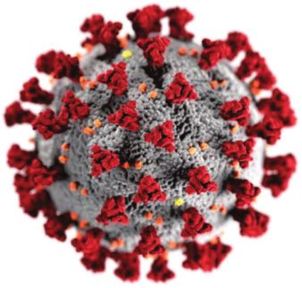
7

development of an acute flare (46). IBD patients can be caught          Patients with high risk include: patients with comorbidities
in a vicious circle in which the growing burden of psychological        (especially diabetes, NASH, cardiovascular or lung diseases),
stress leads to an acute flare that in turn aggravates the anxiety      patients > 70 years, patients under corticosteroid doses > 20
leading to the deterioration of the intestinal inflammation.            mg/day, presence of malnutrition, and combination therapy with
     Patients with IBD treated with a variety of immunosuppressive      biological and immunosuppressive agent. Intermediary risk
therapies, including corticosteroids, biologicals, JAK inhibitors or    shows patients under biologic therapy with anti-TNF-α therapy,
other immunosuppressive agents, are more susceptible for                patients receiving MTX, JAK inhibitors or calcineurin-
infections (47, 48). It seems obvious that many patients have the       inhibitors. Low risk includes use of 5 aminosalicylates,
tendency to discontinue their immunosuppressive treatment. This         budesonide, cholestyramine, lorepamide, luminal antibiotics
discontinuation is associated with an increased risk of developing      such as rifaximin and biologicals with low risk of infection such
an acute flare. For better monitoring the outcomes of COVD-19 in        as ustekinumab and vedolizumab.
patients with IBD, the international pediatric and adult database
Surveillance Epidemiology of Coronavirus Under Research
Exclusion (SECURE-IBD) was launched. According to this                                      COVID-19 AND LIVER
database (30.3.2020), 164 COVID-19 IBD patients were identified
(93 with Crohn’s disease and 66 with ulcerative colitis, 2 IBD               The infection with SARS-CoV2 may be accompanied by the
patients unclassified). 38 patients were hospitalized and the death     abnormal levels of aminotransferases in up to 61.1% of cases.
fatality was 5% (AGA website). These results indicate that patients     This indicates that infection with SARS-CoV2 virus may lead in
with IBD do not show a higher mortality rate despite the                some patients to liver injury. Generally, the changes in the liver
immunosuppressive therapy. The explanation for this may be the          function tests caused by the virus are transitory and the
fact that the main role in the development of “cytokine storm” in       development of severe liver injury is uncommon (53).
course of acute respiratory distress syndrome plays the                      The pathomechanism of the liver injury include direct
overexpression of tumor necrosis factor alpha (TNF-α). This             harmful action of the virus via ACE-2 receptors expressed on
reinforces the hypothesis that anti-TNF-α therapy could be even         cholangiocytes and hepatocytes, drug-induced liver disease and
beneficial and protect the patients against severe forms of SARS-       liver complications due to sepsis (54) (Fig. 7).
CoV2. Recently Tursi et al. (49) described a case of a 30 year old           Huang et al. postulated that the incidence of abnormal levels
male with IBD under treatment with mesalazine 3 g/day and               of alanine and aspartate aminotransferase (ALT, AST) and
adalimumab 40 mg every other week that was admitted to hospital,        hepatic dysfunction is more frequent in patients with severe
tested positively for SARS-CoV2. COVID-19 disease manifested            diseases, especially in those patients that require therapy in the
as mild interstitial inflammation in the chest CT scan and the          ICU (5). In fact, the literature has shown discrepant results.
patient needed only oxygen support. Interestingly, no aggravation       Guan et al. (7) observed elevated levels of AST more commonly
of IBD was observed due to COVID-19 and this patient stayed in          in patients with severe course of COVID-19 as compared to non-
the remission and the fecal calprotectin value remained normal. In      severe cases (39.4% versus 18.2%). In contrast, Wu et al. (55)
order to not to endanger the patient, adalimumab therapy was            demonstrated no significant differences in liver function tests
paused during viral infection. This case indicates that biologic        between severe and non-severe cases. An impairment of liver
therapy in IBD patient does not represent a massive threat in terms     function in patients with COVID-19 depends on different factors
of COVID-19 infection.                                                  like genetics, use of medication leading to drug-induced liver
     These results are surprising, because normally patients with       disease (like hydroxychloroquine, toclizumab, remdesvir,
IBD,       treated     with     corticosteroids,     biologic      or   antibiotics), overproduction of proinflammatory cytokines
immunosuppressive agents, are more susceptible for infections.          (especially “cytokine storm”) and the presence of a pre-existing
Compared to other therapeutic regimens, these drugs increase            liver disease, especially non-alcoholic fatty liver disease that
the risk for different viral infections such as herpes simplex          increases the vulnerability to a viral-induced liver damage. At
virus, varicella zoster virus, cytomegalovirus infection or             the moment there is no evidence for the late onset liver function
hepatitis B reactivation. Age represents an important                   damage due to an SARS-CoV2 infection.
independent risk factor for opportunistic infections in IBD                  There is increasing evidence that patients with chronic liver
patients (50).                                                          diseases (CLD) are at higher risk for severe COVID-19
     Considering the risk for infections the biologic therapy           infections. CLD are characterized by progressive deterioration
shows significant differences. Whereas anti TNF-α antibodies            of liver functions for more than six months. The most common
and JAK inhibitors like tofacitinib show increased risk for             etiologies are alcoholic and metabolic liver disease, autoimmune
infections, both vedolizumab and ustekinumab exhibit a much             liver diseases, chronic viral hepatitis, chronic liver diseases with
better safety profile (51).                                             genetic causes and others. The end-stage of these diseases is
     The Chinese Society of IBD made some recommendations               liver cirrhosis (53).
for managing patients with IBD during the COVID-19 pandemic                  Among other chronic liver diseases, one of the most
(52). Based on the observation made during the epidemic in              prevalent is non-alcoholic fatty liver disease (NAFLD) (recently
Wuhan, the experts recommend continuing current treatment,              renamed as metabolic-associated fatty liver disease-MAFLD)
given that the disease is stable. If a new therapy is started, the      (56, 57). MAFLD is commonly associated with diabetes,
therapy with vedolizumab or ustekinumab should be considered            hypertension and obesity. MAFLD is a highly complexed
as a first line therapy due to their good safety profile. Tofacitinib   disease with multiple pathogenic factors and also extrahepatic
should not be newly prescribed, because this drug significantly         manifestations. It encompasses different stages such as steatosis,
increases the risk for viral infections (especially varicella zoster    steatohepatitis, fibrosis and end-stage liver cirrhosis (58).
virus). During the pandemic, only emergency endoscopy should            Previous publications demonstrated that patients with MAFLD
be performed. Elective endoscopy should be postponed. Every             have an increased risk for pneumonia-related mortality (59).
patient that needs emergency surgery should be screened for                  Generally, patients with obesity with high prevalence of
COVID-19. Patients with acute fever should immediately                  MAFLD are risk patients for the development of severe COVID-
consult their IBD doctor.                                               19. Interestingly, in patients aged less than 60 years, the presence
     In every IBD patient, risk stratification for COVID-19             of MAFLD was associated with the more frequent presence of
should be performed in ambulatory or hospital setting (Fig. 6).         severe COVID-19 accompanied by the “cytokine storm” (60). In
8

             uncontrolled production of                                          immune deficiency
             proinflammatory cytokines

                                                                                                            coagulation
                                                                                                             disorders
        dysbiosis
    intestinal barrier                               liver cirrhosis
       dysfunction

                                                                                                            hepatopulmonary
                                                                                                               syndrome

                   portal
                hypertension                           spontaneous                        hypoalbuminemia
                   ascites                                bacterial
                                                      peritonitis (SBP)
Fig. 8. Different factors and mechanisms responsible for severe course of COVID-19 in patients with liver cirrhosis.

another study, Cai et al. assessed 14 patients with SARS-CoV2         COVID-19 (RR 4.6, 95% CI 2.6 – 8.3, P < 0.0001) among
infection and preexisting NAFLD. Six of these patients showed         patients with liver cirrhosis as compared to patients without liver
a severe disease process (61). Therefore, it is important to          disease. Patients with advanced liver cirrhosis and high risk for
identify the prognostic factors for severe COVID-19 in patients       decompensation should be monitored with a web-based system
with MAFLD. One of the important prognosis factors is the             in order to prevent from unnecessary hospital visits during the
presence of fibrosis in the liver. Just recently, Byrne et al. (58)   pandemic. In addition, patients should be informed about the
presented that the presence of fibrosis in patients with MAFLD        hygienic measures (washing of hands) and the importance of
significantly increases the risk of developing a severe form of       social distancing. Despite these recommendations, the patients
COVID-19. More studies are needed to better understand the            with liver cirrhosis are prone to develop acute decompensation
link between MAFLD and higher risk for SARS-CoV2-related              during the pandemic due to the multiple factors, such as irregular
mortality.                                                            medication, lack of regular follow-up visits, decreased
    Patients with autoimmune liver disease are at increased risk      endoscopic surveillance, increased natrium intake in junk food
of infection due to the use of immunosuppressive therapy,             and alcohol consumption due to lockdown and social isolation,
corticoids and altered immune function. Previous studies              increased stress, and decreased mobility. Urgent procedures
demonstrated that patients treated with immunosuppressive             associated with acute liver decompensation such as paracentesis
medication have an increased risk for viral infections. Against       of ascites or treatment of esophageal varices should be organized
these expectations, the experience data gathered in the epicenters    in hospital on COVID-free wards.
of the infection in Wuhan/China or Lombardy/Italy did not show            There is an established correlation between the PNPLA3
a higher risk for ARDS due to COVID-19 and increased                  rs738409 C > G single nucleotide polymorphism (SNP) and
mortality among these patients. The immunosuppressive therapy         hepatic steatosis and fibrosis in hepatitis C virus (HCV) infected
should not be reduced or stopped to avoid acute flare of disease      patients (65). It has been demonstrated that PNPLA3
and unnecessary admission to the hospital. In patients with           rs738409[G] allele is a reliable predictor for steatosis and
stable chronic autoimmune disease, the follow-up visit could be       fibrosis in patients with chronic hepatitis C. The presence of G
postponed until the pandemic is over. Alternatively a web-based       allele along with severe steatosis and insulin resistance are
or telephone-based consultation can be performed. It is useful to     significant predictors for liver fibrosis progression but whether
send general information and recommendation to the patients.          this exacerbation of disease caused by single nucleotide
Acute flares of acute autoimmune hepatitis, especially                polymorphism is influenced by COVID-19 infection in HCV
obstructive jaundice in primary sclerosing cholangitis (PSC),         patients remains to be studied. For instance, sofosbuvir, the
require hospital treatment (53, 62).                                  clinically approved anti-HCV drug, has recently been
    Patients with liver cirrhosis are at high risk of developing      recommended to suppress other families of positive-strand RNA
serious infections including SARS-CoV2-virus with high                viruses (66) but the promising study with this drug efficacy
mortality due to some favorable pathophysiological mechanisms         against COVID-19 is still awaited.
such as cirrhosis-induced immune deficiency, intestinal
dysbiosis, genetic predisposition, bacterial translocation, porto-
systemic shunting and liver dysfunction (Fig. 8). As a                                 COVID-19 AND PANCREAS
consequence, patients with liver cirrhosis may develop acute-on-
chronic liver failure accompanied by overwhelming                        Pancreatic cells highly express angiotensin converting
inflammatory response (63). In their recent publication, Singh S      enzyme 2 (ACE2) that SARS-CoV2 requires to entry the cells.
et al. (64) have found a significantly risk for mortality due to      This indicates a possible involvement of pancreas in patients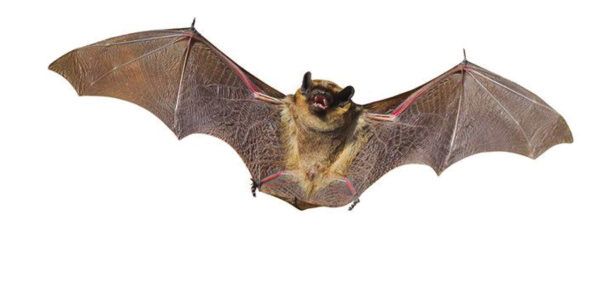
9

with COVID-19 disease (67). Recent reports indicate that some         As shown previously by our group, stress significantly affects the
infected patients may develop acute pancreatitis with elevation       gut microbiota inducing a dysbiosis (74). These effects may be
of serum amylase and lipase (68). In such cases, typical findings     exacerbated by obesity that is recognized as an important risk for
of acute pancreatitis may be observed on abdominal CT. Every          severe progression of COVID-19 (75). In addition, advanced age
physician, especially gastroenterologist should be aware of this      is one of the important risk factors for COVID-19 and increased
complication because patients may present untypical for               mortality. It is well established by studies that the aging is also
COVID-19 abdominal pain and elevation of lipase before the            significantly associated with increasing gut dysbiosis (76). Also,
respiratory symptoms appear. Due to the growing SARS-CoV2             differences in the intestinal microbiota were found in relation to
pandemic, the testing for SARS should be included in the              age in subjects with and without another GI-tract disorder such as
differential diagnosis of patients with acute pancreatitis,           celiac disease (CD) (77). In another study with patients with
especially those patients coming from “hot spots” and having          histamine intolerance, food hypersensitivity, and food allergy, the
had contact to SARS positive patients.                                altered occurrence of Proteobacteria and Bifidobacteriaceae, the
                                                                      reduced alpha-diversity as well as elevated stool zonulin levels
                                                                      have been reported (77). The major message from this study was
             COVID-19 AND GUT MICROBIOTA                              that dysbiosis and intestinal barrier dysfunction are important
                                                                      features in histamine intolerant patients (78). Thus, it is not
     In recent years, there is an emerging evidence for the           excluded that the gut microbiota might be of potential value as a
presence of the bidirectional cross-talk between gut microbiota       diagnostic biomarker and therapeutic target for COVID-19, but
and the lungs termed gut microbiota-lung axis. Negative               further studies on the validation this hypothesis are definitely
alterations of gut microbiota termed dysbiosis may affect             needed.
pulmonary immunity. On the other side, lung inflammation can              Since the gut microbiota influences the systemic immune
induce changes in the gut microbiota. The mechanisms by which         and inflammatory response, the therapy with prebiotics or
gut microbiota affect the development of lung diseases and vice       probiotics to improve the gut microbiota diversity could play an
versa remain poorly understood. As possible candidates, the           important adjuvant role in the prevention of severe progression
involvement of regulatory T cells, toll like receptors, release of    of COVID-19 (79). Finally, the use of antibiotic and antiviral
inflammatory mediators and surfactant protein D have been             substances may also negatively affect the equilibrium of gut
proposed (69).                                                        microbiota (80). The perturbation of the microbiota induced by
     These observations indicate that therapeutic strategies to       antibiotic treatment may have further negative effects on the
improve gut microbiota, including use of prebiotics, probiotics       course of the SARS-CoV2-induced disease.
or synbiotics, could have beneficial effects on the GI symptoms
in COVID-19 patients. Their use is of importance in case of           In conclusion:
antibiotic therapy disrupting the gut microbiome (70). SARS-
CoV2 invades the GI epithelium via ACE2 receptor that is              1) SARS-CoV2 virus invades GI-tract and is associated with a
expressed in the whole GI-tract. The role of ACE2 in the GI-tract         number of GI manifestations, including diarrhea, nausea, the
is not fully understood, but there is evidence that this protein          abdominal pain and vomiting.
regulates the expression of antimicrobial peptides and maintains      2) Viral receptor ACE2 is highly expressed in the GI-tract and
the equilibrium of the gut microbiota. Interestingly, the                 enables the invasion of the GI epithelium.
deficiency of ACE2 results in highly increased susceptibility to      3) COVID-19 is associated with the shedding of the virus via the
intestinal inflammation leading to cancer. For instance, the              GI-tract.
ACE2/MAS receptor pathway might be of great importance in             4) Patients with acute diarrhea should be tested for SARS-CoV2
cancer preventions since the production of Ang1-7 is decreased            infection.
in breast cancer cells in vitro, whereas the expression of MAS        5) COVID-19 is associated with liver manifestations and
receptor was increased in these cells (71). This observation              pancreatic injury.
suggests that agonists of MAS receptor could be useful in the         6) Patients with preexisting liver diseases, particularly NAFLD
treatment of various cancers but the efficacy of Ang1-7 in                and liver cirrhosis, have a higher risk for severe COVID-19
protection against SARS-CoV2 invading gastrointestinal                    course of these diseases and associated mortality.
epithelium requires extensive investigations.                         7) SARS-CoV2-induced dysbiosis is multifaceted and can be
    Importantly, the transplantation of the altered gut microbiota        associated with alterations caused by stress, drug therapy,
from ACE2 mutant mice into germ free wild type host was able              age, and changes in ACE-2 expression.
to transmit the increased propensity for the development of
colitis. With other words, ACE2 is a regulator of immunity and            Conflict of interests: None declared.
gut microbiota ecology (72). This observation indicates that
SARS-CoV2 infection could lead to the disruption of gut
microbiota (dysbiosis). This is not surprising because previous                                REFERENCES
studies have shown the negative effect of viral infections on gut
microbiota. Recently, it has been shown in the murine model that      1. Goyal P, Choi JJ, Pinheiro LC, et al. Clinical characteristics
the antibiotic treatment caused a significant increase in mortality      of Covid-19 in New York City. N Eng J Med 2020; 382:
of C57Bl6 mice infected with paramyxoviral virus type1 (Sendai           2372-2374.
virus). In other murine models, the authors showed effects of         2. Lu R, Zhao X, Li J, et al. Genomic characterization and
influenza and respiratory syncytial virus on the development of          epidemiology of 2019 novel coronavirus: implications for
dysbiosis (73).                                                          virus origins and receptor binding. Lancet 2020; 395: 565-574.
    The outbreak of COVID-19 is not only a global health              3. Sansonetti PJ. COVID-19, chronicle of an expected
problem, but also represents one of the most important stressors.        pandemic. EMBO Mol Med 2020; 12: e12463. doi:
The causes of stress are multifaceted, including fear of infection,      10.15252/emmm.202012463
social distancing, quarantine, lockdown, financial damage, travel     4. Tang D, Comish P, Kang R. The hallmarks of COVID-19
restriction, lack of effective vaccine or treatment etc. All these       disease. In: PLoS Pathog 2020; 16: e1008536. doi:
stressors influence mental health and the function of the GI-tract.      10.1371/journal.ppat.1008536
10

5. Huang C, Wang Y, Li X, et al. Clinical features of patients        26. Freedberg DE, Conigliaro J, Wang TC, et al. Famotidine use
    infected with 2019 novel coronavirus in Wuhan, China.                 is associated with improved clinical outcomes in
    Lancet 2020; 395: 497-506.                                            hospitalized COVID-19 patients: a propensity score matched
6. Wang Y, Tong J, Qin Y, et al. Characterization of an                   retrospective cohort study. Gastroenterology 2020; doi:
    asymptomatic cohort of SARS-COV-2 infected individuals                10.1101/2020.05.01.20086694
    outside of Wuhan, China. Clin Infect Dis 2020; doi:               27. Anand K, Ziebuhr J, Wadhwani P, Mesters JR, Hilgenfeld R.
    10.1093/cid/ciaa629                                                   Coronavirus main proteinase (3CLpro) structure: basis for
7. Guan WJ, Ni ZY, Hu Y, et al. Clinical characteristics of               design of anti-SARS drugs. Science 2020; 300: 1763-1767.
    coronavirus disease 2019 in China. N Eng J Med 2020; 382:         28. Patel KP, Patel PA, Vunnam RR, et al. Gastrointestinal,
    1708-1720.                                                            hepatobiliary, and pancreatic manifestations of COVID-19.
8. Zhou Z, Zhao N, Shu Y, Han S, Chen B; Shu X. Effect of                 J Clin Virol 2020; 128, S. 104386. doi: 10.1016/j.jcv.
    gastrointestinal symptoms on patients infected with coronavirus       2020.104386
    disease 2019. Gastroenterology 2020; 158: 2294-2297.              29. Luo S, Zhang X, Xu H. Don't overlook digestive symptoms
9. Cummings MJ, Baldwin MR, Abrams D, et al.                              in patients with 2019 novel Coronavirus Disease (COVID-
    Epidemiology, clinical course, and outcomes of critically ill         19). Clin Gastroenterol Hepatol 2020; 18: 1636-1637.
    adults with COVID-19 in New York City: a prospective              30. Pan L, Mu M, Yang P, et al. Clinical characteristics of
    cohort study. Lancet 2020; 395: 1763-1770.                            COVID-19 patients with digestive symptoms in Hubei,
10. Giannis D, Ziogas IA, Gianni P. Coagulation disorders in              China: a descriptive, cross-sectional, multicenter study. Am J
    coronavirus infected patients: COVID-19, SARS-CoV-1,                  Gastroenterol 2020; 115: 766-773.
    MERS-CoV and lessons from the past. J Clin Virol 2020;            31. D’Amico F, Baumgart DC, Danese S, Peyrin-Biroulet L.
    127: 104362. doi: 10.1016/j.jcv.2020.104362                           Diarrhea during COVID-19 infection: pathogenesis,
11. Fanelli V, Fiorentino M, Cantaluppi V, et al. Acute kidney            epidemiology, prevention, and management. Clin
    injury in SARS-CoV-2 infected patients. Crit Care 2020; 24:           Gastroenterol Hepatol 2020; doi: 10.1016/j.cgh.2020.04.001
    155. doi: 10.1186/s13054-020-02872-z                              32. Uno Y. Why does SARS-CoV-2 invade the gastrointestinal
12. Asadi-Pooya AA, Simani L. Central nervous system                      epithelium? Gastroenterology 2020; doi: 10.1053/j.gastro.
    manifestations of COVID-19. A systematic review. J Neurol             2020.04.006
    Sci 2020; 413: 116832. doi: 10.1016/j.jns.2020.116832             33. Lamers MM, Beumer J, van der Vaart J, et al. SARS-CoV-2
13. Fulchand S. Covid-19 and cardiovascular disease. BMJ                  productively infects human gut enterocytes. Science 2020;
    2020; 369: m1997. doi: 10.1136/bmj.m1997                              eabc1669. doi: 10.1126/science.abc1669
14. Zaim S, Chong JH, Sankaranarayanan V, Harky A. COVID-             34. Xiao F, Tang M, Zheng X, Liu Ye, Li X, Shan H. Evidence
    19 and multiorgan response. Curr Problems Cardiol 2020;               for    gastrointestinal     infection    of    SARS-CoV-2.
    45: 100618. doi: 10.1016/j.cpcardiol.2020.100618                      Gastroenterology 2020; 158: 1831-1833.e3.
15. Guarneri C, Rullo EV, Pavone P, et al. Silent COVID-19:           35. Repici A, Maselli R, Colombo M, et al. Coronavirus
    what your skin can reveal. Lancet Infect Dis 2020; doi:               (COVID-19) outbreak: what the department of endoscopy
    10.1016/S1473-3099(20)30402-3                                         should know. Gastrointest Endosc 2020; doi:
16. Al-Hajjar S, McIntosh K. Pediatric COVID-19: an update on             10.1016/j.gie.2020.03.019
    the expanding pandemic. Int J Pediatr Adolesc Med 2020;           36. Soetikno R, Teoh AY, Kaltenbach T, et al. Considerations in
    doi: 10.1016/j.ijpam.2020.05.001                                      performing endoscopy during the COVID-19 pandemic.
17. Jones VG, Mills M, Suarez D, et al. COVID-19 and                      Gastrointest Endosc 2020; doi: 10.1016/j.gie.2020.03.3758
    Kawasaki disease: novel virus and novel case. Hosp Pediatr        37. Cavaliere K, Levine C, Wander P, Sejpal DV, Trindade AJ.
    2020; 10: 537-540.                                                    Management of upper GI bleeding in patients with COVID-
18. Bullard J, Dust K, Funk D, et al. Predicting infectious               19 pneumonia. Gastrointest Endosc 2020; doi:
    SARS-CoV-2 from diagnostic samples. Clin Infect Dis 2020;             10.1016/j.gie.2020.04.028
    doi: 10.1093/cid/ciaa638                                          38. MacLeod C, Wilson P, Watson A. Colon capsule endoscopy: an
19. Fraiman JB. Chest CT and coronavirus disease (COVID-19):              innovative method for detecting colorectal pathology during
    a more complete review. Am J Roentgenol 2020; W1. doi:                the Covid-19 pandemic? Colorectal Dis 2020; 22: 621-624.
    10.2214/AJR.20.23428                                              39. Chung SW, Hakim S, Siddiqui S, Cash BD. Update on
20. Kontou PI, Braliou GG, Dimonou NL, Nikolopoulos G;                    flexible    sigmoidoscopy,        computed       tomographic
    Bagos PG. Antibody tests in detecting SARS-CoV-2                      colonography, and capsule colonoscopy. Gastrointest
    infection: a meta-analysis. Diagnostics (Basel) 2020; 10:             Endosc Clin N Am 2020; 30: 569-583.
    319. doi: 10.3390/diagnostics10050319                             40. Repici A, Aragona G, Cengia G, et al. Low risk of Covid-19
21. Gul MH, Htun ZM, Shaukat N, Imran M, Khan A. Potential                transmission in GI endoscopy. Gut 2020; doi:
    specific therapies in COVID-19. Ther Adv Respir Dis 2020;             10.1136/gutjnl-2020-321341
    14: 1753466620926853. doi: 10.1177/1753466620926853               41. Corral JE, Hoogenboom, SA, Kroner PT, et al. COVID-19
22. Mahase E. Covid-19: remdesivir is helpful but not a wonder            polymerase chain reaction testing before endoscopy: an
    drug, say researchers. BMJ 2020; 369: m1798. doi:                     economic analysis. Gastrointest Endosc 2020; doi:
    10.1136/bmj.m1798                                                     10.1016/j.gie.2020.04.049
23. Shen C, Wang Z, Zhao F, et al. Treatment of 5 critically ill      42. Gralnek IM, Hassan C; Dinis-Ribeiro M. COVID-19 and
    patients with COVID-19 with convalescent plasma. JAMA                 endoscopy: implications for healthcare and digestive cancer
    2020; 323: 1582-1589.                                                 screening. Nat Rev Gastroenterol Hepatol 2020; doi:
24. Rogosnitzky M; Berkowitz E, Jadad AR. Delivering benefits             10.1038/s41575-020-0312-x
    at speed through real-world repurposing of off-patent drugs:      43. Binder L, Hogenauer C, Langner C. Gastrointestinal effects
    the COVID-19 pandemic as a case in point. JMIR Public                 of an attempt to ‘disinfect’ from COVID-19. Histopathology
    Health Surveill 2020; 6: e19199. doi: 10.2196/19199                   2020; doi: 10.1111/his.14137
25. Zhang R, Wang X, Ni L, et al. COVID-19: Melatonin as a            44. Ramos GP, Papadakis KA. Mechanisms of disease:
    potential adjuvant treatment. Life Sci 2020; 250: 117583.             inflammatory bowel diseases. Mayo Clinic Proc 2020; 94:
    doi: 10.1016/j.lfs.2020.117583                                        155-165.
11
45. Konturek PC, Brzozowski T, Konturek SJ. Stress and the 65. Crisan D, Grigorescu M, Crisan N, et al. Association
gut: pathophysiology, clinical consequences, diagnostic between PNPLA3[G]/I148M variant, steatosis and fibrosis
approach and treatment options. J Physiol Pharmacol 2011; stage in hepatitis C virus - genetic matters. J Physiol
62: 591-599. Pharmacol 2019; 70: 585-593.
46. Bernstein CN. Psychological stress and depression: risk 66. Sayad B, Sobhani M, Khodarahmi R. Sofosbuvir as
factors for IBD? Dig Dis 2016; 34: 58-63. repurposed antiviral drug against COVID-19: why were we
47. Bonovas S, Fiorino G, Allocca M, et al. Biologic therapies convinced to evaluate the drug in a registered/approved
and risk of infection and malignancy in patients with clinical trial? Arch Med Res 2020; doi:
inflammatory bowel disease: a systematic review and 10.1016/j.arcmed.2020.04.018
network meta-analysis. Clin Gastroenterol Hepatol 2016; 67. Liu F, Long X, Zhang B, Zhang W, Chen X, Zhang Z. ACE2
14: 1385-1397. expression in pancreas may cause pancreatic damage after
48. Shah ED, Farida JP, Siegel, CA, Chong K, Melmed GY. Risk SARS-CoV-2 infection. Clin Gastroenterol Hepatol 2020;
for overall infection with anti-TNF and anti-integrin agents doi: 10.1016/j.cgh.2020.04.040
used in IBD: a systematic review and meta-analysis. 68. Hadi A, Werge M, Kristiansen KT, et al. Coronavirus
Inflamm Bowel Dis 2017; 23: 570-577. Disease-19 (COVID-19) associated with severe acute
49. Tursi A, Angarano G, Monno L, et al. Covid-19 infection in pancreatitis: case report on three family members.
Crohn’s disease under treatment with adalimumab. Gut Pancreatology 2020; 20: 665-667.
2020; 69: 1364-1365. 69. Zhang D, Li S, Wang N, Tan H-Y, Zhang, Z, Feng Y. The
50. Axelrad JE, Cadwell KH, Colombel JF, Shah SC. Systematic cross-talk between gut microbiota and lungs in common
review: gastrointestinal infection and incident inflammatory lung iseases. Front Microbiol 2020; 11: 301. doi:
bowel disease. Aliment Pharmacol Ther 2020; 51: 1222-1232. 10.3389/fmicb.2020.00301
51. Pudipeddi A, Kariyawasam V, Haifer C, Baraty B, Paramsothy 70. Dhar D, Mohanty A. Gut microbiota and Covid-19- possible
S, Leong RW. Safety of drugs used for the treatment of Crohn’s link and implications. Virus Res 2020; 285: 198018. doi:
disease. Expert Opin Drug Saf 2019; 18: 357-367. 10.1016/j.virusres.2020.198018
52. Mao R, Liang J, Shen J, et al. Implications of COVID-19 for 71. Bujak-Gizycka B, Madej J, Bystrowska B, et al. Angiotensin
patients with pre-existing digestive diseases. Lancet 1-7 formation in breast tissue is attenuated in breast cancer -
Gastroenterol Hepatol 2020; 5: 425-427. a study on the metabolism of angiotensinogen in breast
53. Garrido I, Liberal R, Macedo G. COVID-19 and liver cancer cell line. J Physiol Pharmacol 2019; 70: 503-514.
disease - what we know on 1st May 2020. Aliment 72. Hashimoto T, Perlot T, Rehman A, et al. ACE2 links amino
Pharmacol Ther 2020; doi: 10.1111/apt.15813 acid malnutrition to microbial ecology and intestinal
54. Ridruejo E, Soza A. The liver in times of COVID-19: what inflammation. Nature 2012; 487: 477-481.
hepatologists should know. Ann Hepatol 2020; doi: 73. Grayson MH, Camarda LE, Hussain S-R, et al. Intestinal
10.1016/j.aohep.2020.05.001 microbiota disruption reduces regulatory T cells and
55. Wu J, Li W, Shi X, et al. Early antiviral treatment contributes increases respiratory viral infection mortality through
to alleviate the severity and improve the prognosis of patients increased IFNγ production. Front Immunol 2018; 9: 1587.
with novel coronavirus disease (COVID-19). J Intern Med doi: 10.3389/fimmu.2018.01587
2020; doi: 10.1111/joim.1306 74. Konturek PC, Zopf Y. Gut microbiome and psyche:
56. Cichoz-Lach, Celinski K, Konturek PC, Konturek SJ, paradigm shift in the concept of brain-gut axis [in German].
Slomka M. The effect of L-tryptophan and melatonin on MMW Fortschr Med 2016; 158 (Suppl. 4): 12-16.
selected biochemical parameters in patients with 75. Bahlouli W, Breton J, Lelouard M, et al. Stress-induced
steatohepatitis. J Physiol Pharmacol 2010; 61: 577-580. intestinal barrier dysfunction is exacerbated during diet-
57. Eslam M, Sanyal AJ, George J. MAFLD: a consensus-driven induced obesity. J Nutr Biochem 2020; 81: 108382. doi:
proposed nomenclature for metabolic associated fatty liver 10.1016/j.jnutbio.2020.108382
disease. Gastroenterology 2020; 158: 1999-2014. 76. Nagpal R, Mainali R, Ahmadi S, et al. Gut microbiome and
58. Byrne CD, Targher G. NAFLD: a multisystem disease. aging: physiological and mechanistic insights. Nutr Healthy
J Hepatol 2015; 62 (Suppl. 1): 47-64. Aging 2018; 4: 267-285.
59. Nseir WB, Mograbi JM, Amara AE, Abu Elheja OH, 77. Domsa EM, Berindan-Neagoe I, Para I, Munteanu L, Matei
Mahamid MN. Non-alcoholic fatty liver disease and 30-day D, Andreica V. Celiac disease: a multi-faceted medical
all-cause mortality in adult patients with community- condition. J Physiol Pharmacol 2020; 71: 3-14.
acquired pneumonia. QJM 2019; 112: 95-99. 78. Schink M, Konturek PC, Tietz E, et al. Microbial patterns in
60. Gao F, Zheng KI, Wang X-B, et al. Metabolic associated patients with histamine intolerance. J Physiol Pharmacol
fatty liver disease increases COVID-19 disease severity in 2018; 69: 579-593.
non-diabetic patients. J Gastroenterol Hepatol 2020; doi: 79. Pickard JM, Zeng MY, Caruso R, Nunez G. Gut microbiota:
10.1111/jgh.15112 role in pathogen colonization, immune responses, and
61. Cai Q, Huang D, Ou P, et al. COVID-19 in a designated inflammatory disease. Immunol Rev 2017; 279: 70-89.
infectious diseases hospital outside Hubei Province, China. 80. Lange K, Buerger M, Stallmach A, Bruns T. Effects of
Allergy 2020; doi: 10.1111/all.14309 antibiotics on gut microbiota. Dig Dis 2016; 34: 260-268.
62. Di Giorgio, A, Nicastro E, Speziani C, et al. Health status of
patients with autoimmune liver disease during SARS-CoV-2 Author’s address: Prof. Peter C. Konturek, Department of
outbreak in northern Italy. J Hepatol 2020; doi: Internal Medicine II, Thuringia-Clinic Saalfeld, Teaching Hospital
10.1016/j.jhep.2020.05.008 of the University of Jena, Rainweg 68, 07318 Saalfeld, Germany.
63. Ekpanyapong S, Reddy KR Infections in cirrhosis. Curr E-mail: pkonturek@web.de
Treat Options Gastroenterol 2019; 17: 254-270.
64. Singh S, Khan A. Clinical characteristics and outcomes of
COVID-19 among patients with preexisting liver disease in
United States: a multi-center research network study.
Gastroenterology 2020; doi: 10.1053/j.gastro.2020.04.064You can also read