Atopic Dermatitis in Domestic Animals: What Our Current Understanding Is and How This Applies to Clinical Practice
←
→
Page content transcription
If your browser does not render page correctly, please read the page content below
veterinary
              sciences

Review
Atopic Dermatitis in Domestic Animals: What Our Current
Understanding Is and How This Applies to Clinical Practice
Rosanna Marsella

                                          Department of Small Animal Clinical Sciences, College of Veterinary Medicine, University of Florida,
                                          2015 SW 16th Avenue, Gainesville, FL 32610, USA; Marsella@ufl.edu

                                          Abstract: Atopic dermatitis is a clinical syndrome that affects both people and animals. Dogs closely
                                          mimic the complexity of the human skin disease, and much progress has been made in recent years
                                          in terms of our understanding of the role of skin impairment and the identification of new treatments.
                                          Cats and horses also develop atopic syndromes which include both cutaneous and respiratory signs,
                                          yet studies in these species are lagging. It is now recognized that atopic dermatitis is not a single
                                          disease but a multifaceted clinical syndrome with different pathways in various subgroups of patients.
                                          Appreciating this complexity is clinically relevant as we develop more targeted treatments which
                                          may work well in some patients but not in others. Different phenotypes of atopic dermatitis have
                                          been described in dogs, and it is possible that phenotypes related to breed and age may exist in
                                          other animals similar to how they are described in people. The awareness of different mechanisms of
                                          disease leads to the desire to correlate different phenotypes with specific biomarkers and responses
                                          to treatment. In this review, the current understanding and updated information on atopic syndrome
                                          in animals are described, highlighting opportunities for further studies in the future.

                                Keywords: atopic syndrome; dermatitis; dogs; cats; horses


Citation: Marsella, R. Atopic
Dermatitis in Domestic Animals:
What Our Current Understanding Is
                                          1. Introduction
and How This Applies to Clinical
Practice. Vet. Sci. 2021, 8, 124.
                                                Atopic dermatitis is one of the manifestations of atopic disease. In people, dermatitis is
https://doi.org/10.3390/vetsci8070124     typically the first manifestation of atopic disease and can be followed by respiratory disease
                                          later in life as part of what is called the “atopic march” [1]. Atopic dermatitis affects people
Academic Editor: Masashi Yuki             and animals and, in some species (e.g., dogs), is the most prevalent manifestation of atopic
                                          disease. Atopic dermatitis in dogs has become increasingly common as exposure to indoor
Received: 27 May 2021                     environments and processed foods has increased in our pets. Canine atopic dermatitis
Accepted: 26 June 2021                    has characteristics, both clinically and immunologically, that are strikingly similar to the
Published: 2 July 2021                    human counterpart [2,3]. In dogs, progression to respiratory signs has been described in
                                          colonies of atopic dogs [4], but it does not seem to be a common observation in clinical
Publisher’s Note: MDPI stays neutral      practice.
with regard to jurisdictional claims in         Much progress has been made in our understanding of canine atopic dermatitis in
published maps and institutional affil-   recent years [5]. The availability of colonies of atopic research dogs has greatly helped to
iations.                                  shed light on the complex pathogenesis of this condition. In research settings, controlled
                                          studies involving allergen challenges can be done to observe the development of lesions
                                          and better understand the dynamic changes of inflammatory cytokines over time. In these
                                          settings, multiple biopsies can be taken, and the measurement of various inflammatory
Copyright: © 2021 by the author.          mediators at various time points is possible [6,7]. The availability of these models has
Licensee MDPI, Basel, Switzerland.        been instrumental in identifying new potential targets, such as IL-31 [8]. The identification
This article is an open access article    of new targets has led to the development of new drugs currently available in clinics to
distributed under the terms and           provide relief to animals that spontaneously develop this frustrating and chronic disease.
conditions of the Creative Commons
Attribution (CC BY) license (https://
creativecommons.org/licenses/by/
4.0/).

Vet. Sci. 2021, 8, 124. https://doi.org/10.3390/vetsci8070124                                                  https://www.mdpi.com/journal/vetsci
Vet. Sci. 2021, 8, x FOR PEER REVIEW 2 of 20
Vet. Sci. 2021, 8, 124 2 of 18
2. Dogs
2. Dogs
2.1. Evolution of Our Understanding
2.1. Evolution of Our Understanding
Our comprehension of canine atopic dermatitis has greatly improved in the last dec-
Our comprehension of canine atopic dermatitis has greatly improved in the last
ade [9]. The development of the clinical disease is the result of a complex interaction be-
decade [9]. The development of the clinical disease is the result of a complex interaction
tween genetic and environmental factors. While, in the past, IgE was considered the most
between genetic and environmental factors. While, in the past, IgE was considered the
important player in the pathogenesis, and much of the emphasis was placed on mast cells
most important player in the pathogenesis, and much of the emphasis was placed on
and histamine, it is now accepted that IgE may be an epiphenomenon. Many other factors
mast cells and histamine, it is now accepted that IgE may be an epiphenomenon. Many
besides IgE and histamine are now known to play a role in this complex [10]. Recent re-
other factors besides IgE and histamine are now known to play a role in this complex [10].
search research
Recent has focused on the role
has focused on theof the
roleskin barrier
of the both inboth
skin barrier termsin of ultrastructural
terms altera-
of ultrastructural
tions and dysbiosis
alterations and dysbiosis[11] and
[11] the
androle of various
the role lymphocytic
of various lymphocyticpopulations
populations[12–14]. This
[12–14].
evolution in our understanding has been reflected in how clinical
This evolution in our understanding has been reflected in how clinical cases are managed.cases are managed. In
the past, antihistamines were widely advocated for treatment and
In the past, antihistamines were widely advocated for treatment and prevention of flares. prevention of flares.
Currently,the
Currently, theemphasis
emphasison onthetheuseuseofofantihistamines
antihistamineshas hasdecreased
decreased[15], [15],asascontrolled
controlled
studies have failed to show the beneficial effect of antihistamines in a double-blind,
studies have failed to show the beneficial effect of antihistamines in a double-blind, placebo- pla-
cebo-controlled
controlled fashionfashion [16]. Despite
[16]. Despite this, antihistamines
this, antihistamines may
may still still be prescribed
be prescribed in prac-
in practice [17]
tice
as [17]
part ofas part of a multimodal
a multimodal approach.approach.
Aspart
As partofofthe
thenewer
newerapproach,
approach, increased
increased attention
attention hashas been
been given
given to the
to the restoration
restoration of
of the skin barrier through the application of sphingolipid and fatty
the skin barrier through the application of sphingolipid and fatty acid emulsions [18–20]. acid emulsions [18–
20]. approach
This This approach has been
has been shown shown to restore
to restore somesomeof theofabnormalities
the abnormalities
of theofatopic
the atopic skin
skin and
and have
have a positive
a positive effecteffect on clinical
on clinical signs.signs.
TopicalTopical
therapytherapy hasgained
has also also gained
much much
emphasisempha-for
sis treatment
the for the treatment of secondary
of secondary bacterial bacterial infections
infections [21,22] [21,22] dueincrease
due to the to the increase in antimi-
in antimicrobial
crobial resistance.
resistance. Thus, while Thus, while
in the past,inthe
theuse
past, the useantimicrobial
of topical of topical antimicrobial
products was products
consideredwas
considered
an adjunctiveantherapy,
adjunctive
nowtherapy, now it is
it is frequently frequently
advocated asadvocated as a monotherapy
a monotherapy whenever possiblewhen-
ever
to possible
minimize touse
the minimize the use
of systemic of systemic antibiotics.
antibiotics.
2.2.
2.2.Clinical
ClinicalFeatures,
Features,Allergy
AllergyTests,
Tests,and
andHow
HowtotoMake
MakeAADiagnosis
Diagnosis
Atopic
Atopic dermatitis in dogs manifests as a pruriticinflammatory
dermatitis in dogs manifests as a pruritic inflammatorydisease
diseasethat
thataffects
affects
body
body areas where the allergen is more readily absorbed epicutaneously. Examplesofofthese
areas where the allergen is more readily absorbed epicutaneously. Examples these
areas
areasare
arefolds andand
folds areas withwith
areas thinner skin and
thinner skinless
andhair.
lessExamples include the
hair. Examples antebrachial
include the an-
region, theregion,
tebrachial axillae,the
and the inguinal
axillae, area. The
and the inguinal muzzle,
area. periocular
The muzzle, region,region,
periocular pinnae, and
pinnae,
interdigital areas areas
and interdigital are other contact
are other areas where
contact exposure
areas where to the allergen
exposure is common.
to the allergen Thus,
is common.
atopic dermatitis has characteristic predilected sites (Figure
Thus, atopic dermatitis has characteristic predilected sites (Figure 1).1).
Figure1.1.The
Figure Themuzzle,
muzzle,periocular
perioculararea,
area,and
andears
earsare
arepredilected
predilectedsites
sitesfor
foratopic
atopicdermatitis
dermatitisinin dogs,
dogs, as
as shown in this patient.
shown in this patient.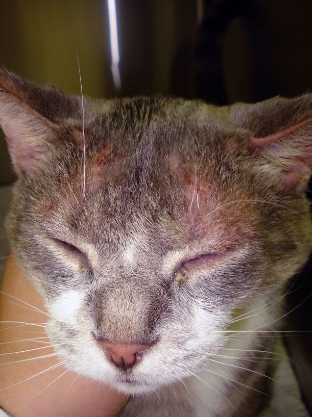
Vet. Sci. 2021, 8, 124                                                                                             3 of 18

                               Each patient has some characteristic body areas that are prone to clinical flares. For
                         example, some dogs present with inflammatory otitis externa, while others show their
                         disease as pododermatitis. Recurrent otitis externa can be, for some patients, the only sign
                         of atopic dermatitis for a while. Thus, it is important to control the underlying allergic
                         process when patients develop secondary bacterial and yeast otitis on a seasonal basis.
                               It is important to realize, however, that the body sites affected by atopic dermatitis
                         are not pathognomonic for this disease. For example, the face and feet can be affected by
                         many other conditions, such as demodicosis or contact allergy. Thus, it is important to
                         consider and rule out other differential diagnoses before making a clinical diagnosis of
                         atopic dermatitis. We still lack a diagnostic test for atopic dermatitis. The diagnosis of
                         atopic dermatitis is clinical and based on compatible history, clinical signs, and exclusion
                         of other pruritic diseases. While we still rely on serology testing and intradermal testing
                         to identify possible offending allergens to include in immunotherapy, it is understood
                         that neither of these “tests” can be used for diagnostic purposes. Instead, “allergy tests”
                         should be used after a clinical diagnosis of atopic dermatitis has been made [23] with the
                         main intent to formulate allergen-specific immunotherapy. This is an important concept
                         in practice as these tests cannot be used to discriminate between an itchy dog due to
                         atopic disease and an itchy dog affected by parasites. When considering serology tests,
                         it is also important to note that the presence of IgEs against cross-reactive carbohydrate
                         determinants has been documented in dogs. These IgEs are clinically irrelevant but can
                         lead to many false positive results when using serology tests. Currently, some companies
                         treat serum samples to block these IgEs to decrease false positive results and improve the
                         accuracy of their serology test [24].

                         2.3. Identification of Triggers
                               While foods can be trigger factors for atopic dermatitis in dogs, as is the case in chil-
                         dren, we still tend to separate a food-induced disease from atopic dermatitis. Typically, the
                         term atopic dermatitis is used to refer to a pruritic skin disease in which food and fleas have
                         been ruled out, and we are left with a diagnosis by exclusion of “environmentally induced
                         allergic dermatitis”. Thus, although foods are potential triggers of atopic dermatitis-type
                         clinical signs, most clinicians still tend to use the term atopic dermatitis as equivalent to
                         environmental allergies. The reality is that atopic dogs in which environmental triggers are
                         important may be clinically indistinguishable to some in which foods are the trigger [25,26].
                         As avoidance of triggers is important in managing clinical flares, the identification of a
                         possible role of foods in affecting the severity of the disease is a critical component of
                         the management of nonseasonal atopic dogs. Importantly, there are some patients that
                         look clinically indistinguishable from our classic atopic dogs but for whom environmental
                         triggers cannot be identified, at least with the current tests available for use. For that subset
                         of patients, we tend to use the terms “intrinsic atopic dermatitis or atopic-like disease” [27].
                         These cases represent an additional challenge as we are not able to use allergen-specific
                         immunotherapy, and we are limited in only considering symptomatic therapies. Based on
                         the current evidence in those cases, it appears that the clinical characteristics of those dogs
                         are similar to the “extrinsic” cases in which an environmental trigger can be identified. The
                         response to treatments also appears to be the same, although we only have very few studies
                         with a low number of cases. This is an area in which we need to further examine this
                         subpopulation to see if they constitute an early stage of the more classic atopic dermatitis
                         or a specific subset. In other words, it is possible that these patients have not had sufficient
                         time to develop an allergic response, and if they were tested later in life, they may be
                         positive in our traditional tests. It may also be that these patients do not have skin barrier
                         impairments, and that is a protective factor against the development of an epicutaneous
                         sensitization toward environmental allergens.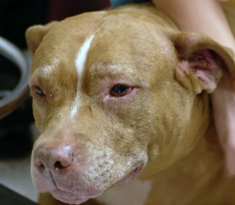
Vet. Sci. 2021, 8, 124                                                                                              4 of 18

                         2.4. The Role of Skin Barrier in Canine Atopic Dermatitis
                               What we have learned in recent years is how important the skin barrier is for atopic
                         dermatitis [28] and the propensity to epicutaneous sensitization to allergens [29]. Indeed,
                         the epicutaneous route of exposure is important for both sensitization [30,31] and for
                         elicitation of clinical signs [31,32]. While we are still lacking definitive evidence of a primary
                         skin barrier impairment in atopic dogs, we do know that secondary skin barrier damage
                         exists, as inflammation and self-trauma deteriorate the barrier function of the skin [33].
                         A damaged skin is more prone to absorb what it encounters and is prone to develop an
                         allergic response to allergens. This is because damaged keratinocytes release cytokines
                         such as thymic stromal lymphopoietin (TSLP), which promotes a T helper 2 response and
                         the development of an allergic response. Thus, frequent removal of allergens from the
                         skin of allergic dogs is crucial to minimize exposure and the worsening of inflammation.
                         We know that the skin of atopic dogs is more alkaline and that it loses more water than
                         normal skin. While in people, atopic skin is accepted to be drier than normal skin, the
                         skin of atopic dogs does not appear to have decreased hydration, at least based on the
                         current studies we have so far [34]. It is possible that our current methodologies are not
                         sensitive enough or that despite the increased loss of water in atopic dogs, the hydration
                         is still within normal limits. This could be linked to the fact that atopic dogs have been
                         reported to have an increased gene expression of proteins such as filaggrin [35,36], whose
                         breakdown products (natural moisturizing factors) are important for hydration.
                               As the interest in the skin barrier and keratinization has increased in the past decade,
                         several studies have investigated the role of filaggrin in canine atopic dermatitis. Filaggrin
                         mutations have been reported as one of the most documented risk factors for atopic
                         dermatitis in people [37,38]; thus, it was natural to address this issue in dogs. We now
                         know that more than one filaggrin-type protein exists in dogs, although the exact function
                         of filaggrin 2 in dogs is not clear [39]. Both filaggrin-type proteins are expressed in the
                         upper layers of the skin and play a role in the differentiation of the epidermis and in the
                         hydration of the skin. While in people, filaggrin loss-of-function mutations have been
                         identified as major risk factors for the development of atopic dermatitis, this does not
                         appear to be case in most breeds of dogs [40]. It is possible that in the future we will
                         discover other players that may be more relevant for dogs. Research toward establishing a
                         correlation between the severity of clinical signs and gene expression in the skin of atopic
                         dogs has shown that genes relevant to skin barrier formation and immune function were
                         altered [41]. Most of the studies on the genetics of canine atopic dermatitis had small
                         sample sizes, which hindered their ability to detect factors given the heterogeneity of this
                         condition and the variation of breeds [42].
                               When challenged with an allergen, atopic dogs attempt to compensate for the insult
                         with an increased production of filaggrin, and an increased expression of enzymes respon-
                         sible for filaggrin degradation has been described in experimental models of canine atopic
                         dermatitis [43]. The increased proteolytic activity of atopic skin has been reported in atopic
                         people, and it is linked to the increased pH [44]. Clinically speaking, it may be important to
                         acidify atopic skin to both decrease the proliferation of bacteria and yeasts and to decrease
                         the proteolytic activity of ceramidases and proteases that degrade lipids in the skin and
                         increase the rate of desquamation.

                         2.5. Factors Playing A Role in the Development of Clinical Disease
                               As mentioned previously, the actual development of clinical signs is the result of a
                         combination of genetic and environmental factors [45]. Many genes have been considered
                         as candidates for canine atopic dermatitis [46]. The increased prevalence of atopic der-
                         matitis may not only be linked to a preferential breeding of atopic individuals but also
                         to a change in environmental conditions. In people, according to the “hygiene theory”, it
                         is documented that decreased exposure to parasites and beneficial bacteria predisposes
                         one to the development of atopic dermatitis [47,48]. A similar situation may apply to
                         our pets. While pets in the past were more exposed to an outdoor environment with less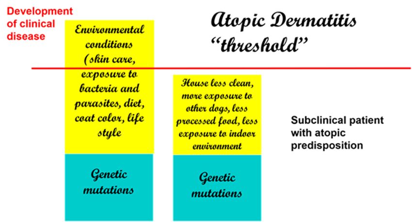
Vet. Sci. 2021, 8, x FOR PEER REVIEW 5 of 20
Vet. Sci. 2021, 8, 124 5 of 18
While pets in the past were more exposed to an outdoor environment with less exposure
to house dust mites and more exposure to parasites and bacteria, the current conditions
of ingesting
exposure processed
to house foodsand
dust mites rather
morethan raw diets,
exposure an increased
to parasites exposure
and bacteria, the to indoor envi-
current
conditions and
ronments of ingesting processed
house dust mites,foods
and arather than raw
decreased diets, an
exposure to increased
beneficialexposure
bacteriatomay con-
indoor environments and house dust mites, and a decreased exposure to beneficial
tribute to an increased development of clinical signs of atopic dermatitis [49,50]. bacteria
may contribute to an increased development of clinical signs of atopic dermatitis [49,50].
If there were an actual “threshold for development of atopic dermatitis” (Figure 2)
If there were an actual “threshold for development of atopic dermatitis” (Figure 2),
we can assume that each factor is additive (from genetic factors to environmental factors)
we can assume that each factor is additive (from genetic factors to environmental factors)
and thatonce
and that oncethe
thethreshold
threshold is achieved,
is achieved, clinical
clinical disease
disease ensues.
ensues. Thus, Thus, when genetic
when genetic factors factors
are
are considered,
considered, ititisispossible
possiblethat
thatmore
more than
than oneone
maymaybe be necessary
necessary to lead
to lead to disease de-
to disease
velopment.
development.
Figure
Figure 2.2.Development
Development of clinical
of clinical disease
disease is the is the of
result result of and
genetic genetic and environmental
environmental factors. De-
factors. Decreased
creased
exposure to beneficial bacteria and increased exposure to indoor allergens and processed foods processed
exposure to beneficial bacteria and increased exposure to indoor allergens and are
foods are
some of thesome of the
reported reported
risk factors inrisk factors in dogs.
dogs.
Therefore,
Therefore,with
withgenetic factors
genetic being
factors considered
being a constant,
considered changeschanges
a constant, in environmental
in environmen-
conditions
tal can, by
conditions can,themselves, determine
by themselves, an increased
determine development
an increased of clinical disease.
development This disease
of clinical
is supported by some studies in the veterinary literature that have linked the development
This is supported by some studies in the veterinary literature that have linked the devel-
of atopic dermatitis in dogs to dietary habits, lifestyle, and living conditions [51,52]. Of
opment of atopic dermatitis in dogs to dietary habits, lifestyle, and living conditions
interest, coat color has been linked to the development of atopic dermatitis, and having
[51,52].
more thanOf50%
interest, coat color
of a white has been
coat color linked
has been to thetodevelopment
reported be a risk factorof[51].
atopic dermatitis, and
having more than 50% of a white coat color has been reported to be a risk factor [51].
2.6. Phenotypes
2.6. Phenotypes
The concept of phenotypes of atopic dermatitis has been investigated in human
medicine for a longoftime.
The concept The termofphenotype
phenotypes is intended
atopic dermatitis hastobeen
emphasize the interaction
investigated in human med-
between genetics and environmental factors. Identification of phenotypes
icine for a long time. The term phenotype is intended to emphasize the interaction is important as be-
we progress to a more personalized approach to treatment [53,54]. As atopic dermatitis is a
tween genetics and environmental factors. Identification of phenotypes is important as we
heterogenous disease, identification of subgroups of patients is important for the sake of
progress
treatment to a more
success. personalized
Phenotypes approach
of atopic to treatment
dermatitis [53,54].
in people have beenAsdescribed
atopic dermatitis
based is a
heterogenous
on age [55] anddisease,
ethnicityidentification of subgroups
[56–58]. In people, of patients
different subgroups is important
appear for the sake of
to have peculiar
treatment
pathways and key cytokines that play a role, thus requiring different treatments. In dogs, based
success. Phenotypes of atopic dermatitis in people have been described
on
ourage [55] andofethnicity
knowledge [56–58].
phenotypes In people,
is limited. Clinicaldifferent
phenotypes subgroups
of canine appear to have peculiar
atopic dermatitis
have been and
pathways described based on that
key cytokines breeds anda distribution
play of lesions,
role, thus requiring but no link
different has beenIn dogs
treatments.
drawn
our to specific of
knowledge markers and responses
phenotypes to treatment
is limited. Clinical[59].
phenotypes of canine atopic dermatitis
have been described based on breeds and distribution
2.7. Strategies for Treatment and Options Available for Atopic Dogs
of lesions, but no link has been
drawn to specific markers and responses to treatment [59].
New research has also focused on the identification of new targets for treatment to
minimize the use of broad-spectrum therapies, such as glucocorticoids and cyclosporine,
2.7.
and Strategies fortargeted
to use more Treatment and Options
approaches, Available
such for Atopic
as biologics Dogs
targeting key cytokines. In this
New research has also focused on the identification of new targets for treatment to
minimize the use of broad-spectrum therapies, such as glucocorticoids and cyclosporine
and to use more targeted approaches, such as biologics targeting key cytokines. In thisVet. Sci. 2021, 8, 124 6 of 18
respect, IL-31 has received much attention for its role in canine atopic dermatitis [60]. IL-31
is produced by TH2 cells, and many cells ranging from immune cells to keratinocytes and
nerve fibers have receptors for this cytokine [61]. Its role in the mediation of pruritus has
gained attention [62], but it important to emphasize that IL-31 also modulates keratinocyte
proliferation and differentiation [63]. Strategies to target IL-31 in dogs have ranged from
the use of a caninized monoclonal antibody [64] to vaccinating dogs against their own IL-
31 [65], although this latter approach is currently only experimental. As atopic dermatitis is
a syndrome with different pathomechanisms, not all dogs treated with a biologic aimed at
targeting this cytokine respond. This is something to be expected as we move toward more
targeted therapies. Nevertheless, this approach is revolutionary in veterinary medicine
as biologics offer the freedom of not having to worry about drug interactions and can
be considered for patients with a prior history of demodicosis or neoplasia, where other
treatments may not be ideal.
Of major importance, we are now appreciating the value of a “proactive approach”
when managing cases rather than a “reactive approach” [66]. As these dogs are very
likely to flare at some point, it is important to do what is possible to prevent the flares
rather than to wait for them to occur and then start treatment. This can be done with
topical therapy in areas prone to flaring to minimize the need of rescue medications [67]. If
we wait for the flares to occur, we may need more medication and of a larger spectrum,
while the proactive approach can now be used with more targeted treatments such as
lokivetmab [68]. Over time, fewer flares and fewer medications are needed to make the
patient comfortable compared to the philosophy of waiting until the animal flares and then
starting the treatment.
Allergen-specific immunotherapy is still the only approach that may potentially alter
the course of the disease and minimize future sensitizations. Different routes of admin-
istration have been reported in the literature, with the subcutaneous and the sublingual
being the most commonly used in practice [69–71]. A recently published study directly
compared the efficacy of subcutaneous with sublingual and intralymphatic and concluded
that subcutaneous and intralymphatic were the most effective routes to improve clinical
signs [72]. Allergen-specific immunotherapy is complementary to other treatments, as the
efficacy takes time to manifest.
Much attention has also been devoted to the identification of biomarkers [73], although
it is not clear at this time if these proposed biomarkers are associated with specific responses
to treatment. Cytokines such as TSLP [74], thymus and activation-regulated chemokine
(TARC) [75], IL-33 [76], and IL-34 [77] have all been described in recent studies as possible
biomarkers, and more studies are necessary to understand how these are relevant to a large
population of atopic dogs. As we move toward a more targeted approach for the treatment
of canine atopic dermatitis, it is reasonable to believe that more biologics targeting these
cytokines will become available for dogs. It is important to emphasize that many of
these cytokines are produced by keratinocytes (e.g., TSLP, IL-33) and that keratinocytes
have the ability to shape the lymphocytic response toward allergens. Cytokines such as
IL-33 and TSLP can promote an allergic/inflammatory response rather than tolerance.
Additionally, some mediators released by keratinocytes such as TSLP and periostin [78] are
able to directly elicit itch by acting on sensory nerves [79]. This is particularly relevant in
chronic disease [80] in which increased density of nerve fibers [81] and enhanced peripheral
sensitization play a role and can contribute to a decreased response to antipruritic therapy.
Thus, keratinocytes are far from being a physical inert barrier, and they are an integral part
of a cross talk with the nervous system and the immune cells.
2.8. Bacteria and Atopic Dermatitis
Much progress has been made in our understanding of the microbiome in atopic dogs
and the importance of restoring biodiversity. We appreciate the role of the microbiome
in modulating immunologic responses in dogs and how a dysbiosis can contribute to
the development of allergic and inflammatory diseases [82,83]. Decreased cutaneousVet. Sci. 2021, 8, 124 7 of 18
biodiversity and predominance of Staphylococcus is a feature of atopic flares [84,85]. As the
antibiotic resistance of Staphylococcus grows and represents a serious challenge for clinicians,
we have embraced more topical therapy rather than broad spectrum antibiotics and are
acutely aware of how important it is to encourage biodiversity and a healthy sustainable
microbiome [86]. Interestingly, in human medicine, topical microbiome transplantation
has shown promising results for decreasing the severity of atopic dermatitis and the
need for anti-inflammatory therapy [87,88]. In veterinary medicine, several studies have
shown a positive effect of probiotic supplementation for modulating immune response in
atopic dogs [89] and for decreasing the severity of clinical disease and the need for rescue
medications [90].
2.9. Take Home Message on Canine Atopic Dermatitis
In summary, it is clear that many different factors play a role in shaping the immune
response in canine atopic dermatitis and that keratinocytes and the skin microbiome play a
crucial role in modulating the immune response to allergens and modulating inflammation
and pruritus. Thus, our management needs to be multimodal with the intent to restore
skin barrier and biodiversity while we provide relief from the itch and work toward the
long-term re-education of the immune system by using allergen-specific immunotherapy.
Each patient will have different thresholds and needs for treatment which may change over
the life time of the patient and throughout the course of the year, as flare factors lower the
threshold of diseases. Thus, the management of these patients becomes an art of applying
our current evidence-based information and tailoring it to the individual needs of our
patients. Importantly, we should strive to implement a proactive approach that is suitable
for the specific case to minimize the need for rescue medications and the development of
secondary infections as part of our efforts to minimize the need for systemic antibiotics.
3. Cats
3.1. Current Understanding on Pathogenesis
Research on feline atopic syndrome has been lagging compared to that on dogs. Re-
cent review papers have focused on cats and atopic syndrome, summarizing the evidence
currently published in cats. These papers have proposed new nomenclature [91] to ad-
dress the fact that cats have their peculiar manifestations of allergic disease which do not
exactly match the human or canine disease. The authors of these papers concluded that
there is sufficient evidence to accept that cats have atopic disease, a disease in which IgE
has been demonstrated to play a role [92–94] and which is amenable to allergen-specific
immunotherapy. Manifestations of atopic disease in cats can include skin, respiratory, and
gastrointestinal disease, as is the case in people. The authors propose using the term “feline
atopic skin syndrome” to designate a complex of pruritic inflammatory skin diseases in
cats that have a variety of patterns and that are linked to allergen-specific IgE to environ-
mental allergens [95]. This terminology is intended to replace the term “non-flea-non-food
hypersensitivity” which was used in the past.
Very little is known at this time about skin barrier dysfunction in atopic cats. Prelimi-
nary studies on skin barrier function in atopic cats with skin disease show that transepider-
mal water loss may be increased, and hydration may, at least in some sites, be decreased [96].
There is limited evidence of any useful correlation between clinical scoring systems and
measurements of hydration [97]. Much work needs to be done to assess skin barrier func-
tion in atopic cats and its potential relevance to the pathogenesis of the disease. It could
be speculated that the development of indolent ulcers and eosinophilic granulomas in the
oral cavity of cats could actually be the result of epicutaneous and oral exposure to the
allergen, as cats can be vigorous groomers and allergens could make prolonged contact
with the perioral and oral mucosa. Currently, there is no study that has documented the
development of such lesions in an experimental model of allergen challenge. No study has
reported on filaggrin and lipid abnormalities in the skin of allergic cats.documented the development of such lesions in an experimental model of allergen cha
lenge. No study has reported on filaggrin and lipid abnormalities in the skin of allergi
Vet. Sci. 2021, 8, 124 cats. 8 of 18
Few studies have reported on the lymphocytic populations [98] in the skin of atopi
cats, and increased numbers of CD4+ and CD8+ T cells have been described. We know
Few studies have reported on the lymphocytic populations [98] in the skin of atopic
that IL-4 plays a role in allergic cats [99], and we have preliminary evidence that IL-3
cats, and increased numbers of CD4+ and CD8+ T cells have been described. We know
may be relevant in allergic cats, as it is in people and dogs. More specifically, circulatin
that IL-4 plays a role in allergic cats [99], and we have preliminary evidence that IL-31
IL-31
may belevels in cats
relevant with acats,
in allergic presumptive diagnosis
as it is in people of allergic
and dogs. More dermatitis
specifically,have been reporte
circulating
IL-31 levels in cats with a presumptive diagnosis of allergic dermatitis have been reported that th
to be significantly higher than those of normal cats [100]. The authors reported
mean
to circulatinghigher
be significantly IL-31 than
levelthose
was 8798 fg/mL
of normal for[100].
cats cats with allergicreported
The authors dermatitisthatcompared
the t
205 fg/mL
mean in age-matched
circulating controls.
IL-31 level was 8798 fg/mLAs eosinophils
for cats withare susceptible
allergic to the
dermatitis effects of IL-3
compared
to 205 fg/mL
[101], in age-matched
IL-31 can represent an controls.
appealingAs eosinophils are susceptible
target for the treatment ofto eosinophilic
the effects of IL-
diseases i
31 [101],
cats. IL-31 can represent an appealing target for the treatment of eosinophilic diseases
in cats.
3.2.Clinical
3.2. ClinicalDisease
Diseaseandand
HowHow to Make
to Make A Diagnosis
A Diagnosis
Thecutaneous
The cutaneous manifestations
manifestations of atopic
of feline felineskin
atopic skin syndrome
syndrome are not
are not the same the same a
as those
those
in atopicindermatitis
atopic dermatitis
in people andin people andcats
dogs. Some dogs.
can Some cats
develop candermatitis
facial develop(Figure
facial 3)
dermatiti
and pruritus, as in atopic people and dogs, but other feline manifestations of environmental
(Figure 3) and pruritus, as in atopic people and dogs, but other feline manifestations o
allergies are peculiar
environmental to catsare
allergies (e.g., indolenttoulcer,
peculiar cats eosinophilic plaque).
(e.g., indolent ulcer, eosinophilic plaque).
Figure3.3.Severe
Figure Severe pruritis
pruritis andand facial
facial dermatitis
dermatitis in cat in cat diagnosed
diagnosed with
with feline feline
atopic atopic
skin skin syndrome.
syndrome. As
As facial
facial pruritus
pruritus can becan be triggered
triggered by many bydiseases,
many diseases, it is important
it is important to rule outto ruledermatophytes,
mites, out mites, dermato-
phytes,
and fleas and fleas
before before considering
considering feline atopic feline atopic skin
skin syndrome as asyndrome as a clinical diagnosis.
clinical diagnosis.
Of
Ofcritical importance
critical importance is toispoint out that
to point out the
thatclinical manifestations
the clinical are not pathog-
manifestations are not pathog
nomonic for a specific trigger [102,103]. Thus, clinicians cannot make conclusions
nomonic for a specific trigger [102,103]. Thus, clinicians cannot make conclusions about the abou
trigger by looking at the clinical presentation of the patient: they need to rule out insects
the trigger by looking at the clinical presentation of the patient: they need to rule out in
and foods as triggers based on the seasonality and history of the patient and consider the
sects andoffoods
diagnosis felineas triggers
atopic based on (also
skin syndrome the seasonality and history
known as dermatitis of to
linked theenvironmental
patient and conside
the diagnosis
triggers) of felineofatopic
as a diagnosis skin just
exclusion, syndrome (also
as it is in known
dogs. as dermatitis
This requires linkedflea
appropriate to environ
mental triggers) as a diagnosis of exclusion, just as it is in dogs. This requires
control in areas where insects are prevalent and an appropriate food trial in patients that appropriatVet. Sci. 2021, 8, 124 9 of 18
are nonseasonal. It is important to point out that although various tests (e.g., salivary
test [104], patch test [105]) have been advocated for the diagnosis of food allergy, food trials
with a novel source of protein or using a hydrolyzed diet are still the common approach in
clinics. The choice of novel protein versus hydrolyzed diet depends on the patient, dietary
history, and availability of diets.
Allergy testing (either by use of serology testing or intradermal skin test) can be used
to select environmental allergens to include for allergen-specific immunotherapy but not for
the purpose of making a diagnosis of feline atopic skin syndrome [106–108]. Intradermal
skin testing can be technically challenging in cats [109], and serology is frequently used
in its place. Cats, similar to people, also have of IgE for cross-reactive carbohydrate
determinants which can be responsible for false positive results on serology. The blocking
of these IgEs has been reported to improve the accuracy of serology testing [110].
3.3. Treatments Available for Atopic Cats
Glucocorticoids and cyclosporine are treatments for which there is the most evidence
of efficacy [111]. As part of the multimodal approach, antihistamines and essential fatty
acids can be added to the regimen, provide relief in cats with milder disease, and minimize
the need for broader spectrum anti-inflammatory therapies [112]. Although oclacitinib is
not labeled for cats, some studies have reported on its use in cats with allergic skin disease.
One study reporting on the pharmacokinetics of oclacitinib in cats [113] demonstrated
that the absorption is variable among individuals and that possibly larger doses may be
needed in feline patients compared to dogs. The authors also pointed out that shorter
dosing intervals would be recommended in cats to achieve similar blood concentrations to
those in dogs. The clinical response to oclacitinib is variable [114] and has overall reported
it to be less effective than methylprednisolone [115].
For young atopic cats with long allergy seasons, it is always beneficial to attempt
allergen-specific immunotherapy. Thus, identification of environmental allergens that
may play a role in that specific patient is important and is done to correlate the results
of the allergy testing with the seasonality and environmental exposure of the patient.
The formulation of a custom-made vaccine is intended to decrease the dependence on
rescue medications. Interestingly, more studies have been published on allergen-specific
immunotherapy for feline asthma than for feline atopic skin syndrome. The subcutaneous
route has been found to be the most reliable route in feline asthma [116]. Both serology and
skin testing were found to be useful for the selection of allergens used for immunotherapy
in cats with respiratory disease [117].
There are few studies on allergen-specific immunotherapy in cats with skin disease.
A recently published study reported on the efficacy of sublingual immunotherapy in
atopic cats sensitized to dust and storage mites [118]. In this prospective open label
study, immunotherapy was given for 12 months and monitoring of IgE and IgG was done
at various intervals. The authors concluded that a significant decrease of the severity
of dermatitis and pruritus was observed at the end of the study, and a decrease of IgE
occurred after 9 months, while IgG did not change throughout the study. The treatment
was well tolerated and can be considered for cats that do not do well with injections.
A monoclonal antibody against feline IL-31 with the ability to block the binding of this
cytokine to its receptor has been described [119], suggesting a promising future biologic
for cats.
3.4. Take Home Message on Feline Atopic Skin Syndrome
In summary, although we have much work ahead to better understand the pathogen-
esis of feline atopic skin syndrome, we have some preliminary evidence that similarities
may exist in the immune dysregulation between cats and dogs and that IL-31 may be a
good target for cats as well. Cats are very much in need of treatments to improve their
quality of life, and identification of key cytokines could prove to be of tremendous benefit.Vet. Sci. 2021, 8, 124 10 of 18
4. Horses
4.1. Our Understanding on the Pathogenesis in Horses
Horses also develop atopic disease, which can manifest as respiratory or cutaneous dis-
ease [120]. The respiratory disease has been recognized to be similar to human asthma [121].
The association between allergen-specific IgE and the presence of dermatitis has been re-
ported in several studies [122,123], and a positive response to allergen-specific immunother-
apy in atopic horses has been documented in the literature [124]. Similar to other species,
IgE levels are influenced by genetic factors in horses [125].
Our understanding of atopic dermatitis in horses is very limited. It is commonly
accepted that this disease is the result of genetic and environmental factors, and it is
frequent to see horses that were raised in colder climates manifest disease only later
in life when moved to a warmer climate with more insect and environmental pressure.
Many of these atopic horses are polysensitized and have hypersensitivity to both insects
and various pollens [126]. Very little is known about skin barrier and atopic disease
in horses. One study in atopic horses showed ultrastructural abnormalities on electron
microscopy when compared to normal horses [127] but it is unclear if this is the result
of inflammation or may be suggestive of some primary impairment of the skin which
could facilitate the epicutaneous absorption of the allergen and increased risk for allergic
sensitization. A recently published study on horses with insect hypersensitivity compared
the transcriptome in the epidermis of allergic horses with that of normal horses [128] and
suggested skin impairment in insect allergic horses. It is possible that some of these allergic
horses were also atopic. Clearly, more work is needed before any conclusion can be made
about the existence of a primary skin impairment and what the pathogenetic relevance
could be for the equine disease.
4.2. Clinical Signs and How to Make A Diagnosis
Atopic dermatitis in horses presents as a relapsing, pruritic inflammatory disease that
typically affects the face (Figure 4), ears, and glabrous areas. Some horses may have a
history of heaves as well. Many atopic horses are also insect allergic. These patients have
clinical signs that are a combination of these allergies: areas like the withers, mane, and tail
can all be affected by pruritus. The combination of allergies is an important concept for
the control of flares. As it is in small animals where the control of flea exposure is critical
to ensure the success of treatment for atopic dermatitis, the prevention of insect bites is
important in atopic horses. Controlling all triggers of pruritus is crucial to bring patients
below a clinical threshold of pruritus and increase the success of therapy.
As is in other species, the diagnosis of atopic dermatitis is a clinical diagnosis based
on suggestive history, compatible clinical signs, and exclusion of other pruritic diseases.
This is an important concept as some positive results on both skin testing and serology
testing can be seen in normal horses, although in lower numbers when compared to
atopics [121,122,129,130]. Nevertheless, allergy testing cannot be used to make a diagnosis
of atopic dermatitis [131] similar to other species.
Older studies reported a poor correlation between serology and skin test [132], while
newer studies have reported a very good correlation [123]. This may be the result of
the more accurate serology testing that is currently available. As we improve our un-
derstanding of how to interpret serology testing, we have learned that horses, similar to
other species, also have IgEs against cross reactive carbohydrate determinants [133]. The
inhibition of these IgEs greatly improves the accuracy of the serology testing.
Treatment of atopic horses primarily still involves the use of glucocorticoids and
antihistamines, but no controlled studies have been done to evaluate the efficacy of these
treatments in a controlled fashion. The reports of these treatments are retrospective and
uncontrolled studies where owners report on the beneficial effects of these strategies [134].
The same limitations hold for reports investigating allergen-specific immunotherapy, which
is recommended for atopic horses with a long allergy season [123,135]. The reported success
rate of allergen-specific immunotherapy in atopic horses ranges from 64% [133] to 84% [136]Vet. Sci. 2021, 8, 124 11 of 18
depending on the study, and the bulk of the improvement is typically visible after the first
year [123]. Some horses can be maintained with allergen-specific immunotherapy alone,
while others still require other medications, although the amounts of medications necessary
to make them comfortable may be decreased. Importantly, the success of immunotherapy
Vet. Sci. 2021, 8, x FOR PEER REVIEW
was not reported to be different based on which allergy testing was used to select 11 the
of 20
allergens [135].
Figure4.4.Horse
Figure Horsediagnosed
diagnosedwith
withenvironmental
environmentaland
andinsect
insectallergies.
allergies.The
Thepruritus
pruritusininthis
thispatient
patientwas
was intense and led to significant self-trauma.
intense and led to significant self-trauma.
As is has
IL-31 in other
been species,
shown to thebediagnosis
a mediator of of
atopic dermatitis
pruritus is a as
in horses, clinical diagnosis
the injection based
of IL-31
on suggestive
protein recombinanthistory,wascompatible clinicalintense
able to trigger signs, and exclusion
pruritus at theofsiteotherof pruritic
injectiondiseases.
in nor-
Thishorses
mal is an important
[137]. Thus, concept
IL-31 as cansome
be a positive
suitable results on the
target for bothtreatment
skin testing and serology
of pruritus as a
testing can be
preliminary seenin
study ininsect
normal horses,horses
allergic although in lowerIn
has shown. numbers
this study,when compared
allergic horses towere
atop-
ics [121,122,129,130].
immunized Nevertheless,
against IL-31 and showed allergy testing in
a reduction cannot be usedoftoclinical
the severity make ascores
diagnosis
when of
compared to a placebo
atopic dermatitis and to their
[131] similar to otherprevious
species.season [138]. The authors propose that im-
munization againstreported
Older studies cytokinesa may poorbe a more cost-effective
correlation between serology strategyandand skinwould also have
test [132], while
the benefit
newer of inducing
studies a polyclonal
have reported a very response rather than
good correlation [123].relying
This may on the administration
be the result of the
of an equinized
more monoclonal
accurate serology testing antibody in the form
that is currently of a biologic.
available. As we improve The long-term poten-
our understand-
tially unwanted consequences of immunizing horses against their
ing of how to interpret serology testing, we have learned that horses, similar to other own cytokines needs
spe-
further consideration, although other studies targeting vaccination
cies, also have IgEs against cross reactive carbohydrate determinants [133]. The inhibition against IL-5 in insect
allergic
of thesehorses seem toimproves
IgEs greatly support safety even when
the accuracy of thehorses received
serology booster vaccinations for
testing.
2 years [139,140].of atopic horses primarily still involves the use of glucocorticoids and an-
Treatment
These strategies
tihistamines, but no are still experimental
controlled studies have and allergic
been donehorses still suffer
to evaluate from a paucity
the efficacy of these
of treatment options. As in other species, controlling flare factors
treatments in a controlled fashion. The reports of these treatments are retrospective andis of crucial importance.
Since many atopic
uncontrolled horses
studies whereare owners
also insect allergic,
report on theprevention
beneficialofeffects
insectof bites
these is very important
strategies [134].
to bring the patient below a threshold of clinical signs. Consistent
The same limitations hold for reports investigating allergen-specific immunotherapy, use of effective repellents
iswhich
key inisgeographical
recommended areas
for with
atopic a high
horsesinsect
withburden. Controlling
a long allergy season secondary
[123,135]. bacterial
The re-
infection is also very important to decrease the level of pruritus
ported success rate of allergen-specific immunotherapy in atopic horses ranges from 64%and prevent self-trauma.
[133]With
to 84%the [136]
goal of providingon
depending more optionsand
the study, for the
allergic
bulkhorses, oclacitinib hasisalso
of the improvement been
typically
tested. A dose of 0.25 mg/kg once daily was reported to result in a
visible after the first year [123]. Some horses can be maintained with allergen-specific im-decrease in severity of
munotherapy alone, while others still require other medications, although the amounts of
medications necessary to make them comfortable may be decreased. Importantly, the suc-
cess of immunotherapy was not reported to be different based on which allergy testing
was used to select the allergens [135].
IL-31 has been shown to be a mediator of pruritus in horses, as the injection of IL-31bacterial infection is also very important to decrease the level of pruritus and prevent sel
trauma.
With the goal of providing more options for allergic horses, oclacitinib has also bee
tested. A dose of 0.25mg/kg once daily was reported to result in a decrease in severity o
Vet. Sci. 2021, 8, 124 12 of 18
signs compared to a placebo starting after 5 days of treatment [141]. No direct compariso
between the effect of oclacitinib and glucocorticoids has been published. The once dail
dosecompared
signs was usedtobased on information
a placebo starting after on the of
5 days pharmacokinetics
treatment [141]. No ofdirect
this drug in horses [142
comparison
which shows a longer half-life compared to dogs.
between the effect of oclacitinib and glucocorticoids has been published. The once daily
In summary,
dose was used based our understanding
on information on theof atopic dermatitis
pharmacokinetics in horses
of this drug in is rudimentary,
horses [142], an
which
muchshowsworka needs
longer half-life compared
to be done to dogs.
to understand the role of the skin barrier and immun
In summary,inour
dysregulation understanding
horses, and how of atopic
this dermatitis
relates to otherinspecies.
horses is rudimentary,
Based on whatand we know,
much work needs to be done to understand the role of the skin barrier and immune
is reasonable to hypothesize that skin barrier impairment may exist in horses. Identifica
dysregulation in horses, and how this relates to other species. Based on what we know, it is
tion of specific targets to decrease the use of glucocorticoids would be of immense benefi
reasonable to hypothesize that skin barrier impairment may exist in horses. Identification
ofIL-31 appears
specific targetsto
tobe a suitable
decrease target.
the use of glucocorticoids would be of immense benefit. IL-31
appears to be a suitable target.
4.3. Dermatitis Linked to Environmental Allergies in Other Animals
4.3. Dermatitis Linked to Environmental Allergies in Other Animals
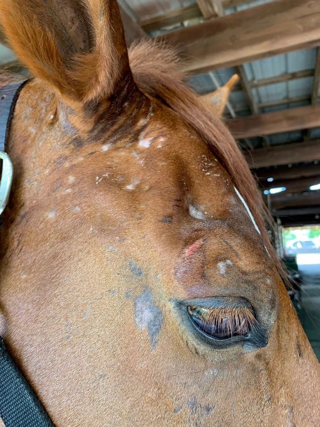
Environmental allergy has been diagnosed in other species ranging from cows to pig
Environmental
(Figure 5), and evenallergy has been
in wildlife diagnosed
species. The in other has
author species
skinranging
tested from cows to
and successfully per
pigs (Figure 5), and even in wildlife species. The author has skin tested and successfully
formed immunotherapy in pigs (subcutaneous) and bats (oral) and is aware of oral im
performed immunotherapy in pigs (subcutaneous) and bats (oral) and is aware of oral
munotherapy done in bears. While mice have been used traditionally as a model for atop
immunotherapy done in bears. While mice have been used traditionally as a model for
dermatitis
atopic in people,
dermatitis thisthis
in people, species does
species doesnot
notspontaneously developatopic
spontaneously develop atopic dermatitis. Mos
dermatitis.
rodent models involve some mutation which leads to the development
Most rodent models involve some mutation which leads to the development of itch of itchoror inflam
mation, but the
inflammation, but dermatitis typically
the dermatitis does
typically doesnot
notmimic
mimic the complexityofofdisease
the complexity disease seen in do
seen
inmestic animals.
domestic animals.
Figure 5. Pot belly pig presenting for pruritic dermatitis of face (periocular and perioral), ventral
abdomen, and legs linked to environmental allergies and responsive to allergen specific immunother-
apy.
5. Conclusions
Atopic dermatitis affects animals in a similar fashion to the human disease. For
animals such as dogs that have embraced lifestyle changes similar to people (e.g., increased
exposure to clean, indoor environments and increased consumption of processed foods),
these changes have increased the risk of development of allergic disease. Whether increased
allergies also are developing in horses and cats due to the fact that animals are dewormed
more frequently and therefore have less exposure to parasites than they did in the past
remains to be established. Our approach to atopic dermatitis has changed over time to
become more holistic and more focused on restoring rather than suppressing the immune
system. Sustainability and safety are key for long-term management. Regardless of the
species, allergen-specific immunotherapy remains the most desirable long-term option
with the intent of re-educating the immune system and decreasing the need of rescue
medications. Of all cytokines, IL-31 seems to be a key player across species and the future
target of treatment for cats and horses.Vet. Sci. 2021, 8, 124 13 of 18
Funding: This research received no external funding.
Conflicts of Interest: Marsella has received multiple pharma research funding.
References
1. Paller, A.S.; Spergel, J.M.; Mina-Osorio, P.; Irvine, A. The atopic march and atopic multimorbidity: Many trajectories, many
pathways. J. Allergy Clin. Immunol. 2019, 143, 46–55. [CrossRef] [PubMed]
2. Arcique, M.A.; Bajwa, J. Atopic dermatitis in humans and dogs. Can. Vet. J. 2020, 61, 82–84, PMC6909410. [PubMed]
3. Marsella, R.; De Benedetto, A. Atopic Dermatitis in Animals and People: An Update and Comparative Review. Vet. Sci. 2017, 4,
37. [CrossRef] [PubMed]
4. Rudolph, K.; Yellowhair, T.R.; Dearing, J.J.; Barrett, E.G. Durability of the atopic dermatitis phenotype in a laboratory colony of
beagles. Vet. Dermatol. 2020, 31, 60.
5. Nuttall, T.J.; Marsella, R.; Rosenbaum, M.R.; Gonzales, A.J.; Fadok, V.A. Update on pathogenesis, diagnosis, and treatment of
atopic dermatitis in dogs. J. Am. Vet. Med Assoc. 2019, 254, 1291–1300. [CrossRef]
6. Marsella, R.; Olivry, T.; Nicklin, C.; Lopez, J. Pilot investigation of a model for canine atopic dermatitis: Environmental house dust
mite challenge of high-IgE-producing beagles, mite hypersensitive dogs with atopic dermatitis and normal dogs. Vet. Dermatol.
2006, 17, 24–35. [CrossRef] [PubMed]
7. Pucheu-Haston, C.M.; Jackson, H.A.; Olivry, T.; Dunston, S.M.; Hammerberg, B. Epicutaneous sensitization with Der-
matophagoides farinae induces generalized allergic dermatitis and elevated mite-specific immunoglobulin E levels in a canine
model of atopic dermatitis. Clin. Exp. Allergy 2008, 38, 667–679. [CrossRef] [PubMed]
8. Olivry, T.; Mayhew, D.; Paps, J.S.; Linder, K.E.; Peredo, C.; Rajpal, D.; Hofland, H.; Cote-Sierra, J. Early Activation of Th2/Th22
Inflammatory and Pruritogenic Pathways in Acute Canine Atopic Dermatitis Skin Lesions. J. Investig. Dermatol. 2016, 136,
1961–1969. [CrossRef]
9. Marsella, R. Advances in our understanding of canine atopic dermatitis. Vet. Dermatol. 2021. [CrossRef]
10. Pucheu-Haston, C.M.; Bizikova, P.; Eisenschenk, M.N.C.; Santoro, D.; Nuttall, T.; Marsella, R. Review: The role of antibodies,
autoantigens and food allergens in canine atopic dermatitis. Vet. Dermatol. 2015, 26, 115. [CrossRef]
11. Santoro, D.; Marsella, R.; Pucheu-Haston, C.M.; Eisenschenk, M.N.C.; Nuttall, T.; Bizikova, P. Review: Pathogenesis of canine
atopic dermatitis: Skin barrier and host-micro-organism interaction. Vet. Dermatol. 2015, 26, 84-e25. [CrossRef]
12. Pucheu-Haston, C.M.; Bizikova, P.; Marsella, R.; Santoro, D.; Nuttall, T.; Eisenschenk, M.N.C. Review: Lymphocytes, cytokines,
chemokines and the T-helper 1-T-helper 2 balance in canine atopic dermatitis. Vet. Dermatol. 2015, 26, 124. [CrossRef]
13. Früh, S.P.; Saikia, M.; Eule, J.; Mazulis, C.A.; Miller, J.E.; Cowulich, J.M.; Oyesola, O.O.; Webb, L.; Peng, S.A.; Cubitt, R.L.; et al.
Elevated circulating Th2 but not group 2 innate lymphoid cell responses characterize canine atopic dermatitis. Vet. Immunol.
Immunopathol. 2020, 221, 110015. [CrossRef]
14. Van der Lee, A.J.; Rutten, V.P.M.G.; Bruijn, J.; Willemse, T.; Broere, F. CD4+ and CD8+ skin-associated T lymphocytes in canine
atopic dermatitis produce interleukin-13, interleukin-22 and interferon-γ and contain a CD25+FoxP3+subset. Vet. Dermatol. 2014,
25, 456-e72. [CrossRef] [PubMed]
15. Saridomichelakis, M.N.; Olivry, T. An update on the treatment of canine atopic dermatitis. Vet. J. 2016, 207, 29–37. [CrossRef]
[PubMed]
16. Hsiao, Y.-H.; Chen, C.; Willemse, T. Effects of cetirizine in dogs with chronic atopic dermatitis: A randomized, double blind,
placebo-controlled trial. J. Vet. Sci. 2016, 17, 549–553. [CrossRef]
17. Zur, G.; Ihrke, P.J.; White, S.D.; Kass, P.H. Antihistamines in the management of canine atopic dermatitis: A retrospective study of
171 dogs (1992–1998). Vet. Ther. Res. Appl. Vet. Med. 2002, 3, 88–96. [PubMed]
18. Marsella, R.; Segarra, S.; Ahrens, K.; Alonso, C.; Ferrer, L. Topical treatment with SPHINGOLIPIDS and GLYCOSAMINOGLY-
CANS for canine atopic dermatitis. BMC Vet. Res. 2020, 16, 1–10. [CrossRef]
19. Blaskovic, M.; Rosenkrantz, W.; Neuber, A.; Sauter-Louis, C.; Mueller, R. The effect of a spot-on formulation containing
polyunsaturated fatty acids and essential oils on dogs with atopic dermatitis. Vet. J. 2014, 199, 39–43. [CrossRef]
20. Tretter, S.; Mueller, R.S. The Influence of Topical Unsaturated Fatty Acids and Essential Oils on Normal and Atopic Dogs. J. Am.
Anim. Hosp. Assoc. 2011, 47, 236–240. [CrossRef]
21. Jeffers, J.G. Topical Therapy for Drug-Resistant Pyoderma in Small Animals. Vet. Clin. N. Am. Small Anim. Pract. 2013, 43, 41–50.
[CrossRef]
22. Loeffler, A.; Lloyd, D. What has changed in canine pyoderma? A narrative review. Vet. J. 2018, 235, 73–82. [CrossRef]
23. Hensel, P.; Santoro, D.; Favrot, C.; Hill, P.B.; Griffin, C.E. Canine atopic dermatitis: Detailed guidelines for diagnosis and allergen
identification. BMC Vet. Res. 2015, 11, 1–13. [CrossRef] [PubMed]
24. Piccione, M.L.; DeBoer, D.J. Serum IgE against cross-reactive carbohydrate determinants (CCD) in healthy and atopic dogs. Vet.
Dermatol. 2019, 30, 507. [CrossRef]
25. Favrot, C.; Steffan, J.; Seewald, W.; Picco, F. A prospective study on the clinical features of chronic canine atopic dermatitis and its
diagnosis. Vet. Dermatol. 2010, 21, 23–31. [CrossRef]
26. Olivry, T.; DeBoer, D.J.; Prélaud, P.; Bensignor, E. The International Task Force on Canine Atopic Dermatitis Food for thought:
Pondering the relationship between canine atopic dermatitis and cutaneous adverse food reactions. Vet. Dermatol. 2007, 18,
390–391. [CrossRef] [PubMed]You can also read