Mechanically Induced Cavitation in Biological Systems - MDPI
←
→
Page content transcription
If your browser does not render page correctly, please read the page content below
life

Review
Mechanically Induced Cavitation in Biological Systems
Chunghwan Kim, Won June Choi, Yisha Ng and Wonmo Kang *

                                          School for Engineering of Matter, Transport and Energy, Arizona State University, Tempe, AZ 85281, USA;
                                          ckim110@asu.edu (C.K.); wchoi37@asu.edu (W.J.C.); ywng1@asu.edu (Y.N.)
                                          * Correspondence: wonmo.kang@asu.edu

                                          Abstract: Cavitation bubbles form in soft biological systems when subjected to a negative pressure
                                          above a critical threshold, and dynamically change their size and shape in a violent manner. The
                                          critical threshold and dynamic response of these bubbles are known to be sensitive to the mechanical
                                          characteristics of highly compliant biological systems. Several recent studies have demonstrated
                                          different biological implications of cavitation events in biological systems, from therapeutic drug
                                          delivery and microsurgery to blunt injury mechanisms. Due to the rapidly increasing relevance
                                          of cavitation in biological and biomedical communities, it is necessary to review the current state-
                                          of-the-art theoretical framework, experimental techniques, and research trends with an emphasis
                                          on cavitation behavior in biologically relevant systems (e.g., tissue simulant and organs). In this
                                          review, we first introduce several theoretical models that predict bubble response in different types
                                          of biological systems and discuss the use of each model with physical interpretations. Then, we
                                          review the experimental techniques that allow the characterization of cavitation in biologically
                                          relevant systems with in-depth discussions of their unique advantages and disadvantages. Finally,
                                          we highlight key biological studies and findings, through the direct use of live cells or organs, for
                                each experimental approach.


Citation: Kim, C.; Choi, W.J.; Ng, Y.;    Keywords: cavitation; soft matter; blunt injury mechanism; dynamic bubble behaviors; acceleration-
Kang, W. Mechanically Induced             induced pressure gradients
Cavitation in Biological Systems. Life
2021, 11, 546. https://doi.org/
10.3390/life11060546
                                          1. Introduction
Academic Editors: Pietro Mascheroni
                                                When a homogeneous liquid is subjected to a transient pressure drop below its
and Haralambos Hatzikirou
                                          saturated vapor pressure at a given temperature, small vapor cavities, referred to as
                                          Cavitation [1], can be formed inside the liquid media. Generally, cavitation can be classified
Received: 6 May 2021
Accepted: 7 June 2021
                                          into two types: Inertial and Non-inertial. The former describes rapid bubble dynamics
Published: 10 June 2021
                                          that involve unstable bubble expansion and collapse typically triggered by a rapid change
                                          of pressure with a relatively large amplitude. The latter refers to much gentler bubble
Publisher’s Note: MDPI stays neutral
                                          dynamics, e.g., the stable oscillation of a bubble around its equilibrium radius, typically
with regard to jurisdictional claims in
                                          driven by small periodic external pressure. Inertial cavitation dynamics, the focus of the
published maps and institutional affil-   current review, involves multiple steps including nucleation, expansion, oscillation, and
iations.                                  collapse. During the bubble expansion, the bubble works against the resistance of the
                                          surrounding media, i.e., liquid. During the bubble collapse, the energy stored in the media
                                          is released. This collapse is violent in nature because the energy release is very localized at
                                          very high rates, a phenomenon known as microjetting.
                                                Traditionally, cavitation in liquid has been of great interest to many researchers due
Copyright: © 2021 by the authors.
Licensee MDPI, Basel, Switzerland.
                                          to its important implications for many industrial and military applications. For example,
This article is an open access article
                                          sudden pressure drops in liquid media can occur in many engineering systems that involve
distributed under the terms and           rapid acceleration of the media, such as propellers of submarines and ships, hydraulic
conditions of the Creative Commons        pumps, water turbines, and industrial piping systems. Due to its violent nature, cavitation
Attribution (CC BY) license (https://     can damage even the strongest man-made materials and structures over time, significantly
creativecommons.org/licenses/by/          shortening the life of these systems. Therefore, traditional research has focused on pre-
4.0/).

Life 2021, 11, 546. https://doi.org/10.3390/life11060546                                                         https://www.mdpi.com/journal/life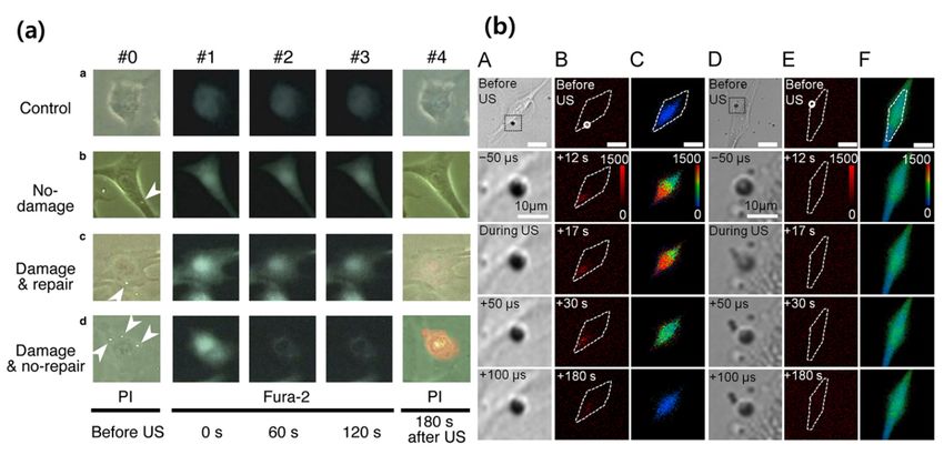
Life 2021, 11, 546                                                                                             2 of 17

                     venting cavitation-induced damage by predicting and avoiding the critical conditions that
                     trigger cavitation nucleation.
                          There have been increasing research efforts to investigate cavitation in biological
                     systems, e.g., a human body or tissue simulant. For example, significant progress has
                     been made in shockwave lithotripsy (SWL) [2–6] by understanding the contribution of
                     cavitation dynamics for biomedical applications [7–11]. Similarly, laser-induced cavitation
                     has been used in ophthalmic microsurgery [12,13]. Another biomedical application of
                     cavitation is targeted drug delivery [14–16]. In these works, cavitation was used to release
                     an encapsulated drug within a carrier, such as a liposome or polymeric nanoparticle, when
                     the carriers were near the target site, e.g., tumor or cancer. Other than that, a microfluidic
                     system with highly controllable bubbles also gives us several advantages associated with
                     understanding of cell injury mechanism or mechanotransduction via calcium signaling
                     processes [17–19]. In the viewpoint of being possible for single-cell analysis, it is helpful to
                     characterize shear stress-induced membrane deformation and the level of its poration.
                          More recently, several studies have reported that injuries that involve rapid accelera-
                     tion of the human body by mechanical impact, e.g., car crash, collisions during sporting
                     events, and bullet wounds [20–23], can induce cavitation in the human body or a tissue
                     simulant. Among the instances of cavitation in the human body, cavitation-induced trau-
                     matic brain injury (TBI) has received much increased attention, because cavitation bubbles
                     inside the human skull can result in tremendous brain damage [24–26]. Therefore, it is
                     essential to understand the behavior of bubbles from the nucleation of the cavity to the
                     collapse of the bubble and its effect on biological systems.
                          With biomedical applications of cavitation, there have been rapidly increasing de-
                     mands for theoretical and experimental characterization of cavitation dynamics in bio-
                     logical systems to capture the unique interplay between cavitation and soft biological
                     systems. Unlike homogeneous pure liquid, cavitation in soft biological systems exhibits
                     highly complex behavior due to the viscoelastic properties [27–29] and heterogeneous
                     microstructures [30,31] of biological systems. In this regard, we reviewed recent research
                     progress on theoretical and experimental approaches for investigating cavitation dynamics
                     in biological systems and biomedical applications. First, we introduce various strain en-
                     ergy function-based constitutive models that delineate bubble behavior in a wide range
                     of biological matters. Each model is described with its physical implications. Then, we
                     consider four different types of experimental methods—needle/acoustic/laser-induced
                     cavitation and an integrated drop tower system—to investigate cavitation phenomenon in
                     the scope of biological applications. Finally, we highlight key in vitro and in vivo studies.

                     2. Theoretical Background: Static and Dynamic Approaches
                           Following the seminal work by Rayleigh [32], it has been shown that the response of
                     inertial cavitation bubbles in media (e.g., liquid or soft materials) depends on the material
                     properties of the media, such as its surface tension [33], viscosity [34] and material stress
                     tensor (σ) associated with the deformation of the media due to change in bubble size
                     and shape. Two different theoretical approaches (i.e., static and dynamic) are available to
                     analyze cavitation bubbles. These two approaches offer crucial theoretical frameworks for
                     interpreting experimental observations from recently developed experimental techniques.
                     It is worth noting that our emphasis is on the dynamic approach since detailed review on
                     the static approach is available elsewhere [31,35–39].

                     2.1. Static Approach
                           The static approach is mostly used to predict the critical bubble size that corresponds
                     with the onset of unstable bubble growth, known as bubble burst, without considering
                     the time-dependent behavior of cavitation. This approach is applicable when bubble size
                     changes very slowly and, therefore, dynamic effects can be ignored.
                           When a spherical cavitation bubble changes its size in a soft material sample, the stress
                     tensor is developed due to the interplay between the bubble and soft material. Using a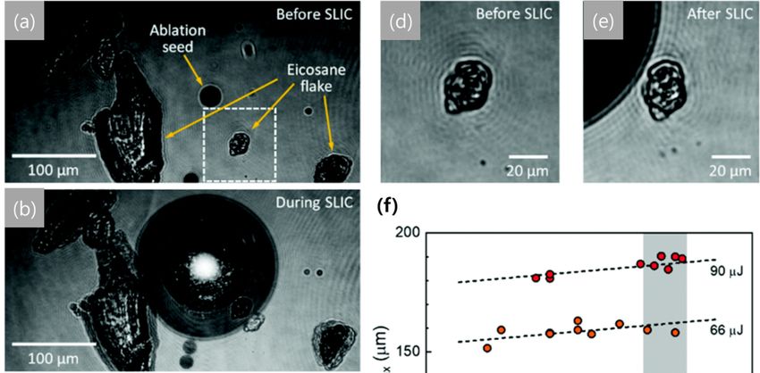
Life 2021, 11, 546                                                                                                    3 of 17

                     nonlinear Kelvin–Voight model [40,41], the tensor consists of elastic stress (σe ) and viscous
                     stress (σv ) as follows:
                                                           σ = σe + σv                                           (1)
                     σe depends on the current deformation of the soft material sample and σv is strain-rate-
                     dependent (i.e., time-dependent). In the static approach (i.e., a bubble in a soft material
                     sample deforms very slowly), the second term in Equation (1) (i.e., σv ) is not considered.
                          When the soft material sample is hyperelastic, isotropic, and incompressible, the
                     elastic stress tensor (σe ) in the sample can be defined by a function of strain energy density
                     (W) as follows [42]:
                                                               ∂W       ∂W          ∂W
                                                  σe,ij = 2        + I1       Vij −    V V                       (2)
                                                               ∂I1      ∂I2         ∂I2 ik kj
                     where Ii is an i-th invariant, V is the left Cauchy–Green strain tensor (V = FFT , where F is
                     the deformation gradient tensor [42]), i and j are free indices, and k is a dummy index.
                             To predict the behavior of cavitation bubbles in different types of soft materials, several
                     constitutive models have been developed and utilized for the invariant of the Cauchy–
                     Green strain tensor (see Table 1). For the neo-Hookean (NH) model [43], the strain energy
                     density is expressed only by the first invariant (I1 , i.e., hydrostatic stress) of the tensor V.
                     In addition to I1 , the Mooney–Rivlin (MR) model [44,45] includes the second invariant
                     (I2 , i.e., distortional stress), where the strain energy of isotropic material is a symmetric
                     function of I1 , I2 and I3 where I3 = 1. By including the second invariant, the MR model
                     provides a wider range of responses of hyperelastic material compared to NH [46], as
                     it considers the deformation of a soft gel by both the mean normal stress tensor (i.e., I1 )
                     and the deviatoric component (i.e., the stress deviator tensor or I2 ). Gent [47] developed
                     a new model that defines the maximum value of I1 (referred to as Im ) in the NH model.
                     Im is introduced to describe the state of polymer chains in a hydrogel. As I1 approaches
                     Im , the entangled polymer chains are straightened, aligned, and axially stretched, which
                     results in rapid stiffening. Another model is the Ogden model [48,49], which consists
                     of polynomial terms that capture the material deformation in the principal directions
                     (see Table 1 for more details). It has been experimentally shown that the Ogden model
                     captures cavitation dynamics in gelatin gels, commonly used as tissue simulant, as well as
                     in different types of organs [46,50,51]. Fung [52,53] developed a constitutive model that
                     takes the strain hardening effect [54] into account, e.g., the effect of pre-stretched soft tissues
                     on elastic shear measurements [54]. Table 1 summarizes the mathematical expressions of
                     the constitutive models discussed here.

                     Table 1. The equation of elastic stress tensor and constitutive strain functions with different types of
                     models with relevant material parameter.

                             Name of Model                 Strain Energy Density (W)                  Reference
                                                                       µ
                          Neo-Hookean Model                            2 ( I1− 3)                         [43]
                                                          µ
                          Mooney–Rivlin Model                      − 3) +(1 − c)(
                                                          2 [ c ( I1                  I2 − 3)]          [44,45]
                              Gent Model                           µ           Im                         [47]
                                                                     Im ln Im − I1 +3
                                                                  2
                               Ogden Model                    2µ     N + λN + λN − 3                     [49]
                                                              N 2  λ r     θ        φ
                                                                       µ α( I1 −3)
                               Fung Model                              2α e
                                                                                                        [52,53]

                          When a soft gel is incompressible, the principal stretch of a cavitation bubble due
                     to applied pressure, p, can be described in the spherically symmetric coordinate as
                     follows [55,56]:
                                                           2γ         W (λ)
                                                                 Z λ
                                                  p = po +     +              dλ                           (3)
                                                            a     1 ( λ3 − 1)

                     where po is the ambient pressure, a is the deformed bubble radius, γ is the surface tension
                     of gel, and λ is the normalized radius of the cavitation bubble. Note that a different
                     constitutive model can be substituted into Equation (3). The applied pressure p is balanced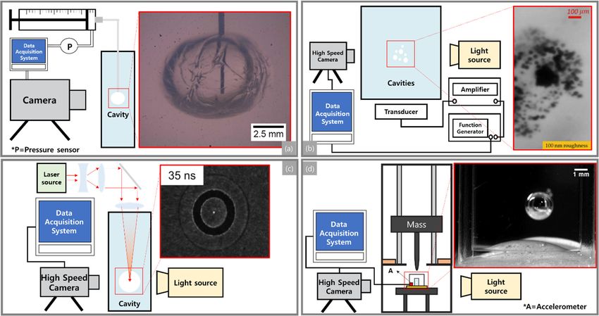
Life 2021, 11, 546                                                                                                4 of 17

                     with the ambient pressure, the Laplace pressure due to surface tension, and the stress
                     term associated with the deformation of gel [55]. it is worth noting that there have been
                     recent studies that consider additional effects from pH [57], temperature [58], nonlinear
                     elasticity [59], humidity [60,61], and energy dissipation level [62], which is beyond the
                     scope of this review.

                     2.2. Dynamics
                          Here, the focus is placed on the time-dependent behavior of a spherical cavitation
                     bubble in soft material (i.e., σv in Equation (1)). The viscous stress σv is a deviatoric and
                     linearly dependent on a strain rate as follows [41]:

                                                          σv = ν ∇u + ∇uT                                       (4)

                     where u = dr/dt, t is time, and ν is the viscosity coefficient of the soft material. Substituting
                     u(r, t) and σ into the radial component of the momentum equation, the governing equation
                     of a spherical cavitation bubble in soft material can be written as follows [63]:

                                                                                         2σrr − σθθ − σφφ

                                     ∂u      ∂u       ∂p                  ∂p ∂σrr
                                 ρ       +u       = − + (∇·σ)r = − +                  +                             (5)
                                     ∂t      ∂r       ∂r                  ∂r      ∂r             r

                     where r is the radial coordinate from the center of the bubble at the deformed state, ρ is
                     density, θ, and φ are the polar and azimuthal angle in the spherical coordinate configuration.
                     σrr , σθθ , and σφφ are the components of the Cauchy stress tensor in the spherical-polar rep-
                     resentation. Integrating the above Equation (5) with the stress tensor given in Equation (1)
                     results in
                                                     3 .2  p( a) − p∞ (t) 1 ∞
                                                                             Z
                                                  ..
                                                aa + a =                 +       (∇·σ)r dr                      (6)
                                                     2            ρ        ρ a
                     where p∞ (t) is the pressure in the medium far from the bubble and p( a) is the pressure
                     in the medium at the bubble-medium interface. Note that the integration in Equation (6)
                     is evaluated over an infinitely large medium, i.e., from the current bubble radius (r = a)
                     to infinity (r = ∞). Finally, the following governing equation can be obtained from
                     Equation (6):
                                                                            .
                                     p B − p∞ (t)
                                                                                  Z
                                                     .. 3 . 2 2γ 4µ a 1                    λ     W (λ)
                                                  = aa + a +     +    +                                  dλ          (7)
                                          ρ             2     ρa   ρa   ρ              1       ( λ3 − 1)

                     where p B is the internal bubble pressure and µ is the viscosity of the medium, and the over
                     dot indicates the derivative of a with respect to time. When p B is a polytropic process, it can
                     be expressed as p B = pv + ( p∞ (t = 0) + 2γ/A − pv )(λ)3k , where pv is the vapor pressure,
                     k is the ratio of the specific heat, i.e., the polytropic index. Equation (7) is the Rayleigh–
                     Plesset (RP) equation, where the last term considers the effect of soft material deformation
                     on the bubble dynamics. As discussed above, a different constitutive material model can
                     be utilized to analyze different biological soft materials. For example, the RP equation
                     with the neo-Hookean (NH) model has been widely utilized to analyze experimentally
                     measured cavitation bubble behaviors in soft hydrogels [10,27–29,64,65].
                           So far, we have introduced the governing equation of single bubble dynamics in the
                     Kelvin–Voigt-type constitutive model, represented by a viscous damper and an elastic
                     spring in parallel, that captures the creep behavior of soft media [40]. Here we discuss
                     other available linear viscoelastic models: the linear Maxwell and solid models. The
                     Maxwell model, composed of a purely elastic spring and a purely viscous damper in
                     series, is applicable for liquid-dominant materials [40] (see Table 2). The linear solid
                     model, a combination of the Kelvin–Voigt and Maxwell models, is used to describe creep,
                     deformation recovery, and stress relation in soft media.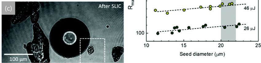
Life 2021, 11, 546                                                                                                                           5 of 17

                                               Table 2. Summary of linear constitutive models [66].

         Name of Model                Strain Energy Density (W)                           Description                        Reference
                                                            .                 Viscous stresses linearly dependent
            Newtonian                               σrr = 2νεrr                                                                  [67]
                                                                                    on the local strain rate
                                                                  .          A spring and a dashpot in parallel;
           Kelvin–Voigt                   σrr = 2 µε rr + νεrr                                                                [41,68,69]
                                                                               Viscoelastic solid; Creep behavior
                                                .       ν .                    A spring and a dashpot in series;
             Maxwell                       2νεrr =      µ σrr   + σrr                                                          [70,71]
                                                                              Viscoelastic liquid; Stress relaxation
     Standard Linear solid             v .                                .
                                                                                Both creep and stress relaxation                 [72]
                                       µ σrr   + σrr = 2µε rr + 2νεrr

                                          It is worth noting that there have also been continuous research efforts to modify the
                                     RP equation to include mass and thermal transfer, compressibility [73,74], non-spherical
                                     perturbations [75], and larger deformation of soft materials, e.g., by developing more
                                     complex nonlinear constitutive models [76–79]. The details are not discussed as these
                                     topics are beyond the scope of the current review.

                                     3. Experimental Methods for Cavitation-Induced Damage to the Biological Systems
                                             Here, we review experimental techniques for triggering and analyzing cavitation bub-
                                       bles in biologically relevant systems. We categorize the available experimental techniques
                                       into two groups: static (needle-induced cavitation) and dynamic (laser- and acoustic-
                                       induced cavitation and the integrated drop tower system). We directly compare the
                                       advantages and disadvantages of these newly developed techniques with an emphasis on
        Life 2021, 11, x FOR PEER REVIEW                                                                              6 of 18
                                       cavitation-induced damage to biological systems (i.e., mechanisms of blunt injuries). A few
                                       key biological advances utilizing each technique are also highlighted.

                                     3.1.3.1. Needle-InducedCavitation
                                           Needle-Induced    Cavitation
                                             Needle-induced cavitation
                                          Needle-induced      cavitation (NIC),
                                                                          (NIC),shown
                                                                                  shown in in
                                                                                            Figure  1a [31,36],
                                                                                               Figure            was was
                                                                                                       1a [31,36],   developed  by
                                                                                                                         developed by
                                        Crosby’s research group  [31]. A cavitation bubble  in a soft material  sample was created
                                     Crosby’s research group [31]. A cavitation bubble in a soft material sample was created
                                     by by applying pressure through a narrow needle inserted into the sample. The needle was
                                        applying   pressure through a narrow needle inserted into the sample. The needle was
                                        connected to a syringe and a pressure sensor by small tubes so that the applied pressure
                                     connected to a syringe and a pressure sensor by small tubes so that the applied pressure was
                                        was precisely controlled by concurrently utilizing a syringe pump and pressure sensor.
                                     precisely controlled by concurrently utilizing a syringe pump and pressure sensor. Bubble
                                        Bubble size was monitored by using a microscope as applied pressure incrementally in-
                                     size was  monitored
                                        creased.           by using a microscope as applied pressure incrementally increased.

      FigureFigure  1. Schematic
              1. Schematic        diagrams
                              diagrams   andand  cavitationimages
                                              cavitation    imagesof
                                                                   of (a)
                                                                      (a) NIC
                                                                          NIC(Needle-induced
                                                                              (Needle-induced cavitation), (b) AIC
                                                                                                cavitation),       (Acoustically
                                                                                                               (b) AIC           induced
                                                                                                                       (Acoustically   induced
            cavitation), (c) LIC (Laser-induced cavitation), and (d) integrated drop tower system. Regenerated by permission from the
      cavitation), (c) LIC   (Laser-induced    cavitation),
            following references [28,31,36,80–83].           and (d) integrated drop  tower  system.   Regenerated   by permission    from the
      following references [28,31,36,80–83].
                                                The NIC method allows experimental characterization, which correlates the mechan-
                                          ical properties of soft material samples (e.g., elastic modulus and viscosity) with their crit-
                                          ical pressure at the onset of the bubble burst [31,37]. To improve the accuracy of the char-
                                          acterization, more detailed studies have followed, including the study of cavitation be-
                                          havior (cavitation and/or fracture) resulting from differing needle diameters ranging from
Life 2021, 11, 546 6 of 17
The NIC method allows experimental characterization, which correlates the mechan-
ical properties of soft material samples (e.g., elastic modulus and viscosity) with their
critical pressure at the onset of the bubble burst [31,37]. To improve the accuracy of the
characterization, more detailed studies have followed, including the study of cavitation
behavior (cavitation and/or fracture) resulting from differing needle diameters ranging
from 30 to 205 µm and differing polymer compositions [35,36,39].
Due to its simple working mechanism, the applications of the NIC method have been
expanded to biological organs, e.g., eyes, skin, and bone marrow, at relatively low strain
rates (10−1 –10−3 s−1 ). Zimberlin et al. [31] first demonstrated the use of NIC for in vivo
samples [84] by measuring the elastic modulus of the bovine eye (more specifically, the
vitreous body in an eye (shown in Figure 2) [84,85]. Similarly, it has been reported that
biological organs have location-dependent elastic moduli (e.g., the elastic moduli measured
Life 2021, 11, x FOR PEER REVIEW in the areas of the nucleus and cortex parts in an extracted bovine eye (see Figure7 of
2) 18
were
11.8 and 0.8 kPa, respectively [85]).
Figure
Figure 2. 2. Images
Images (a)(a)and
andschematic
schematicdiagrams
diagrams (b)
(b) of
of the
the needle-induced
needle-induced cavitation
cavitationmethod
method (NIC) introduced
(NIC) by by
introduced Crosby’s
Crosby’s
research group. Using the NIC method, cavitation behavior and mechanical properties of the bovine eye were
research group. Using the NIC method, cavitation behavior and mechanical properties of the bovine eye were investi-investigated
[84,85].
gated [84,85].
NIC-based methods, unlike conventional shear rheometry for bulk elastic modulus,
NIC-based methods, unlike conventional shear rheometry for bulk elastic modulus,
allow the measurement of localized elastic modulus in heterogeneous soft material sam-
allow the measurement of localized elastic modulus in heterogeneous soft material samples.
ples. In addition, the method is relatively simple and cheap [31]. Furthermore, the size of
In addition, the method is relatively simple and cheap [31]. Furthermore, the size of a
a void can be controlled by the needle radius, gas pressure, and pressure rate. Despite
void can be controlled by the needle radius, gas pressure, and pressure rate. Despite these
these advantages, it is difficult to use the NIC method when the length scale of the defects
advantages, it is difficult to use the NIC method when the length scale of the defects is in
is in the same order as the cavitation size. In addition, the NIC method is mainly for quasi-
the same order as the cavitation size. In addition, the NIC method is mainly for quasi-elastic
elastic behavior due to its slow strain rate (about 10−1–10−3 s−1).
behavior due to its slow strain rate (about 10−1 –10−3 s−1 ).
3.2. Acoustically Induced Cavitation
3.2. Acoustically Induced Cavitation
Acoustically induced cavitation (AIC) uses ultrasound as the driving force of cavita-
Acoustically induced cavitation (AIC) uses ultrasound as the driving force of cavitation
tion nucleation and oscillation. Typically, the AIC system (see a schematic in Figure 1b
nucleation and oscillation. Typically, the AIC system (see a schematic in Figure 1b [82,83])
[82,83]) consists of transducers, signal amplifiers, and waveform generators for generating
consists of transducers,
and controlling signal amplifiers,
desired ultrasonic and waveform
inputs to biological generators
samples. for generating
When a liquid is subjectedand
controlling desired ultrasonic inputs to biological samples. When a liquid is
to an ultrasound field, alternating expansion and compression cycles occur in the media.subjected to an
ultrasound
If the intensity of the alternations is sufficiently large, pressure decreases rapidly and gas- the
field, alternating expansion and compression cycles occur in the media. If
intensity of the
eous bubbles alternations
nucleate [86]. is sufficiently large, pressure decreases rapidly and gaseous
bubbles nucleate [86].
It is important to note that the AIC method utilizes acoustic fields in soft materials
and,Itasisaimportant to note nucleates
result, it typically that the AIC
manymethod utilizes
cavitation acoustic
bubbles. fields
Because in soft
of this materials
feature, it
and,
is not trivial to use the AIC method to characterize material properties of soft materials. it
as a result, it typically nucleates many cavitation bubbles. Because of this feature,
isTonot trivial tothis
overcome useexperimental
the AIC method to characterize
challenge, Mancia et al.material properties
proposed of soft
a cavitation materials.
rheometry
To overcome
technique this
that usesexperimental challenge,
highly focused Mancia
ultrasound et al. proposed
to generate a bubble,a cavitation
named for rheometry
inertial
technique that uses highly
microcavitation-based high focused ultrasound
strain rate rheometryto(IMR),
generate a bubble,
which has the named for inertial
high strain rate
microcavitation-based
range from 103 to 108 s−1high strain
. Using thisrate
newrheometry
method, the (IMR), which properties
mechanical has the high strain rate
of agarose
hydrogel were quantified [87].
The AIC method has garnered significant attention especially in biological systems
due to its ability to focus energy on a small volume. One of the early uses of ultrasound
in biological applications was reported by Brohult et al. to study the degradation of bio-
logical polymers [88]. This pioneering work gave rise to increasing efforts to characterizeLife 2021, 11, 546 7 of 17
range from 103 to 108 s−1 . Using this new method, the mechanical properties of agarose
hydrogel were quantified [87].
The AIC method has garnered significant attention especially in biological systems
due to its ability to focus energy on a small volume. One of the early uses of ultrasound in
biological applications was reported by Brohult et al. to study the degradation of biological
polymers [88]. This pioneering work gave rise to increasing efforts to characterize how
ultrasound interacts with biological systems in the scope of establishing the criteria for
safe use of ultrasound in medical applications. For example, Pohlman et al. investigated
the diminishing intensity of ultrasound beams when transmitted through several layers of
tissue [89]. Carstensen and Schwan et al. focused on the reduced intensity of ultrasound
waves as they propagated through blood [90]. Owing to this prior work, the AIC method
has been used for many biomedical applications such as disintegration of kidney and
gallstones (shockwave lithotripsy) [5,91–94] and intracellular delivery of molecules to a
target site (drug delivery) [95].
In shockwave lithotripsy (SWL), several studies revealed the importance of cavitation
collapse for in vitro applications [9,96–101]. Ikeda et al. proposed high-intensity focused ul-
trasound (HIFU) for lithotripsy to maximize the cavitation effect using two acoustic waves
with different frequencies: one to nucleate multiple bubbles, called bubble clouds, and the
other one to excite the bubble dynamics [8]. HIFU also showed great potential as a non-
invasive treatment as SWL with the accurate control of cavitation behavior [11,102–104].
It is worth noting that excessive energy generated by SWL may result in considerable
damage to organs, e.g., rupture of injury-prone blood vessels [105]. For example, it has
been shown that bubble growth and collapse can lead to vessel stretching and vascular
rupture [106–108]. To reveal these injury mechanisms, Chen et al. developed an experi-
mental setup (Applied pressure: 4–7 MPa) that consists of a high-speed camera and an
inverted microscope for spatial–temporal observations of cavitation bubbles near blood
vessels [109].
For in vitro demonstrations of targeted drug delivery, it has been shown that mi-
crobubbles driven by ultrasound influenced the membrane permeability of live cells [110],
perforation for endothelial cells [111], and the shear stress on cell walls [112], as shown in
Figure 3. Other studies also showed that ultrasound is an effective way to transfer thera-
peutic agents to rats’ hearts [113] and the epidermal growth factor receptor (EGFR)-direct
small inhibitory RNA to target cells for slowing tumor growth [114].
One notable advantage of the AIC method is that it can be utilized in noninvasive med-
ical applications by controlling the frequency and amplitude of input acoustic waves from
medical imaging to lithotripsy. Despite these applications, multifaceted bioeffects [115–117]
and the fundamental root of in vivo cavitation are still not well understood, even with low-
intensity ultrasound. The foremost reason is that the generation of a single cavitation bubble
using AIC is quite challenging as it requires highly focused acoustic waves and precisely
controlled wave frequency, amplitude, and damping. The analysis of bubble dynamics in
biological samples is rather complex due to continuous bubble-to-bubble interactions.
In response to the challenges above, a theoretical model (e.g., Bilayer Sonophore
(BLS)) has been developed. The model underscores the capability of transferring oscillat-
ing ultrasound waves to the expanded or contracted intramembrane compartment [118].
In addition, Iida et al. measured bubble size and distribution using a laser diffraction
method and compared their experimental results with computational predictions [119].
Furthermore, the size and lifetime of bubbles has been investigated to reveal the behavior
of clustered bubbles in bubble clouds [120–123].For in vitro demonstrations of targeted drug delivery, it has been shown that mi-
crobubbles driven by ultrasound influenced the membrane permeability of live cells [110],
perforation for endothelial cells [111], and the shear stress on cell walls [112], as shown in
Figure 3. Other studies also showed that ultrasound is an effective way to transfer thera-
Life 2021, 11, 546 8 of 17
peutic agents to rats’ hearts [113] and the epidermal growth factor receptor (EGFR)-direct
small inhibitory RNA to target cells for slowing tumor growth [114].
Figure 3. Cell
Figure 3. Cellmembrane
membranedamage
damageininthethe
presence of aofmicrobubble
presence a microbubbleoscillated by ultrasound.
oscillated (a) Damage
by ultrasound. and repair
(a) Damage and of bovine
repair of
endothelial monolayer
bovine endothelial cells measured
monolayer over time
cells measured overintime
propidium iodideiodide
in propidium (PI) and
(PI)Fura
and 2Fura
fluorescence [111].[111].
2 fluorescence (b) Time-lapse
(b) Time-
results of PI (B,E)
lapse results of PIand
(B,E)Calcium (C,F) changes
and Calcium in bEnd.3
(C,F) changes cells with
in bEnd.3 cells(A,B,C) or without
with (A,B,C) shear stress
or without shear (D,E,F) [112]. [112].
stress (D,E,F)
3.3. Laser-Induced Cavitation
One notable advantage of the AIC method is that it can be utilized in noninvasive
Since
medical the development
applications of light amplification
by controlling the frequencyby andstimulated
amplitude emission
of inputofacoustic
radiation (i.e.,
waves
laser) [124,125], there have been many attempts to apply laser techniques
from medical imaging to lithotripsy. Despite these applications, multifaceted bioeffects in biological
applications.
[115–117] andThe
themeasurement
fundamentalmethodroot of using
in vivolasers, so-called
cavitation arethe laser-induced
still cavitation
not well understood,
(LIC) method, was introduced by several researchers as early as the 1970s
even with low-intensity ultrasound. The foremost reason is that the generation of a single [126].
The focused
cavitation bubble laser
usingbeam
AIC is(see Figure
quite 1c) [80,81])
challenging transmits
as it requiresenergy
highly to a specific
focused area
acoustic
within a sample. When the temperature increases above the critical
waves and precisely controlled wave frequency, amplitude, and damping. The analysisthreshold temperature,
cavitation
of bubbles form
bubble dynamics in the soft
in biological sample.
samples Then, complex
is rather the cavitation
due tobubbles are monitored
continuous bubble-to-
through a high-speed
bubble interactions. camera.
Recently,
In responsethetoLIC
themethod has been
challenges above,applied to the characterization
a theoretical model (e.g., Bilayer of the dynamic
Sonophore
response of soft material at high strain rates (101 –108 ) with an emphasis on underlying
(BLS)) has been developed. The model underscores the capability of transferring oscillat-
injury mechanisms in the human body including for traumatic brain injuries [27]. Because
ing ultrasound waves to the expanded or contracted intramembrane compartment [118].
the LIC is based on the focused laser beam, it can be used to probe the dynamics of
In addition, Iida et al. measured bubble size and distribution using a laser diffraction
cavitation bubbles at different locations within a sample. This is an attractive feature
for characterizing localized material properties of soft gels. For example, experimentally
measured bubble dynamics over time have been analyzed and compared with theoretical
analysis to predict material properties at 103 –108 strain rates [27]. Brujan et al. investigated
the interaction of a single bubble with hydrogel and showed the relationship between
the elastic modulus and bubble dynamics such as jetting behavior, jet velocity, bubble
oscillation time, bubble migration, and bubble erosion. For example, polyacrylamide gel
with 0.25 MPa elastic modulus has a maximum liquid jet velocity of 960 ms−1 , which
can infiltrate the elastic boundary thickness [127]. This jetting ejection and the tensile
stress from the bubble collapse can influence the ablation process during short-pulsed
laser surgery.
Laser-induced cavitation has been widely applicable as a useful tool for probing the
physics of ablation in soft tissues [128–130], microsurgery in vivo [131], medical diagnos-
tic [132], cell lysis [133], etc. Short-pulsed lasers such as holmium and erbium have been
particularly studied since they have high absorption coefficients in water and pass fairly
well through a low concentration of hydroxide quartz fibers [128]. Some studies focus
on cavitation dynamics during pulsed laser ablation. Asshauer et al. focused on acoustic
transients after bubble collapse since this rapidly changing pressure might inflict direct or
indirect damage on adjacent tissues [129]. Several studies [131–133] have been conducted
to determine potential uses of LIC for therapeutic purposes. The findings of these studiesLife 2021, 11, 546 9 of 17
are: (i) the critical cavitation formation values are lower in vivo and bubble growth can
be restricted by the biological matrix [131], (ii) the onset of cavitation occurs below the
medical safety range when gold nanoparticles are used as a seed for lowering the cavitation
threshold, conducive to reducing the thermal effect to surrounding tissue based on their
in vitro study [132], and (iii) cavitation bubble growth was one of the main reasons for cell
lysis such that the extent of growth was characterized with respect to the pulse energy and
cellular surface density [133].
Advantages of the LIC method include its noncontact process, highly focused lo-
calization, and use of electromagnetic radiation with uniform wavelength, phase, and
polarization. The focused energy of the LIC method formed bubbles with higher pressure
compared to other methods [38] (see in Table 3). However, the LIC method is also limited
due to the thermal effect during bubble generation, which can cause permanent damage
in biological systems [134]. Additionally, dielectric breakdown of the surrounding mate-
rial can render it unstable, resulting in uncertain shifts of intrinsic properties in confined
areas [135,136].
Table 3. Comparison of characteristics of cavitation methods (NIC, AIC, LIC, and drop-tower test) [38]. This table is
modified from [38] with additional information.
NIC AIC LIC Drop Tower Test
Driving force Pressure energy Wave energy Potential energy
Strain rate 10−4 –103 103 –108 101 –108 100 –105
Scale (µm) 100 –105 103 10−1 –102 102 –105
Pressure (Pa) ≤105 ≤107 ≤108 ≤106
Cavity type Single Multiple Single and Multiple Single and Multiple
Level of accessibility Low High High Intermediate
Approach type Contact Noncontact Noncontact Noncontact
Thermal effect Low Intermediate High Low
Drug delivery,
Lithotripsy Microsurgery, High strain rate
Application Drug delivery Imaging, Medical diagnostic, material properties
Drug delivery High strain rate (Isothermal)
material properties
Recent efforts to address the mentioned disadvantages are the following. Quinto-Su et al.
examined the thermal distribution of the bubble after collapse using a high-speed cam-
era [134]. There are several attempts to differ the laser source, i.e., using laser wavelengths
from the near-infrared range, for example Nd:YAG source laser (1064 nm) to ArF ex-
cimer laser (193 nm), depending on the absorbance of materials and applications [137].
In addition, double- or multiple-pulse LIC methods, which use two or more laser pulses
simultaneously, have been used recently to resolve limits of detection [138,139] and im-
prove emission signal [140–143]. By minimizing the unpredictable inhomogeneous local
material properties, Tiwari et al. made the best use of the geometrical flexibility of seeded
laser-induced cavitation (SLIC), uncovering physical and dynamic bubble-to-tissue inter-
actions over temporal and positional resolutions without disrupting any surroundings.
As shown in Figure 4 [81], cavitation occurred at the aimed ablation seed, and the cavity
expanded according to the increase in time. In this research, it was demonstrated that the
SLIC method is an effective way to quantify the mechanical properties of soft matter, and
in addition, when considering the shape and movement of seed before and after SLIC in
Figure 4, the effect of the laser on temperature was not significant due to the low thermal
diffusivity of the specimen and short time of laser application [81].Life 2021, 11, 546 10 of 17
Life 2021, 11, x FOR PEER REVIEW 11 of 18
Figure
Figure 4.
4. (a–c)
(a–c) Procedure
Procedure of seeded laser-induced
of seeded laser-inducedcavitation
cavitation(SLIC)
(SLIC)from
fromcavitation
cavitationnucleation
nucleationtoto collapse.
collapse. Magnified
Magnified im-
image
age of eicosane flakes around an ablation seed (d) before and (e) after SLIC. (f) Maximum cavity size with respect
of eicosane flakes around an ablation seed (d) before and (e) after SLIC. (f) Maximum cavity size with respect to seed to seed
diameter and laser-pulse energy [81].
diameter and laser-pulse energy [81].
3.4.
3.4.Integrated
IntegratedDropDropTower
Tower System
System
Characterizing
Characterizing and understanding
understandingcell celland
andtissue
tissueresponse
response totorapidrapid mechanical
mechanical im-
impact
pact are crucial
are crucial to thetoaccurate
the accurate assessment
assessment of potential
of potential blunt injuries
blunt injuries and elucidating
and elucidating un-
underlying
derlying injury mechanisms.
injury mechanisms. When a When humana body
human is body
exposed is exposed to mechanical
to mechanical impact, impact,
the human the
human skin, or
skin, brain, brain,
liverorisliver
rapidlyis rapidly accelerated,
accelerated, potentially
potentially resulting
resulting in acceleration-in-
in acceleration-induced
duced cavitation
cavitation bubbles.
bubbles. As a As a result,
result, mimicking
mimicking the mechanical
the mechanical signatures
signatures of blunt
of blunt inju-
injuries
becomes
ries becomes essential for quantitative
essential for quantitative characterization
characterization of cell response
of cell response under rapid
under pressure
rapid pres-
changes
sure and and
changes cavitation events.
cavitation events.
A A recently developed
developed experimentalapproach,
experimental approach, called the drop-tower-based
called the drop-tower-based integrated
inte-
grated system, allows the probing of the transient dynamic response of soft tissueand
system, allows the probing of the transient dynamic response of soft tissue simulant live
simu-
cells and
lant under well-controlled
live cells under mechanical
well-controlledinputs. The drop-tower-based
mechanical inputs. The method (Figure 1d)
drop-tower-based
consists(Figure
method of a unique sample of
1d) consists holder and asample
a unique series of effective
holder and springs
a seriesand damperssprings
of effective which
mimic
and common
dampers blunt
which injurycommon
mimic events [144].
bluntAinjury
known weight
events is lifted
[144]. A known to a specific
weightdrop height
is lifted to
and then released to apply impact to a sample. Each impact results
a specific drop height and then released to apply impact to a sample. Each impact results in acceleration-induced
pressure
in gradients in thepressure
acceleration-induced sample. The response
gradients of the
in the sample
sample. is recorded
The responsewith a high-speed
of the sample is
camera. This innovative method has been utilized to explore
recorded with a high-speed camera. This innovative method has been utilized to explore the effect of initial bubble
size, shear modulus, and surface tension on cavitation bubble dynamics
the effect of initial bubble size, shear modulus, and surface tension on cavitation bubble [28,29,144].
dynamicsFor example,
[28,29,144].Kang et al. have experimentally shown that impact-induced pressure
gradients (100–400
For example, Kang kPa) et
in al.
softhave
gels experimentally
are sensitive to the shownsize that
of the sample (proportional
impact-induced pressure to
the sample height squared). Furthermore, the critical transition in
gradients (100–400 kPa) in soft gels are sensitive to the size of the sample (proportional to the material response
from
the small height
sample deformations
squared). to Furthermore,
sudden bubble thebursts,
criticalalso knownin
transition asthe cavitation
materialnucleation
response
from small deformations to sudden bubble bursts, also known as cavitationsize
to growth, depends on the gel’s stiffness (3–200 kPa) as well as the initial and shape
nucleation to
of the bubbles.
growth, dependsThe on key
the biologically
gel’s stiffnessrelevant conclusions
(3–200 kPa) as well are
as the(1) initial
the establishment
size and shape of the
ofLife 2021, 11, x FOR PEER REVIEW 12 of 18
Life 2021, 11, 546 11 of 17
the bubbles. The key biologically relevant conclusions are (1) the establishment of the crit-
ical bounds of mechanical inputs which will likely result in cavitation-induced damage to
biological systems; (2) that the size of biological systems, e.g., head size, should be appro-
critical bounds of mechanical inputs which will likely result in cavitation-induced damage
priately considered
to biological systems; for (2)
accurate
that theassessment of potential
size of biological injuries,
systems, because
e.g., acceleration-in-
head size, should be
duced local pressure strongly depends on the size of the
appropriately considered for accurate assessment of potential injuries, becausesample. Fu et al. adopted the
acceleration-
drop-tower-based method and introduced a microbubble into a
induced local pressure strongly depends on the size of the sample. Fu et al. adopted sample by utilizing a mi-
crofluidic system [145].method
the drop-tower-based This new and approach
introduced allows control of the
a microbubble initial
into bubble
a sample bysize and its
utilizing a
effect on bubble dynamics during mechanical impact.
microfluidic system [145]. This new approach allows control of the initial bubble size and
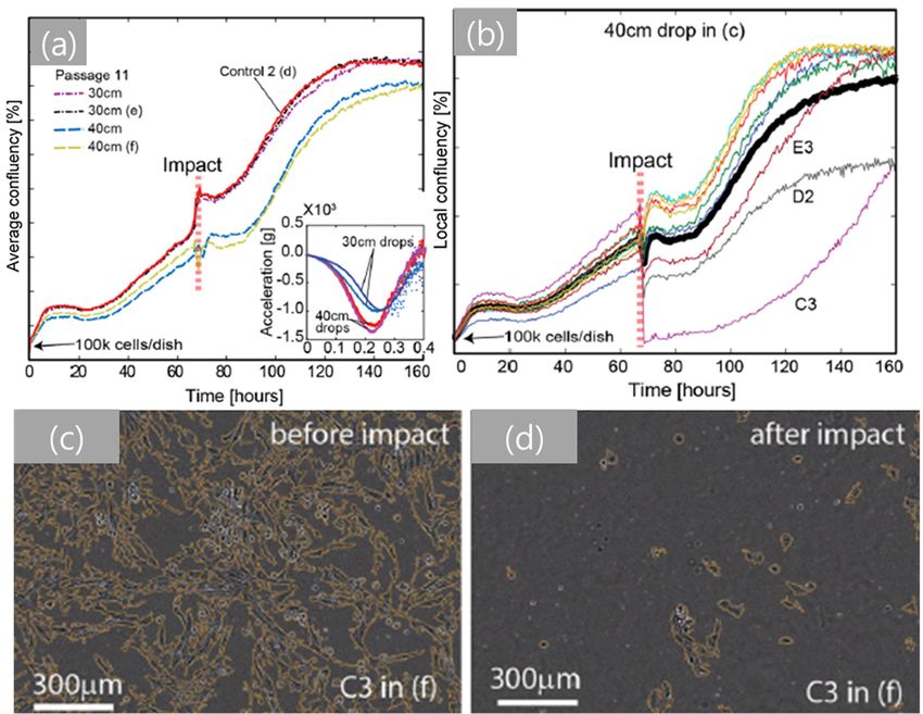
The on
its effect drop-tower-based
bubble dynamics system
duringhas been recently
mechanical modified for in vitro studies of live
impact.
cells The
[146]. The innovative experimental setup
drop-tower-based system has been recently modified allows the characterization of the experi-
for in vitro studies of live
mental
cells [146]. The innovative experimental setup allows the characterization ofstudy,
correlations between mechanical impact and cell damage/injury. This using
the experi-
fibroblast cells as a model, showed that input acceleration alone
mental correlations between mechanical impact and cell damage/injury. This study, using does not result in cell
damage,
fibroblastascells
shownas aby Figureshowed
model, 5. However, cell membrane
that input acceleration damage
alone and
doesa not
sudden
resultdecrease
in cell
in cell population
damage, as shown by were observed
Figure above cell
5. However, a material-dependent
membrane damage critical pressure
and a sudden value.
decrease
These results indicate that the critical pressure is associated with the
in cell population were observed above a material-dependent critical pressure value. These onset of cavitation
bubbles in a cellthat
results indicate culture chamber
the critical and that
pressure the dynamics
is associated withofthe
cavitation
onset of bubbles
cavitation inbubbles
the cham-in
ber induces localized compressive pressure cycles that significantly
a cell culture chamber and that the dynamics of cavitation bubbles in the chamber induces damage cells. This
innovative technique could
localized compressive leadcycles
pressure to new thatscientific findings
significantly on impact-induced
damage cellular
cells. This innovative
pathways that may
technique could leadtrigger
to new uncontrolled cell death
scientific findings (e.g., necrosiscellular
on impact-induced and apoptosis).
pathwaysSuch that
findings
may trigger would be an important
uncontrolled step(e.g.,
cell death towards innovative
necrosis technicalSuch
and apoptosis). advances in designing
findings would be
effective
an importantprotective equipment
step towards and new
innovative biomedical
technical technology
advances for post-injury
in designing treatment
effective protective
of Service members.
equipment and new biomedical technology for post-injury treatment of Service members.
Figure 5.
Figure 5. Characterization of cell
Characterization of cell injury
injury depending
depending on
on drop
drop height
height using
using Hs27
Hs27 fibroblasts
fibroblasts (x-axis:
(x-axis: time
time and
and y-axis:
y-axis: average
average
confluency). (a) The average confluency graph for 30 and 40 cm drops. (b) The local confluency graph for 40 cm drop. (c,d)
confluency). (a) The average confluency graph for 30 and 40 cm drops. (b) The local confluency graph for 40 cm drop.
Live cell images during 40 cm drop (c) before and (d) after impact [146].
(c,d) Live cell images during 40 cm drop (c) before and (d) after impact [146].Life 2021, 11, 546 12 of 17
In contrast to other methods such as AIC and LIC, the drop-tower-based system can
correlate a physical or mechanical property, i.e., acceleration or its gradients, to cavitation
nucleation in different types of soft matters. This capability to directly correlate the onset of
cavitation with acceleration would be quite helpful to understanding the underlying injury
mechanism of biomaterials that are known to be sensitive to strain rates. The drop weight
impact test is currently the only method that allows control of the input acceleration profile.
This unique feature is crucial to revealing blunt injury mechanisms as one can mimic exact
acceleration profiles for actual blunt injuries and study the biological responses of live
cells or tissues. Furthermore, this integrated system is coupled with a sample holder and
high-speed cameras to avoid direct contact between the biological sample and the impactor
while optically observing the real-time material deformation of soft gels. Despite the key
findings of cellular damage at the population level correlated with changes in transient
acceleration and the following bubble growth [146], there are still remaining questions,
i.e., how this system can be used to analyze deformation and damage of individual cells
during impact loading. Therefore, an effort to add high-resolution real-time imaging
techniques, i.e., single-cell-level observations, to the current drop-tower-based system for
in vitro studies would be critical to gain a fundamental understanding of the cavitation
damage mechanisms at the single-cell/subcellular level.
4. Conclusions
The characterization, analysis, and interpretation of cavitation within biological matter
are becoming inevitably important since they are increasingly relevant to medical applica-
tions such as lithotripsy, microsurgery, and medical imaging as well as to understanding
blunt injury mechanisms. Due to emerging interests, there has been rapid technical ad-
vancement in the field of cavitation in biological systems. In this regard, we have reviewed
cavitation in soft materials with an emphasis on biological implications of cavitation. First,
the two main theoretical frameworks (static and dynamic approaches) have been discussed.
Second, the experimental methods, i.e., needle-, laser-, and acoustically induced cavitation
and the integrated drop tower system, have been discussed and directly compared with
each other for different use cases and evaluated for their unique advantages and limitations.
Author Contributions: W.K., C.K., and W.J.C. outlined the manuscript and all authors wrote the
manuscript together. C.K., W.J.C., and W.K. performed literature review. C.K., Y.N. and W.K. edited
manuscript. All authors have read and agreed to the published version of the manuscript.
Funding: This research was funded by the Office of Naval Research (N00014-20-1-2409) and Arizona
State University’s Fulton Undergraduate Research Initiative.
Institutional Review Board Statement: Not applicable.
Informed Consent Statement: Not applicable.
Data Availability Statement: 3rd Party Data.
Acknowledgments: Not applicable.
Conflicts of Interest: The authors declare no conflict of interest.
References
1. Brennen, C.E. Cavitation and Bubble Dynamics; Cambridge University Press: New York, NY, USA, 2014.
2. Sass, W.; Bräunlich, M.; Dreyer, H.P.; Matura, E.; Folberth, W.; Preismeyer, H.G.; Seifert, J. The mechanisms of stone disintegration
by shock waves. Ultrasound Med. Biol. 1991, 17, 239–243. [CrossRef]
3. Sauerbruch, T.; Delius, M.; Paumgartner, G.; Holl, J.; Wess, O.; Weber, W.; Hepp, W.; Brendel, W. Fragmentation of gallstones by
extracorporeal shock waves. N. Engl. J. Med. 1986, 314, 818–822. [CrossRef] [PubMed]
4. Holmer, N.G.; Almquist, L.O.; Hertz, T.G.; Holm, A.; Lindstedt, E.; Persson, H.W.; Hertz, C.H. On the mechanism of kidney stone
disintegration by acoustic shock waves. Ultrasound Med. Biol. 1991, 17, 479–489. [CrossRef]
5. Chaussy, C.H.; Brendel, W.; Schmiedt, E. Extracorporeally Induced Destruction of Kidney Stones by Shock Waves. Lancet 1980,
316, 1265–1268. [CrossRef]Life 2021, 11, 546 13 of 17
6. Kaude, J.V.; Williams, C.M.; Millner, M.R.; Scott, K.N.; Finlayson, B. Renal morphology and function immediately after extracor-
poreal shock-wave lithotripsy. AJR Am. J. Roentgenol. 1985, 145, 305–313. [CrossRef] [PubMed]
7. Sokolov, D.L.; Bailey, M.R.; Crum, L.A. Use of a dual-pulse lithotripter to generate a localized and intensified cavitation field. J.
Acoust. Soc. Am. 2001, 110 Pt 1, 1685–1695. [CrossRef]
8. Ikeda, T.; Yoshizawa, S.; Tosaki, M.; Allen, J.S.; Takagi, S.; Ohta, N.; Kitamura, T.; Matsumoto, Y. Cloud cavitation control for
lithotripsy using high intensity focused ultrasound. Ultrasound Med. Biol. 2006, 32, 1383–1397. [CrossRef]
9. Pishchalnikov, Y.A.; Sapozhnikov, O.A.; Bailey, M.R.; Williams, J.C., Jr.; Cleveland, R.O.; Colonius, T.; Crum, L.A.; Evan, A.P.;
McAteer, J.A. Cavitation bubble cluster activity in the breakage of kidney stones by lithotripter shockwaves. J. Endourol. 2003, 17,
435–446. [CrossRef]
10. Johnsen, E.; Colonius, T. Shock-induced collapse of a gas bubble in shockwave lithotripsy. J. Acoust. Soc. Am. 2008, 124, 2011–2020.
[CrossRef]
11. Yoshizawa, S.; Ikeda, T.; Ito, A.; Ota, R.; Takagi, S.; Matsumoto, Y. High intensity focused ultrasound lithotripsy with cavitating
microbubbles. Med. Biol. Eng. Comput. 2009, 47, 851–860. [CrossRef] [PubMed]
12. Kennedy, P.K.; Hammer, D.X.; Rockwell, B.A. Laser-induced breakdown in aqueous media. Prog. Quantum Electron. 1997, 21,
155–248. [CrossRef]
13. Zysset, B.; Fujimoto, J.G.; Deutsch, T.F. Time-resolved measurements of picosecond optical breakdown. Appl. Phys. B 1989, 48,
139–147. [CrossRef]
14. Husseini, G.A.; Pitt, W.G. Micelles and nanoparticles for ultrasonic drug and gene delivery. Adv. Drug Deliv. Rev. 2008, 60,
1137–1152. [CrossRef] [PubMed]
15. Graham, S.M.; Carlisle, R.; Choi, J.J.; Stevenson, M.; Shah, A.R.; Myers, R.S.; Fisher, K.; Peregrino, M.B.; Seymour, L.; Coussios, C.C.
Inertial cavitation to non-invasively trigger and monitor intratumoral release of drug from intravenously delivered liposomes. J.
Control. Release 2014, 178, 101–107. [CrossRef]
16. Husseini, G.A.; Diaz de la Rosa, M.A.; Richardson, E.S.; Christensen, D.A.; Pitt, W.G. The role of cavitation in acoustically
activated drug delivery. J. Control. Release 2005, 107, 253–261. [CrossRef] [PubMed]
17. Li, F.; Yang, C.; Yuan, F.; Liao, D.; Li, T.; Guilak, F.; Zhong, P. Dynamics and mechanisms of intracellular calcium waves elicited by
tandem bubble-induced jetting flow. Proc. Natl. Acad. Sci. USA 2018, 115, E353–E362. [CrossRef]
18. Lo, C.-W.; Chen, S.-F.; Li, C.-P.; Lu, P.-C. Cavitation Phenomena in Mechanical Heart Valves: Studied by Using a Physical
Impinging Rod System. Ann. Biomed. Eng. 2010, 38, 3162–3172. [CrossRef] [PubMed]
19. Chao, T.-C.; Ros, A. Microfluidic single-cell analysis of intracellular compounds. J. R. Soc. Interface 2008, 5 (Suppl. 2), S139–S150.
[CrossRef]
20. Goldsmith, W. The state of head injury biomechanics: Past, present, and future: Part 1. Crit. Rev. Biomed. Eng. 2001, 29, 441–600.
[CrossRef] [PubMed]
21. Lubock, P.; Goldsmith, W. Experimental cavitation studies in a model head-neck system. J. Biomech. 1980, 13, 1041–1052.
[CrossRef]
22. Farjo, L.A.; Miclau, T. Ballistics and mechanisms of tissue wounding. Injury 1997, 28, C12–C17. [CrossRef]
23. Hu, J.; Lee, J.B.; Yang, K.H.; King, A.I. Injury patterns and sources of non-ejected occupants in trip-over crashes: A survey of
NASS-CDS database from 1997 to 2002. Annu. Proc. Assoc. Adv. Automot. Med. 2005, 49, 119–132.
24. Estrada, J.B.; Scimone, M.T.; Cramer, H.C.; Mancia, L.; Johnsen, E.; Franck, C. Microcavitation as a Neuronal Damage Mechanism
in an In Vitro Model of Blast Traumatic Brain Injury. Biophys. J. 2017, 112 (Suppl. 1), 159a. [CrossRef]
25. Wu, Y.-T.; Adnan, A. Effect of Shock-Induced Cavitation Bubble Collapse on the damage in the Simulated Perineuronal Net of the
Brain. Sci. Rep. 2017, 7, 5323. [CrossRef]
26. Kanagaraj, J.; Chen, B.; Xiao, S.; Cho, M. Reparative Effects of Poloxamer P188 in Astrocytes Exposed to Controlled Microcavitation.
Ann. Biomed. Eng. 2018, 46, 354–364. [CrossRef]
27. Estrada, J.B.; Barajas, C.; Henann, D.L.; Johnsen, E.; Franck, C. High strain-rate soft material characterization via inertial cavitation.
J. Mech. Phys. Solids 2018, 112, 291–317. [CrossRef]
28. Kang, W.; Adnan, A.; O’Shaughnessy, T.; Bagchi, A. Cavitation nucleation in gelatin: Experiment and mechanism. Acta Biomater.
2018, 67, 295–306. [CrossRef]
29. Kang, W.; Raphael, M. Acceleration-induced pressure gradients and cavitation in soft biomaterials. Sci. Rep. 2018, 8, 15840.
[CrossRef] [PubMed]
30. Jansen, L.E.; Birch, N.P.; Schiffman, J.D.; Crosby, A.J.; Peyton, S.R. Mechanics of intact bone marrow. J. Mech. Behav. Biomed. Mater.
2015, 50, 299–307. [CrossRef] [PubMed]
31. Zimberlin, J.A.; Sanabria-DeLong, N.; Tew, G.N.; Crosby, A.J. Cavitation rheology for soft materials. Soft Matter 2007, 3, 763–767.
[CrossRef] [PubMed]
32. Rayleigh, L., VIII. On the pressure developed in a liquid during the collapse of a spherical cavity. Lond. Edinb. Dublin Philos. Mag.
J. Sci. 1917, 34, 94–98. [CrossRef]
33. Beeching, R. Resistance to cavitation erosion. Trans. Instn. Engrs. Shipb. Scot. 1942, 85, 210–276.
34. Gilmore, F.R.; California Institute of Technology; Hydrodynamics Laboratory. The Growth or Collapse of a Spherical Bubble in a
Viscous Compressible Liquid; California Institute of Technology: Pasadena, CA, USA, 1952.You can also read