Molecular Regulations and Functions of the Transient Receptor Potential Channels of the Islets of Langerhans and Insulinoma Cells - MDPI
←
→
Page content transcription
If your browser does not render page correctly, please read the page content below
cells
Review
Molecular Regulations and Functions of the Transient
Receptor Potential Channels of the Islets
of Langerhans and Insulinoma Cells
Md. Shahidul Islam 1,2
 1    Karolinska Institutet, Department of Clinical Science and Education, Södersjukhuset, Research Center,
      5th floor, SE-118 83 Stockholm, Sweden; shahidul.islam@ki.se
 2    Department of Emergency Care and Internal Medicine, Uppsala University Hospital, Uppsala University,
      SE-751 85 Uppsala, Sweden

 Received: 2 February 2020; Accepted: 8 March 2020; Published: 11 March 2020

 Abstract: Insulin secretion from the β-cells of the islets of Langerhans is triggered mainly by nutrients
 such as glucose, and incretin hormones such as glucagon-like peptide-1 (GLP-1). The mechanisms
 of the stimulus-secretion coupling involve the participation of the key enzymes that metabolize the
 nutrients, and numerous ion channels that mediate the electrical activity. Several members of the
 transient receptor potential (TRP) channels participate in the processes that mediate the electrical
 activities and Ca2+ oscillations in these cells. Human β-cells express TRPC1, TRPM2, TRPM3, TRPM4,
 TRPM7, TRPP1, TRPML1, and TRPML3 channels. Some of these channels have been reported to
 mediate background depolarizing currents, store-operated Ca2+ entry (SOCE), electrical activity,
 Ca2+ oscillations, gene transcription, cell-death, and insulin secretion in response to stimulation
 by glucose and GLP1. Different channels of the TRP family are regulated by one or more of the
 following mechanisms: activation of G protein-coupled receptors, the filling state of the endoplasmic
 reticulum Ca2+ store, heat, oxidative stress, or some second messengers. This review briefly compiles
 our current knowledge about the molecular mechanisms of regulations, and functions of the TRP
 channels in the β-cells, the α-cells, and some insulinoma cell lines.

 Keywords: TRP channels of islets; TRP channels and insulin; TRPC1; TRPM2; TRPM3; TRPM4;
 TRPM5; TRPM7; TRPA1; TRP channels and GLP-1

1. Introduction
       The islets of Langerhans contain mainly the insulin-secreting β-cells, glucagon-secreting α-cells,
and somatostatin-secreting δ-cells [1]. Because of the difficulty in obtaining pure human β-cells,
it is common to use a variety of rodent insulinoma cells and glucagonoma cells for basic research in
this field. The β-cells secrete insulin in response to stimulation by nutrients such as glucose, amino
acids, and free fatty acids, neurotransmitters such as acetylcholine, and incretin hormones such as
glucagon-like peptide-1 (GLP-1) [2]. The molecular mechanisms of stimulus-secretion coupling in the
β-cells involve the intermediary metabolism of the nutrients in the cytoplasm and in the mitochondria,
the participation of some G protein-coupled receptors (GPCR), and many ion channels [2]. Crucial
events in the stimulus-secretion coupling are electrical activities, and increase in the concentration of
Ca2+ in the cytoplasm ([Ca2+ ]i ), in the form of spikes, bursts, and oscillations [3]. The electrical activities
and the [Ca2+ ]i oscillations are generated by concerted participation of a unique repertoire of ion
channels present in the β-cells [4]. These include different K+ channels, Ca2+ channels, Na+ channels,
Cl− channels, volume-sensitive anion channels, hyperpolarization-activated cyclic nucleotide-gated
channels, store-operated Ca2+ entry (SOCE) channels, and the transient receptor potential (TRP)

Cells 2020, 9, 685; doi:10.3390/cells9030685                                          www.mdpi.com/journal/cells
Cells 2020, 9, 685 2 of 22
channels [4]. It is not meaningful to debate which of these ion channels are more important than the
others. Study of the ion channels of the islets is important because of their roles in the secretion of the
hormones and in the impairment of such secretions in the pathogenesis of diabetes mellitus, which is
a major public health problem.
Many studies have demonstrated the presence of different TRP channels in the islets,
different insulinoma cells, and glucagonoma cells by different methods including functional studies
using pharmacological tools, RT-PCR, RNA-sequencing, Western blot, immuno-histochemistry,
immuno-fluorescence, and electrophysiology. Interpretation of some of the results, especially those
obtained by antibody-based methods may be difficult. Demonstration of expression of TRP channels
at RNA or protein level does not necessarily mean that they are translocated to the plasma membrane
and form functional channels. Interpretation of the mRNA expression data can also be difficult.
For instance, mRNA level may be low while the protein level may still be high because of high RNA
degradation rate, but slow protein turnover rate. Similarly, mRNA level may be high, but the protein
level may be very low due to repression of translation. Moreover, the methods used for purification of
the e.g., fluorescence activated cell sorting (FACS) may alter the mRNA expression level. Some TRP
channels are expressed in rodent islets and rodent insulinoma or glucagonoma cell lines but are almost
absent in human islets.
During recent years, interest in the understanding the roles of the TRP channels in the physiology
and pathology of islets in the context of diabetes has increased [5]. The availability of newer
pharmacological tools and knockout mouse models have enabled the investigators elucidate the
regulations and the functions of these channels in the islets. Today, our knowledge about these channels
in the islets is substantially more than that about a decade back [6]. This review describes the essential
background information, and the recent advances in our understanding of the regulation of these
channels, and their roles in mediating β-cell functions.
2. TRPC1
Among the channels of the TRPC family, only TRPC1 can be detected at mRNA level in human
β-cells (Figure 1) [7]. TRPC1 mRNA can be detected in mouse islets [8,9], MIN6 cells [8,9], rat islets [10],
rat beta cells [11], and INS-1 cells [10–12]. Rat primary β-cells express more TRPC1 compared
to the INS-1 cells [11]. In MIN6 cells and mouse islets, four splice variants of TRPC1 have been
identified, the β-variant being the most abundant one [9]. Protein kinase C (PKC) is important for
glucose-stimulated insulin secretion. In INS-1E cells, glucose increases insulin secretion by stimulation
of PKCα, which induces phosphorylation of TRPC1 [12].
SOCE plays an important role in mediating insulin secretion [13]. In rat β-cells, TRPC1 and
Orai1 form the non-selective cation channel that mediates SOCE and is regulated by STIM1 [10].
Orai1-mediated Ca2+ entry stimulates recruitment of TRPC1 into the plasma membrane. Orai1 and
STIM1 form channels that are gated by STIM1 [14]. STIM1 gates TRPC1 by intermolecular electrostatic
interaction between the positively charged poly-lysine domain in the C-terminus of STIM1 with the
negatively charged aspartates in the TRPC1 [15]. SOCE is impaired in the β-cells obtained from patients
with type 2 diabetes (T2D) [16]. The human TRPC1 gene is located on the chromosome 3q23;the band
3q is associated with T2D [17,18]. Genetic polymorphisms of TRPC1 are associated with T2D and
its complications in some populations [19]. In the Han Chinese population, the SNP rs7638459 has
been suspected as a risk factor for T2D without diabetic nephropathy. The CC genotype of rs7638459
significantly increases risk compared with the TT genotype. In the same population another SNP,
rs953239, is protective against development of nephropathy in T2D [19]. The CC genotype of rs953239
significantly reduces the risk of getting T2D without nephropathy compared to the AA genotype [19].
3. TRPC2, TRPC3, TRPC4, TRPC5 and TRPC6
In humans, TRPC2 is a pseudogene and the protein is not expressed in human cells. TRPC2 is
present in mouse insulinoma MIN6 cells [8].Cells 2020, 9, 685 3 of 22
TRPC3 is expressed in mouse and rat β-cells where it is triggered upon activation of some GPCRs.
Activation of the G protein-coupled receptor 40 (GPR40) of rat β-cells by fatty acids potentiates
glucose-induced insulin secretion. Activation of the GPR40 activates the TRPC3; this is mediated by
activation of phospholipase C and the PKC pathway [20]. Activation of the TRPC3 channel induces
a non-selective cation current that leads to depolarization of the membrane potential of the β-cell [20].
TRPC3 also plays a role in the development and proliferation of β-cells. The transcription factor
pancreatic and duodenal homeobox 1 (Pdx-1) increases proliferation of islet cells partly by upregulating
the expression of TRPC3 and TRPC6, and also by increasing the activity of these channels [21].
TRPC4 is not expressed in human β-cells (Figure 1) [7] but is expressed in rat and mouse primary
β-cells and insulinoma cells (Table 1). At least two major isoforms of TRPC4 are known. TRPC4β
lacks 84 amino acids in the C-terminus and TRPC4α is the full-length form. In INS-1 cells, TRPC4α is
the main isoform, whereas in rat β-cells TRPC4β is the main isoform [11]. In βTC3 cells, TRPC4 is
activated by store depletion, and this activates a non-selective cation current, which contributes to the
generation of glucose-stimulated oscillations of membrane potential and [Ca2+ ]i [8].
Leptin signaling activates the TRPC4 channel of β-cells through phosphorylation of the channel
by phosphoinositide 3-kinase [22]. Reversible histidine phosphorylation plays an important role in the
activation of the TRPC4 channel of β-cells. In INS-1 cells, protein histidine phosphatase 1 (PHPT-1)
activates TRPC4 by dephosphorylating a histidine residue in the C-terminus of the channel [23].
Activation of TRPC4 increases [Ca2+ ]i , which leads to the translocation of the ATP-sensitive K+ (KATP )
channels to the plasma membrane. In Phpt-1−/− mice, TRPC4 is inhibited and the KATP current
in the plasma membrane is decreased. Consistent with these observations, the Phpt-1−/− mice are
hypoglycemic during the perinatal period [23].
Small GTPase Rasd1 activates TRPC4β, but not TRPC4α. Glucocorticoids increase Rasd1 in INS-1
cells, and by that way increase the TRPC4 current in these cells [24].
4. TRPM2
Early studies reported a non-selective Ca2+ -permeable cation channel activated by β-NAD+ ,
H2 O2 , and alloxan, and inhibited by AMP, in CRI-G1 rat insulinoma cells [25,26]. The current described
has characteristics of TRPM2, e.g., unusually long single channel open times, linear current-voltage
relationship, and the requirement of cytoplasmic Ca2+ for activation of the current [26,27]. Later on,
it was demonstrated that the CRI-G1 cells express TRPM2 at a high level and adenosine 50 -diphosphate
ribose (ADPR) activates the characteristic TRPM2 current [28]. Other rodent insulinoma cells that
express TRPM2 are the INS-1E cells [29], the RIN-5F cells [30–32], and theHIT-T15 cells [33]. Primary
mouse [31,34], rat [31], and human β-cells [7,29,35] express the TRPM2 channel. The α-cells do not
express TRPM2 [31]. Human islets express two isoforms of the TRPM2 channel: the full length or
the long form of the channel (TRPM2-L) and a short form (TRPM2-S) where the four C-terminal
transmembrane domains, the pore region, and the entire C-terminus are truncated [29]. TRPM2-S,
which does not form a channel, acts as a dominant negative of TRPM2-L [36]. The relative proportion
of the two isoforms may determine the extent of the TRPM2-mediated Ca2+ influx.
ADPR together with the co-agonist Ca2+ activates TRPM2 [37]. The NUDT9 homology (NUDT9-H)
domain of human TRPM2 plays crucial roles in mediating expression of the channel in the plasma
membrane and in channel gating. ADPR binds to both the NUDT9-H domain and the TRPM homology
regions (MHR) 1 and 2 (MHR1/2) [38]. The NUDT9-H domain of human TRPM2 binds ADPR and
promotes channel opening, but does not degrade ADPR. Channel opening also requires binding of Ca2+
to the transmembrane domains [38]. 8-Br-cADPR binds only to the MHR1/2 domain and stabilizes the
channel at the resting state.
Cyclic ADP ribose (cADPR) can also directly activate human TRPM2, but the EC50 of cADPR for
activation of TRPM2 is much higher than that of ADPR. This is in spite of the fact that the binding
affinity of cADPR to the NUDT9-H domain is higher than that of ADPR [39]. cADPR binds to the
same pocket of NUDT9-H that binds ADPR, but the interaction pattern of ADPR and cADPR with theCells 2020, 9, 685 4 of 22
binding sites are different [39]. It should be noted that some batches of commercially available cADPR
contain 25–50% ADPR as contaminant. Activation of TRPM2 by cADPR reported in some papers may
partly be due to the contaminant ADPR [40].
ADPR is the best known agonist of TRPM2, but a recent study reports that 20 -deoxy-ADPR is the
most efficient endogenous agonist of the channel [41]. Compared to ADPR, 20 -deoxy-ADPR produces
10-fold more TRPM2 current. 20 -deoxy-ADPR is thought to be produced by CD38 from cytosolic
2’-deoxy-NAD. It has been speculated that 20 -deoxy-ADPR is the principal agonist of TRPM2 for
mediating the physiological signaling functions [41]. ADPR-2’-phosphate is a partial agonist of TRPM2
that activates TRPM2 with reduced efficacy [40,41].
Activation of the TRPM2 by reactive oxygen species (ROS) is generally attributed to the formation
of ADPR by the actions of poly(ADP-ribose) polymerase (PARP) and Poly(ADP-ribose) glycohydrolase
(PARG) [42]. DNA damage activates PARP leading to the synthesis of poly (ADP-ribose) (PAR). PARG
catalyzes the degradation of PARs to yield free ADPR, which activates TRPM2 leading to increase in
the [Ca2+ ]i and cell death [43].
N-(p-amylcinnamoyl) anthranilic acid, an inhibitor of TRPM2, inhibits H2 O2 -induced Ca2+
increase in the INS-1E cells [29]. Newer, more potent inhibitors of TRPM2 are curcumin, JNJ-28583113,
some derivatives of 2,3-dihydroquinazolin-4(1H)-one, scalaradial, and 12-deacetylscalaradial [44–47].
JNJ-28583113 inhibits human TRPM2 with IC50 of 126 nM, scalaradial inhibits with IC50 of 210 nM,
and the most potent derivative of 2,3-dihydroquinazolin-4(1H)-one inhibits with IC50 of 3.7 µM.
4.1. Role of the TRPM2 Channel in Stimulus-Secretion Coupling
The permeability ratio pCa :pCs of TRPM2 is low (~0.54), but the permeability for Ca2+ increases
(pCa :pNa = 5.83) when the channel is activated by heat, and activation of the channel increases
[Ca2+ ]i [28,29,31]. Ca2+ activates TRPM2 and its alternatively spliced isoforms, including the ones
that do not bind ADPR [33,37]. In the presence of ADPR, TRPM2 behaves like a Ca2+ -activated
channel [33,48]. Extracellular Ca2+ entering through the TRPM2 channel activates the channel by
binding to the Ca2+ -binding sites located in the vicinity of the pore region. This mechanism prolongs
the activation of the channel in a self-sustained manner [33,48]. Receptor-activation-induced Ca2+
release can also activate the TRPM2 channel [33].
TRPM2 is involved in mediating insulin secretion in response to stimulation by glucose [49,50].
Glucose-stimulated TRPM2 current and insulin secretion are reduced in rat β-cells transfected with
shTRPM2 RNA indicating that TRPM2 is involved in the coupling process [51]. GLP-1-induced
insulin secretion is increased in transfected β-cells where TRPM2 is overexpressed, and this increase is
inhibited by 2-aminoethyl diphenyl borate. GLP-1, at nanomolar concentrations, activates the TRPM2
channel through the cAMP-Epac pathway [50–52]. In addition, cADPR is also involved in the activation
of the TRPM2 by glucose and GLP-1 [31,53]. GLP-1 releases Ca2+ from the intracellular store—it is
possible that this may be partly due to the release through the TRPM2, since TRPM2 channels are
also present on the membrane of the Ca2+ stores [34,53]. TRPM2 knockout mice have higher blood
glucose levels. Ca2+ -response and insulin secretion upon stimulation by glucose and nanomolar
concentrations of GLP-1 is impaired in the β-cells obtained from TRPM2 knockout mice [50]. The role
of TRPM2 in stimulating insulin secretion by picomolar concentrations of GLP-1 has not been reported.
Low concentration of adrenaline inhibits glucose- and GLP-1-induced insulin secretion by activating
the α2A adrenoceptor, inhibiting cAMP signaling and thereby inhibiting the TRPM2 channel [54].
Nanomolar concentrations of ghrelin inhibit glucose-induced insulin secretion by inhibiting cAMP
formation and thereby reducing the TRPM2 current [55].
Acidic cytoplasmic pH inhibits TRPM2 and such inhibition is abolished by high pH [56]. In this
context, it is noteworthy that stimulation of β-cells by glucose increases cytoplasmic pH [57], which is
supposed to favor the activation of the TRPM2 by ADPR and Ca2+ .Cells 2020, 9, 685 5 of 22
4.2. Heat as a Regulator of TRPM2
TRPM2 is a thermosensitive TRP channel (Q10 = 15.6). In the presence of ADPR the Q10 value
increases to 44. The temperature threshold and the temperature for optimal activity are ~34 ◦ C and
~37 ◦ C respectively. At the physiological body temperature, the TRPM2 channels are constitutively
active contributing to the background depolarizing current. β-cells are rich in mitochondria, and it is
known that stimulation of β-cells by glucose generates heat, which may possibly increase the local
temperature and increase the activity of the TRPM2 channel [58,59]. Heat-evoked increase in [Ca2+ ]i
in mouse β-cells is abolished in TRPM2 knockout mice [50]. The steep temperature dependence of
glucose-induced insulin secretion may partly be mediated by the temperature sensitivity of the TRPM2
channels of β-cells [60]. It is noteworthy that β-cells have other TRP channels including TRPM4,
TRPM5, TRPV1, TRPV2, and TRPV4 that are temperature sensitive. Although there is much skepticism,
it has been demonstrated in other systems that temperatures inside the cells can increase dramatically,
but it remains unclear whether such increases in the temperature have any signaling functions [61].
4.3. TRPM2 and β-Cell Death
Oxidative stress increases the concentration of ADPR and Ca2+ in the cytoplasm, which synergize
to activate TRPM2 and increase Ca2+ influx [29,62]. In rat insulinoma cell lines, H2 O2 and TNFα cause
cell death, which can be inhibited by treatment with antisense-TRPM2 [30,34,63]. Alloxan-induced
β-cell death is probably mediated by Ca2+ influx through the TRPM2 channel [26]. Human β-cells
express a short isoform of TRPM2 (TRPM2-S) [29], which does not form a channel, but inhibits the full
length isoform of the channel (TRPM2-L), and by that way inhibits cell death [36,64]. This could possibly
be one of the many reasons why human β-cells are relatively resistant to alloxan [65]. Production
of reactive oxygen species by free fatty acids and cytokines causes β-cell death by mechanisms that
involve TRPM2 [66]. It is possible that TRPM2 provides the β-cells a mechanism to undergo apoptosis
when they are severely damaged by oxidative stress [67].
TRPM2 plays a role in mediating free fatty acid- and cytokine-induced β-cell death [66]. Palmitate
activates NADPH-oxidase-2 and thereby generates reactive oxygen species leading to the activation of
TRPM2. This leads to an increase in the concentration of Zn2+ by activation of the TRPM2 channels
located on the lysosomes [34,68]. This, in turn, leads to an increase in the concentration of Zn2+ in the
mitochondria, and recruitment of dynamin-related protein Drp-1 to mitochondria, which catalyzes
mitochondrial fission, loss of mitochondrial membrane potential, and mitochondrial fragmentation [66].
TRPM2 knockout mice are resistant to β-cell loss and hyperglycemia caused by multiple low dose
streptozotocin [68].
4.4. TRPM2 Channels Located on the Intracellular Membranes
In β-cells TRPM2 channels are located also on the membranes of the lysosomes. Activation of
these TRPM2 channels releases Ca2+ from the lysosomal Ca2+ store [34]. Ca2+ released from lysosomal
stores externalizes phosphatidyl serine, a distinct feature of apoptosis [69].
Activation of the lysosomal TRPM2 channel also releases Zn2+ from the lysosomal stores. It is
possible that Zn2+ , rather than Ca2+ , plays a more important role in mediating apoptosis of β-cells [68].
TRPM2 mediates free fatty acid- and cytokine-induced β-cell death by releasing Zn2+ from the
intracellular stores, and by increasing the concentration of Zn2+ in the mitochondria [66].
5. TRPM3
TRPM3 has numerous isoforms generated by alternative splicing. The TRPM3α2 isoform is more
permeable for Ca2+ and Mg2+ than the TRPM3α1, which is more monovalent cation selective [70].
Extracellular monovalent cations inhibit the channel. TRPM3α2 is present in mouse β-cells and is
absent in α-cells [71]. TRPM3 is present also in human β-cells but it is not known which isoforms of
TRPM3 are present in these cells (Figure 1) [7].Cells 2020, 9, 685 6 of 22
TRPM3 channel can be activated by extracellular application of micromolar concentrations
of pregnenolone sulphate, an endogenous steroid, which is also able to change activity of several
other ion channels. A more potent synthetic activator of TRPM3 is 3,4-dihydro-N-(5-methyl-3-
isoxazolyl)-a-phenyl-1(2H)-quinolineacetamide (CIM0216) (pEC50 0.77 µM) [72]. Activation of the
TRPM3 by pregnenolone or CIM0216 increases [Ca2+ ]i and stimulates insulin secretion, and these
effects are lost in the islets obtained from Trpm3−/− mice [71–73]. TRPM3 is permeable to both Ca2+
and Na+ (pCa :pNa = 1.57). It is possible that pregnenolone sulphate depolarizes the β-cell first by
inducing a Na+ current through the TRPM3 channel, and the resulting depolarization activates the
voltage-gated Ca2+ channels. In INS-1 cells and mouse islets, mefenamic acid selectively inhibits the
[Ca2+ ]i -increase triggered by pregnenolone sulphate, but not that triggered by glucose [73].
Pregnenolone sulphate-induced activation of TRPM3 increases the expression of the transcription
factor Egr-1 which binds to the regulatory region of the transcription factor Pdx-1 gene leading to
increased insulin gene transcription [74].
TRPM3 may be involved in mediating activation of the β-cells by insulin secretagogues
in several ways. TRPM3 shows constitutive activity [75], and by that way could provide the
background depolarizing current necessary for membrane depolarization and electrical activity of
the agonist-stimulated β-cells. Like several other channels, the TRPM3α2 channel is also positively
regulated by phosphatidylinositol 4,5-biphosphate (PIP2 ) [76]. It is conceivable that the glucose-induced
increase in the concentration of PIP2 in the plasma membrane of β-cells [77] favors activation of the
channel by unidentified agonists. TRPM3 is also a thermosensitive channel [72]—it is conceivable that
glucose-induced heat production [59] could promote increased activity of the TRPM3 channel.
The physiological importance of TRPM3 in human β-cells remains unclear. The channel is
also highly permeable for Zn2+ ions and β-cells take up Zn2+ through this channel even when the
concentration of Zn2+ in the extracellular solution is low [78]. In Trpm3−/− mice, fasting blood glucose
is normal and they appear generally healthy [79]. This is not surprising given that β-cells have many
other TRP channels, which could possibly compensate for the loss of the TRPM3 channel in the
knockout mice. It will be useful to know if these mice develop signs of pre-diabetes or diabetes when
put on a high fat diet. Primidone, a medicine used in the treatment of epilepsy and essential tremor,
inhibits TRPM3 with an IC50 of 0.6 µM [80]. This drug does not cause impaired glucose tolerance,
pre-diabetes, or diabetes, suggesting that TRPM3 is not essential for insulin secretion.
6. TRPM4
TRPM4 is a nonselective monovalent cation channel impermeable to divalent cations [81]. It is
activated by Ca2+ , followed by a fast desensitization to activation by Ca2+ . In CRI-G1 rat insulinoma
cells, a ~25 pS TRPM4-like current activated by Ca2+ was first described by Sturges et al. in 1986 [82].
It was inhibited by different adenine nucleotides but the potency sequence for inhibition (AMP >
ADP > ATP > adenosine) was different from that for the inhibition of the cloned TRPM4 expressed in
heterologous systems (ADP > ATP > AMP >> adenosine) [83]. Leech and Habener reported a ~25 pS
nonselective cation current activated by Ca2+ and inhibited by ATP in HIT-T15 cells [84]. This current,
which is also activated by GLP-1, appears to be mediated by TRPM4.
There are at least three isoforms of TRPM4: the full length TRPM4 (TRPM4b), an N-terminal
174 amino acid deletion isoform (TRMM4a), and an isoform lacking 537 amino acids (TRPM4c) [85].
It is not known which of these are expressed in the β-cells. TRPM4 protein has two ABC transporter
signature-like motifs, and four nucleotide binding domains. Consistent with these, TRPM4 is inhibited
by the sulphonylurea drug glibenclamide in some cells [86]. The TRPM4-like channel of the β-cells is
not inhibited by glibenclamide [87]. TRPM4 is also inhibited by cytoplasmic ATP and other adenine
nucleotides without requiring Mg2+ . While ATP inhibits TRPM4, it also inhibits the desensitization
of the channel by Ca2+ [88]. TRPM4 has phosphorylation sites for protein kinase A (PKA), PKC,
and binding sites for PIP2 . Consistent with these, TRPM4 is regulated by PKC and PIP2 [88,89]. PKC
phosphorylation enhances the sensitivity of the TRPM4 for activation by Ca2+ . Different PKC isoformsCells 2020, 9, 685 7 of 22
regulate β-cell functions [43]. PIP2 moves the voltage-activation curve of TRPM4 towards negative
voltages and prevents desensitization of the channel by Ca2+ [88]. It is noteworthy that stimulation
of the β-cells by glucose increases the concentration of PIP2 in the plasma membrane. This is likely
to promote inward depolarizing currents through the TRPM4 and activation of the voltage-gated
Ca2+ channels [77,90].
TRPM4 is present in human β-cells [7,91], rodent islets, and a variety of rodent insulinoma cells
(Table 1). It is also present in α-cells, where it plays a role in mediating glucagon secretion [92].
TRPM4 is activated by an elevated [Ca2+ ]i (EC50 ~ 0.57–1.25 µM). Activation of the TRPM4 current
by [Ca2+ ]i is biphasic with a first phase that develops within seconds, and a second phase that
develops slowly. The latter phase appears to be due to incorporation of TRPM2 into the plasma
membrane following exocytosis [91,93]. TRPM4 is involved in mediating agonist-induced insulin
secretion [91,93,94]. Suppression of the TRPM2 by a dominant negative construct inhibits the magnitude
of the [Ca2+ ]i -increase and insulin secretion [91,93]. Inhibition of the TRPM4 by 9-Phenanthrol inhibits
glucose- and GLP-1-stimulated insulin secretion from rat islets [94].
TRPM4 is involved in mediating stimulation of insulin-secretion by picomolar concentrations of
GLP-1 [94,95]. Picomolar concentrations of GLP-1 activate TRPM4 and TRPM5 through activation
of PKC leading to extracellular Na+ -dependent membrane depolarization [95]. PKC-dependent
phosphorylation of TRPM4 increases the sensitivity of the channel to activation by Ca2+ [88]. Islets
obtained from the Trpm4-/- mice respond normally to stimulation by glucose, but not to stimulation by
picomolar concentrations of GLP-1 [95,96].
TRPM4 is present in the glucagon-secreting α-TC1-6 cells and INR1G9 cells [91,92]. [Ca2+ ]i
activates TRPM4-like Na+ current in the α-TC1-6 cells. Agonist-induced Ca2+ -response and glucagon
secretion is reduced in cells where TRPM4 is knocked down by TRPM4 shRNA [92].
7. TRPM5
TRPM5, which is closely related to TRPM4, is another nonselective cation channel activated by
[Ca2+ ]I [81]. Unlike TRPM4, TRPM5 is not inhibited by adenine nucleotides or glibenclamide. TRPM5
is abundant in the taste buds and is best known for its role in mediating taste signaling. It is expressed,
together with the TRPM4, in rodent islets and rodent insulinoma cell lines (Table 1). In human islets,
TRPM5 is almost absent in the β-cells, but it is expressed in the non-β-cells of the islet (Figure 1) [7].
However, the RNA-sequencing data were obtained from only two preparations of human β-cells and
should therefore be interpreted with some caution [7].
In mice, TRPM5 is involved in mediating the glucose-induced oscillations in the membrane
potential, and [Ca2+ ]i . In Trpm5−/− mice the frequency of the glucose-induced fast oscillations in the
membrane potential, and the fast oscillations in [Ca2+ ]i are reduced. TRPM5 contributes a depolarizing
current during an inter-burst interval to change the membrane potential to the threshold for starting
a new burst activity. It reduces the inter-burst interval and increases the amplitude and frequency
of the membrane depolarizations and action potentials [97]. Consistent with these, glucose-induced
insulin secretion is reduced in the Trpm5−/− mice, and these mice have impaired glucose tolerance [98].
In in vitro experiments, insulin secretion in response to glucose from islets isolated from Trpm5−/−
remains normal, but insulin secretion in response to GLP-1 becomes impaired [95]. Impairment of
glucose-induced insulin secretion and glucose intolerance observed in in vivo experiments using
Trpm5−/− mice could partly be due to the inability of GLP-1 to trigger the downstream signals in
the β-cells.
Factors that couple glucose stimulation to the activation of TRPM5 may include a glucose-induced
increase in the membrane potential, [Ca2+ ]i , concentration of cytoplasmic arachidonic acid [99,100],
and the concentration of PIP2 [77,101]. GLP-1-induced stimulation of insulin secretion is coupled to
the activation of the TRPM5 channels by PKC [95]. TRPM5 activators improve insulin secretion from
mouse islets. Steviol glycosides potentiate Ca2+ -dependent activity of the TRPM5 channel, and by thatCells 2020, 9, 685 8 of 22
way improve glucose-induced insulin secretion, and prevent high-fat-diet-induced hyperglycemia
in mice [102].
Wolfram syndrome (diabetes insipidus, diabetes mellitus, optic atrophy, DIDMOAD) caused
by mutation in the Wolframin gene (WFS1), is an autosomal recessive disorder. In Wfs1−/− mice the
number
Cells 2020, 9, xof islets in the pancreas is reduced, and insulin secretion from the individual islets8 is of also
22
reduced. In these islets the trpm5 gene is downregulated [103].
Sweet-taste
Sweet-taste receptors
receptors are
arepresent
presentnot notonly
onlyininthe
thetongue
tonguebutbutalso
alsoininthe
theβ-cells.
β-cells.These
TheseGPCRs
GPCRs areare
heterodimers
heterodimers ofof
T1R2
T1R2 (taste receptor
(taste receptor type
type1 member
1 member 2) 2)
and T1R3
and T1R3(taste
(tastereceptor
receptortype 1 member
type 1 member 3).3).
Fructose
Fructose stimulates
stimulates insulin
insulin secretion
secretionbybyactivating
activatingthe thesweet-taste
sweet-tastereceptors
receptorsofofthe theβ-cells.
β-cells.TRPM5
TRPM5
mediates
mediates the effects
the effectsofofactivation
activationofofthe thesweet-taste
sweet-tastereceptor
receptorininmouse
mouseislets
islets[104]. Trpm5−/−−/−
[104].Trpm5 mice lack
mice lack
sweet-taste
sweet-taste preference.
preference.These
Thesemice
micegain
gainless weight
less weight when
when put onon
put a high-calorie
a high-calorie diet, and
diet, andtheir glucose
their glucose
tolerance
tolerance remains
remains better
better than
thanthat
thatofofthe
thewild
wildtype
typemice
mice[105−106].
[105,106].
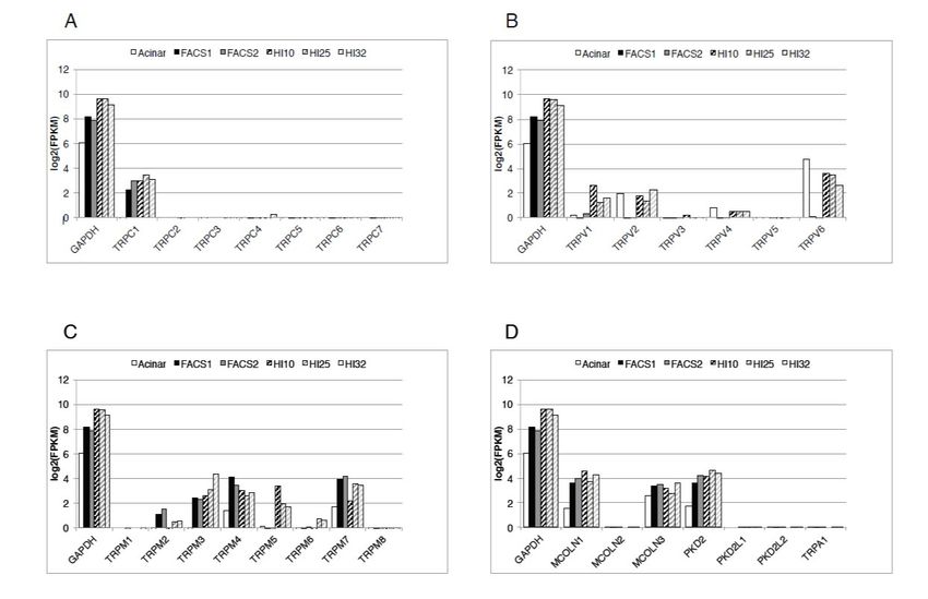
Figure 1. Expression of the transient receptor potential (TRP) channels in human β-cell and human
Figure 1. Expression of the transient receptor potential (TRP) channels in human β-cell and human
islets. Expression levels are shown as bar plots on a log2 (FPKM) scale. The bars in each group represent
islets. Expression levels are shown as bar plots on a log2(FPKM) scale. The bars in each group
(from left to right) human pancreatic acinar cells; purified human β-cells preparations 1 and 2 (FACS1,
represent (from left to right) human pancreatic acinar cells; purified human β-cells preparations 1 and
FACS2); and human islet preparations HI10, HI25, and HI32. Relative levels of expressions of the
2 (FACS1,
channelsFACS2); and human
of the TRPC isletTRPV
family (A), preparations HI10,
family (B), HI25,
TRPM and HI32.
family Relative
(C), and levels of expressions
the remaining TRP channels
of(TRPA1,
the channels of the TRPC family (A), TRPV family (B), TRPM family (C),
members of the TRPP, and TRPML families) (D), are shown. GAPDH expression and the remaining TRPfor
is shown
channels (TRPA1, members of the TRPP, and TRPML families) (D), are shown. GAPDH
comparison. A log2 (FPKM) = 0 was considered as a minimum threshold for expression. MCOLN1 expression is =
shown for comparison. A log 2(FPKM) = 0 was considered as a minimum threshold for expression.
TRPML1, MCOLN2 = TRPML2, MCOLN3 = TRPML3, PKD2 = TRPP1, PKD2L1 = TRPP2, PKD2L2 =
MCOLN1
TRPP3. FPKM= TRPML1, MCOLN2
= Fragments Per=Kilobase
TRPML2,Million.
MCOLN3 = TRPML3,
Reproduced PKD2
with = TRPP1,
permission PKD2L1
from = TRPP2,
[7], Marabita and
PKD2L2 = TRPP3.
Islam, 2017. FPKM = Fragments Per Kilobase Million. Reproduced with permission from [7],
Marabita and Islam, 2017.
Insulin downregulates TRPM5 in the islets [107]. Hyperinsulinemia reduces mRNA expression of
Insulin
Trpm5 downregulates
in the islets of mouse TRPM5
modelsin of
theobesity
islets [107]. Hyperinsulinemia
and diabetes reduces
(ob/ob and db/db mRNA
mice) [107].expression
In humans,
ofsome
Trpm5genetic
in the islets of mouse
variations models
within the of obesitylocus
TRPM5 and diabetes (ob/ob and
are associated db/db
with mice) [107].
impaired In humans,
insulin secretion,
some genetic variations within the TRPM5 locus are associated with impaired insulin
increased plasma glucose concentration, reduced concentration of GLP-1, and decreased insulin secretion,
increased plasma
sensitivity glucose
[108,109]. concentration,
In the white German reduced concentration
population, of GLP-1,isand
the SNP rs2301699 decreasedassociated
significantly insulin
sensitivity [108−109]. In the white German population, the SNP rs2301699 is significantly
with glucose-stimulated insulin secretion in women. The minor allele carriers of the SNPs rs800344, associated
with glucose-stimulated insulin secretion in women. The minor allele carriers of the SNPs rs800344,
rs800345, and rs2301699 show significantly higher glucose level during an oral glucose tolerance test
and show reduced insulin sensitivity [108]. In the Turkish population, the SNP rs4929982
polymorphism is associated with metabolic syndrome. In this population an increase in the A allele
and decrease in the G allele of rs4929982 polymorphism increase susceptibility to metabolicCells 2020, 9, 685 9 of 22
rs800345, and rs2301699 show significantly higher glucose level during an oral glucose tolerance test and
show reduced insulin sensitivity [108]. In the Turkish population, the SNP rs4929982 polymorphism is
associated with metabolic syndrome. In this population an increase in the A allele and decrease in the
G allele of rs4929982 polymorphism increase susceptibility to metabolic syndrome [109].
8. TRPM6 and TRPM7
TRPM7 (formerly called LTRPC7) is a “chanzyme” containing a serine-threonine α-kinase domain
on its intracellular C-terminus. It is a nonspecific divalent cation channel that is permeable to Ca2+ ,
Mg2+ , and Zn2+ . TRPM7 is constitutively active and by that way it provides a mechanism for
background entry of divalent cations into the cells. TRPM7 is one of the most abundant TRP channels
expressed in human β-cells (Figure 1) [7]. This is not surprising given that expression of TRPM7 is
almost ubiquitous. In mouse islets, expression of TRPM7 is eight times higher than that of TRPM6,
which is the other Mg2+ channel present in the mouse islets [110]. In human β-cells TRPM6 is not
expressed. TRPM7 is thought to regulate intracellular Mg2+ concentration. Deficiency of TRPM7
reduces total cellular Mg2+ at least in some cells [111]. Knockdown of Trpm7 in INS-1 cells by siRNA
increases insulin secretion in response to glucose [110]. It appears that TRPM7 plays a role in mediating
glucose-induced insulin secretion possibly by regulating the cytoplasmic Mg2+ concentration.
In some cells, TRPM7 is also present on some special intracellular vesicles (called M7 vesicles or
M7V) that contain Zn2+ [112]. It is not known whether such vesicles exist in β-cells. If so, TRPM7 of
β-cells could release Zn2+ from such stores in response to oxidative stress and could damage the β-cells.
TRPM7 provides a mechanism for entry of divalent trace metal ions into the cells with a permeability
sequence of Zn2+ ≈ Ni2+ >> Ba2+ > Co2+ > Mg2+ ≥ Mn2+ ≥ Sr2+ ≥ Cd2+ ≥ Ca2+ [113]. It is conceivable
that entry of toxic heavy metal ions through the TRPM7 channels of the β-cells may damage these
cells, leading to the development of diabetes [114]. There is no evidence that variations in the TRPM6
or TRPM7 genes are associated with T2D [115]. TRPM6 mRNA is expressed at a low level in mouse
islets but not in human β-cells [7,110].
9. TRPV1
Human islets and human insulinoma cells do not express TRPV1 [7,116]. TRPV1 is expressed in the
rat insulinoma INS-1E cells [116,117]. In the RINm5F cells, TRPV1 can be detected at the mRNA level,
but TRPV1 currents cannot be demonstrated [118]. According to most studies, primary rodent β-cells
do not express TRPV1 [116,118–120]. TRPV1 immunoreactivity has been demonstrated in primary rat
β-cells [121], but no TRPV1 current can be detected in these cells [118]. Some studies have shown that
the TRPV1 channel is involved in mediating insulin secretion from isolated rodent islets or β-cells, but
this is not a universal finding [118,121,122]. It is possible that capsaicin, an agonist of TRPV1, stimulates
insulin secretion from these cells by some non-specific mechanisms [118,121]. Glucose-induced insulin
secretion from isolated β-cells obtained from Trpv1−/− mice is not impaired [118].
TRPV1 is also expressed in the sensory neurons that innervate the pancreas and the islets of mice
and rats [119,120]. However, in adult human pancreas and islets, we cannot detect TRPV1-positive
neurons by immunohistochemistry [116]. In the non-obese diabetic (NOD) mice, these neurons appear
to control access of lymphocytes to the islets and play a role in the pathogenesis of autoimmune
diabetes [120]. Two missense mutations in the Trpv1 gene are associated with autoimmune diabetes in
these mice. In humans, the SNP rs222747 (M315I) variant of the TRPV1 gene is significantly increased
in the type 1 diabetic patients in an Ashkenazi Jewish population, suggesting that TRPV1 may be
a susceptible gene for type 1 diabetes in some ethnic groups [123].
In Trpv1−/− mice, both normal [118] and impaired [124] glucose tolerance upon intraperitoneal
injection of glucose has been reported. This difference can be due to the difference in the bodyweight
of the Trpv1−/− mice used in these studies [118,124]. TRPV1 of the sensory neurons that innervate
the pancreas and the islets participate in the regulation of insulin secretion through the release of
calcitonin-gene-related peptide (CGRP) and substance P [124]. In the Trpv1−/− mice, insulin secretionCells 2020, 9, 685 10 of 22
in response to intraperitoneal injection of glucose is reduced, and these mice have impaired glucose
tolerance [124]. It should be noted that the results obtained in Trpv1−/− mice are different from those
obtained in mice where TRPV1-positive nerve fibers are ablated by chemicals. Whole body denervation
of TRPV1-positive sensory neurons by capsaicin enhances glucose-induced insulin secretion in the
male mice [125,126]. CGRP and substance P can exert stimulatory or inhibitory effect on insulin
secretion depending on concentrations of the peptides, glucose concentration, and animal species [124].
High concentration of CGRP inhibits insulin secretion. Ablation of the TRPV1-positive neurons
increases insulin secretion by removing the inhibition.
Another mechanism by which TRPV1 increases insulin secretion involves the incretin hormone
GLP-1. Activation of the TRPV1 channels in the GLP-1-secreting L-cells in the ileum stimulates
GLP-1 secretion, and by that way increases insulin secretion in mice [127]. Chronic dietary capsaicin
increases plasma GLP-1 and lowers plasma glucose in the diabetic db/db mice [127]. In animal
experiments, activation of TRPV1 by pharmacological agents stimulates insulin secretion in normal
mice but not in Trpv1−/− mice by mechanisms that may involve GLP-1 or peptides released from the
nerve terminals [121,122,124].
In mice, loss of TRPV1 increases obesity and insulin resistance induced by a high-fat-diet and
aging [128]. TRPV1 gene polymorphism is associated with a risk of developing T2D in humans.
The minor alleles of two TRPV1 variants rs161364 and rs8065080 are associated with reduced insulin
resistance and decreased risk of T2D [129]. People with the major allele of the TRPV1 variants rs161364
and rs8065080 have a high risk of developing T2D if their fat intake is high.
10. TRPV2
TRPV2 is not expressed in human β-cells but is expressed in the non-β-cells of human islets [7].
In isolated mouse β-cells and MIN6 cells, glucose-induced osmotic cell swelling activates TRPV2 leading
to membrane depolarization and insulin secretion [130]. TRPV2 also displays some spontaneous activity
and contributes to the background depolarizing current. In MIN6 cells, insulin accelerates exocytosis by
translocation of TRPV2 to the plasma membrane, which is mediated by phosphatidylinositol 3 kinase
(PI3K) [131,132]. Glucose-stimulated insulin secretion promotes translocation of TRPV2 to the plasma
membrane providing a positive feedback mechanism for increased insulin secretion [132]. In MIN6
cells, the anti-aging gene Klotho enhances glucose-induced Ca2+ -response and insulin secretion by
translocating TRPV2 to the plasma membrane [133].
11. TRPV3 and TRPV4
TRPV3 and TRPV4 are absent in the human β-cells (Figure 1) [7]. TRPV4 is expressed, at low
levels, in the non-β-cells of human islets [7]. TRPV4 is a thermosensitive, mechanosensitive and,
osmo-sensitive channel. The difference between the mechanosensitive channels and the thermosensitive
molecules lies in the size and the organization of the exciting agent [134]. The thermal stimuli represent
a lot of non-coordinated events and mechanical stimuli represent net stretch. This explains why some
members of the TRPV family are thermosensors, osmo-sensors and mechanosensors [134].
In MIN6 cells, TRPV4 acts as a stretch-activated ion channel. In these cells aggregated human
islet amyloid polypeptide increases [Ca2+ ]i by activating the mechanosensitive TRPV4 channel [135].
In INS-1E cells and rat islet cells, activation of the TRPV4 channel by thermal stimulation, hypotonic
solution, or by pharmacological agonist 4α-phorbol 12,13-didecanoate (4-αPDD) increases [Ca2+ ]i
and stimulates insulin secretion [136]. In INS-1E cells and rat islets, short activation of TRPV4 by
pharmacological agonist GSK1016790A increases insulin mRNA expression by increasing ERK1/2
phosphorylation, but prolonged activation of TRPV4 suppresses the expression of insulin mRNA,
and causes death of the cells by increased production of nitric oxide [137].Cells 2020, 9, 685 11 of 22
12. TRPV5 and TRPV6
TRPV5 and TRPV6 are structurally related highly Ca2+ -selective TRP channels present mostly in
the Ca2+ -transporting epithelial cells. TRPV5 is not present in human islets, but TRPV6 is expressed
in the non-β-cells of human islets, which are mostly the α-cells [7]. It is expressed in the α-cells of
mouse islets, rat β-cells, MIN6 cells, and INS-1E cells [130,138]. In INS-1E cells Ca2+ influx through the
TRPV6 channel regulates insulin gene expression, cell viability, and cell proliferation [138].
13. TRPML
The three members of the transient receptor potential mucolipin (TRPML) channels are TRPML1,
TRPML2, and TRPML3. TRPML1 and TRPM3 are highly expressed in human β-cells, and in other
cells of human islets, but TRPML2 is not expressed in human islets (Figure 1) [7]. It is known that
TRPML1 and TRPML3 are expressed almost ubiquitously, while the expression of TRPML2 is more
restrictive. These channels can form hetero-multimers.
TRPML channels are located on the intracellular vesicles, especially on the late endolysosomes,
but the channels are translocated to the plasma membrane in an activity-dependent manner. These
channels are permeable to many cations including Na+ , Ca2+ , Fe2+ , and Zn2+ . These channels
are activated by phosphatidylinositol 3,5-bisphosphate, and this phosphoinositide is enriched in
the endolysosomes. Low pH in the lysosomes favors the activation of TRPML1 and high pH in
the extracellular space favors the inhibition of the channel [139]. These channels play important
roles in vesicular trafficking, lysosomal biogenesis, lysosomal exocytosis, and autoohagy [140].
Inactivating mutations in TRPML1 impair lysosomal functions causing accumulation of heterogenous
macromolecules in the lysosomes giving rise to a severe disease called mucolipidosis type IV [141].
Some mutations of TRPML1 increase activity of the channel causing constitutive activation of lysosomal
exocytosis, and increased plasma membrane localization of the channel [142]. Overactivity of the
TRPML channels located on the plasma membrane can damage the cells by Ca2+ overload [143].
14. TRPP
The transient receptor potential polycystic (TRPP) family has three members: TRPP1 (product
of the gene PKD2; previously called TRPP2), TRPP2 (product of the gene PKD2L1; previously called
TRPP3), and TRPP3 (product of the gene PKD2L2; previously called TRRR5). Human β-cells and other
cells of the islet express TRPP1 (Figure 1), but TRPP2 and TRPP3 are not expressed in human islets [7].
TRPP1 is a nonspecific cation channel with high permeability for Ca2+ . It is constitutively active, and it
is possible that it may contribute to the background depolarizing current for depolarization of β-cells.
Mutation of the PKD2 gene that encodes TRPP1 causes autosomal dominant polycystic kidney disease,
but not diabetes or impaired glucose tolerance [144].
15. TRPA1
Transient receptor potential ankyrin 1 (TRPA1) is a non-selective, highly Ca2+ permeable cation
channel. Numerous compounds of diverse structures, including many irritants, environmental
toxins, natural products, endogenous reactive mediators, and pharmaceutical agents can activate this
channel. Many of these compounds are thiol-reactive electrophiles that activate the channel by covalent
modification of the channel. Others are non-reactive, and they activate the channel by binding without
covalent modifications [145].
TRPA1 is expressed in the sensory neurons and in many other tissues. The TRPA1 channel
is expressed in rodent β-cells and rodent insulinoma cells, where it mediates insulin secretion
when stimulated by the agonists of the channel (Table 1) [146,147]. Activators of the TRPA1
4-hydroxy-2-nonenal, allylisothiocyanate, and 15-deoxy-∆12,14 -prostaglandin J2 increase [Ca2+ ]i in
RINm5F cells by activating the channel [32]. Cinnamaldehyde, an agonist of TRPA1, stimulates insulin
secretion from rat islets [148]. Activators of the TRPA1 channel induce membrane currents, membraneCells 2020, 9, 685 12 of 22
depolarization, action potentials, and insulin secretion in primary rat β-cells, and all these can be
blocked by selective TRPA1 inhibitors [146]. The antidiabetic sulphonylurea drug glibenclamide and
its derivatives activate the TRPA1 channel by interacting with some reactive cysteines, and stimulate
insulin secretion from rat islets [149,150].
In mouse β-cells and INS-1 cells, catechol estrogens activate the TRPA1 channels, increase [Ca2+ ]i ,
and stimulate insulin secretion in a glucose-dependent manner [147]. These effects are inhibited by
pharmacological inhibitors of TRPA1 and siRNA. 2-hydroxyestradiol, a catechol estrogen, increases
insulin secretion from human islets [147]. This is in apparent contradiction to our finding that human
β-cells do not express this channel [7]. It should be noted that our mRNA expression data are based on
only two preparations of purified human β-cells, and it will be more informative to perform similar
analysis using β-cells obtained from a larger number of human donors.
In the islets of the GK rats (a model of T2D mellitus) the expression of the TRPA1 channels is
reduced [151]. The expression of the TRPA1 channels in the islets of GK rats increases when the rats
are treated by a Roux-en-Y gastric bypass surgery [151]. Gastric bypass surgery leads to an increase in
the plasma concentration of bile acids, which activate the nuclear farnesoid X receptor (FXR) [151].
FXR recruits histone acetyltransferase steroid receptor coactivator-1, which promotes acetylation of
histone H3 and the promotion of TRPA1 leading to increased expression of the channel [151].
Streptozotocin, a toxin used for inducing diabetes in animal models, activates TRPA1 by oxidizing
the critical cysteines by peroxynitrite [152]. However, β-cell damage by streptozotocin does not require
the presence of TRPA1 channels since hyperglycemia of similar magnitude develops both in wild type
and Trpa1−/− mice [152].
16. Conclusions
Studies of the TRP channels of the islets have increased our understanding of the mechanisms
of signal transduction that leads to insulin secretion. Based on the analysis of the RNA-sequencing
data obtained from human β-cells, it appears that these cells express TRPC1, TRPM4, TRPM7, TRPM2,
TRPM3, TRPP1, TRPML1, and TRPML3. Some of these channels are constitutively active and contribute
to the background depolarization currents. Activation of these channels increases [Ca2+ ]i either directly,
or through promoting membrane depolarization, which activates the voltage-gated Ca2+ channels.
When the input resistance of the β-cells is high, small currents through the TRP channels can cause
marked depolarization of the β-cell membrane-potential. TRPC1 acts as an SOCE channel. TRPM2 acts
as a redox sensor that may help removal of the damaged β-cells. TRPM2, TRPM4, and TRPM5 have
been implicated in mediating GLP-1-induced stimulation of insulin secretion. More studies will be
needed to elucidate the mechanisms by which these channels are regulated by different intermediary
metabolites, hormones, neurotransmitters, and other ligands of receptors present in the islet cells. It is
important to understand whether impaired regulation and functions of these channels contribute to
the pathogenesis of human diabetes.
Table 1. TRP channels of native and transformed islet cells.
Channel Cell Type Method References
human β-cell RNA sequencing [7]
TRPC1 INS-1 cells, rat β-cell, rat islet RT-PCR, WB [10–12]
MIN6 cells, mouse islet RT-PCR, NB [8,9]
TRPC2 MIN6 cells RT-PCR [8]
rat β-cells EP [20]
TRPC3 mouse β-cells pharmacological tools [20]
RT-PCR pharmacological tools,
mouse and rat islets [8,20,21]
microarrayCells 2020, 9, 685 13 of 22
Table 1. Cont.
Channel Cell Type Method References
mouse β-cell, INS-1 cell EP [22–24]
TRPC4
MIN6, βTC3, INS-1, rat β-cell RT-PCR, NB [8,11]
TRPC5 βTC3 RT-PCR [8]
MIN6 RT-PCR [8]
TRPC6 rat islet microarray [21]
INS-1E WB [21]
human β-cell RNA sequencing [7]
human islet RT-PCR, WB [29,35]
INS-1E EP [29,34,44]
RIN-5F EP, IF, WB, RT-PCR [30–32,62]
TRPM2
CRI-G1 EP, RT-PCR [28]
HIT-T15 IF, EP [33]
mouse β-cell IF, EP [31,34,50,54]
rat β-cell Ca2+ imaging [31]
human β-cell RNA sequencing [7]
TRPM3 EP, RT-PCR, NB, WB, shRNA,
INS-1, mouse islet [71–73]
KO mice
mouse β-cell EP [71]
human β-cell RNA sequencing, IF, EP [7,84,91,95]
INS-1, RINm5F, HIT-T15,
RT-PCR, WB, EP [84,91,93,153]
MIN-6, βTC3
TRPM4
rat islet pharmacological tool [94]
mouse islet KO mice, EP [95]
αTC1-6, INR1G9 EP [91,92]
CRI-G1 EP [82]
MIN6, INS-1, human islet RT-PCR, RNA sequencing [7,98,107,153]
TRPM5 RT-PCR, IF, KO, EP, Ca2+ ,
Mouse β-cell [95,98,102,104]
insulin secretion
rat islet pharmacological tool [154]
TRPM6 mouse islet RT-PCR [110]
human β-cell RNA-sequencing [7]
TRPM7 INS-1 RT-PCR, SiRNA [110]
mouse islet RT-PCR [110]
human islet RNA-sequencing, WB [7,116]
WB, pharmacological tool,
INS-1 [116,121]
TRPV1 RT-PCR, EP
mouse islet KO mice [122]
rat islet RT-PCR [121]
RINm5F RT-PCR [121]
mouse islet RT-PCR, WB, IF [130]
human islet RNA-sequencing [7]
TRPV2
MIN6 RT-PCR, WB [131–133]
mouse β-cell IF [132]
MIN6 RT-PCR, Ca2+ imaging [135]
TRPV4
INS-1E, rat islet RT-PCR, WB, Ca2+ imaging [136,137]
INS-1E RT-PCR, WB, IF [138]
rat islet, rat β-cell RT-PCR, WB, IF [138]
TRPV6
human islet RNA sequencing [7]
mouse α-cells IF [130]
TRPML1 human β-cell RNA sequencing [7]
(MCOLN1) human islet RNA sequencing [7]Cells 2020, 9, 685 14 of 22
Table 1. Cont.
Channel Cell Type Method References
TRPML3 human β-cell RNA sequencing [7]
(MCOLN3) human islets RNA sequencing [7]
human β-cell RNA sequencing [7]
TRPP1 (PKD2)
human islet RNA sequencing [7]
INS-1 pharmacological tool shRNA [147,151]
mouse islet pharmacological tool [147]
TRPA1 RINm5F, rat islet RT-PCR, WB, siRNA [32,146]
rat β-cell IF, EP [146]
pharmacological tool, WB,
rat islet [150,151]
RT-PCR, EP
EP: electrophysiology; WB: Western blot; IF: immunofluorescence; KO: knockout.
Funding: This work received no external funding.
Acknowledgments: Financial support was obtained from the Karolinska Institutet and the Uppsala County
Council, Department of Emergency Care and Internal Medicine, Uppsala University Hospital, Uppsala University.
Conflicts of Interest: The author declares no conflict of interest.
References
1. Islam, M.S.; Gustafsson, A.J. Islets of Langerhans: Cellular structure and physiology. In Chronic Allograft
Failure: Natural History, Pathogenesis, Diagnosis and Management; Ahsan, N., Ed.; Landes Bioscience: Austin,
TX, USA, 2007; pp. 229–232.
2. Islam, M.S. Stimulus-Secretion Coupling in Beta-Cells: From Basic to Bedside. Adv. Exp Med. Biol. 2020,
1131, 943–963. [PubMed]
3. Islam, M.S. Calcium Signaling in the Islets. In Islets of Langerhans, 2nd ed.; Islam, M.S., Ed.; Springer:
Dordrecht, The Netherlands, 2014; pp. 605–632.
4. Drews, G.; Krippeit-Drews, P.; Düfer, M. Electrophysiology of Islet Cells. In Islets of Langerhans, 2nd ed.;
Islam, M.S., Ed.; Springer: Dordrecht, The Netherlands, 2015; Volume 1, pp. 249–303.
5. Philippaert, K.; Vennekens, R. The Role of TRP Channels in the Pancreatic Beta-Cell. In Neurobiology of TRP
Channels, 2nd ed.; Emir, T.L.R., Ed.; CRC Press/Taylor & Francis: Boca Raton, FL, USA, 2017; pp. 229–250.
6. Islam, M.S. TRP channels of islets. Adv. Exp. Med. Biol. 2011, 704, 811–830. [PubMed]
7. Marabita, F.; Islam, M.S. Expression of Transient Receptor Potential Channels in the Purified Human
Pancreatic beta-Cells. Pancreas 2017, 46, 97–101. [CrossRef]
8. Roe, M.W.; Worley, J.F., 3rd; Qian, F.; Tamarina, N.; Mittal, A.A.; Dralyuk, F.; Blair, N.T.; Mertz, R.J.;
Philipson, L.H.; Dukes, I.D. Characterization of a Ca2+ release-activated nonselective cation current
regulating membrane potential and [Ca2+]i oscillations in transgenically derived beta-cells. J. Biol. Chem.
1998, 273, 10402–10410. [CrossRef]
9. Sakura, H.; Ashcroft, F.M. Identification of four trp1 gene variants murine pancreatic beta-cells. Diabetologia
1997, 40, 528–532. [CrossRef] [PubMed]
10. Sabourin, J.; Le Gal, L.; Saurwein, L.; Haefliger, J.A.; Raddatz, E.; Allagnat, F. Store-operated Ca2+ Entry
Mediated by Orai1 and TRPC1 Participates to Insulin Secretion in Rat beta-Cells. J. Biol. Chem. 2015, 290,
30530–30539. [CrossRef] [PubMed]
11. Li, F.; Zhang, Z.M. Comparative identification of Ca2+ channel expression in INS-1 and rat pancreatic beta
cells. World J. Gastroenterol. 2009, 15, 3046–3050. [CrossRef]
12. Xu, J.; Zhang, W.; Cui, W.; Shi, B.; Wang, H. PKCα promotes insulin secretion via TRPC1 phosphorylation in
INS-1E cells. Biosci. Biotechnol. Biochem. 2019, 83, 1676–1682. [CrossRef]
13. Sabourin, J.; Allagnat, F. Store-operated Ca2+ entry: A key component of the insulin secretion machinery.
J. Mol. Endocrinol. 2016, 57, F35–F39. [CrossRef]
14. Ambudkar, I.S.; de Souza, L.B.; Ong, H.L. TRPC1, Orai1, and STIM1 in SOCE: Friends in tight spaces.
Cell Calcium 2017, 63, 33–39. [CrossRef]You can also read


