The Role of Tocotrienol in Preventing Male Osteoporosis-A Review of Current Evidence - MDPI
←
→
Page content transcription
If your browser does not render page correctly, please read the page content below
International Journal of
           Molecular Sciences

Review
The Role of Tocotrienol in Preventing Male
Osteoporosis—A Review of Current Evidence
Kok-Yong Chin *           and Soelaiman Ima-Nirwana
 Department of Pharmacology, Faculty of Medicine, Universiti Kebangsaan Malaysia, Jalan Yaacob Latif,
 Bandar Tun Razak, 56000 Cheras, Malaysia; imasoel@ppukm.ukm.edu.my
 * Correspondence: chinkokyong@ppukm.ukm.edu.my; Tel.: +603-9145-9573

 Received: 11 February 2019; Accepted: 13 March 2019; Published: 18 March 2019

 Abstract: Male osteoporosis is a significant but undetermined healthcare problem. Men suffer from a
 higher mortality rate post-fracture than women and they are marginalized in osteoporosis treatment.
 The current prophylactic agents for osteoporosis are limited. Functional food components such as
 tocotrienol may be an alternative option for osteoporosis prevention in men. This paper aims to
 review the current evidence regarding the skeletal effects of tocotrienol in animal models of male
 osteoporosis and its potential antiosteoporotic mechanism. The efficacy of tocotrienol of various
 sources (single isoform, palm and annatto vitamin E mixture) had been tested in animal models of
 bone loss induced by testosterone deficiency (orchidectomy and buserelin), metabolic syndrome,
 nicotine, alcoholism, and glucocorticoid. The treated animals showed improvements ranging from
 bone microstructural indices, histomorphometric indices, calcium content, and mechanical strength.
 The bone-sparing effects of tocotrienol may be exerted through its antioxidant, anti-inflammatory,
 and mevalonate-suppressive pathways. However, information pertaining to its mechanism of actions
 is superficial and warrants further studies. As a conclusion, tocotrienol could serve as a functional
 food component to prevent male osteoporosis, but its application requires validation from a clinical
 trial in men.

 Keywords: antioxidant; inflammation; men; mevalonate; osteopenia; skeleton; tocochromanol;
 vitamin E

1. Introduction
     Osteoporosis, a metabolic skeletal disease reflected by decreased bone mass, microarchitectural
deterioration, and impaired bone strength, affects both men and women. The ultimate consequence
of osteoporosis is bone fracture [1]. Women are more susceptible to bone fracture compared to men
owing to the difference in bone strength and the presence of menopause in women [2]. However,
bone fractures in men, which constitute 29% of all fragility fractures occurring worldwide, still pose a
significant healthcare burden to society [3]. Of the 16.9 billion USD medical cost related to fracture,
4.15 billion USD was contributed by men [4]. Besides, men suffer from greater morbidity and mortality
post-fracture compared to women [5,6]. They are also less likely to receive osteoporosis treatment
compared women after a bone fracture [7].
     Male osteoporosis can be classified into primary and secondary osteoporosis. The cause of
primary male osteoporosis is age-related bone loss (senile osteoporosis) or unknown (idiopathic
osteoporosis). Secondary male osteoporosis is caused by lifestyles, medical conditions, or medications
harmful to the bone. Some of the lifestyle behaviors contributing to male osteoporosis are
excessive consumption of alcohol and caffeinated beverages, cigarette-smoking, and physical
inactivity. Male osteoporosis can occur secondary to other diseases, such as hypogonadism,
gastrointestinal disease, hyperparathyroidism, and thyrotoxicosis. Prolonged use for medications,

Int. J. Mol. Sci. 2019, 20, 1355; doi:10.3390/ijms20061355                       www.mdpi.com/journal/ijms
Int. J. Mol. Sci. 2019, 20, 1355 2 of 20
such as glucocorticoids,
Int. J. Mol. Sci. 2019, 20, x FORantineoplastic
PEER REVIEW agents, and anticonvulsants, will also cause osteoporosis [8–10]. 2 of 20
The underlying pathology of osteoporosis is imbalanced bone turnover, in which the bone
[8-10]. Therate
resorption underlying
(mediated pathology of osteoporosis
by osteoclasts) exceeds the is imbalanced
bone formation bonerate turnover,
(mediated in which the bone
by osteoblasts).
resorption rate (mediated by osteoclasts) exceeds the bone formation rate
The reasons for this imbalance vary, but it mainly stems from the disturbance of calcium homeostasis, (mediated by osteoblasts).
The reasons
hormonal for this
changes, imbalance
chronic vary, and
inflammation but increased
it mainlyoxidative
stems from stress the disturbance
resulted from the of riskcalcium
factors
homeostasis, hormonal
aforementioned [11,12]. changes, chronic inflammation and increased oxidative stress resulted from
the risk factors aforementioned [11,12].
The current pharmaceutical interventions for osteoporosis are mainly targeted at postmenopausal
women The[2].current pharmaceutical
The currently approved drugs interventions for osteoporosis
for male osteoporosis are mainly targeted
include bisphosphonates, teriparatide at
postmenopausal women [2]. The currently approved drugs
and denosumab [13]. However, studies revealed that most bone fractures occur in patients with for male osteoporosis include
bisphosphonates,
osteopenia teriparatide
rather than those with and denosumab[14].
osteoporosis [13].
The However, studies revealed
current prophylactic thatosteoporosis
agent for most bone
is limited to calcium with or without vitamin D. Thus, functional food components may be acurrent
fractures occur in patients with osteopenia rather than those with osteoporosis [14]. The viable
prophylactic
option to preventagentdeterioration
for osteoporosis is limited
of bone health.to calcium with or without vitamin D. Thus, functional
food Tocotrienol
components(T3) mayisbeonea viable
of theoption to prevent
functional deterioration being
food components of bone health. investigated for
intensively
its bone-sparing properties [15–17]. T3 and tocopherol (TP) belong to the familyinvestigated
Tocotrienol (T3) is one of the functional food components being intensively of tocochromanol for its
bone-sparing properties [15-17]. T3 and tocopherol (TP) belong to the
(vitamin E), which is chemically characterized by a chromanol ring and a long carbon tail. Three carbon family of tocochromanol
(vitamin
bonds at E),
the which
positionis chemically
of 3, 7, 11 on characterized
the carbon by taila are
chromanol
double bondsring and for aT3,
longin carbon
contrasttail. Three
to single
carbon bonds at the position of 3, 7, 11 on the carbon tail are double
bonds for TP (Figure 1). This property enables T3 to integrate with the lipid bilayer and recycle bonds for T3, in contrast to
single
free bonds better,
radicals for TPthus(Figure 1). Thisitsproperty
explaining superiorenables
antioxidantT3 toeffect.
integrate
T3 alsowithpossesses
the lipid suppressive
bilayer and
effects on the mevalonate pathway responsible in cholesterol production, a property not possesses
recycle free radicals better, thus explaining its superior antioxidant effect. T3 also observed
suppressive
with TP. There effects on the
are four mevalonate
different isoforms pathway
of T3, responsible
i.e., α, β, γ andin cholesterol production,
δ-T3, depending on the a property
number and not
observedofwith
position the TP. There
methyl are four
group on the different
chromanolisoforms
ring of T3,Natural
[18]. i.e., α, β, γ and δ-T3, depending
tocochromanols usually existon thein
number and position of the methyl group on the chromanol ring
mixtures of varying composition in natural sources, such as botanical oil from palm kernel, annatto [18]. Natural tocochromanols
usually
seed, riceexist
bran, inbarley
mixturesandofwheat
varying[19].composition
α-TP is the mostin natural sources,
abundant suchEasinbotanical
vitamin food and oil from
in the palm
human
kernel,after
body annatto seed, rice bran,
supplementation withbarley
palmand wheatE[19].
vitamin α-TP is the most
supplementation, abundant
followed by vitamin
α-T3 [20]. E in food
This is
and in the human body after supplementation with palm vitamin E supplementation,
related to the binding of tocochromanols on α-TP transfer protein, which dictates their bioavailability followed by
α-T3
in the[20].
blood This is related to the binding of tocochromanols on α-TP transfer protein, which dictates
[21].
their bioavailability in the blood [21].
Figure 1. The
Figure 1. The molecular
molecular structure
structure of
of tocopherol
tocopherol (TF)
(TF) and
and tocotrienol
tocotrienol (T3).
(T3). The
The images
images are
are obtained
obtained
from https://pubchem.ncbi.nlm.nih.gov/.
from https://pubchem.ncbi.nlm.nih.gov/.
Several broad reviews on the bone-protective effects of T3 have been published previously [15,17,22].
Several broad reviews on the bone-protective effects of T3 have been published previously
Briefly, T3 Briefly,
[15,17,22]. showedT3 promising bone-sparing
showed promising effects on effects
bone-sparing animalonmodels
animal ofmodels
bone loss due to
of bone estrogen
loss due to
deficiency induced by
estrogen deficiency bilateral
induced by ovariectomy [23–25]. T3
bilateral ovariectomy was able
[23-25]. to prevent
T3 was able to deterioration of skeletal
prevent deterioration of
microarchitecture, mineral density,
skeletal microarchitecture, mineralstrength
density,and calcium
strength andcontent [26–28].
calcium The[26-28].
content issues on
Thethe mechanism
issues on the
of bone protection
mechanism of bonebyprotection
T3, whether bythrough decreasing
T3, whether bone
through resorptionbone
decreasing or increasing bone
resorption formation
or increasing
or both,
bone is still debatable
formation or both, is[15]. Recent studies
still debatable highlighted
[15]. Recent that
studies T3 also protect
highlighted that T3male
also osteoporosis
protect male
osteoporosis models [29,30]. Of note, T3 prevented gonadotropin-releasing hormone agonist
(GnRH) [30,31] and metabolic syndrome-induced bone loss [32-34]. In view of the limited options of
prophylactic agents for male osteoporosis, T3 may aid in the prevention of bone loss in high-risk
men. This review aimed to provide an updated summary of the antiosteoporotic effects of T3 in maleInt. J. Mol. Sci. 2019, 20, 1355 3 of 20
models [29,30]. Of note, T3 prevented gonadotropin-releasing hormone agonist (GnRH) [30,31] and
metabolic syndrome-induced bone loss [32–34]. In view of the limited options of prophylactic agents
for male osteoporosis, T3 may aid in the prevention of bone loss in high-risk men. This review aimed
to provide an updated summary of the antiosteoporotic effects of T3 in male osteoporosis. Some of the
causes of male osteoporosis will be elaborated further in this review because they are related to the
bone loss models used to test T3. Since most of the studies are preclinical, the following discourse will
be divided according to the models of bone loss in which the efficacy of T3 has been tested.
2. The Composition of T3 Used
The T3 investigated was usually extracted from natural sources, such as palm and annatto oil
with varying composition of vitamin E isomers (Table 1). They were called palm vitamin E [33,35–39],
palm/annatto T3 [29–32,40,41], T3 enriched fraction [42,43]. In some studies, pure T3 isomers were
also used [42,44,45].
Table 1. The composition of tocotrienol (T3) mixture used in experiments
Composition of Vitamin E (%)
Reference Vitamin E Used
αTP αT3 γT3 δT3
[35] Palm vitamin E 24.4 21.6 27.7 11
[36,37] Palm vitamin E 24.83 20.73 26.68 13.32
[38] Palm vitamin E 22.48 23.16 36.89 12.57
[33,39] Palm vitamin E 21.9 24.7 36.9 12
[46] Palm T3 18.43 14.62 32.45 23.93
[29–32,40,41] Annatto T3 10 90
[42,43] Palm T3-enriched fraction 43 31 14
3. Effects of T3 on Bone Growth
Peak bone mass in humans is achieved during the third decade of life. A higher peak bone
mass is protective against fragility fracture later in life. Apart from genetic factors, physical exercise,
nutrition and health conditions may all play a role in determining the individual peak bone mass [47].
Studies were conducted to compare the effects of T3 isomers and αTP on the skeleton of normal male
rats [44,45]. γT3 (oral, 60 mg/kg body weight (bw)/day, 4 months) was found to improve the bone
structural, cellular and dynamic parameters better than α-TP at the same dose in normal male rats [45].
γT3 also performed better than δT3 (oral, 60 mg/kg bw/day, 4 months) in improving the cellular and
dynamic histomorphometric indices in these rats [44]. Ultimately, rats treated with γT3 had greater
bone strength than those treated with αTP [45].
On the other hand, an earlier study demonstrated that normal male rats (3 months old) treated
with palm T3 (oral, 30 mg/kg bw/day, 8 months) did not change bone mass density (BMD) in any
bone sites. However, it reduced the level of serum tartrate acid phosphatase (TRAP), a marker of
bone resorption in these animals [35]. Since the dose used in this study was half of the previous
studies [44,45], it might not be sufficient to promote growth. In another study, supplementation of
high-dose palm T3 (500 mg/kg diet) for 18 weeks in normal male rats (10 weeks old) also did not
alter the bone microstructure, BMD, bone mineral content, mineral apposition and bone formation
rate [48]. The study also revealed that expression of genes associated with osteoblast differentiation,
number and activity was not significantly altered by high-dose T3 supplementation [48]. The skeletal
effects of high-dose T3 and α-TP were comparable [48]. The discrepancy in the skeletal effects of
high-dose and low-dose T3 supplementation reflected that the bone anabolic effects of T3 diminish at
high-dose, indicating a potential U-shaped relationship between bone and vitamin E, which has been
hypothesized earlier [49]. At high-dose, T3 was reported to be toxic to bone cells [50].
Another study supplemented normal male rats using palm T3 mixture (oral, 100 mg/kg bw/day,
4 months) and showed evidence of decreased oxidative stress in the bone, marked by increasedInt. J. Mol. Sci. 2019, 20, 1355 4 of 20
glutathione peroxidase and decreased lipid peroxidation product (malondialdehyde) [46]. αTP at
the same dose did not change the level of both indicators in the bone [46]. Since free radicals induce
osteoclast differentiation and oxidative damage on osteoblasts [51–53], the bone anabolic effects of the
T3 could be attributed to its antioxidative activity.
4. Effects of T3 on Bone Loss Due to Androgen Deficiency (Late-Onset or Drug-
Induced Hypogonadism)
Androgen plays an important role in maintaining the bone health of men [54,55]. During bone
growth, androgen is responsible for periosteal apposition in men. Androgen promotes the proliferation
and differentiation of pre-osteoblasts [56]. It also enhanced the survival of osteoblasts [57]. Androgen
deficiency induced through orchidectomy increases the proliferation of osteoclast and promotes
bone resorption through increasing receptor activator of nuclear factor kappa-B ligand (RANKL)
synthesis [58]. Androgen can be transformed into estrogen via the aromatase enzyme and part
of its skeletal effects are exerted through interaction with estrogen receptors [59]. This is clearly
demonstrated by men with dysfunctional estrogen receptors or aromatase who are reported to suffer
from severe osteoporosis [60–63].
Elderly men are susceptible to late-onset hypogonadism (LOH) and its complications. LOH is
caused by dysregulation of the hypothalamic-gonadal axis, degeneration of the Leydig cells in testes
and reduced bioavailability of testosterone due to increased sex-hormone binding globulin (SHBG) in
ageing men [64]. However, unlike the menopause in women, testosterone deficiency is not universal
in elderly men [65]. Epidemiological studies showed that age-related decline of free and bioavailable
testosterone is associated with bone loss [66,67]. Besides, men receiving gonadotropin-releasing
hormone (GnRH) agonist (a form of androgen ablation therapy) for prostate cancer are also susceptible
to osteoporosis and bone loss [68].
Palm vitamin E mixture rich in T3 (oral, 30 m/kg bw/day, 8 months) was shown to prevent the
decline in BMD and lumbar calcium content in orchidectomized rats [35]. Compared with rats fed
with diet mixed with palm olein, serum alkaline phosphatase (ALP), a bone formation marker of rats
supplemented with palm vitamin E was reduced, probably reflecting a reduced bone turnover rate [35].
The efficacy of annatto T3 mixture rich in δ-T3 (oral, 60 mg/kg bw/day, 8 weeks) was compared with
testosterone replacement (intramuscular, 7 mg/kg weekly, 8 weeks) in orchidectomized rats. Annatto
T3 reduced trabecular separation at the proximal tibia and the distal femur, as well as increased bone
volume and trabecular number at the distal femur [29]. It also increased the calcein double-labelled
surface of these rats but did not alter mineralizing surface and bone formation rate, indicating the
increase in bone formation was marginal [29]. Osteoblast number increased, and osteoclast number was
reduced in supplemented orchidectomized rats, but these were not reflected in the bone remodeling
markers [29,40]. Their serum total calcium level was reduced and tibial calcium content increased [41].
However, this did not translate to an improvement in bone mechanical strength, indicating the
changes at the microscopic level and mineral content was not enough to impact bone strength [41].
The authors postulated that given sufficient dose or treatment period, more promising results might
be observed [41]. Mechanistically, mRNA expression of markers related to osteoblasts (ALP, collagen I
α 1, β-catenin and osteopontin) was up-regulated prominently but there were no changes in markers
related to osteoclasts in this study, showing that the action of annatto T3 may be more anabolic than
anti-resorptive [29]. In contrast, the testosterone supplemented group showed significantly better
improvement in bone structural parameters, serum bone remodeling markers and bone mechanical
strength compared to annatto T3 [29,41]. It should be noted that the dose of testosterone used was
supraphysiological, thus partly explaining the more prominent skeletal effects [69].
Buserelin is a GnRH agonist commonly used in androgen ablation therapy [70]. A rat study
showed that buserelin (subcutaneous, 75µg/kg bw/day, 3 months) induced testosterone deficiency
and bone loss defined by deterioration in microstructure and strength comparable to, or even worse
than, orchidectomy after treatment for three months [71]. The skeletal effects of annatto T3 (oral, 60 andInt. J. Mol. Sci. 2019, 20, 1355 5 of 20
100 mg/kg bw/day, 3 months) and calcium supplement (1% in drinking water) on male rats treated
with buserelin had been compared [30,31]. Annatto T3 at both doses increased proximal tibial bone
volume and cortical thickness, as well as reduced trabecular separation. It also increased distal femoral
bone volume and trabecular thickness [30,31]. They also increased osteoblast number without affecting
osteoclast number [31]. Besides, both doses lowered serum C-terminal telopeptide of type 1 collagen
(CTX-1) level but did not affect the osteocalcin level [31]. Considering the cellular results together,
this may reflect that the bone resorption rate was reduced, but the bone formation rate was maintained.
Annatto T3 also increased maximum load, stress, and elastic modulus of the bone, but only the dose of
60 mg/kg increased strain and calcein double-labelled surface [30,31]. The efficacy of annatto T3 was
superior to calcium supplementation in many parameters tested.
5. Effects of T3 on Bone Loss Due to Metabolic Syndrome
Metabolic syndrome is a collection of five medical conditions, including central obesity,
hypertension, hyperglycemia, hypertriglyceridemia, and low high-density lipoprotein cholesterol,
which present together, increase the risk of cardiovascular disease and diabetes mellitus
exponentially [72]. The relationship between metabolic syndrome and osteoporosis is complicated
because some components are harmful to bone health while others are protective [73]. This is
reflected in the heterogeneity of the results obtained from previous epidemiological studies, whereby
positive, negative and nil relationships between bone health and metabolic syndrome have been
reported [74–76]. For instance, obesity is protective against osteoporosis because of mechanical loading
and the expression of aromatase enzyme in adipose tissue, which produces estrogen peripherally.
At the same time, chronic low-grade inflammation and bone marrow adiposity associated with obesity
are detrimental to bone health [77]. Other components, such as increased calcium elimination due to
hypertension [78], impaired osteoblast survival and function, as well as increased osteoclast formation
due to oxidative stress associated with hyperglycemia [79–81] and oxidized lipoprotein [82–84], have a
more straightforward, negative association with bone health. A previous study showed that metabolic
syndrome induced by high-carbohydrate high-fat diet decreased bone volume, osteoblast number
and osteoid surface but increased eroded surface and serum CTX-1 level in rats [85]. In another
study, rats with metabolic syndrome induced by similar diet suffered from reduced tibial bone
volume, trabecular number, connectivity density, cortical area, but increased trabecular separation [34].
However, the calcium content of tibial did not alter in these rats [34]. Biomechanically, the tibiae of
these rats endured less load but higher strain and displacement [34]. Hence, this animal model is
suitable as a bone loss model due to metabolic syndrome.
The rats fed with high-carbohydrate high-fat diet were supplemented with T3 from annatto
and palm to assess changes that occurred to the skeletal system [32,33,39]. The supplementation
was initiated 8 weeks after the diet was introduced to the rats. Researchers found that annatto T3
at 60 and 100 mg/kg bw/day for 16 weeks increased femoral bone volume, trabecular number,
connectivity density and reduced trabecular separation and structural model index as assessed using
micro-computed tomography [32]. Annatto T3 at both doses also increased osteoblast number and
mineral apposition rate at the femur, implying that it could be anabolic to the bone [32]. Only annatto
T3 at 100 mg/kg bw raised the load and Young’s modulus of the femur [32]. Another study used palm
T3 at similar doses and showed that it prevented the decline in bone volume and trabecular number,
as well as reducing structural model index and trabecular separation of the rats’ femurs [33]. Palm T3
at both doses also elevate load and Young’s modulus of the femur, but only the dose of 60 mg/kg bw
improved femoral stiffness [33]. Similarly, palm T3 might be anabolic because it enhanced osteoblast
surface and osteoid surface on the trabecular bone, but this was not reflected in the bone remodeling
markers [39]. Both annatto T3 and palm T3 lowered the interleukin-1α and interleukin-6 [32,39].Int. J. Mol. Sci. 2019, 20, 1355 6 of 20
6. Effects of T3 on Bone Loss Due to Cigarette-Smoking
Cigarette-smoking is recognized as an independent risk factor for bone loss and fracture [86,87].
In vitro studies showed that one of the major components of cigarette smoke, nicotine, decreased
the formation of osteoblasts from human bone marrow mesenchymal stem cells [88]. Another study
demonstrated that proliferation and formation of mineralized nodules were reduced in rat primary
osteoblasts [89]. Gene expression analysis on these cells revealed two pathways related to bone
metabolism affected by nicotine, i.e., Hedgehog and Notch pathways [89]. Other studies found that
the effects of nicotine on osteogenesis might be bi-phasic in nature, whereby negative impacts were
only observed with high doses [90]. Rats exposed to nicotine (intraperitoneal, 7 mg/kg/6 days a week,
4 months) showed decreased bone volume and trabecular number, bone mineralization and formation
rate [91]. This might be contributed by increased bone resorption as shown through higher osteoclast
number and eroded surface on the trabecular bone in the nicotine-treated rats compared to control [91].
The higher bone resorption was mediated by increased expression of proinflammatory cytokines,
such as interleukin-1 and interleukin-6 [91]. The adverse effects of smoking on bone cannot be easily
reversed even after cessation. This is illustrated by another animal, whereby the skeletal negative
impacts of nicotine (intraperitoneal, 7 mg/kg/6 days a week, 2 months) could not be reversed after
cessation for two months [92]. Changes in bone remodeling could be observed as early as 2 months,
indicated by increased bone resorption marker (serum osteocalcin) and decreased bone resorption
marker (serum pyridinoline), and they persisted after cessation [92].
Comparison of the effects of palm T3-enriched fraction, γ-T3 and α-TP in reversing bone damage
due to exposure to nicotine has been attempted in male rats [42]. All treatment groups demonstrated
improved bone volume, bone formation and reduction in osteoclast surface [42]. Groups treated
with T3 experienced additional improvement in trabecular thickness, osteoblast number and eroded
surface [42]. The mineralization rate and bone formation rate of rats receiving γ-T3 were higher
compared to those receiving T3-enriched fraction and α-TP [42]. Further studies showed that
T3-enriched fraction and γ-T3 prevented nicotine-induced interleukin-1 and interleukin-6, as well
as halting the increase in serum pyridinoline [43]. Serum osteocalcin was also increased with both
treatments [43]. These changes were not seen with α-TP treatment [43]. A gene expression study
showed that palm vitamin E (oral, 60 mg/kg/day, 2 months) rescued suppression of RUNX2, OSX and
BMP-2 mRNA expression in the femur of rats post-nicotine administration (intraperitoneal, 7 mg/kg/
6 days a week, 2 months) in male rats [93].
7. Effects of T3 on Bone Loss Due to Alcohol
Epidemiological studies have shown that light alcohol intake may be beneficial to the bone but
heavy alcohol use is associated with bone loss in men [94]. The mechanism of alcohol-induced bone
loss is complex [95]. Male alcohol abusers were reported to have a lower testosterone level and higher
oxidative stress compared to healthy control [96]. Chronic alcohol use also increases the production
of proinflammatory cytokines, such as tumor necrosis factor-α and interleukin-1β, which will impair
bone formation [97]. Besides, high-dose alcohol was found to regulate mammalian target of rapamycin
(mTOR) pathway and reduce osteoblast and bone formation (indicated by reduced runt-related factor-2
(RUNX2) and ALP expression) and increase adipocyte formation (indicated by increased peroxisome
proliferator-activated receptor-γ expression) in the bone marrow [98]. Serum sclerostin level was
reported to increase in alcoholic patients, and it correlated negatively with serum osteocalcin (a bone
formation marker) and positively with telopeptide (a bone resorption marker) [99]. Other bone related
pathways that may be impacted by chronic alcohol use include vitamin D-parathyroid (PTH) axis and
insulin-like growth factor-1 (IGF1) - growth hormone (GH) signaling [95].
A previous study compared the bone-sparing effects of palm vitamin E and α-TP (oral, 60 mg/kg
bw/day, 2 months after the last alcohol ingestion) in a binge-drinking male rat model (oral, 3 g/kg
bw 20% ethanol in saline, 3 days a week for 4 weeks). Both palm vitamin E and α-TP increased tibial
calcium and magnesium compared to the vehicle-treated group. Palm vitamin E but not α-TP wasInt. J. Mol. Sci. 2019, 20, 1355 7 of 20
shown to improve the biomechanical strength of the tibiae as evidenced by increased maximum force,
ultimate stress and Young’s modulus values. However, the authors did not investigate the mechanism
of action of palm vitamin E in reducing the skeletal damage of alcohol but suggested the antioxidative
and anti-inflammatory properties of T3 might play a role [38].
8. Effects of T3 on Bone Loss Due to Glucocorticoid
Long-term glucocorticoid use is a major risk factor of osteoporosis. Glucocorticoid mainly
affects bone formation, whereby it impairs the differentiation and function of osteoblasts [100].
High-dose glucocorticoid reduces the expression of Wnt and increases the expression of Wnt inhibitors
secreted frizzled-related protein and dickkopf-related protein 1 (DKK1) in mature osteoblasts [101].
It also increases serum sclerostin level that correlates negatively with bone formation markers [102].
Glucocorticoid activates glycogen synthase kinase-3β in the Wnt-signaling pathway, thereby increases
the phosphorylation of β-catenin by GSK-3 and inhibits nuclear translocation of β-catenin [103].
It also antagonizes Runx2 signaling and reduces the expression of several genes related to
osteoblastogenesis [104]. The effects of glucocorticoid on osteoclastogenesis are mainly a derivative of
decreased osteoprotegerin (OPG) production by osteocytes, which promotes osteoclastogenesis [105].
Indirectly, glucocorticoid reduces blood testosterone level and calcium absorption, as well as altering
IGF-1-GH axis, which altogether cause bone loss [106–108].
The effects of T3 in preventing bone loss due to glucocorticoid have been explored in a few studies.
Male rats were adrenalectomized and supplemented with dexamethasone to mimic glucocorticoid
replacement therapy in human post-adrenalectomy. γ-T3 (oral, 60 mg/kg bw/day for 8 weeks) was
shown to preserve bone lumbar calcium content in rats given low (intramuscular, 120 mg/kg/bw)
and high-dose dexamethasone (intramuscular, 240 mg/kg/bw), although it did not affect the BMD
of the rats [37]. It also prevented the dexamethasone-induced increase in fat mass assessed by DXA.
α-TP at the same dose did not demonstrate similar effects in the same study [37]. In another study,
dexamethasone (intramuscular, 120 mg/kg/bw) reduced BMD gained in 8 weeks, femoral length
and calcium content [36]. Palm vitamin E (oral, 60 mg/kg bw/day for 8 weeks) prevented all these
changes [36].
A summary of the bone-sparing properties of T3 is presented in Table 2.Int. J. Mol. Sci. 2019, 20, 1355 8 of 20
Table 2. Skeletal properties affected by tocotrienol (T3).
Skeletal Properties Affected by T3
Biomechanical Strength
Bone Calcium Content
Connectivity Density
Cortical Indices
Ob.N or Ob.S
Oc.N or Oc.S
Induction of
BFS/MS
dLS/BS
OV/BV
sLS/BS
BV/TV
OS/BS
Ref Treatment Period
Tb.Th
ES/BS
Tb.Sp
MAR
BMD
Tb.N
SMI
Bone Loss
MS
AnT3
[29] Orchidectomy 2 months ↑ ↑ ↔ ↓ ↔ ↔ ↓ ↑ ↔ ↔ ↔
60 mg/kg
AnT3
[40] Orchidectomy 2 months ↑ ↓ ↓ ↑ ↑
60 mg/kg
AnT3 ↑
[41] Orchidectomy 2 months ↔
60 mg/kg (tibia)
PVE ↑
[35] Orchidectomy 8 months ↑
30 mg/kg (lumbar)
Chemical castration AnT3 60 or ↑ ↑
[30] 3 months ↑ ↑ ↔ ↓ ↔ ↑ ↑
by buserelin 100 mg/kg thickness (femur)
Chemical castration AnT3 60 or ↑
[31] 3 months ↑ ↔ ↑ ↔ ↓ ↔ ↔ ↔ ↓ ↔ ↔ ↔
by buserelin 100 mg/kg (60 mg/kg only)
AnT3 60 or ↑ ↑ ↔
[32] Metabolic syndrome 4 months ↑ ↑ ↔ ↓ ↓ ↑ ↔ ↑ ↔ ↔ ↔ ↔ ↔ ↔ ↔ ↑
100 mg/kg (60 mg/kg) (60 mg/kg only) (femoral)
Palm T3 60 or ↔
[33] Metabolic syndrome 4 months ↑ ↑ ↔ ↓ ↓ ↔ ↔ ↑
100 mg/kg (femoral)
Palm T3 60 or ↓ ↓
[39] Metabolic syndrome 4 months ↑ ↔ ↔ ↔ ↑ ↔ ↑ ↔ ↔ ↔ ↔ ↔ ↔
100 mg/kg (100 mg/kg) (60 mg/kg)
γ-T3 ↑
[37] Glucocorticoid 2 months ↔
60 mg/kg (lumbar)
PVE ↑
[36] Glucocorticoid 2 months ↑
60 mg/kg (femoral)
Palm T3
enriched
[42] Nicotine 2 months ↑ ↔ ↑ ↑ ↓ ↓ ↓ ↑ ↑
fraction, γ-T3
60 mg/kg
PVE ↑
[38] Alcohol 2 months ↑
60 mg/kg (tibial)
Abbreviation: BV/TV, bone volume; Tb.N, trabecular number; Tb.Th, trabecular thickness; Tb.Sp, trabecular separation; SMI, structural model index; Ob.N. or Ob.S, osteoblast
number or surface; Oc.N or Oc.S, osteoclast number or surface; ES/BS, eroded surface; OS/BS, osteoid surface; OV/BV, osteoid volume; sLS/BS, single-labelled surface; dLS/BS,
double-labelled surface; MS, mineralizing surface; MAR, mineral apposition rate; BFS/MS, bone formation rate; BMD, bone mineral density; AnT3, annatto tocotrienol; PVE, palm vitamin
E; ↑, increase/improve; ↓ reduce; ↔ no change.Int. J. Mol. Sci. 2019, 20, 1355 9 of 20
9. Mechanism of Action of T3 in Protecting Bone Health
9.1. Oxidative Stress
The role of oxidative stress in bone remodeling has been established in preclinical studies.
Free radical species, especially hydrogen peroxide, facilitate intracellular signaling in pre-osteoclasts
and promote their differentiation into mature osteoclasts and bone resorption activity through nuclear
factor-κB (NF-κB) and mitogen-activated protein kinase (MAPK) pathways [51]. Oxidative stress
also decreases the survival of osteoblasts and osteocytes, encourages their apoptosis and diminishes
bone formation [109]. However, the relationship between oxidative stress and bone is complicated
in vivo. Smoking [110], alcohol drinking [111] and metabolic syndrome [112] are contributors to
oxidative stress in vivo regardless of sex. Estrogen is regarded as an antioxidant and postmenopausal
osteoporosis is suggested to be partly attributable to increased oxidative stress [113]. On the other
hand, the role of androgen on oxidative stress is controversial, whereby some studies reported a
protective role of androgen against oxidative stress [114–116] while others demonstrated that androgen
further enhanced oxidative stress in various systems [117–119]. Currently, the contribution of oxidative
stress in bone loss due to androgen deficiency is uncertain.
In vitro studies showed that oxidative stress induced by hydrogen peroxide on primary
osteoblasts overwhelmed their antioxidative enzyme defense. This subsequently promotes the
apoptosis of these osteoblasts. γ-T3 prevented the decrease in the activity of antioxidative enzymes
and apoptosis of osteoblasts [120]. However, high-dose γ-T3 was demonstrated to be cytotoxic to
osteoblasts [50,120]. In contrast, α-TP did not prevent the apoptotic effects of hydrogen peroxide
in osteoblasts [50]. It is not known whether T3 achieved its protective activities by acting as a free
radical scavenging agent or by regulating the NF-E2-related factor 2 (NRF2)-associated antioxidant
responsive element in the cells. A previous study demonstrated that γ-T3 was able to stabilize the
expression of NRF2 in keratinocytes and promoted the mRNA expression of heme oxygenase-1 and
NAD(P)H:quinone oxidoreductase-1 [121]. Hence, the mechanism of antioxidant action on T3 in
protecting bone cells against oxidative damage should be examined further.
9.2. Inflammation
The NF-κB pathway plays a very significant role in bone remodeling. It is a family of transcription
factors involved in inflammatory and immune response [122]. The pathway can be divided into
canonical and non-canonical. In the canonical pathway, IKB kinase (IKK), upon activation by stimuli,
will phosphorylate IKBα and subject it to degradation, subsequently enable nuclear translocation of
NF-κB [122]. Activation of NF-κB pathway leads to transcription of genes related to inflammation,
including cytokines, cell adhesion molecules and chemokines [123]. IKKβ (a component of IKK) is
critical in the survival and differentiation of osteoclast progenitors [124]. It prevented apoptosis of
osteoclast progenitors induced by Jun N-terminal kinase (JNK) activation [124]. Deletion of IKKβ
caused osteopetrosis in mice due to defective osteoclast formation [125]. IKKβ also rescued the
osteoclast progenitors from tumor necrosis factor (TNF) α-induced apoptosis [125]. Deletion of
IKKβ also prevented endotoxin-induced inflammatory bone loss in mice [125]. Activation of the
non-canonical pathway promotes TNF-induced osteoclast formation through increased RelB. Inhibition
of TNF receptor-associated receptor (TRAF) 3 and p100 also reduced osteoclastogenesis [126,127].
With regards to bone formation, inhibition of NF-κB through IKK mutation stimulates osteoblast
function but not its differentiation [128]. Ovariectomized mice with mutated IKK were protected
against bone loss. These effects were mediated through up-regulation of JNK/Fra-1 pathway [128].
The regulation of NF-κB by various stimuli would explain the pathogenesis of bone loss secondary to
inflammatory diseases.
T3 could suppress the activation of NFκB pathway, as evidenced from previous studies using
cancer cells or macrophages [129–131]. Particularly, γ-T3 and δ-T3 suppressed NF-κB activity
better than other T3 isomers in a study using pancreatic cells [132]. T3 inhibited IKK activation,Int. J. Mol. Sci. 2019, 20, 1355 10 of 20
phosphorylation and degradation of IKBα and prevented nuclear translocation of NF-κB [130,131,133].
γ-T3 was shown to block NF-κB reporter gene transcription induced by TNF, TRAF2 and
NF-κB-inducing kinase. This demonstrated that T3 can block the non-canonical pathway [133]. T3 was
shown to lower inflammatory cytokines levels in an animal model of osteoporosis induced by metabolic
syndrome [32,33], nicotine [43] and ferric nitrilotriacetate [134]. However, there is no direct evidence
demonstrating the suppression of NF-κB pathway by T3 in bone cells. The closest evidence is the
suppression of LPS-induced IKBα in the bone marrow-derived macrophages, which could serve as
precursors to osteoclasts, by γ-T3 [130]. This could explain the inhibitory effects of γ-T3 on osteoclast
formation from bone marrow macrophages in a separate study [135].
9.3. Mevalonate Pathway
The mevalonate pathway, known to be responsible for cholesterol synthesis, also produces
isoprenoids involved in prenylation of signaling proteins, known as GTPases [136]. These proteins
are known to regulate bone remodeling [137]. For instance, PTH-induced osteoblastogenesis was
shown to be mediated by suppression of the mevalonate pathway and Rho-associated protein kinase
inhibition [138]. Osteoclast functions, including the formation of ruffled border and sealing zone,
depend on RhoA, Rac, Cdc42, RhoU, and Arf6. Ras regulates the survival of osteoclasts [139].
Nitrogen-containing bisphosphonates, a class of antiosteoporosis agents, exert their functions by
inhibiting farnesyl-diphosphate synthase [140]. Statins, a class of cholesterol-lowering agents, inhibits
3-hydroxy-3-methyl-glutaryl-CoA reductase (HMGR), the rate-determining enzyme of the mevalonate
pathway to achieve its therapeutic effects [141]. Although statins are not used in the treatment of
osteoporosis, its prolonged consumption has been associated with increased hip and lumbar BMD and
reduced hip fracture risk in humans [142]. Several preclinical studies have established that T3 is a potent
suppressor of the mevalonate pathway. T3 was shown to decrease the protein expression of HMGR
post-transcriptionally by increasing its degradation rate in hepatocytes [143]. This was echoed by
studies using cancer cells, demonstrating down-regulation of HMGR expression by T3, which partially
explains its anticancer activities [144–146]. As a result, of HMGR suppression, metabolites along the
mevalonate pathway, such as farnesyl-diphosphate, squalene and cholesterol were reduced [147].
The involvement of mevalonate pathway in the bone-sparing effects of T3 has been implied
in several studies. Studies in ovariectomized rats showed that supplementation of annatto T3 in
combination with lovastatin prevented bone loss better than individual treatments, as evaluated
through bone histomorphometry, bone calcium content and bone strength [26–28]. The combination
therapy also enhanced the mRNA expression of BMP-2 in the femur of the rats better than annatto T3
or lovastatin alone [28]. In an ovariectomized mice model, daily supplementation of mevalonate (oral,
25 mg/kg bw, daily for three months) diminished the bone-protective effects of γ-T3 (intraperitoneal,
100 mg/kg bw, once monthly for three months) as evaluated by bone structural and histomorphometric
indices [148]. Mevalonate also suppressed γ-T3 induced elevation of transcription factors mRNA
expression for osteoblastogenesis, such as osterix and RUNX2. Up-regulation of OPG mRNA and
down-regulation of RANKL mRNA by γ-T3 in mice was abolished by mevalonate co-treatment [148].
The observation was validated in an in vitro study using bone marrow cells and UAMS-32P cells.
Treatment with γ-T3 significantly reduced the intracellular farnesyl pyrophosphate and geranylgeranyl
pyrophosphate level, both of which are intermediates in the mevalonate pathway. γ-T3 mediated
suppression of osteoclast-like cell formation stimulated by parathyroid was also abrogated by
mevalonate [148].
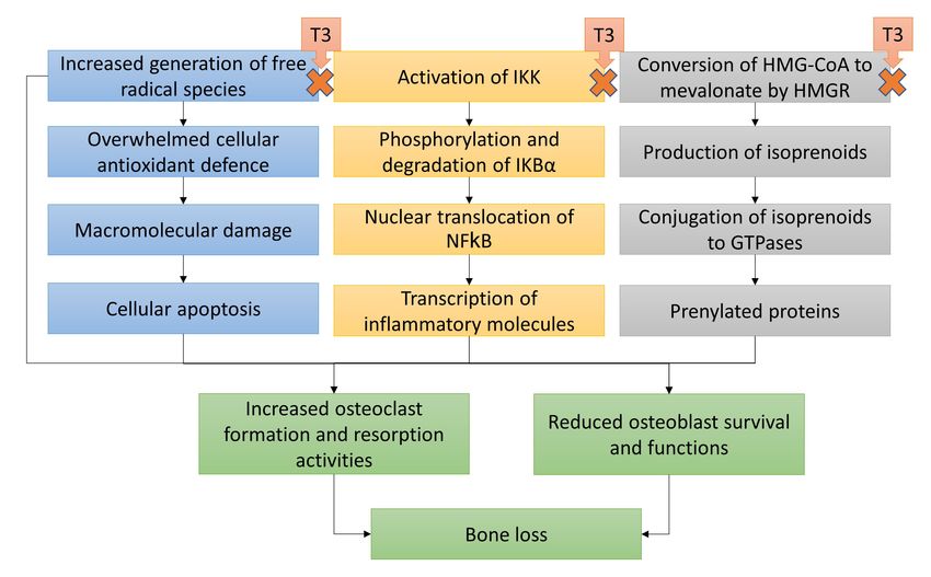
A summary of the suggested mechanism of actions of T3 in preventing bone loss is presented
in Figure 2.Int. J.J.Mol.
Int. Mol.Sci.
Sci.2019, 20,x1355
2019,20, FOR PEER REVIEW 211ofof 20
20
Figure 2. The proposed mechanism of action of tocotrienol (T3) in protecting bone health. Abbreviation:
Figure 2. The3-hydroxy-3-methylglutaryl-CoA;
HMG-CoA, proposed mechanism of action of tocotrienol (T3) in protecting bone health. Abbreviation:
HMGR, 3-hydroxy-3-methylglutaryl-CoA reductase;
HMG-CoA, 3-hydroxy-3-methylglutaryl-CoA; HMGR, 3-hydroxy-3-methylglutaryl-CoA reductase;
IKB, nuclear factor of kappa light polypeptide gene enhancer in B-cells inhibitor, alpha; IKK, IKB kinase;
IKB, nuclear
NF-κB, factor
nuclear of kappa-light-chain-enhancer
factor kappa light polypeptide gene enhancerB-cells.
of activated in B-cells inhibitor, alpha; IKK, IKB
kinase; NF-κB, nuclear factor kappa-light-chain-enhancer of activated B-cells.
10. Perspectives on the Use of T3
10. Perspectives on the Use of T3
Three major concerns on the application of T3 as a bone-sparing agent are its safety, bioavailability,
and marketability.
Three major In terms ofon
concerns safety,
the one study in postmenopausal
application of T3 as a bone-sparing women with osteopenia
agent are itsrevealed
safety,
that annatto T3and
bioavailability, at 600 mg for 12 weeks
marketability. In termsdid not affect their
of safety, liver and
one study kidney functionswomen
in postmenopausal [149]. Itwith
also
suppressedrevealed
osteopenia the high thatturnover markers
annatto T3 at among
600 mgthe forsubjects,
12 weeksindicated
did not by reduced
affect serum
their liver bone
and ALP
kidney
and urinary
functions N-terminal
[149]. telopeptide.
It also suppressed the This
high was accompanied
turnover markers by amonga reduced level of
the subjects, 8-hydroxy-
indicated by
2’-deoxyguanosine,
reduced serum bone an ALP oxidative stress
and urinary marker [150].
N-terminal However,
telopeptide. Thissimilar studies on the
was accompanied by ause of T3
reduced
for bone
level protection in men is absent. Toxicity
of 8-hydroxy-2'-deoxyguanosine, studiesstress
an oxidative in female
marker mice revealed
[150]. However,increased
similarbleeding
studiesrisk
on
withuse
the high-dose
of T3 forpalmbonevitamin E (oral,
protection in men 500isand 1000 Toxicity
absent. mg/kg for 14 and
studies in 42 days)mice
female [151], but thisincreased
revealed dose was
much higher
bleeding thanhigh-dose
risk with the desired dose
palm for bone
vitamin strengthening
E (oral, 500 and 1000 effects.
mg/kgSimilar
for 14toxicity studies
and 42 days) in male
[151], but
animals
this doseorwas menmuchwere higher
limited.than the desired dose for bone strengthening effects. Similar toxicity
In in
studies terms
maleofanimals
bioavailability, T3 generally
or men were limited. showed much lower bioavailability compared to α-TP
due toIn the
termscompetitive binding T3
of bioavailability, at α-TP transport
generally showed protein,
muchwhichlowerregulates the circulating
bioavailability compared vitamin
to α-TPE
levelto[21].
due the However,
competitive a previous
binding study
at α-TP established
transport that a single-dose
protein, of emulsified
which regulates γ-T3 was vitamin
the circulating deposited E
in the[21].
level rat femur and spine
However, 14 days after
a previous studysupplementation
established that[152]. The treatment
a single-dose also exerts γ-T3
of emulsified biological
was
effects on the
deposited bone,
in the ratby up-regulating
femur and spineOPG mRNA
14 days afterand down-regulating
supplementation RANKL
[152]. mRNA [152].
The treatment alsoVarious
exerts
means to increase
biological effects on thethebioavailability of T3 are being
bone, by up-regulating OPG studied,
mRNAincluding the use of the RANKL
and down-regulating self-emulsifying
mRNA
system
[152]. [153] and
Various nanoparticles
means to increase[154].
the bioavailability of T3 are being studied, including the use of the
There are some
self-emulsifying commercial
system [153] andchallenges
nanoparticles in developing
[154]. T3 as a pharmaceutical product to prevent
osteoporosis
There areinsome men.commercial
A patent search revealed
challenges two relevant
in developing T3patents pertaining toproduct
as a pharmaceutical the use ofto T3 as an
prevent
antiosteoporotic
osteoporosis agentA[155,156].
in men. However,
patent search the T3
revealed twosupplements
relevant patents are prevalent
pertaining in the
to market,
the use andof T3natural
as an
composition is usually
antiosteoporotic used. It might
agent [155,156]. However, hamperthe the interest of pharmaceutical
T3 supplements are prevalent companies
in the market,to invest
and
and develop
natural it as pharmaceutical
composition is usually used. products.
It mightHowever,
hamperitthe stillinterest
has great potential to be developed
of pharmaceutical companiesinto to
invest and develop it as pharmaceutical products. However, it still has great potential to be
functional foods and nutraceuticals for men to halt bone loss. In addition, T3 also showed beneficial
developed
effects againstinto other
functional foods and
age-related nutraceuticals
diseases, for men to
such as metabolic halt bone[157,158],
syndrome loss. In addition, T3 also
neurogenerative
showed beneficial effects against other age-related diseases, such as metabolic syndrome [157,158],Int. J. Mol. Sci. 2019, 20, 1355 12 of 20
disease [159], arthritis [160,161], sarcopenia [162,163], which may be attractive to elderly men suffering
from multiple conditions concurrently.
11. Limitations
This review is not without its limitations. The ultimate consequence of male osteoporosis is
fragility fracture, which carries significant morbidity and mortality to the patients. Fragility fracture is
not only predicted by reduced bone mass, but also muscle and cognitive functions of an individual,
which are related to the gait and tendency to fall [164]. This review has only addressed the effects
of T3 on bone mass, structure, and strength, but not on evidence for muscle and cognitive functions.
However, osteoporosis remains the most treatable predictors of fracture, and it can be prevented by
T3 supplementation.
12. Conclusions
As a conclusion, evidence accumulated thus far has demonstrated promising effects of T3
as a preventive agent against male osteoporosis in models of bone loss induced by testosterone
deficiency (surgical or chemical ablation), metabolic syndrome, cigarette-smoking, glucocorticoids
and alcoholism. Men with these risk factors or osteopenia could benefit from T3 supplementation to
stop the progression of bone loss. The human equivalent dose of T3 proven to prevent osteoporosis
is approximately 600 mg/day, to be taken after meals to enhance absorption. However, there is no
clinical trial to study the skeletal effects of T3 in men so far. Thus, the efficacy of T3 in preventing
the progression of male osteoporosis still awaits new clinical evidence in humans. The bone-sparing
effects of T3 could be mediated by its antioxidant, anti-inflammatory, and mevalonate-suppressive
effects. However, more studies are needed to illustrate its mechanism of actions, especially the cell
signaling pathways involved.
Author Contributions: K.-Y.C. drafted the manuscript. S.I.-N. provided critical review for the manuscript.
Funding: The authors are funded by Ministry of Education, Malaysia through grant FRGS/1/2018/SKK10/
UKM/03/1 and Universiti Kebangsaan Malaysia through grant GUP-2017-060.
Acknowledgments: We thank Ministry of Education, Malaysia and Universiti Kebangsaan Malaysia for funding
the studies through grant FRGS/1/2018/SKK10/UKM/03/1 and GUP-2017-060.
Conflicts of Interest: The authors declare no conflict of interest.
References
1. Edwards, M.H.; Dennison, E.M.; Aihie Sayer, A.; Fielding, R.; Cooper, C. Osteoporosis and sarcopenia in
older age. Bone 2015, 80, 126–130. [CrossRef] [PubMed]
2. Cawthon, P.M. Gender differences in osteoporosis and fractures. Clin. Orthop. Relat. Res. 2011, 469, 1900–1905.
[CrossRef] [PubMed]
3. Johnell, O.; Kanis, J. An estimate of the worldwide prevalence and disability associated with osteoporotic
fractures. Osteoporos. Int. 2006, 17, 1726–1733. [CrossRef] [PubMed]
4. Burge, R.; Dawson-Hughes, B.; Solomon, D.H.; Wong, J.B.; King, A.; Tosteson, A. Incidence and economic
burden of osteoporosis-related fractures in the united states, 2005–2025. J. Bone Miner. Res. 2007, 22, 465–475.
[CrossRef] [PubMed]
5. Chatterton, B.D.; Moores, T.S.; Ahmad, S.; Cattell, A.; Roberts, P.J. Cause of death and factors associated with
early in-hospital mortality after hip fracture. Bone Jt. J. 2015, 97-B, 246–251. [CrossRef] [PubMed]
6. Mizrahi, E.H.; Arad, M.; Fleissig, Y.; Adunsky, A. Gender differences in functional outcome of elderly hip
fracture patients. Geriatr. Gerontol. Int. 2014, 14, 845–850. [CrossRef]
7. Wang, C.C.; Wu, C.H.; Farley, J.F. Patterns of pharmacological treatment for osteoporosis among
patients qualified for pharmacotherapy according to the national osteoporosis foundation guidelines.
Ann. Pharmacother. 2015, 49, 995–1003. [CrossRef]
8. Willson, T.; Nelson, S.D.; Newbold, J.; Nelson, R.E.; LaFleur, J. The clinical epidemiology of male osteoporosis:
A review of the recent literature. Clin. Epidemiol. 2015, 7, 65–76.Int. J. Mol. Sci. 2019, 20, 1355 13 of 20
9. Amelio, P.; Isaia, G.C. Male osteoporosis in the elderly. Int. J. Endocrinol. 2015, 2015, 8.
10. NIH Osteoporosis and Related Bone Diseases-National Resource Center. Osteoporosis in Men. Available
online: https://www.bones.nih.gov/health-info/bone/osteoporosis/men (accessed on 12 March 2019).
11. Feng, X.; McDonald, J.M. Disorders of bone remodeling. Annu. Rev. Pathol. 2011, 6, 121–145. [CrossRef]
12. O’Brien, C.A.; Almeida, M. Basic biology of skeletal aging: Role of stress response pathways. J. Gerontol.
2013, 68, 1197–1208.
13. Giusti, A.; Bianchi, G. Treatment of primary osteoporosis in men. Clin. Interv. Aging 2015, 10, 105–115.
[PubMed]
14. Bliuc, D.; Alarkawi, D.; Nguyen, T.V.; Eisman, J.A.; Center, J.R. Risk of subsequent fractures and mortality in
elderly women and men with fragility fractures with and without osteoporotic bone density: The dubbo
osteoporosis epidemiology study. J. Bone Miner Res. 2015, 30, 637–646. [CrossRef] [PubMed]
15. Chin, K.Y.; Ima-Nirwana, S. The biological effects of tocotrienol on bone: A review on evidence from rodent
models. Drug Des. Dev. Ther. 2015, 9, 2049–2061. [CrossRef] [PubMed]
16. Chin, K.-Y.; Ima-Nirwana, S. Vitamin e as an antiosteoporotic agent via receptor activator of nuclear factor
kappa-b ligand signaling disruption: Current evidence and other potential research areas. Evid. Based
Complement Altern. Med. 2012, 2012, 747020. [CrossRef] [PubMed]
17. Chin, K.-Y.; Mo, H.; Soelaiman, I.-N. A review of the possible mechanisms of action of tocotrienol—A
potential antiosteoporotic agent. Curr. Drug Targets 2013, 14, 1533–1541. [CrossRef] [PubMed]
18. Ahsan, H.; Ahad, A.; Siddiqui, W.A. A review of characterization of tocotrienols from plant oils and foods.
J. Chem. Biol. 2015, 8, 45–59. [CrossRef]
19. Aggarwal, B.; Sundaram, C.; Prasad, S.; Kannappan, R. Tocotrienols, the vitamin e of the 21st century: It’s
potential against cancer and other chronic diseases. Biochem. Pharmacol. 2010, 80, 1613–1631. [CrossRef]
[PubMed]
20. Fairus, S.; Nor, R.M.; Cheng, H.M.; Sundram, K. Alpha-tocotrienol is the most abundant tocotrienol isomer
circulated in plasma and lipoproteins after postprandial tocotrienol-rich vitamin e supplementation. Nutr. J.
2012, 11, 5. [CrossRef]
21. Hosomi, A.; Arita, M.; Sato, Y.; Kiyose, C.; Ueda, T.; Igarashi, O.; Arai, H.; Inoue, K. Affinity for
alpha-tocopherol transfer protein as a determinant of the biological activities of vitamin e analogs. FEBS Lett.
1997, 409, 105–108. [CrossRef]
22. Shen, C.L.; Klein, A.; Chin, K.Y.; Mo, H.; Tsai, P.; Yang, R.S.; Chyu, M.C.; Ima-Nirwana, S. Tocotrienols for
bone health: A translational approach. Ann. N. Y. Acad. Sci. 2017, 1401, 150–165. [CrossRef]
23. Muhammad, N.; Luke, D.A.; Shuid, A.N.; Mohamed, N.; Soelaiman, I.N. Two different isomers of vitamin
e prevent bone loss in postmenopausal osteoporosis rat model. Evid. Based Complement Altern. Med. 2012,
2012, 161527. [CrossRef]
24. Muhammad, N.; Razali, S.; Shuid, A.N.; Mohamed, N.; Soelaiman, I.N. Membandingkan kesan
antara fraksi-kaya tokotrienol, kalsium dan estrogen terhadap metabolisme tulang tikus terovariektomi.
Sains Malays. 2013, 42, 1591–1597.
25. Soelaiman, I.N.; Ming, W.; Abu Bakar, R.; Hashnan, N.A.; Mohd Ali, H.; Mohamed, N.; Muhammad, N.;
Shuid, A.N. Palm tocotrienol supplementation enhanced bone formation in oestrogen-deficient rats.
Int. J. Endocrinol. 2012, 2012, 532862. [CrossRef]
26. Abdul-Majeed, S.; Mohamed, N.; Soelaiman, I.-N. Effects of tocotrienol and lovastatin combination on
osteoblast and osteoclast activity in estrogen-deficient osteoporosis. Evid. Based Complement Altern. Med.
2012, 2012, 960742. [CrossRef]
27. Abdul-Majeed, S.; Mohamed, N.; Soelaiman, I.N. The use of delta-tocotrienol and lovastatin for
anti-osteoporotic therapy. Life Sci. 2015, 125, 42–48. [CrossRef]
28. Chin, K.Y.; Abdul-Majeed, S.; Mohamed, N.; Ima-Nirwana, S. The effects of tocotrienol and lovastatin
co-supplementation on bone dynamic histomorphometry and bone morphogenetic protein-2 expression in
rats with estrogen deficiency. Nutrients 2017, 9, 143. [CrossRef]
29. Chin, K.Y.; Ima Nirwana, S. The effects of annatto-derived tocotrienol supplementation in osteoporosis
induced by testosterone deficiency in rats. Clin. Interv. Aging 2014, 9, 1247–1259. [CrossRef]
30. Mohamad, N.V.; Ima-Nirwana, S.; Chin, K.Y. Effect of tocotrienol from bixa orellana (annatto) on bone
microstructure, calcium content, and biomechanical strength in a model of male osteoporosis induced by
buserelin. Drug Des. Dev. Ther. 2018, 12, 555–564. [CrossRef]Int. J. Mol. Sci. 2019, 20, 1355 14 of 20
31. Mohamad, N.V.; Soelaiman, I.N.; Chin, K.Y. Effects of tocotrienol from bixa orellana (annatto) on bone
histomorphometry in a male osteoporosis model induced by buserelin. Biomed. Pharmacother. 2018,
103, 453–462. [CrossRef]
32. Wong, S.K.; Chin, K.Y.; Suhaimi, F.H.; Ahmad, F.; Ima-Nirwana, S. Exploring the potential of tocotrienol from
bixa orellana as a single agent targeting metabolic syndrome and bone loss. Bone 2018, 116, 8–21. [CrossRef]
33. Wong, S.K.; Chin, K.-Y.; Suhaimi, F.H.; Ahmad, F.; Ima-Nirwana, S. The effects of palm tocotrienol on
metabolic syndrome and bone loss in male rats induced by high-carbohydrate high-fat diet. J. Funct Foods
2018, 44, 246–254. [CrossRef]
34. Wong, S.K.; Chin, K.-Y.; Suhaimi, F.H.; Ahmad, F.; Jamil, N.A.; Ima-Nirwana, S. Osteoporosis is associated
with metabolic syndrome induced by high-carbohydrate high-fat diet in a rat model. Biomed. Pharmacother.
2018, 98, 191–200. [CrossRef]
35. Ima Nirwana, S.; Kiftiah, A.; Zainal, A.; Norazlina, M.; Gapor, M.; Khalid, B.A.K. Palm vitamin e prevents
osteoporosis in orchidectomised growing male rats. Nat. Prod. Sci. 2000, 6, 155–160.
36. Ima-Nirwana, S.; Fakhrurazi, H. Palm vitamin e protects bone against dexamethasone-induced osteoporosis
in male rats. Med. J. 2002, 57, 133–141.
37. Ima Nirwana, S.; Suhaniza, S. Effects of tocopherols and tocotrienols on body composition and bone calcium
content in adrenalectomized rats replaced with dexamethasone. J. Med. Food 2004, 7, 45–51. [CrossRef]
38. Zakaria, S.; Mat-Husain, S.Z.; Ying-Hwey, K.; Xin-Kai, K.; Mohd-Badawi, A.; Abd-Ghani, N.A.; Aziz, M.A.;
Mohamed, N. Vitamin e improved bone strength and bone minerals in male rats given alcohol. Iran J. Basic
Med. Sci. 2017, 20, 1360–1367.
39. Wong, S.; Chin, K.-Y.; Suhaimi, F.; Ahmad, F.; Ima-Nirwana, S. The effects of vitamin e from elaeis guineensis
(oil palm) in a rat model of bone loss due to metabolic syndrome. Int. J. Environ. Res. Public Health 2018,
15, 1828. [CrossRef]
40. Chin, K.Y.; Abdul-Majeed, S.; Fozi, N.F.; Ima-Nirwana, S. Annatto tocotrienol improves indices of bone
static histomorphometry in osteoporosis due to testosterone deficiency in rats. Nutrients 2014, 6, 4974–4983.
[CrossRef]
41. Chin, K.Y.; Gengatharan, D.; Mohd Nasru, F.S.; Khairussam, R.A.; Ern, S.L.; Aminuddin, S.A.; Ima-Nirwana, S.
The effects of annatto tocotrienol on bone biomechanical strength and bone calcium content in an animal
model of osteoporosis due to testosterone deficiency. Nutrients 2016, 8, 808. [CrossRef]
42. Hermizi, H.; Faizah, O.; Ima-Nirwana, S.; Ahmad Nazrun, S.; Norazlina, M. Beneficial effects of tocotrienol
and tocopherol on bone histomorphometric parameters in sprague–dawley male rats after nicotine cessation.
Calcif. Tissue Int. 2009, 84, 65–74. [CrossRef]
43. Norazlina, M.; Hermizi, H.; Faizah, O.; Ima-Nirwana, S. Vitamin e reversed nicotine-induced toxic effects on
bone biochemical markers in male rats. Arch. Med. Sci. 2010, 6, 505–512. [CrossRef]
44. Mehat, M.; Shuid, A.; Mohamed, N.; Muhammad, N.; Soelaiman, I. Beneficial effects of vitamin e isomer
supplementation on static and dynamic bone histomorphometry parameters in normal male rats. J. Bone
Miner Metab. 2010, 28, 503–509. [CrossRef]
45. Shuid, A.; Mehat, Z.; Mohamed, N.; Muhammad, N.; Soelaiman, I. Vitamin e exhibits bone anabolic actions
in normal male rats. J. Bone Miner Metab. 2010, 28, 149–156. [CrossRef]
46. Maniam, S.; Mohamed, N.; Shuid, A.N.; Soelaiman, I.N. Palm tocotrienol exerted better antioxidant activities
in bone than α-tocopherol. Basic Clin. Pharmacol. Toxicol. 2008, 103, 55–60. [CrossRef]
47. Gordon, C.M.; Zemel, B.S.; Wren, T.A.; Leonard, M.B.; Bachrach, L.K.; Rauch, F.; Gilsanz, V.; Rosen, C.J.;
Winer, K.K. The determinants of peak bone mass. J. Pediatr. 2017, 180, 261–269. [CrossRef]
48. Tennant, K.G.; Leonard, S.W.; Wong, C.P.; Iwaniec, U.T.; Turner, R.T.; Traber, M.G. High-dietary
alpha-tocopherol or mixed tocotrienols have no effect on bone mass, density, or turnover in male rats
during skeletal maturation. J. Med. Food 2017, 20, 700–708. [CrossRef]
49. Chin, K.Y.; Ima-Nirwana, S. The effects of alpha-tocopherol on bone: A double-edged sword? Nutrients 2014,
6, 1424–1441. [CrossRef]
50. Abd Manan, N.; Mohamed, N.; Shuid, A.N. Effects of low-dose versus high-dose gamma-tocotrienol on the
bone cells exposed to the hydrogen peroxide-induced oxidative stress and apoptosis. Evid. Based Complement
Altern. Med. 2012, 2012, 680834. [CrossRef]
51. Callaway, D.A.; Jiang, J.X. Reactive oxygen species and oxidative stress in osteoclastogenesis, skeletal aging
and bone diseases. J. Bone Miner Metab. 2015, 33, 359–370. [CrossRef]You can also read