From Dysbiosis to Healthy Skin: Major Contributions of Cutibacterium acnes to Skin Homeostasis - MDPI
←
→
Page content transcription
If your browser does not render page correctly, please read the page content below
microorganisms

Review
From Dysbiosis to Healthy Skin: Major Contributions
of Cutibacterium acnes to Skin Homeostasis
Miquel Rozas 1,2 , Astrid Hart de Ruijter 1 , Maria Jose Fabrega 2 , Amine Zorgani 1 , Marc Guell 1,2 ,
Bernhard Paetzold 1 and Francois Brillet 1, *

                                          1   S-Biomedic, JLABS, Turnhoutseweg 30, 2340 Beerse, Belgium; miquel@sbiomedic.com (M.R.);
                                              astrid@sbiomedic.com (A.H.d.R.); amine@sbiomedic.com (A.Z.); marc.guell@upf.edu (M.G.);
                                              bernhard.paetzold@sbiomedic.com (B.P.)
                                          2   Department of Experimental and Health Sciences, Universitat Pompeu Fabra (UPF),
                                              C. Dr. Aiguader 88, 08003 Barcelona, Spain; maria-jose.fabrega@upf.edu
                                          *   Correspondence: francois@sbiomedic.com

                                          Abstract: Cutibacterium acnes is the most abundant bacterium living in human, healthy and sebum-
                                          rich skin sites, such as the face and the back. This bacterium is adapted to this specific environment
                                          and therefore could have a major role in local skin homeostasis. To assess the role of this bacterium
                                          in healthy skin, this review focused on (i) the abundance of C. acnes in the skin microbiome of
                                          healthy skin and skin disorders, (ii) its major contributions to human skin health, and (iii) skin
                                          commensals used as probiotics to alleviate skin disorders. The loss of C. acnes relative abundance
                                          and/or clonal diversity is frequently associated with skin disorders such as acne, atopic dermatitis,
                                          rosacea, and psoriasis. C. acnes, and the diversity of its clonal population, contributes actively to the

                                   normal biophysiological skin functions through, for example, lipid modulation, niche competition
                                          and oxidative stress mitigation. Compared to gut probiotics, limited dermatological studies have
Citation: Rozas, M.; Hart de Ruijter,
                                          investigated skin probiotics with skin commensal strains, highlighting their unexplored potential.
A.; Fabrega, M.J.; Zorgani, A.; Guell,
M.; Paetzold, B.; Brillet, F. From
Dysbiosis to Healthy Skin: Major
                                          Keywords: skin microbiota; microbiome dysbiosis; skin disorders; Cutibacterium acnes; topical bacte-
Contributions of Cutibacterium acnes      riotherapy
to Skin Homeostasis. Microorganisms
2021, 9, 628. https://doi.org/
10.3390/microorganisms9030628
                                          1. Cutibacterium and Cutibacterium acnes in the Skin Microbiome
Academic Editor: Roberto Di Marco               The skin is the largest (2 m2 ) and most visible organ of the human body [1]. Consti-
                                          tuted by three immunologically active layers [2]—the epidermis (75 to 150 µm), the dermis
Received: 3 March 2021
                                          (
Microorganisms 2021, 9, 628                                                                                                 2 of 18

                              genera Staphylococcus and Corynebacterium, and the oily/sebaceous sites (e.g., face and
                              back) largely predominated by the genus Propionibacterium, followed by Staphylococcus
                              and Corynebacterium (Figure 1C) [16]. In these lipid-rich microenvironments, although
                              not exclusively, the relative abundance of bacterial populations is influenced by gender,
                              age, and geographical origin. Nonetheless, Propionibacterium still predominates, with a
                              relative stability in the sebaceous sites of healthy individuals (Figure 1D) [17–19]. It cohabi-
                              tates with the other bacterial populations but also with the viruses (e.g., bacteriophages),
                              fungi (e.g., Malassezia), and parasites (e.g., Demodex) colonizing specific environments—
                              for example, the human hair follicle [20]. This genus, recently reclassified and renamed
                              Cutibacterium [21], is a sentinel of the healthy human skin microbiome [22]. Alteration
                              of its structure is associated with dysbiosis and disease, including atopic dermatitis (AD),
                              psoriasis, rosacea, and acne [23,24]. Systemic antibiotics targeted at reducing C. acnes can
                              also disrupt the equilibrium, which may lead to opportunistic infections in the hair follicle
                              by competing species (e.g., Pseudomonas species) [25].
                                    AD is a chronic inflammatory disease characterized by itchy skin, affecting children
                              (10-20%) and adults (1−2%) in industrial countries [26,27]. There is evidence that human
                              skin microbiome dysbiosis promotes AD [28]. Through sampling the skin microbiome of
                              lesional and non-lesional skin regions of AD and healthy patients, authors showed that
                              Cutibacterium acnes (C. acnes) abundance correlates inversely with Staphylococcus aureus
                              (Figure 1E) [29,30]. The presence of this pathogen is known as the main factor that exac-
                              erbates AD and it has been suggested that antimicrobials from human skin commensal
                              bacteria protect against S. aureus and are deficient in AD patients [31]. Rosacea is another
                              dermatological disorder characterized by chronic inflammation of the face in adults, mostly
                              women, after the age of 30 years [32]. Colonization of the skin by mites such as Demodex
                              is a major pathogenic factor associated with rosacea skin conditions [33,34]. Interestingly,
                              this mite has an associated microbiome, namely endosymbionts such as Corynebacterium
                              kroppenstedtii subsp. demodicis [35], Bacillus oleronius, and Bacillus cereus [36], which are sug-
                              gested to play a role in the pathogenicity of this disease [37]. However, the dysbiosis and
                              the loss of relative abundance of Cutibacterium acnes has also been observed and correlated
                              with the severity of the disease (Figure 1F) [38,39]. Psoriasis is also a chronic inflammatory
                              disease characterized by hyperproliferation of keratinocytes and increased inflammation,
                              affecting approximately 2% of the population [40]. Psoriasis is one of the most common
                              immune-mediated skin disorders [41]. Alteration of microbial communities is observed
                              within the bacterial populations of lesional and non-lesional skin regions of psoriasis versus
                              healthy patients [42–44]. This strongly associates the dysbiosis with the progression of
                              the disease [45,46], particularly with an imbalance of the dominant genera, Cutibacterium
                              (decreased), Corynebacterium, and Staphylococcus (Figure 1G) [47,48]. Acne vulgaris is the
                              most common dermatological condition worldwide [49]. Aggregating several risk factors
                              (e.g., age, skin type, hormonal changes) and prevalent in the sebaceous/lipid-rich skin
                              sites (e.g., face and back) [50,51], acne involves chronic inflammation of the pilosebaceous
                              unit [52], often persisting into adulthood [53]. Extensively described and studied in past
                              centuries [54], and firmly associated with the skin microbiome and Cutibacterium acnes
                              proliferation [55,56], the paradigm of this dysbiosis is now changing. Recently, many
                              authors observed that the relative abundance and bacterial load of C. acnes are not signifi-
                              cantly different between acne and healthy patients (Figure 1H,I) [57–62]. In line with these
                              findings, advances in sequencing technologies and in research in the past few decades have
                              revealed the diversity of C. acnes at subspecies (C. acnes subsp. acnes (phylotype I), C. acnes
                              subsp. defendens (phylotype II), C. acne subsp. elongatum (phylotype III)) and subtype levels
                              (phylogenetic groups IA1 , IA2 , IB, IC, II, III) (Figure 2) [63]. This is responsible for the recent
                              paradigm shift: rather than a sudden proliferation in the skin appendages, a decrease in the
                              diversity between the six phylogenetic groups, rather than C. acnes proliferation, has been
                              associated with acne progression (Figure 1J,K) [64,65].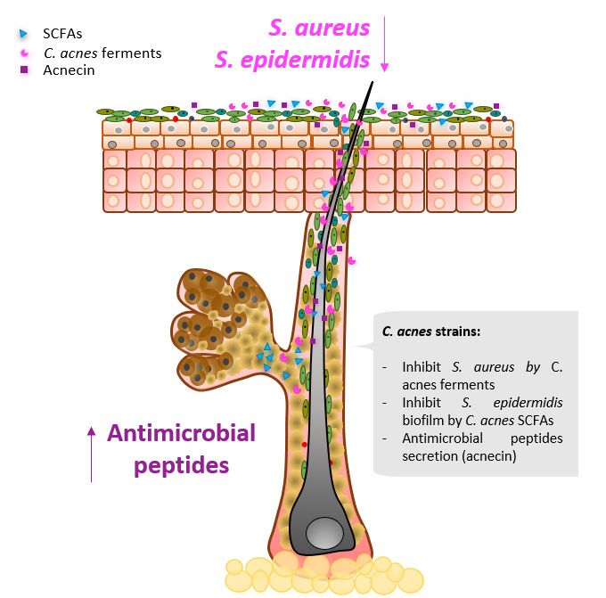
Microorganisms 2021, 9, x FOR PEER REVIEW 3 of 18
Microorganisms 2021, 9, 628 3 of 18
rather than C. acnes proliferation, has been associated with acne progression (Figure 1J,K)
[64,65].
a c HV1
Sebaceous
1.0
HV1
Moist
1.0
HV1
Dry
1.0
HV2 HV2 HV2
0.8 0.8 0.8
HV3 HV3 HV3
HV4 HV4 HV4
0.6 0.6 0.6
b
HV5 HV5 HV5
HV6 HV6 HV6
0.4 0.4 0.4
HV7 HV7 HV7
HV8 HV8 HV8
0.2 0.2 0.2
HV9 HV9 HV9
HV10 HV10 HV10
0 0 0
Ct St Co βP Other Co βP St Fl Ct βP Co Fl Ct γP
d
e f Rosacea g Psoriasis
Healthy Healthy
n=22 n=26
ETR ✱ Non-lesional ✱
✱ n=21 ✱ ✱
n=28
PPR Lesional
n=15 n=28
0.0 0.5 1.0 0.0 0.5 1.0
i j
IA1
IA2
IB
IC
II
III
k
h Face Back MSLT8 Database
1.0 1.0
0.5 0.5
0.0 0.0
Healthy Acne
ne
n = ne
n= lthy
y
lth
Ac
Ac
12
24
ea
ea
H
H
Figure 1.
Figure 1. Cutibacterium
Cutibacterium acnes acnesininthe
thehuman
humanskinskinmicrobiome.
microbiome.(a) (a)Relative
Relativeabundance
abundance ofof skin
skin microbiota
microbiota across
across kingdoms
kingdoms in
in 15 heathy volunteers (HV) (9 males, 6 females) sampled in 18 different skin sites, adapted from Oh et al., 2014 [7].
15 heathy volunteers (HV) (9 males, 6 females) sampled in 18 different skin sites, adapted from Oh et al., 2014 [7]. Bacterial
Bacterial genomes predominate at most sites. (b) The analysis of 16S ribosomal RNA of 10 HV (20 sites sampled, upper
genomes predominate at most sites. (b) The analysis of 16S ribosomal RNA of 10 HV (20 sites sampled, upper bar graph)
bar graph) and 9 HV (27 sites sampled, lower bar graph) shows that most of the sequences are attributed to four phyla,
and 9 HVfrom
adapted (27 sites
Gricesampled, lower
et al., 2009 [13]bar
andgraph) shows
Costello et al.,that
2009most
[14],ofrespectively.
the sequences are this,
Over attributed to four phyla, adapted
skin microenvironments alsofrom
vary
Grice et al.,in
drastically 2009 [13]
their andofCostello
level bacterialetdiversity.
al., 2009 [14],
(c) Inrespectively.
10 HV sampled Overinthis, skin microenvironments
sebaceous, moist, and dry sites, alsothe
vary drastically
relative abun-
in theiroflevel
dance of bacterial(Ct,
Cutibacterium diversity.
formerly(c)Propionibacterium),
In 10 HV sampled Staphylococcus
in sebaceous, moist, and dry sites, the(Co),
(St), Corynebacterium relative abundance of
Betaproteobacteria
(βP), Flavobacteria
Cutibacterium (Ct, (Fl), and Gammaproteobacteria
formerly (γP) is strongly(St),
Propionibacterium), Staphylococcus shaped by the niche, adapted
Corynebacterium from Grice et al., (βP),
(Co), Betaproteobacteria 2009
[13]. (d) The predominating
Flavobacteria relative abundance
(Fl), and Gammaproteobacteria of is
(γP) Cutibacterium
strongly shaped in sebaceous
by the niche,sitesadapted
is relatively
fromstable
Grice over
et al.,the
200922[13].
HV
from different genders, ages (Ado: teenager, Adu: adult, Eld: elderly), and locations (U:
(d) The predominating relative abundance of Cutibacterium in sebaceous sites is relatively stable over the 22 HV from urban, R: rural), adapted from
Findley etgenders,
different al., 2013ages
[17] (Ado:
and Ying et al., 2015
teenager, Adu:[18]. However,
adult, this relative
Eld: elderly), abundance
and locations of Cutibacterium
(U: urban, genus isfrom
R: rural), adapted significantly
Findley
decreased (*: p-value < 0.05) in (e) atopic dermatitis lesional regions (2 sebaceous (interscapular and retroauricular) and 1
et al., 2013 [17] and Ying et al., 2015 [18]. However, this relative abundance of Cutibacterium genus is significantly decreased
moist (antecubital fossa) sites sampled in 18 to 60yo males (n: number of volunteers)), adapted from Francuzik et al., 2018
(*: p-value < 0.05) in (e) atopic dermatitis lesional regions (2 sebaceous (interscapular and retroauricular) and 1 moist
[29], (f) rosacea lesional regions (ETR: erythematotelangiectatic rosacea, PPR: papulopustular rosacea, 1 sebaceous site
(antecubital
(cheek) sampled fossa)insites
18 tosampled
64yo malesin 18andto 60yo males
females), (n: number
adapted from of volunteers)),
Wang et al., 2020adapted
[39], andfrom Francuziknon-lesional
(g) psoriasis et al., 2018 [29],
and
(f) rosacea
lesional lesional
regions regionsin(ETR:
sampled erythematotelangiectatic
1 sebaceous (scalp), 2 moist (axillarosacea, PPR: papulopustular
and gluteal), and 3 dry (trunk,rosacea,
arm,1leg)
sebaceous siteadapted
skin sites, (cheek)
sampled
from Chang in 18ettoal.,
64yo
2018 males
[47]. and females), adapted
(h) Interestingly, from Wang
no significant et al., 2020
alteration [39],relative
of the and (g)abundance
psoriasis non-lesional and lesional
of Cutibacterium genus
was observed
regions sampled over
in67 patients (22-23
1 sebaceous year
(scalp), 2 old)
moistwith no and/or
(axilla different
and gluteal), acne
and 3 dryseverity
(trunk, sampled
arm, leg)onskin
the cheek, adapted from
sites, adapted from
Li et al.,et2019
Chang [61]. [47].
al., 2018 (i) No(h)difference in theno
Interestingly, bacterial loadalteration
significant (log colony of forming units
the relative (CFU) andoflog
abundance genomic units
Cutibacterium (GU)was
genus per
strip) between
observed over 67 15-30yo
patients male andyear
(22-23 female
old)healthy
with noor acne patients
and/or differentsampled on thesampled
acne severity face, adapted from Pecastaings
on the cheek, adapted from et al.,
Li
2018 [62]. The sharp difference between healthy and acne patients is in the decrease in the diversity
et al., 2019 [61]. (i) No difference in the bacterial load (log colony forming units (CFU) and log genomic units (GU) per of C. acnes phylotypes.
(j) Comparison of the relative abundance of the 6 phylotypes of C. acnes between 16-35yo male and female healthy and
strip) between 15-30yo male and female healthy or acne patients sampled on the face, adapted from Pecastaings et al.,
acne patients, adapted from Dagnelie et al., 2017 [65]. (k) Relative abundance and statistically significant enrichment of
2018 [62]. The sharp difference between healthy and acne patients is in the decrease in the diversity of C. acnes phylotypes.
type IA1 with acne is confirmed by the analysis of the current MSLT8 isolate database, adapted from McLaughlin et al.,
(j)
2019Comparison
[64]. of the relative abundance of the 6 phylotypes of C. acnes between 16-35yo male and female healthy and acne
patients, adapted from Dagnelie et al., 2017 [65]. (k) Relative abundance and statistically significant enrichment of type IA1
with acne is confirmed by the analysis of the current MSLT8 isolate database, adapted from McLaughlin et al., 2019 [64].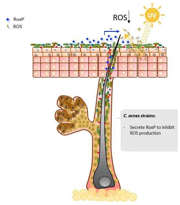
Microorganisms 2021, 9, 628 4 of 18
Microorganisms 2021, 9, x FOR PEER REVIEW 4 of 18
For the
For the four
four skin
skindisorders
disorderssuccinctly
succinctlydescribed
describedabove,
above,associated
associated dysbiosis
dysbiosis is is corre-
corre-
lated with a loss of Cutibacterium relative abundance and/or diversity at the
lated with a loss of Cutibacterium relative abundance and/or diversity at the genus and/orgenus and/or
species levels.
species levels. These
Thesealterations
alterationshave
haveaanegative
negativeimpact
impacton onskin
skinbiology
biologybybygenerating
generatinga a
loss of biological functions essential for skin homeostasis, possibly leading to
loss of biological functions essential for skin homeostasis, possibly leading to inflamma-inflammation.
Therefore,
tion. in this
Therefore, inreview, two main
this review, questions
two main are addressed:
questions (1) How
are addressed: doesdoes
(1) How Cutibacterium
Cutibac-
contribute
terium to the physiology
contribute of healthy
to the physiology skin? and
of healthy (2)and
skin? How (2)could
How probiotics better prevent
could probiotics better
and/or treat
prevent and/or the dysbiosis
treat disorders
the dysbiosis “on site”
disorders “ontosite”
restore the natural
to restore healthy
the natural functions
healthy func-of
the skin?
tions of the skin?
a b
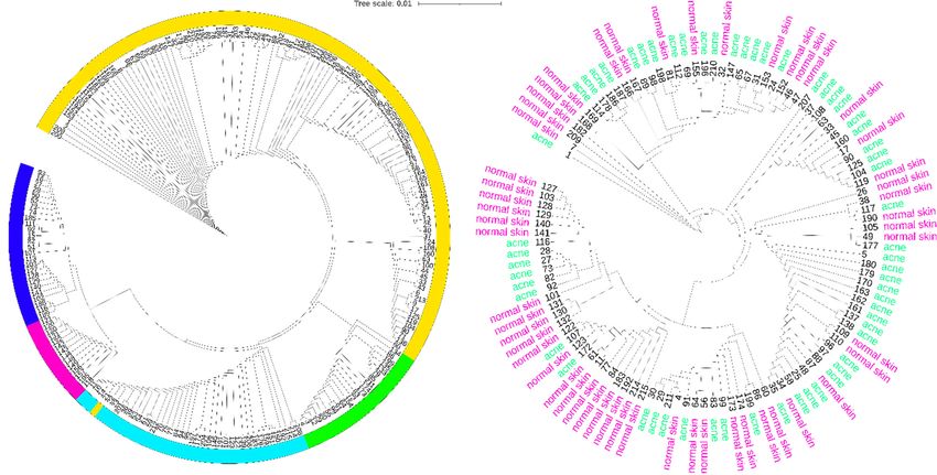
Figure 2. Cutibacterium acnes clonal population structure and distribution on normal and acne skin. (a) Neighbor-joining tree
Figure 2. Cutibacterium acnes clonal population structure and distribution on normal and acne skin. (a) Neighbor-joining
created using iTOL tool [66] from concatenated nucleotide sequences of MLST schemes [67] from all the current strains in
tree created using iTOL tool [66] from concatenated nucleotide sequences of MLST schemes [67] from all the current strains
the database published by Jolley et al., 2018 [68]. Over the 215 different isolates, 114 (53%) are classified in the subtype IA1 ,
in the database published by Jolley et al., 2018 [68]. Over the 215 different isolates, 114 (53%) are classified in the subtype
22
IA(10%) in the subtype IA2 , and
1, 22 (10%) in the subtype IA2,39and(18%) in the in
39 (18%) subtype IB of the
the subtype IB major
of thephylogenetic group type
major phylogenetic groupI. The
typerest are classified
I. The rest are
in the phylogenetic
classified groups IIgroups
in the phylogenetic (26 isolates, 12%) and
II (26 isolates, III (14
12%) andisolates, 6%). Considering
III (14 isolates, the sources
6%). Considering of all of
the sources theallstrains in the
the strains
database, the majority,
in the database, 112 (52%),
the majority, have been
112 (52%), haveisolated from skin
been isolated fromsamples. The rest
skin samples. The of the
rest ofstrains havehave
the strains beenbeenisolated from
isolated
from samples
samples categorized
categorized as eye,as eye, prostate,
prostate, other, soft
other, blood, blood, soft medical
tissue, tissue, medical
device, device, spinal
spinal disc, disc, and
dental, dental,
boneand(19,bone (19,13,
18, 14, 18,12,
14,10,
10, 13,6,12,
and 10,1%,
10,respectively).
6, and 1%, respectively). (b) Neighbor-joining
(b) Neighbor-joining tree from concatenated
tree from concatenated nucleotideofsequences
nucleotide sequences MLST schemes of MLSTfrom
schemes
all from all
the isolates the isolates
sourced as skinsourced
samples as(acne
skin samples
and normal(acneskin).
and normal
From theskin).
totalFrom thestrains,
of 112 total of59
112 strains,
have been59 have been
isolated from
isolated from acne skin and 53 from normal skin. For acne skin, 44 (74%) are classified in the subtype IA1, 7 (11%) in the
acne skin and 53 from normal skin. For acne skin, 44 (74%) are classified in the subtype IA1 , 7 (11%) in the subtype IA2 ,
subtype IA2, 2 (3%) in the subtype IB, 6 (10%) in the phylogenetic group II, and 0 in the phylogenetic group III. For normal
2skin,
(3%)27 in (50%)
the subtype IB, 6 (10%)
are classified in thein the phylogenetic
subtype groupinII,the
IA1, 6 (11%) andsubtype
0 in the IA
phylogenetic group III. For normal skin, 27 (50%)
2, 10 (18%) in the subtype IB, 7 (13%) in the
are classified in the subtype
phylogenetic group II, and 3 (6%) IA 1 , 6 (11%) in the subtype
in the phylogenetic group IA , 10
2 III.(18%) in the subtype IB, 7 (13%) in the phylogenetic group II,
and 3 (6%) in the phylogenetic group III.
2. Major Contributions of Cutibacterium acnes to Skin Homeostasis
2. Major Contributions
The human of Cutibacterium
microbiome plays a crucialacnes to human
role in Skin Homeostasis
health. Different gut and skin
Thehave
diseases humanbeen microbiome playsmicrobiome
associated with a crucial role in human
dysbiosis, health. Different
“elucidating gut and skin
by contraposition”
diseases
its have been
importance in theassociated
maintenance withofmicrobiome dysbiosis,
a healthy state “elucidating
[69]. The by contraposition”
gut microbiome is the most
its importance
extensively in the
studied maintenance
human of a healthy
microbiome state
[70]. It has [69]. The
essential gut microbiome
functions is thestate
in the healthy most
extensively
of studied
the gut, such human microbiome
as protection [70]. It has
against pathogen essential
invasion, functionsthe
nourishing in the
hosthealthy state
cells with
of themetabolic
their gut, suchproducts,
as protection againstthe
reinforcing pathogen
intestinalinvasion, nourishing
barrier, and trainingthe
andhost cells with
modulating
theirimmune
the metabolic products,
system reinforcing
[71]. Since the intestinal
some skin barrier,
diseases relate toand training
dysbiosis andnatural
of the modulating
micro-the
immune
biome, it system [71]. Since
is also expected some
that diseases relate
the skin microbiome to dysbiosis
plays a central of the
role innatural microbiome,
skin homeostasis.
it is also
One expected
of the that the
key players in skin
skinmicrobiome
homeostasisplays a central roleacnes,
is Cutibacterium in skin homeostasis. One
a Gram-positive rodof
the key players
bacterium in aerotolerant
that is skin homeostasis is Cutibacterium
anaerobic and does not acnes, a Gram-positive
produce rodItbacterium
spores [72]. has co-
that is aerotolerant anaerobic and does not produce spores [72]. It has coevolved with theMicroorganisms 2021, 9, x FOR PEER REVIEW 5 of 18
Microorganisms 2021, 9, 628 5 of 18
evolved with the host to reside in the pilosebaceous units, where oxygen and easily acces-
sible nutrients are scarce [21]. To survive in the harsh and lipid-rich environment of the
host to reside in the pilosebaceous units, where oxygen and easily accessible nutrients are
pilosebaceous units, C. acnes acquired genes to modulate and metabolize, inter alia, host
scarce [21]. To survive in the harsh and lipid-rich environment of the pilosebaceous units,
skin lipids [73].
C. acnes acquired genes to modulate and metabolize, inter alia, host skin lipids [73].
2.1.2.1.
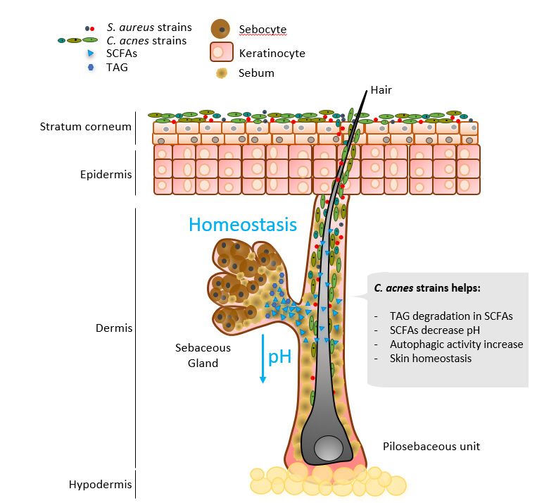
Lipid Modulation
Lipid Modulation
To To
obtain energy
obtain energyfrom
fromthetheabundant
abundant triacyclglycerols in sebum,
triacyclglycerols in sebum, C. acnes
C. acnessecretes a a
secretes
triacylglycerol lipase, GehA [74]. As a product of triacylglycerol fermentation,
triacylglycerol lipase, GehA [74]. As a product of triacylglycerol fermentation, C. acnes C. acnes
secretes short-chain
secretes fattyfatty
short-chain acidsacids
(SCFAs) (Figure
(SCFAs) 3). The
(Figure 3).major
The SCFAs are acetate
major SCFAs (C2), pro-
are acetate (C2),
pionate (C3), and
propionate butyrate
(C3), (C4) [75].
and butyrate Less
(C4) is known
[75]. about the
Less is known effect
about theofeffect
SCFAs ofon the skin,
SCFAs on the
although SCFAs metabolized
skin, although by the gut
SCFAs metabolized microbiome
by the have been
gut microbiome haveshown to contribute
been shown to the to
to contribute
maintenance of the of
the maintenance colonic epithelium
the colonic barrier
epithelium by modulating
barrier by modulatingepithelial tight
epithelial junctions,
tight junctions,
colonocyte
colonocyteproliferation, final
proliferation, differentiation,
final differentiation,and apoptosis
and [76].
apoptosis [76].
Figure
Figure 3. Selected
3. Selected Cutibacterium
Cutibacterium acnes
acnes major
major contributions
contributions in skin
in skin homeostasis.
homeostasis.
As As a result
a result of its
of its metabolism,
metabolism, C. acnes
C. acnes predominantly
predominantly produces
produces propionic
propionic acid,
acid, from
from
its former name, Propionibacterium,
which its former name, Propionibacterium, comes [21]. The role of propionic acid in the skinthe
which comes [21]. The role of propionic acid in
skin
is yet to is
beyet to be uncovered,
uncovered, although it although it contributes
contributes to maintaining to maintaining the on
the acidic layer acidic layer on
the skin’s
the skin’s surface [77]. The physiological pH of healthy skin ranges
surface [77]. The physiological pH of healthy skin ranges from 4.1 to 5.8 [78]. Increased from 4.1 to 5.8 [78].
pH has been reported in AD, irritant contact dermatitis, ichthyosis, rosacea, and acne, butand
Increased pH has been reported in AD, irritant contact dermatitis, ichthyosis, rosacea,
alsoacne, but also
in aged and indryaged
skinand
[78].dry skin [78].
Enzymes Enzymes
involved involved in maintaining
in maintaining skin barrier skin barrier
function,
epidermal cell differentiation, and lipid production and accumulation are pH-dependentpH-
function, epidermal cell differentiation, and lipid production and accumulation are
dependent
[78]. For example, [78].acid
For example, acid sphingomyelinase
sphingomyelinase hydrolyzes sphingolipids
hydrolyzes sphingolipids to liberate
to liberate ceramides
ceramides on the stratum corneum, an essential molecule on the skin
on the stratum corneum, an essential molecule on the skin[79]. Its activity is pH-regulated [79]. Its activity is
andpH-regulated
AD has beenand AD haswith
correlated beenimpaired
correlated with impaired sphingomyelinase
sphingomyelinase activity and highactivity
skin pHand
high skin pH [80,81].
[80,81].
2.2. Follicular Niche Competition
2.2. Follicular Niche Competition
Skin commensals are highly adapted to live in specific niches, in which they thrive
and outcompete pathogens for nutrient acquisition [15]. Different strategies of niche
modulation by C. acnes have been described in the literature (Figure 4).Microorganisms 2021, 9, x FOR PEER REVIEW 6 of 18
Skin commensals are highly adapted to live in specific niches, in which they thrive
Microorganisms 2021, 9, 628 6 of 18
and outcompete pathogens for nutrient acquisition [15]. Different strategies of niche mod-
ulation by C. acnes have been described in the literature (Figure 4).
Figure
Figure4.4.Selected
SelectedCutibacterium
Cutibacteriumacnes
acnesmajor
majorcontributions
contributionsininniche
nichemodulation.
modulation.
Recently,
Recently,aa biosynthetic
biosyntheticgenegene(BSG)
(BSG) cluster
clustercapable
capableof of producing
producing the the antimicrobial
antimicrobial
thiopeptide
thiopeptidecutimycin
cutimycinwas wasidentified
identifiedininspecific
specificC.C.acnes
acnesstrains
strainsfrom
fromphylotypes
phylotypesIB IBand
and
III
III[82].
[82].Interestingly,
Interestingly,onlyonly88ofof219
219screened
screenedC. C.acnes
acnesisolates
isolatescontained
containedthe thecutimycin
cutimycinBGC BGC
inintheir
theirgenome.
genome.Highlighting
Highlighting thethe
selective
selectiveniche competition
niche competition of skin commensals,
of skin commensals, the ex-
the
pression levels of the BSG cluster producing cutimycin were increased
expression levels of the BSG cluster producing cutimycin were increased when C. acnes when C. acnes was
anaerobically
was anaerobically cocultured with with
cocultured strains fromfrom
strains the genus Staphylococcus,
the genus Staphylococcus, and BSG cluster
and BSG ex-
cluster
pression
expression levels were
levels decreased
were decreased when
when C.C.acnes
acneswas
wasanaerobically
anaerobicallycocultured
coculturedwithwithstrains
strains
from
fromthethegenus
genusCorynebacterium
Corynebacterium[82]. [82].Cutimycin
Cutimycinwas wastested
testedininvitro
vitroand
andwaswasshown
shownto to
possess
possessantimicrobial
antimicrobialactivity
activityagainst
againstStaphylococcus
Staphylococcusaureus,
aureus,but
butnot
notActinobacteria
Actinobacteriaphyla. phyla.
InInvivo
vivosampling
samplingshowed
showedaadecreased
decreasedratioratioofofStaphylococcus
Staphylococcusto C.acnes
toC. acnesininindividual
individualhair hair
follicles positive for cutimycin BCG compared to hair follicles where
follicles positive for cutimycin BCG compared to hair follicles where the cutimycin BSG the cutimycin BSG
clusterwas
cluster wasnotnotdetected.
detected.
C.acnes
C. acnesisisthe
themain
maincolonizer
colonizerof ofthe
thepilosebaceous
pilosebaceousunit unitniche,
niche,where
whereititproduces
producesthe the
SCFAspropionic
SCFAs propionicacid,
acid,isobutyric,
isobutyric,andandisovaleric
isovalericacid
acidin inanaerobic
anaerobicconditions
conditions[83].
[83]. These
These
SCFAs were
SCFAs shown to restore S.
were shown S. epidermidis
epidermidisantibiotic
antibioticsensitivity
sensitivityby byreducing
reducingitsitscapacity
capacity to
form biofilms [83]. S. epidermidis biofilm
to form biofilms [83]. S. epidermidis biofilm formationformation is related to skin disorders and
skin disorders and rarely rarely
reportedin
reported inhealthy
healthyskin
skin [84].
[84]. An
Anolder
olderstudy
studyinvestigated
investigatedthe the antimicrobial
antimicrobialactivity
activityof of
acnecin, a peptide produced by a subset of C. acnes strains. It was
acnecin, a peptide produced by a subset of C. acnes strains. It was reported that acnecinreported that acnecin
inhibitsthe
inhibits thegrowth
growthof ofother
otherC.C.acnes
acnesstrains
strainsthat
thatdodonot
notproduce
produceacnecin
acnecin [85].
[85].
2.3.Immune
2.3. ImmuneModulation
Modulation
Different skin-resident immune cells contribute to tissue homeostasis [2]. The major
Different skin-resident immune cells contribute to tissue homeostasis [2]. The major
skin-resident immune cells are the myeloid and lymphoid cell subsets, which include a vari-
skin-resident immune cells are the myeloid and lymphoid cell subsets, which include a
ety of specialized cell families that contribute to skin homeostasis, inflammation, and tissue
variety of specialized cell families that contribute to skin homeostasis, inflammation, and
reconstruction [86]. The immune cells on the skin participate in tight interactions with the
tissue reconstruction [86]. The immune cells on the skin participate in tight interactions
skin microbiome to keep the skin healthy. It has been shown that germ-free mice had lower
immune responses to pathogens, which were rescued by S. epidermidis recolonization on
the skin [87]. Furthermore, S. epidermidis activates keratinocytes innate immune signaling
pathways, triggering an increase in antimicrobial peptides (AMPs) targeting S. aureus [88].Microorganisms 2021, 9, 628 7 of 18
Two different studies investigated immune interactions among C. acnes with keratinocytes
and sebocytes. It was reported that C. acnes would not trigger an immune response unless
environmental changes triggered higher production of SCFAs. These studies highlighted
the immune tolerance of the skin towards C. acnes on homeostasis [89,90]. Another study
explored the immune response of the skin to different C. acnes phylotypes, showing very
different immune response patterns for acne and health-associated C. acnes phylotypes [91].
Finally, other research has described the enhanced autophagy activity of keratinocytes
upon C. acnes interaction [92]. Autophagy is a major cellular defense mechanism to fight
pathogen invasion [93]. It was suggested that low levels of hair follicle colonization by
C. acnes could bring antimicrobial protection by locally enhancing autophagic activity
in keratinocytes [92]. In the studies described above, it is reflected that in healthy skin,
associated distribution of C. acnes phylotypes is tolerated and trains the host immune
system. C. acnes has been described to promote the activation of T helper type 1 (Th1)
cells in vivo [94]. Th1 cells belong to the cluster of differentiation 4 (CD4) Th group and
are essential for intracellular pathogens eradication, such as Listeria monocytogenes. The
other CD4 Th cells are type 2 (Th2) and are necessary for antibody production against
extracellular organisms. The equilibrium of Th1/Th2 response is crucial for proper immune
functioning [95]. Interestingly, AD is characterized by a shift in the Th1/Th2 equilibrium
towards strong Th2 cytokine expression, triggering an allergic reaction on the skin [96].
In a study on mice with AD, C. acnes injections increased the Th1/Th2 ratio by enhancing
the Th1 response. Th1 stimulation increased interleukin 12 (IL-12) and interferon gamma
(INF-γ) expression, which have been described to counteract the effects of Th2 cells. Th1
stimulation in mice resulted in reduced development of AD compared to the control [97].
In another experiment performed on mice, using in vivo injections of C. acnes in malignant
melanoma (MM), Th1 cells activated by C. acnes produced the antitumor cytokines IL-12,
tumor necrosis factor alpha (TNF-α), and INF-γ [98]. Subcutaneous granuloma formation,
which plays a major role in antitumor immunotherapy, was also observed. The tumor
size in mice with malignant melanoma treated with C. acnes was significantly smaller
than in the control mice [98]. This study concluded that intratumoral injection of C. acnes
vaccine (ITPV) suppressed MM by inducing IL-12, TNF-α, and IFN-γ expression, along
with granuloma formation.
2.4. Oxidative Stress Mitigation
The skin is constantly exposed to UV radiation, which triggers the formation of
reactive oxygen species (ROS) [99]. ROS oxidize lipids, proteins, and DNA, leading to
cell damage and contributing to skin carcinogenesis [100]. Epithelial cells have multiple
defense mechanisms to reduce ROS levels on the skin [101], such as the production of
melanin and enzymes with antioxidant properties [102]. In addition to the ROS-mitigating
strategies of epithelial cells, the most abundant secreted protein of C. acnes, radical oxy-
genase of Propionibacterium acnes (RoxP), has been reported to exert antioxidant activity
(Figure 5) [74,103]. RoxP is the first extracellular bacterial antioxidant enzyme to be char-
acterized [103]. RoxP is constitutively expressed and two variants with 83% amino acid
identity have been described, one for C. acnes clade I and the other for clades II and III,
both showing similar levels of antioxidant activity. RoxP was shown to increase the vi-
ability of ROS-stressed monocytes and keratinocytes in vitro, even to higher levels than
the non-stressed group [104]. Furthermore, in actinic keratosis (AK), an initial stage of
non-melanoma skin cancer, host cells antioxidant function is shown to be deficient [105].
In AK-affected sites, 16sRNA analysis showed a decrease in C. acnes populations, and
RoxP levels detected in vivo by a capacity biosensor were lower compared to healthy
areas [106,107].Microorganisms 2021, 9, 628 8 of 18
Microorganisms 2021, 9, x FOR PEER REVIEW 8 of 18
Figure
Figure5.5.Selected
SelectedCutibacterium
Cutibacteriumacnes
acnesmajor
majorcontributions
contributionsin
inoxidative
oxidativestress.
stress.
3.3.How
HowtotoPrevent
Preventand/or
and/orTreat
Treat“On“OnSite”
Site”Dysbiosis
DysbiosisDisorders
Disorders
Dysbiosisof
Dysbiosis ofmicrobiome
microbiomecomposition
compositionand and function
function makemake microbiome-modulating
microbiome-modulating
strategiesan
strategies aninteresting
interestingnovel
novelfield
fieldofofresearch
researchfor
fortreatment
treatmentofofdysbiotic
dysbioticconditions.
conditions.Cur- Cur-
rently,microbiome-modulating
rently, microbiome-modulatingstrategies
strategieshave
havebeen
beenmainly
mainlyaimed
aimedat atmodulating
modulatingthe thegut
gut
microbiota to redress dysbiotic patterns of the microbiome associated
microbiota to redress dysbiotic patterns of the microbiome associated with disease [108]. with disease [108].
Strategies aimed
Strategies aimed atat modulating
modulatingthe thegut
gutmicrobiota
microbiota involve
involve using probiotics,
using prebiotics,
probiotics, prebiotics,and
synbiotic and fecal microbiota transplants [109]. Microbiota transplantations
and synbiotic and fecal microbiota transplants [109]. Microbiota transplantations are are based
on transferring
based the microbiome
on transferring the microbiome fromfrom
a healthy subject
a healthy to the
subject dysbiotic
to the dysbioticreceiver.
receiver.Fecal
Fe-
microbiota transplants (FMT) have been proven to be effective for restoring
cal microbiota transplants (FMT) have been proven to be effective for restoring the phy- the phyloge-
netic richness
logenetic of the
richness of recipient’s intestinal
the recipient’s microbiota,
intestinal effectively
microbiota, treating
effectively gastric
treating Clostridium
gastric Clos-
difficile infections [110].
tridium difficile infections [110].
Microbiomemodulation
Microbiome modulationthrough
throughthe theusage
usageof ofprobiotics,
probiotics,prebiotics,
prebiotics,and
andsynbiotics
synbiotics
already has a long history of health claims through oral usage. Probiotics are defined
already has a long history of health claims through oral usage. Probiotics are defined as
as “live microorganisms which when administered in adequate amounts confer a health
“live microorganisms which when administered in adequate amounts confer a health ben-
benefit on the host” [111]. Microorganism genera most commonly used are Lactobacillus,
efit on the host” [111]. Microorganism genera most commonly used are Lactobacillus,
Bifidobacterium, Enterococcus, Lactococcus, Streptococcus, Bacillus, and yeast species such
Bifidobacterium, Enterococcus, Lactococcus, Streptococcus, Bacillus, and yeast species
as Saccharomyces boulardii [112]. In light of the research conducted mainly in the last 10 years,
such as Saccharomyces boulardii [112]. In light of the research conducted mainly in the last
the health benefits of these probiotics, administered through the gastrointestinal tract, act
10 years, the health benefits of these probiotics, administered through the gastrointestinal
through four different mechanisms of action: (i) improvement of the epithelial barrier
tract, act through four different mechanisms of action: (i) improvement of the epithelial
function, (ii) interference with pathogenic bacteria, (iii) immunomodulation, and (iv) in-
barrier function, (ii) interference with pathogenic bacteria, (iii) immunomodulation, and
fluence on other organs of the body through the immune system [112]. Different strains
(iv) influence on other organs of the body through the immune system [112]. Different
of microbial species have specialized enzymatic activities and varied metabolic strategies,
strains of microbial species have specialized enzymatic activities and varied metabolic
even within one species [113,114]. The mechanistic basis of probiotics is not associated with
strategies,
the genus even within
or species of one species [113,114].
a microorganism, The mechanistic
but claims can only bebasis
made offor
probiotics is not
a few specially
associated with the
selected strains of agenus or species
particular speciesof[115].
a microorganism,
Prebiotics arebut claims can
substrates only beutilized
selectively made for by
ahost
few microorganisms,
specially selectedconferring
strains of ahealth
particular species [115]. Prebiotics are substrates
benefits [116]. Such selectivity was shown for selec-
the
tively utilized byBifidobacterium,
gut commensal host microorganisms, whichconferring health benefits
can be promoted [116]. Such
by the ingestion selectivity
of substances
such as fructo-saccharides, oligosaccharides, and other prebiotics [117]. Synbiotics areMicroorganisms 2021, 9, 628 9 of 18
products containing both prebiotics and probiotics. The implied synergism in the term
reserves the definition “for products in which the prebiotic compound selectively favors
the probiotic compound” [109]. Under this definition, a combination of an oligofructose
and Bifidobacterium would classify as a synbiotic. Postbiotics refer to non-viable microor-
ganisms (mainly heat-treated probiotic cells), lysed microbial cells, cell-free supernatants
containing metabolites produced by probiotic strains, or purified key components from
this supernatant [118]. These non-viable products have been tentatively termed postbi-
otics, but international consensus has yet to be reached [119]. The effects of non-viable
microbial components such as heat-killed or UV-inactivated cells and cellular components
when the cell is destroyed through lysis (DNA, RNA, proteins/peptides, polysaccharides,
lipids, and others) have been shown to be immunomodulatory [120–123]. The metabolites
derived from microbial metabolism include synthesized metabolites (SCFAs, bacteriocins,
antioxidants, and others).
3.1. Skin Microbiome Modulation Strategies
Dysbiosis of the skin microbiome, associated with skin disorders, could be altered
via multiple mechanisms: skin microbiome transplant, prebiotics, probiotics, synbiotics,
and, putatively, postbiotics. Whole skin microbiome transplantation would require col-
lecting the skin microbial community, which is challenging to achieve compared to fecal
microbiota samples, as a culturing step is always required. Another ongoing challenge
regards the uncultivability of microorganisms in vitro, also known as the “great plate count
anomaly” [124]. Therefore, the performance of skin microbiome transplantation analogous
to FMT is not scalable or industry applicable.
Recent years have seen a sharp increase in clinical investigations of probiotics and
postbiotics used in dermatology. Processes to produce such non-viable fermentation
products (postbiotics) are industrially scalable, can easily be formulated into products, and
are widely used in the cosmetic industry. Probiotics, on the other hand, pose a challenge in
terms of formulation and packaging to ensure the viability of the microorganism [125]. As a
result, most suppliers formulate using postbiotics. Nevertheless, while postbiotics of skin
commensals may protect against UV-induced oxidative damage, hyperpigmentation [126],
and pathogens such as S. aureus [127], reintroducing viable microorganisms into their
adapted niche has the potential to modulate the microbiome [128]. Skin microbiome
modulation could mitigate or potentially eliminate pathological skin conditions, analogous
to strategies in the gut. Callewaert et al. recently (2021) reviewed efforts undertaken in
skin microbiome modulation strategies [128] and reintroduced the term bacteriotherapy to
describe probiotics and postbiotics.
3.2. Bacteriotherapy in Dermatology
A recent review conducted in 2019 by Yu et al. investigated the interventional out-
comes of oral probiotics and topical bacteriotherapy for dermatological conditions. Oral
probiotics can be either consumed as encapsulated freeze-dried bacteria [129] or consumed
through fermented foods such as yogurt, kefir, kimchi, and others [130]. In addition to
alleviating gastrointestinal disorders, recent studies have investigated the usage of encap-
sulated probiotics in dermatology. It was noted that most clinical studies investigating
probiotics in dermatology have investigated the effects of orally consumed probiotics for
their interventional outcome, mainly aimed at alleviating acne and AD [131]. Although
initial interventional outcomes of oral probiotics on dermatological conditions show promis-
ing results, we focus in this review on the underexplored potential of topical probiotics as
a novel route for the prevention or treatment of “on site” dysbiosis disorders. Excluding
duplicates from meta-analyses on oral and topical probiotics used in dermatology, only
9 out of 72 studies focused on topical bacteriotherapy. In addition to those reviewed by
Yu et al., five studies were added that matched our search criteria on PubMed (“topical”,
“probiotic”, “bacteriotherapy”, “dermatology”). Two were recent interventional studies in-
vestigating the outcome of topical bacteriotherapy in acne24 and AD25 and three additionalMicroorganisms 2021, 9, 628 10 of 18
studies [133–135] investigated the application of bacteriotherapy on healthy [132,133] and
sensitive skin [134]. The first is a recent study by Karoglan et al. (2019) [135], who investi-
gated the application of a mixture of probiotic strains (SLST types: C3, K8, A5, F4) from the
skin commensal C. acnes in 14 patients with acne. Prior to topically applying the different
mixtures of commensal probiotic strains, the skin microbial community was reduced using
benzoyl peroxide to enhance the potential for transplantation [135]. As it has been noted
that mixtures of strains from different phylogenetic lineages have synergistic effects on
transplantation, two different mixtures of strains were assessed. The strains applied were
selected based upon their production of linoleic isomerase, a biomarker putatively asso-
ciated with inflammation [135]. This open-label study, without a control group, reported
a significant reduction in total lesions compared to baseline. Interestingly, the applied
probiotics could be detected after treatment in 50% of the treated patients, suggesting their
ability to engraft onto the skin. No adverse events were reported by the patients, indicating
that topical probiotics with C. acnes are safe. The second study included for revision is a
randomized, placebo-controlled study conducted by Butler et al. (2020) [136], where the
effect of the gut commensal Lactobacillus Reuteri (DSM 17938) strain was investigated in
36 adults with AD. When comparing the probiotic to the control, there was no significant
improvement in scoring atopic dermatitis (SCORAD) index score. Thirdly, an interven-
tional placebo-controlled study by Di Marzio et al. (2008) [132] was included, in which the
authors investigated the application of non-viable commensal Streptococcus thermophilus
cells on healthy skin of 20 elderly subjects. After receiving twice-daily dosages for 15 days,
they reported a significant increase in hydration (p = 0.001), although no significant differ-
ence in transepidermal water loss (TEWL) was reported. The fourth study included is an
interventional study by Nodake et al. (2015) [133] applying viable commensal S. epidermis
strains to the healthy skin of 21 elderly subjects. They received twice-weekly dosages for
4 weeks in a double-blinded, placebo-controlled study. A significant reduction in TEWL
(p < 0.05), significant increase in lipid content (p < 0.05), and significant increase in hydra-
tion (p < 0.05) were observed. Furthermore, they reported a significant reduction in the
pH of the skin of elderly participants (p < 0.05). Interestingly, a meta-analysis of 63 ran-
domized controlled trials revealed that the use of products with a low pH (4) is currently
the most effective strategy for improving the skin barrier [137]. The fifth additional study
is a randomized, placebo-controlled study by Gueniche et al. (2010) [134]. They applied
lysed Bifidobacterium longum reuteri postbiotics to participants with sensitive skin twice
daily for 2 months. The treatment group showed significantly decreased skin sensitivity
as assessed with a lactic acid test (p < 0.01). Furthermore, they reported significantly
improved resistance to physical aggression in the form of tape-stripping (p < 0.01). With the
addition of these five recent studies, a total of 14 studies investigating the interventional
outcomes of topical probiotics and postbiotics were reviewed. All interventional studies
are summarized in Supplementary Table S1, including strain origin, viability, dose, and
main results. Furthermore, bacterial phages naturally present on the skin microbiome are
being studied and investigated for their microbiome-modulating properties by targeting
specific species such as C. acnes [138].
3.3. Probiotics Used in Dermatology
The use of probiotics in dermatology involves mainly oral probiotics (80%), and of
the 14 studies included in this review, only eight investigated viable bacterial probiotics.
Only 6 out of 14 studies investigated interventional outcomes of skin commensal strains,
as the other studies included strains isolated from the gastrointestinal tract (Lactobacillus
reuteri DSM 17938, Lactobacillus plantarum, Bifidobacterium longum reuteri), urogenital tract
(Lactobacillus johnsonii), thermal water (Vitreoscilla filiformis), and the skin of Amerindian
peoples (Nitrosomonas eutropha). Despite the promising results of the clinical investigations
presented in this review (mainly targeted towards acne and AD), research on topical
probiotics is still in its very early stages. There is an underexploited opportunity to apply
viable commensal strains that have the potential to restore the dysbiosis of skin disorders.Microorganisms 2021, 9, 628 11 of 18
There is an unmet need for patients to find effective treatments with limited side-effects.
Commensal skin probiotics should be further explored to provide patients with novel
treatment strategies with strain-associated beneficial properties that are adapted to the
environmental habitat of application.
3.4. Safety Functionality and Technical Feasibility for Skin Probiotics
As topical probiotics are nascent in the cosmetic and dermatological industry, their
regulatory classification is unclear and may fall between cosmetics, medical devices, and
pharmaceuticals [139]. Rigorous standards for the selection of specific strains according to
their safety, functionality, and technical feasibility have been established for gut probiotics
through years of research and development [140]. A review published in 2017 outlined
the safety, functional, and technological aspects that must be considered when selecting
probiotic strains for usage in the gastrointestinal tract. These were compiled from recom-
mendations by the Food and Agricultural Organization, World Health Organization, and
European Food Safety Authority [140]. Adapting similar rigorous selection criteria for skin
probiotic strains would be desirable for skin probiotics, to protect and deliver value to
patients, as depicted in Table 1 using probiotics with C. acnes as an example.
Table 1. Selection criteria for skin probiotic strains (adapted from and Katarzyna Śliżewska [140]).
Safety Statement
The novel genus Cutibacterium contains the human cutaneous species
Human or animal origin formerly known as Propionibacterium acnes, Propionibacterium avidum,
Propionibacterium granulosum [21].
C. acnes is a prevalent bacterial commensals on healthy skin [19]. Studies
Isolated from the skin
investigating probiotic C. acnes strains used isolates from the skin of healthy
of healthy individuals
individuals [135,141].
C. acnes has no long history of use yet, see “no adverse effects” for safety
History of safe use
indication [135].
Precise diagnostic identification
SLST analysis allows the strain-level identification of C. acnes species [142].
(phenotype and genotype traits)
Strains of C. acnes have been reported as opportunistic pathogens. Virulence is
Absence of data regarding an
related to certain phylotypes or strains (e.g., phylotype IA has been associated
association with infective disease
with acne [143]).
No adverse events were reported in two studies applying C. acnes
No adverse effects
probiotics [135,141].
Absence of genes responsible for antibiotic No detectable differences in the MIC of 21 antibiotics were observed between
resistance localized in non-stable elements parent strains and their plasmid-cured derivatives for C. acnes [144].
Functionality Statement
Competitiveness with respect to the microbiota
Antagonistic activity between S. epidermis and C. acnes strains [145].
inhabiting the ecosystem of the skin
Ability to survive and maintain the metabolic Applied probiotic strains of C. acnes could be detected well beyond application
activity and to grow in the target site days, indicating their ability to survive in the target site [141].
C. acnes is adapted to lipid-rich anaerobic environment of sebaceous
Resistance to skin salts and enzymes
follicles [146].
Resistance to low pH of the skin C. acnes can compensate for the low pH by utilizing arginine deiminase [147].
Competitiveness with respect to microbial
C. acnes inhibits biofilm formation of S. epidermidis [83]. C. acnes produces an
species inhabiting the ecosystem of the skin
antibiotic that targets specific C. acnes strains [85].
(including closely related species)
C. acnes produces an antibiotic against S. aureus and C. acnes ferments have
been shown to inhibit methicillin-resistant S. aureus [82,127]. S. epidermis
Antagonistic activity towards pathogens
degrades proteins associated with S. aureus biofilm formation [148].
(e.g., S. aureus)
S. lugdunensis inhibits growth of S. aureus through production of AMP
lugdunin [149].Microorganisms 2021, 9, 628 12 of 18
Table 1. Cont.
Functionality Statement
Resistance to bacteriocins and acids produced C. acnes and S. epidermidis coexist in the skin as stable heterogeneous
by the endogenic skin microbiota communities of strains [19].
Adherence and ability to colonize some
Different body sites are shown to be colonized by different multi-phyletic
particular sites within the host organism,
communities of C. acnes [15].
and an appropriate survival rate on the skin
Technological feasibility Statement
Easy production of large amounts of biomass
Steady-state continuous culture of Propionibacterium acnes was achieved [150].
and high productivity of cultures
Viability and stability of the desired properties
of probiotic bacteria during the fixing process
Not yet established.
(freezing, freeze-drying), preparation, and
distribution of probiotic products
High storage survival rate in finished products C. acnes probiotic solution was stable for at least 1.5 months at room
(in aerobic and micro-aerophilic conditions) temperature [141]
Guarantee of desired sensory properties of
finished products (in the case of the Not yet established
cosmetics industry)
The sequenced strain exhibited 100% identity on the 16S ribosomal RNA to
Genetic stability
several isolated C. acnes [73].
C. acnes phylotypes show selective resistance or sensitivity to
Resistance to bacteriophages
bacteriophages [151].
4. Conclusions
C. acnes is a commensal bacterium of human skin. As reviewed in this paper, the rela-
tive abundance and/or diversity of clonal population is associated with skin health and is
involved in maintaining the essential biophysiological functions of the skin. The history of
this genus is constantly evolving as research and technology progress. Mainly regarded
as the cause of disease due to the virulence and pathogenic factors associated with a
large part of its clonal population, the paradigm of C. acnes colonization is now shifting.
From a uniquely opportunistic pathogen, the nascent alternative vision encourages the
potential use of some specific strains of C. acnes as efficient probiotics to restore the natural
equilibrium of a disbalanced skin microbiome.
Supplementary Materials: The following are available online at https://www.mdpi.com/2076-260
7/9/3/628/s1.
Author Contributions: M.R., A.H.d.R., F.B. and M.J.F. wrote, reviewed, and edited the manuscript.
B.P., M.G. and A.Z. reviewed and edited the manuscript. All authors have read and agreed to the
published version of the manuscript.
Funding: This research received no external funding.
Conflicts of Interest: The authors declare no conflict of interest.
References
1. Mosteller, R.D. Simplified Calculation of Body-Surface Area. N. Engl. J. Med. 1987, 317, 1098. [CrossRef] [PubMed]
2. Kabashima, K.; Honda, T.; Ginhoux, F.; Egawa, G. The Immunological Anatomy of the Skin. Nat. Rev. Immunol. 2019, 19, 19–30.
[CrossRef] [PubMed]
3. Wong, R.; Geyer, S.; Weninger, W.; Guimberteau, J.-C.; Wong, J.K. The Dynamic Anatomy and Patterning of Skin. Exp. Dermatol.
2016, 25, 92–98. [CrossRef] [PubMed]
4. Gallo, R.L. Human Skin Is the Largest Epithelial Surface for Interaction with Microbes. J. Investig. Dermmatol. 2017, 137, 1213–1214.
[CrossRef] [PubMed]Microorganisms 2021, 9, 628 13 of 18
5. Jahns, A.C.; Alexeyev, O.A. Three Dimensional Distribution of Propionibacterium Acnes Biofilms in Human Skin. Exp. Dermmatol.
2014, 23, 687–689. [CrossRef]
6. Nakatsuji, T.; Chiang, H.-I.; Jiang, S.B.; Nagarajan, H.; Zengler, K.; Gallo, R.L. The Microbiome Extends to Subepidermal
Compartments of Normal Skin. Nat. Commun. 2013, 4. [CrossRef]
7. Oh, J.; Byrd, A.L.; Deming, C.; Conlan, S.; Kong, H.H.; Segre, J.A. Biogeography and Individuality Shape Function in the Human
Skin Metagenome. Nature 2014, 514, 59–64. [CrossRef] [PubMed]
8. Carlotta De, L.R. Mycobiota: Micro-Eukaryotes Inhabiting Our Body as Commensals or Opportunistic Pathogens. Fungal. Genom.
Biol. 2015, 5. [CrossRef]
9. Underhill, D.M.; Iliev, I.D. The Mycobiota: Interactions between Commensal Fungi and the Host Immune System. Nat. Rev.
Immunol. 2014, 14, 405–416. [CrossRef]
10. Hannigan, G.D.; Meisel, J.S.; Tyldsley, A.S.; Zheng, Q.; Hodkinson, B.P.; SanMiguel, A.J.; Minot, S.; Bushman, F.D.; Grice, E.A.
The Human Skin Double-Stranded DNA Virome: Topographical and Temporal Diversity, Genetic Enrichment, and Dynamic
Associations with the Host Microbiome. mBio 2015, 6. [CrossRef]
11. Lecuit, M.; Eloit, M. The Human Virome: New Tools and Concepts. Trends Microbiol. 2013, 21, 510–515. [CrossRef]
12. Grice, E.A.; Kong, H.H.; Renaud, G.; Young, A.C.; Bouffard, G.G.; Blakesley, R.W.; Wolfsberg, T.G.; Turner, M.L.; Segre, J.A.
A Diversity Profile of the Human Skin Microbiota. Genome Res. 2008, 18, 1043–1050. [CrossRef] [PubMed]
13. Grice, E.A.; Kong, H.H.; Conlan, S.; Deming, C.B.; Davis, J.; Young, A.C.; Bouffard, G.G.; Blakesley, R.W.; Murray, P.R.; Green,
E.D.; et al. Topographical and Temporal Diversity of the Human Skin Microbiome. Science 2009, 324, 1190–1192. [CrossRef]
[PubMed]
14. Costello, E.K.; Lauber, C.L.; Hamady, M.; Fierer, N.; Gordon, J.I.; Knight, R. Bacterial Community Variation in Human Body
Habitats Across Space and Time. Science 2009, 326, 1694–1697. [CrossRef]
15. Byrd, A.L.; Belkaid, Y.; Segre, J.A. The Human Skin Microbiome. Nat. Rev. Microbiol. 2018, 16, 143–155. [CrossRef]
16. Grice, E.A.; Segre, J.A. The Skin Microbiome. Nat. Rev. Microbiol 2011, 9, 244–253. [CrossRef] [PubMed]
17. Findley, K.; Oh, J.; Yang, J.; Conlan, S.; Deming, C.; Meyer, J.; Schoenfeld, D.; Nomicos, E.; Park, M.; Becker, J.; et al. Topographic
Diversity of Fungal and Bacterial Communities in Human Skin. Nature 2013, 498. [CrossRef] [PubMed]
18. Ying, S.; Zeng, D.-N.; Chi, L.; Tan, Y.; Galzote, C.; Cardona, C.; Lax, S.; Gilbert, J.; Quan, Z.-X. The Influence of Age and Gender on
Skin-Associated Microbial Communities in Urban and Rural Human Populations. PLoS ONE 2015, 10, e0141842. [CrossRef]
19. Oh, J.; Byrd, A.L.; Park, M.; Kong, H.H.; Segre, J.A. Temporal Stability of the Human Skin Microbiome. Cell 2016, 165, 854–866.
[CrossRef] [PubMed]
20. Lebeer, S.; Spacova, I. Exploring Human Host–Microbiome Interactions in Health and Disease—How to Not Get Lost in
Translation. Genome Biol 2019, 20, 56. [CrossRef]
21. Scholz, C.F.P.; Kilian, M. The Natural History of Cutaneous Propionibacteria, and Reclassification of Selected Species within
the Genus Propionibacterium to the Proposed Novel Genera Acidipropionibacterium Gen. Nov., Cutibacterium Gen. Nov. and
Pseudopropionibacterium Gen. Nov. Int. J. Syst. Evol. Microbiol. 2016, 66, 4422–4432. [CrossRef] [PubMed]
22. Fournière, M.; Latire, T.; Souak, D.; Feuilloley, M.G.J.; Bedoux, G. Staphylococcus Epidermidis and Cutibacterium acnes: Two Major
Sentinels of Skin Microbiota and the Influence of Cosmetics. Microorganisms 2020, 8, 1752. [CrossRef] [PubMed]
23. Schommer, N.N.; Gallo, R.L. Structure and Function of the Human Skin Microbiome. Trends Microbiol. 2013, 21, 660–668.
[CrossRef] [PubMed]
24. Grice, E.A. The Skin Microbiome: Potential for Novel Diagnostic and Therapeutic Approaches to Cutaneous Disease. Semin.
Cutan. Med. Surg. 2014, 33, 98–103. [CrossRef]
25. Chien, A.L.; Tsai, J.; Leung, S.; Mongodin, E.F.; Nelson, A.M.; Kang, S.; Garza, L.A. Association of Systemic Antibiotic Treatment
of Acne With Skin Microbiota Characteristics. JAMA Dermmatol. 2019, 155, 425–434. [CrossRef] [PubMed]
26. Weidinger, S.; Novak, N. Atopic Dermatitis. Available online: https://pubmed.ncbi.nlm.nih.gov/26377142/ (accessed on 26
January 2021).
27. Larsen, F.S.; Hanifin, J.M. Epidemiology of Atopic Dermatitis. Immunol. Allergy Clin. North. Am. 2002, 22, 1–24. [CrossRef]
28. Williams, M.R.; Gallo, R.L. Evidence That Human Skin Microbiome Dysbiosis Promotes Atopic Dermatitis. J. Investig. Dermatol.
2017, 137, 2460–2461. [CrossRef] [PubMed]
29. Francuzik, W.; Franke, K.; Schumann, R.; Heine, G.; Worm, M. Propionibacterium Acnes Abundance Correlates Inversely with
Staphylococcus Aureus: Data from Atopic Dermatitis Skin Microbiome. Acta Dermmatol. Venereol. 2018, 98. [CrossRef]
30. Kong, H.H.; Oh, J.; Deming, C.; Conlan, S.; Grice, E.A.; Beatson, M.A.; Nomicos, E.; Polley, E.C.; Komarow, H.D.; Murray, P.R.;
et al. Temporal Shifts in the Skin Microbiome Associated with Disease Flares and Treatment in Children with Atopic Dermatitis.
Genome Res. 2012, 22, 850–859. [CrossRef]
31. Nakatsuji, T.; Chen, T.H.; Narala, S.; Chun, K.A.; Two, A.M.; Yun, T.; Shafiq, F.; Kotol, P.F.; Bouslimani, A.; Melnik, A.V.; et al.
Antimicrobials from Human Skin Commensal Bacteria Protect against Staphylococcus Aureus and Are Deficient in Atopic
Dermatitis. Sci. Transl. Med. 2017, 9. [CrossRef]
32. Gether, L.; Overgaard, L.K.; Egeberg, A.; Thyssen, J.P. Incidence and Prevalence of Rosacea: A Systematic Review and Meta-
Analysis. Br. J. Dermatol. 2018, 179, 282–289. [CrossRef]You can also read