Non-Nutritive Sweeteners and Their Implications on the Development of Metabolic Syndrome - MDPI
←
→
Page content transcription
If your browser does not render page correctly, please read the page content below
nutrients
Review
Non-Nutritive Sweeteners and Their Implications on
the Development of Metabolic Syndrome
Iryna Liauchonak 1,† , Bessi Qorri 2,† , Fady Dawoud 1,† , Yatin Riat 1,†,‡ and
Myron R. Szewczuk 2, *
 1   Graduate Diploma and Professional Master in Medical Sciences, Postgraduate Medical Education, School of
     Medicine, Queen’s University, Kingston, ON K7L 3N6, Canada; iryna.liauchonak@gmail.com (I.L.);
     Fady.Dawoud@hotmail.com (F.D.); riatyatin@gmail.com (Y.R.)
 2   Department of Biomedical and Molecular Sciences, Queen’s University, Kingston, ON K7L 3N6, Canada;
     bessi.qorri@queensu.ca
 *   Correspondence: szewczuk@queensu.ca; Tel.: +1-613-533-2457; Fax: +1-613-533-6796
 †   These authors contributed equally to this work.
 ‡   Current address: Emergency Medicine, Resident Physician, Brooklyn Hospital Centre, 121 DeKalb Avenue,
     Brooklyn, NY 11201, USA.

 Received: 28 February 2019; Accepted: 13 March 2019; Published: 16 March 2019

 Abstract: Individuals widely use non-nutritive sweeteners (NNS) in attempts to lower their overall
 daily caloric intake, lose weight, and sustain a healthy diet. There are insufficient scientific
 data that support the safety of consuming NNS. However, recent studies have suggested that
 NNS consumption can induce gut microbiota dysbiosis and promote glucose intolerance in
 healthy individuals that may result in the development of type 2 diabetes mellitus (T2DM). This
 sequence of events may result in changes in the gut microbiota composition through microRNA
 (miRNA)-mediated changes. The mechanism(s) by which miRNAs alter gene expression of different
 bacterial species provides a link between the consumption of NNS and the development of metabolic
 changes. Another potential mechanism that connects NNS to metabolic changes is the molecular
 crosstalk between the insulin receptor (IR) and G protein-coupled receptors (GPCRs). Here, we aim
 to highlight the role of NNS in obesity and discuss IR-GPCR crosstalk and miRNA-mediated changes,
 in the manipulation of the gut microbiota composition and T2DM pathogenesis.

 Keywords: non-nutritive sweeteners; type 2 diabetes mellitus; gut microbiota; GPCR; insulin receptor
 signaling; miRNAs

1. Introduction
      Artificial sweeteners have gained increasing attention as dietary assessment tools to help combat
the obesity epidemic by providing a sweet taste without the extra calories [1]. Taste has a significant
role in human perception of food quality, contributing to its overall pleasure and enjoyment. To this
end, the development of sweeteners as food additives that mimic the sweet taste of natural sugars
suggest promise [2]. These artificial sweeteners are classified as nutritive or nonnutritive, both of
which enhance the flavor and texture of food. Nutritive sweeteners contain carbohydrates and provide
calories (energy). Non-nutritive sweeteners (NNS) are very low calorie or zero calorie alternatives that
provide minimal or no carbohydrates or energy [3].

Nutrients 2019, 11, 644; doi:10.3390/nu11030644                              www.mdpi.com/journal/nutrients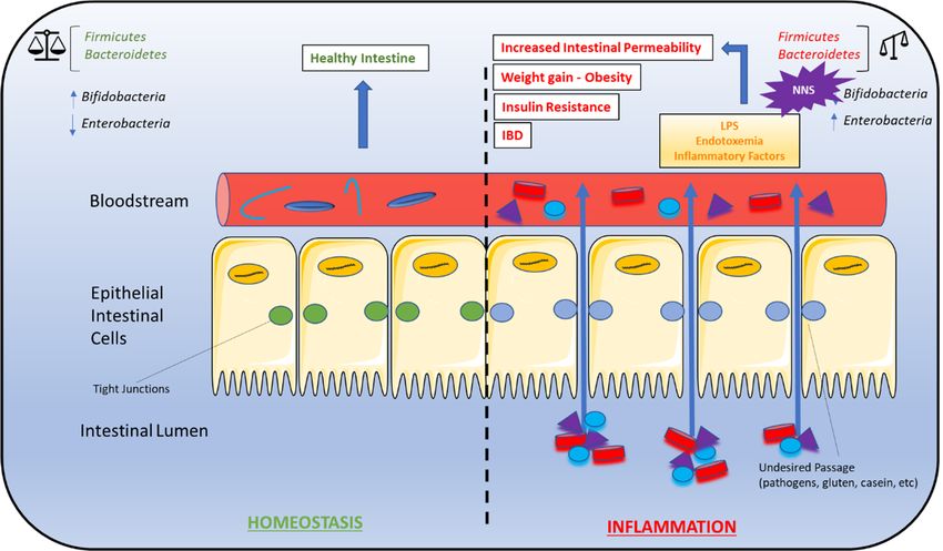
Nutrients 2019, 11, 644                                                                              2 of 19

      As part of dietary intake, NNS consumption can modulate energy balance, and metabolic
functions through several peripheral and central mechanisms, suggesting that NNS are not inert
compounds as once thought [4]. However, the specific mechanism(s) and details of the effects of NNS
consumption on host metabolism and energy homeostasis remain to be elucidated. This is particularly
relevant as NNS have been an option for individuals to improve their health; yet, NNS consumption
has been associated with increased risk factors for metabolic syndrome [5]. Here, metabolic syndrome
refers to the collection of physiological, biochemical, clinical, and metabolic factors that contribute
to the increased risk of cardiovascular disease and type 2 diabetes melitus (T2DM) [6]. Based on
measurements and laboratory tests, metabolic syndrome can also contribute to hypertension, glucose
intolerance, proinflammatory state, atherogenic dyslipidemia, prothrombic state [7], and kidney
disease [8]. It is noteworthy that the cause of these health-related issues may be due to emerging
contaminants in the environment worldwide and their associated risks to human health and the
environment [9]. Interestingly, one study identified a total of 24 non-nutritive artificial sweeteners
studies to their occurrence in the environment from 38 locations globally across Europe, including
the United Kingdom, Canada, United States, and Asia. Overall, the findings of the study indicated
that non-nutritive artificial sweeteners are present in surface water, tap water, groundwater, seawater,
lakes, and atmosphere [9]. Furthermore, in a Norwegian pregnancy cohort study, sucrose-sweetened
soft beverages were reported to increase the risk of congenital heart defects (CHDs) in offspring, while
fruit juices, cordial beverages, and artificial sweeteners had no associations with CHD [10].
      In this review, we discuss the current status on the use of non-nutritive sweeteners, or non-caloric
artificial sweeteners (NAS) which are used interchangeably and the future use of NNS in the food
industry. This review has a particular focus on identifying the underlying mechanisms that can
be responsible for the development of metabolic syndrome associated with NNS consumption.
The physiological effects of NNS including NNS-induced metabolic changes will be discussed. We will
highlight the development of metabolic syndrome that collectively involves the potential role of
GPCR-IR crosstalk in the development of glucose intolerance and insulin resistance, the development
of T2DM and the pathological mechanisms by which microRNAs (miRNAs) may mediate the changes
in gut microbiota composition.

2. Current Status on the Use of Non-Nutritive Sweeteners
      Currently, the Food and Drug Administration (FDA) has approved the use of acesulfame-potassium
(Ace-K), aspartame, neotame, saccharin, sucralose, and stevia (https://www.fda.gov/food/
ingredientspackaginglabeling/foodadditivesingredients/ucm397725.htm). Saccharin was discovered
as early as 1876 and was the “original” artificial sweetener used in the food industry. Unfortunately,
saccharin and many of its sweet alternatives have been considered to be health hazards, and as a result,
are banned in many countries. Recently, other sweeteners have been developed and implemented within
the food industry. In general, there are three primary types of sweeteners used in the food industry
today: high-intensity sweeteners (e.g., acesulfame potassium, advantame, aspartame, neotame, saccharin,
and sucralose), sugar alcohols (e.g., erythritol, glycerol, mannitol, sorbitol, and xylitol), and natural
sweeteners (e.g., honey, lucuma powder, maple syrup, monk fruit known as Siraitia grosvenorii swingle
fruit extract, stevia, and yacon syrup) [11]. These sweeteners and their uses in the food industry are
summarized in Table 1. The high-intensity sweeteners can be synthetic or natural and are classified into
two categories: nutritive and non-nutritive. The majority of high-intensity sweeteners used today fall into
the non-nutritive category, with the exception of aspartame. Sugar alcohols are found naturally in small
amounts in fruits and vegetables but are produced commercially in larger quantities.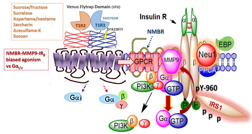
Nutrients 2019, 11, 644                                                                                                     3 of 19

              Table 1. Classification of Food and Drug Administration (FDA)-approved sweeteners.

                                                                                                             Relative Sweetness
          Name                  Brand Names                      Applications in Food Industry                 (Measured to
                                                                                                                  Sucrose)
                                                     High-intensity Sweeteners
                           Sweet and Low® , Sweet
                                                           Beverages, bases, and mixes for many food
        Saccharin          Twin® , Sweet’N Low® ,                                                                200–700×
                                                                products, table sugar substitute
                               Necta Sweet®
                                                           Soft drinks, chewing gum, pudding, cereals,
                            Nutrasweet® , Equal® ,                         instant coffee
       Aspartame *                                                                                                 200×
                                Sugar Twin®                  Also distributed as a “General Purpose
                                                                            Sweetener”
  Acesulfame-potassium                                   Beverages, candy, frozen desserts, baked goods
                            Sunett® , Sweet One®                                                                   200×
         (Ace-K)                                             Heat stable so it can be used in baking
        Sucralose                 Splenda®                                                                         600×
         Neotame                 Newtame®                            Beverages, candy gum                      7000–13,000×
                                                        Baked goods, beverages, frozen desserts, frosting,
       Advantame                    N/A                    chewing gum, candy, pudding, jelly and                 20,000×
                                                                         jam, gelatin
                                                          Sugar Alcohols
                                                          Fondant, ice cream, gum, tabletop sweeteners,
        Erythritol                                                                                              0.60×–0.70×
                                                            chocolate, dairy products, jelly, beverages
                                                          Dairy products, processed fruits, energy bars,
                                                                           jam, fondant
         Glycerol
                                                         Often used as a thickening agent and to provide
                                                                          texture to food
                                                          Infant formula, frozen fish, precooked pasta,
        Mannitol                                                                                                0.50×–0.70×
                                                               butter, chocolate flavored coatings
   Sorbitol (Glucitol) *                                               Used as emulsifier                          0.66×
                                                          Hard candy, chewing gum, mints, ice cream,
          Xylitol                                            chocolate, cookies, beverages, table                   1×
                                                                       sugar substitute
                                                        Natural Sweeteners
                            Natural constituents of
                                leaves of Stevia
    Steviol glycosides        rebaudiana (Bertoni)              Beverages, chewing gum, candy                    200–400×
                           plant, commonly known
                                    as Stevia
                              Siraitia grosvenorii
   Luo Han Guo Monk
                             Swingle fruit extract                             Tea                               100–250×
      fruit extracts
                                    (SGFE)
                                                              Beverages, pudding, granola, pastry,
     Lucuma powder
                                                                         baked goods
      * Nutritive sweetener. Content taken in part from the FDA approval of artificial sweeteners. https://www.fda.gov/
      food/ingredientspackaginglabeling/foodadditivesingredients/ucm397725.htm and Shwide-Slavin et al. [11].

     Although sugar substitutes have been around since the 1880s, artificial sweetener consumption
has dramatically increased over the last two decades as they are favorable alternatives to sucrose and
other sugar substitutes. NNS can be several hundred to thousands times sweeter than sucrose with
negligible caloric value, making them favorable health tools in attempts to control caloric intake and
to assist in weight loss [12,13]. This trend has resulted in NNS becoming a staple in the Western diet,
with cross-sectional studies reporting that 25% of children and 41% of adults consume low-calorie
sweeteners. Consumption of NAS is found to be higher amongst females, obese individuals, and
non-Hispanic white individuals as well as those with higher incomes [12,14].
     Although these low-calorie sugar substitutes seem promising, NAS consumption has been
associated with several inconsistent reports regarding their effects on the body. Due to the
up-and-down history surrounding sweeteners used in the food industry, it can be quite confusing
to understand what they are and how they are used. The greatest concerns are regarding the safety
Nutrients 2019, 11, 644 4 of 19
and side effects associated with NAS consumption [15]. For example, artificial sweeteners were once
thought to be good options for diabetic or obese individuals where they were safe to use, providing
sweetness without added calories [3,16]. However, most sweeteners have been shown to have no
beneficial effects on diabetes mellitus, with the possibility of increasing risk of the disease diabetes.
There are also some concerns with regard to the increased risk of developing cancer [16] and kidney
disease [8]. NAS safety and health benefits remain to be a topic of controversy due to the increased
incidence of obesity and T2DM that parallel increased consumption of artificial sweeteners over the
past decade [14,17]. Using the rapid evidence mapping (rEM) approach, Lam et al. identified a lack
of studies assessing appetite and dietary intake-related outcomes in people with diabetes [18]. This
approach required approximately 100 person-hours conducted over seven calendar months. It is
thought that non-nutritive sweeteners provide fewer calories per gram than sucrose as they are not
entirely absorbed by the digestive system [19].
3. Future of Artificial Sweeteners in the Food Industry
There are now growing concerns over obesity and other health issues, and as a result, there will
be a demand for sweet alternatives. Consumers can be classified broadly into two categories:
1. Those that are interested in having low-sugar, low-calorie options to promote a healthy lifestyle
and to avoid some of the health issues associated with consuming high amounts of sugar, such as
obesity, diabetes, and heart disease.
2. Those who already have with one or more of these health issues and are looking for ways to
improve their diet and manage their health.
While the demand for artificial sweetener options in the beverage industry has been high, the
demand for low-calorie sweeteners in place of sugar in baked goods, candies, and ice cream is
increasing [20]. This high consumer pool opens a larger market for food manufacturers, making
it increasingly important to understand artificial sweeteners and the roles they play in the lives of
consumers worldwide. The preferences for specific sweeteners may impact food and beverage sales,
so it is important that manufacturers stay abreast of the scientific developments surrounding each
sweetener and what their impact may have on the demand for that specific sweetener.
Despite FDA approval of several sucrose alternatives marked as Generally Recognized As Safe
(GRAS), there remains growing concern about the potentially harmful side effects associated with
NNS consumption. Although several epidemiologic studies are focusing on artificial sweetener use
and weight gain, it is critical that when interpreting such studies we consider factors that affect
causality, and control for confounding factors such as age, diet, and environment, as well as additional
stressors that may modify microbiota composition [21]. The gaps in our knowledge regarding how
NNS consumption is implicated in host metabolism reinforces the importance of research needed to
understand the mechanistic action of NNS on the body.
4. Physiological Effects of Non-Nutritive Sweeteners
Increased incidence of obesity and diabetes make NNS and their low caloric value even more
favorable diet supplements. It is generally accepted that high sugar diets contribute to metabolic
disorders [22]. The National Heart, Lung and Blood Institute (NHLBI) define metabolic syndrome
as the group of risk factors that would increase heart disease and other health problems such as
diabetes and stroke. The metabolic risk factors include abdominal obesity, high triglyceride level,
low HDL cholesterol level, high blood pressure, and high fasting blood sugar. High sugar diets have
been associated with the development of insulin resistance, T2DM, and additional cardiovascular
diseases that fall within the realm of metabolic syndrome [23]. Briefly, these conditions are a result
of dietary sugar upregulating hepatic uptake and metabolism of fructose, which leads to liver lipid
accumulation, dyslipidemia, decreased insulin sensitivity, and increased uric acid levels [24]. The role
of non-nutritive sweeteners in metabolic syndrome has been discussed in other reviews, with a focusNutrients 2019, 11, 644 5 of 19
Nutrients 2019, 11, x FOR PEER REVIEW 5 of 19
on three potential mechanisms: NNS interacting with sweet taste receptors, NNS interfering with gut
with gut microbiota
microbiota composition,
composition, and NNS interfering
and NNS interfering with
with learned learnedtoresponses
responses sweetnessto[25].
sweetness [25].
These three
These three mechanisms
mechanisms are depictedare
in depicted in Figure
Figure 1 and 1 and
will be will beofthe
the focus thefocus of the remainder
remainder of this review.
of this review.
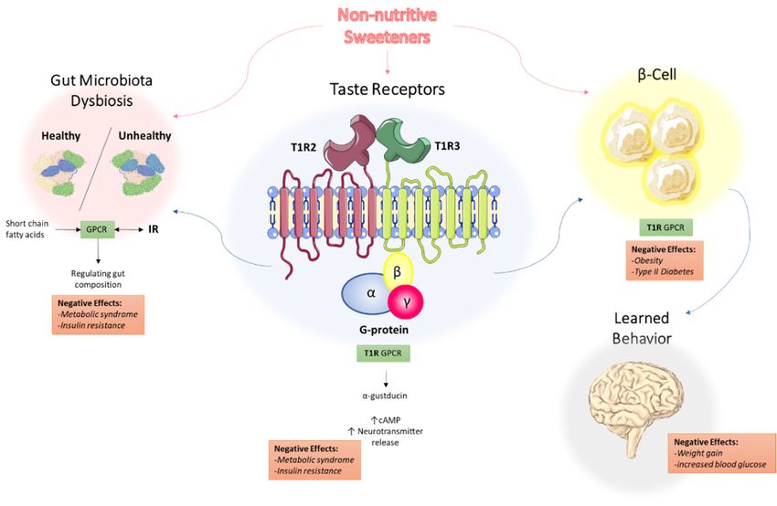
Figure 1. Proposed mechanisms of the underlying effects of non-nutritive sweeteners on the
Figure 1. Proposed
development mechanisms
of metabolic syndrome.of the
NNS underlying effects
interact with the of non-nutritive
T1R sweetenersreceptors
family of sweet-taste on the
development of metabolic
through associated G proteinsyndrome.
α-gustducin,NNSwhich
interact withinthe
results T1R family
increased of sweet-taste
intracellular receptors
cAMP levels and
through associated G protein α-gustducin, which results in increased intracellular cAMP
increased neurotransmitter release. Through the associated GPCR signaling, this may explain how NNS levels and
increased neurotransmitter
can contribute release. Through
to metabolic syndrome the associated
and insulin resistance.GPCR signaling,
NNS also this
interfere may
with explain
gut how
microbiota
NNS can contribute
composition, to metabolic
with short-chain syndrome
fatty acids (SCFAs) and
frominsulin resistance.
dietary NNS
intake acting also interfere
as ligands for GPCRswithin gut
the
microbiota composition,
gastrointestinal with short-chain
tract, regulating fatty acids
NNS permeability and(SCFAs) from dietary
gut microbiota intake acting
composition. as ligands
Additionally, for
NNS
GPCRs in the gastrointestinal tract, regulating NNS permeability and gut microbiota
are associated with insulin and other hormone secretion, which ultimately impact learned behavior composition.
Additionally,
and response toNNS are associated
sweetness. with insulin
Abbreviations: NNS,and other hormone
non-nutritive secretion,GPCR,
sweeteners; whichGultimately impact
protein-coupled
receptor;behavior
learned SCFA, short-chain
and responsefattytoacid.
sweetness. Abbreviations: NNS, non-nutritive sweeteners; GPCR,
G protein-coupled receptor; SCFA, short-chain fatty acid.
5. Non-Nutritive Sweeteners Interact with Sweet-Taste Receptors
5. Non-Nutritive
5.1. Sweeteners
Sweet-Taste Receptors in theInteract with Sweet-Taste
Mouth: Perception Receptors
of Sweetness
The innate Receptors
5.1. Sweet-Taste universal in
preference forPerception
the Mouth: sweetnessofonce served to support survival as it was associated
Sweetness
with food reward and energy (calories) in the form of carbohydrates; however, sweetness is now often
The innate universal preference for sweetness once served to support survival as it was
delivered via added sugars [26]. Sweet taste perception first begins at the level of type 2 taste receptor
associated with food reward and energy (calories) in the form of carbohydrates; however, sweetness
cells (TRCs) clustered in taste buds on the tongue that are G protein-coupled receptors (GPCRs) [27].
is now often delivered via added sugars [26]. Sweet taste perception first begins at the level of type 2
There are two classes of GPCRs that have been identified: the taste 1 receptor (T1R) and taste 2 receptor
taste receptor cells (TRCs) clustered in taste buds on the tongue that are G protein-coupled receptors
(T2R) families [28]. Within the T1R family, the T1R2 and T1R3 subtypes have been found to form
(GPCRs) [27]. There are two classes of GPCRs that have been identified: the taste 1 receptor (T1R) and
heterodimers that act as sweet-taste receptors [29].
taste 2 receptor (T2R) families [28]. Within the T1R family, the T1R2 and T1R3 subtypes have been
Interestingly, the T1R2/T1R3 receptors recognize all of the chemically diverse compounds that
found to form heterodimers that act as sweet-taste receptors [29].
are perceived as sweet by humans, including nutritive and non-nutritive sweeteners [29]. Given the
Interestingly, the T1R2/T1R3 receptors recognize all of the chemically diverse compounds that
vast number of compounds that can bind to the sweet-taste receptors, it is not surprising that there
are perceived as sweet by humans, including nutritive and non-nutritive sweeteners [29]. Given the
are different functional roles of T1R2 and T1R3 with multiple ligand binding sites corresponding
vast number of compounds that can bind to the sweet-taste receptors, it is not surprising that there
to the many possible ligands [30,31]. Sweet-taste receptor signaling has been extensively studied
are different functional roles of T1R2 and T1R3 with multiple ligand binding sites corresponding to
and reported [32–34]. Since sweet-taste receptors are GPCRs, they can induce the downstream
the many possible ligands [30,31]. Sweet-taste receptor signaling has been extensively studied and
activation of second messenger systems that ultimately result in increased intracellular calcium
reported [32–34]. Since sweet-taste receptors are GPCRs, they can induce the downstream activation
levels and neurotransmitter release [31,35]. Briefly, when a sweet-tasting compound binds to the
of second messenger systems that ultimately result in increased intracellular calcium levels and
neurotransmitter release [31,35]. Briefly, when a sweet-tasting compound binds to the T1R2/T1R3
receptors, α-gustducin is activated. The GPCR Gα-gustducin was previously identified as the firstNutrients 2019, 11, 644 6 of 19
T1R2/T1R3 receptors, α-gustducin is activated. The GPCR Gα-gustducin was previously identified as
the first protein molecularly associated with taste cells [36], but its role in taste signal transduction is
still not completely understood. Gustducin has considerable sequence homology to transducin,
which is also expressed in taste buds [37,38]. Both α-gustducin and α-transducin are known
to activate a phosphodiesterase (PDE) and decrease intracellular cAMP levels. There is also an
increase in phospholipase Cβ2 (PLCβ2) concentration which in turn increases production of inositol
1,4,5-trisphosphate and diacylglycerol. These compounds, in turn, activate the transient receptor
potential cation channel subfamily M member 5 (TRPM5), which subsequently increase intracellular
calcium and neurotransmitter release [32,39].
5.2. Sweet-Taste Receptors in the Gut: Effect of Sweeteners on Hormone Secretion
Sweet-taste receptors have also been found throughout the gastrointestinal (GI) tract, the biliary
tract, and the respiratory tract, suggesting that non-nutritive sweeteners have additional effects in the
body and may not be the inert compounds that they were once thought to be [31,40–42]. Within the
GI tract, sweet-taste receptors were primarily found in enteroendocrine L and K cells which secrete
specific hormones, as well as in pancreatic β-islet cells [31,39]. These studies have shown that ligand
binding to sweet taste receptors on enteroendocrine cells (EECs) in part affects hormone secretion.
In particular, the use of a sweet-taste inhibitor decreased glucagon-like peptide-1 (GLP-1) and peptide
YY (PYY) secretion by L cells, without affecting cholecystokinin (CCK) secretion from I cells, which are
known to not express sweet-taste receptors [41,43]. Thus, it appears that this network of sweet taste
signaling pathways in the oral cavity and the GI tract mediate the hormonal responses that orchestrate
the hunger–satiety cycle [44].
Enteroendocrine cells comprise 90% of all intestinal epithelial cells and are polarized such that they
permit the transport of nutrients from the gut lumen through apical sodium-glucose cotransporter-1
(SGLT-1) and into circulation through glucose transporter-2 (GLUT2) [45]. The hormones secreted
by EECs such as GLP-1, PYY, and CCK can act locally as paracrine factors, neurotransmitters, and
neuromodulators, or enter the bloodstream and act as classical hormones at distant sites [46,47]. It has
been established that SGLT-1 based transport is critical for GLP-1 release in humans which enhances
glucose-induced insulin secretion from pancreatic β-cells [45,48]. In animals, several sweet stimuli
including NNS have been shown to upregulate SGLT-1 expression and function, suggesting that
SGLT-1 activity is modulated by an upstream and broad sweet taste receptor [49,50]. Thus, it is thought
that NNS can potentiate SGLT-1 function and glucose absorption [51]. NNS including sucralose and
Ace-K demonstrate high levels of GLP-1 secretion in in vitro studies, with many inconclusive results in
human studies [52]. Given the collective effects of these hormones, it is likely that they contribute to the
pathogenesis of metabolic disorders, including obesity and T2DM [19,47,53]. Thus, it is possible that
NNS can stimulate sweet-taste receptors on intestinal EECs to promote the release of these hormones
involved in glucose homeostasis [26,48].
6. Non-Nutritive Sweeteners Interfere with Gut Microbiota Composition
The gut microbiota consists of millions of bacteria, viruses, and fungi that exist symbiotically
within the gut and begins to develop at birth [54]. The composition and function of the microbiota
varies not only amongst individuals, but also changes throughout an individual’s life, affected by
external factors such as environmental stressors, antibiotics and diet [55]. It is thought that diet is
responsible for approximately 10% of the influence on intestinal microbiota, a substantial amount when
considering the high variability in lifestyle and genetics amongst individuals [56]. Aberrations in the
gut microbiota have been associated with the development of insulin resistance, obesity, and metabolic
syndrome; however, the details are still in the process of being understood [46,57]. In particular, it has
been reported that T2DM is associated with alterations in microbiota composition [58].
In the human gut, the most common phyla are the Gram-positive Firmicutes and the
Gram-negative Bacteroidetes [59]. Analysis of the gut microbiota in lean and obese individuals hasNutrients 2019, 11, 644 7 of 19
revealed differences in the phyla present. There are several reports on a higher ratio of Firmicutes to
Bacteroidetes in obese individuals compared to lean individuals, with the proportion of Bacteroidetes
increasing with weight loss [60–62]. As a result, it has been speculated that the differences in the phyla
present may be associated with the development of obesity, a component of metabolic syndrome.
However, there are conflicting results, and specific roles of phyla have not yet been fully established [60].
Given the differences in microbiota composition amongst lean and obese individuals and the negligible
caloric value of NNS, it is surprising that NNS consumption may induce changes in microbiota
composition [52]. There have been several forms of dysbiosis that have been observed following NNS
consumption, mainly an increased ratio of Firmicutes:Bacteroidetes and an increase in Lactobacilli spp.,
such that the microbiota composition resembles that of obese individuals [63].
Suez and colleagues first reported the dysbiosis that occurs as a result of NNS consumption
in animal studies [63]. There are several diet-induced animal models of metabolic syndrome,
in which the animals are fed a single type or a combination of diets, investigating the whole-body
effects of metabolic syndrome such as through hormones, glucose metabolism and lipid metabolism
pathways [64]. Suez et al. reported on the cooperation between microbial species in the gut being
linked to enhanced energy harvest that promotes lipogenesis in mice through glycan degradation
pathways [63]. Interestingly, the metagenomes of saccharin-consuming mice were found to be enriched
with pathways such as sphingolipid metabolism and lipopolysaccharide biosynthesis, both of which
have been associated with T2DM and obesity [65,66]. Perhaps the most intriguing result of the study
was that the Bacteroidetes to Firmicutes ratio was positively correlated with reduced glucose tolerance,
and the reverse tendency was observed for overweight people, and the deleterious metabolic effects
were transferable to germ-free mice [67]. Thus, it is essential that we consider the gut microbiota
composition when developing treatment strategies for T2DM and obesity within the metabolic
syndrome platform.
NAS-induced gut microbiota composition changes have been linked to the phenomenon of
metabolic endotoxemia, the development of a low-grade inflammatory state by the gut microbiota that
ultimately promotes the development of insulin resistance (Figure 2) [68]. Briefly, dead bacteria result
in the release lipopolysaccharides (LPS) into the gut. LPS is absorbed into circulation where it binds to
CD14 proteins (modulators of insulin sensitivity in animals with hyperglycemia, hyperinsulinemia,
and weight gain), nucleotide oligomerization domains (NODs), and Toll-like receptors (TLRs) on the
surface of the macrophages and dendritic cells. The activation of these innate immune cells initiates
several inflammatory processes through the release of inflammatory cytokines [69]. Overproduction
of inflammatory cytokines, in turn, activates additional signaling pathways in metabolic cells that
ultimately result in insulin desensitization, altered expression of proteins responsible for glucose
transport, increased intestinal permeability, LPS infiltration, oxidative stress, and adipose tissue
inflammation [68]. Metabolic endotoxemia may be a driving force behind NAS-induced obesity and
insulin resistance.
6.1. The Role of GPCR-IR Crosstalk in Metabolic Syndrome
There has been extensive research elucidating the role of diet in modulating gut microbiota
composition in humans and animals [55,70,71]. Specifically, it has been suggested that NNS
consumption modulates gut microbiota composition as it has been associated with an increased
risk of obesity, T2DM, and metabolic syndrome [7,72]. As a result, there is increasing interest to
understand the signaling pathways implicated in metabolic syndrome, with a particular focus on the
novel phenomenon of biased agonism of GPCRs. Here, specific substrates or metabolites can induce
the preferential activation of these specific GPCRs [68,73]. Indeed, the gut microbiota can produce
short-chain fatty acids (SCFAs) as metabolites from the host diet that bind to specific GPCRs [74–76]
and confer insulin resistance through biased activation of insulin receptor (IR) signaling [77]. Also, diet-
and NNS-induced gut microbiota composition changes have been linked to metabolic syndrome. This
process may be mediated at least in part by miRNAs that can enter bacterial mitochondria, regulatingNutrients 2019, 11, 644 8 of 19
their gene expression and ultimately promoting a state of dysbiosis that can result in the development
of insulin resistance and T2DM in the host [78].
Nutrients 2019, 11, x FOR PEER REVIEW 8 of 19
Figure
Figure 2. Gut
2. Gut microbiotadysbiosis
microbiota dysbiosisand
andmetabolic
metabolic syndrome.
syndrome. Dysbiosis
Dysbiosisofofthe
theFirmicutes:Bacteroidetes
Firmicutes:Bacteroidetes
ratio is associated with several conditions characteristic of metabolic syndrome,including
ratio is associated with several conditions characteristic of metabolic syndrome, includingweight
weight
gain/obesity, insulin resistance, high-fat diets, gut permeability, and inflammatory
gain/obesity, insulin resistance, high-fat diets, gut permeability, and inflammatory bowel bowel disease
disease (IBD).
As (IBD). AsNNS
a result, a result, NNS consumption
consumption may contribute
may contribute to the development
to the development of these conditions
of these conditions due to
due to alterations
alterations in the Firmicutes:Bacteroidetes ratio. A bifidobacteria decrease combined with an enterobacteria
in the Firmicutes:Bacteroidetes ratio. A bifidobacteria decrease combined with an enterobacteria increase
increase leads to endotoxemia that causes a chronic low-grade inflammation associated with some
leads to endotoxemia that causes a chronic low-grade inflammation associated with some pathological
pathological conditions such as insulin resistance and increased gut permeability. A right balance in
conditions such as insulin resistance and increased gut permeability. A right balance in the microbiota
the microbiota may be considered in gut homeostasis and maintaining the microbiota can be
may be considered in gut homeostasis and maintaining the microbiota can be considered prebiotics
considered prebiotics and restore eubiosis in some pathological conditions. Abbreviations: IBD,
and restore eubiosis in some pathological conditions. Abbreviations: IBD, inflammatory bowel disease;
inflammatory bowel disease; NNS, non-nutritive sweeteners.
NNS, non-nutritive sweeteners.
As discussed, the development of insulin resistance, obesity, and overall metabolic syndrome
As discussed, the development of insulin resistance, obesity, and overall metabolic syndrome has
has been associated with changes in gut microbiota composition. The sweet taste receptors are
been associated with changes in gut microbiota composition. The sweet taste receptors are associated
associated with G protein α-gustducin, and GPCR crosstalk with several receptors, and particularly,
with G protein α-gustducin, and GPCR crosstalk with several receptors, and particularly, the insulin
the insulin receptor (IR) has been implicated in the development of metabolic syndrome [77]. The
receptor (IR)ofhas
receptors thebeen
tasteimplicated in the development
buds are coupled to G proteins of metabolic
(T1R2 syndrome
and T1R3), forming [77]. The
part of receptors
the C classofofthe
taste buds are
GPCRs, whichcoupled to G proteins
are structurally (T1R2
similar andglutamate
to the T1R3), forming part of receptors.
metabotropic the C class of GPCRs, which are
structurally similar to the glutamate metabotropic receptors.
GPCR-IR crosstalk has been associated with altering gut motility and permeability of SCFAs
GPCR-IR
[79]. Briefly,crosstalk
SCFA bind hastobeen
GPCRsassociated with altering
and ultimately get intogutsystemic
motilitycirculation
and permeability
and activateof SCFAs [79].
several
Briefly, SCFAand
metabolic bindinflammatory
to GPCRs andprocesses
ultimately getThe
[79]. into crucial
systemic circulation
role of GPCRand activate
proteins fromseveral metabolic
studies with
andGpr41-deficient
inflammatory processes
mice which [79].
were The crucial roleleaner
significantly of GPCR thanproteins
control from
mice studies with Gpr41-deficient
have validated the role of
mice which
GPCR were significantly
proteins [73]. Gpr41 and leaner
Gpr43 than
arecontrol
a pair ofmice have validated
mammalian orphanthe role of
GPCRs GPCR proteins
expressed in human [73].
adipocytes,
Gpr41 and Gpr43 colonareepithelial
a pair cells, and peripheral
of mammalian blood
orphan mononuclear
GPCRs cells.inThese
expressed human GPCRs are activated
adipocytes, colon
by SCFAs
epithelial cells,such
and as acetate, blood
peripheral propionate, and butyrate,
mononuclear whichGPCRs
cells. These are produced during
are activated by dietary fiber as
SCFAs such
fermentation by resident gut bacteria. SCFA stimulation of Gpr41 increases
acetate, propionate, and butyrate, which are produced during dietary fiber fermentation by resident gut leptin release, which
slows gut
bacteria. SCFAmotility and promotes
stimulation of Gpr41lipogenesis
increases[80]. Gpr43,
leptin another
release, SCFA
which receptor,
slows was found
gut motility andtopromotes
inhibit
insulin signaling
lipogenesis in adipose
[80]. Gpr43, another tissue,
SCFAsuppressing
receptor, was fat found
accumulation
to inhibitand promoting
insulin weight
signaling loss in mice
in adipose tissue,
[81]. Indeed, GPCRs are closely linked to microbe–microbe and microbe–host
suppressing fat accumulation and promoting weight loss in mice [81]. Indeed, GPCRs are closely interactions that play a
role in processes such as energy harvest, storage, and expenditure. Since NAS
linked to microbe–microbe and microbe–host interactions that play a role in processes such as energy play a role in changing
the gut microbiota composition, it is possible that GPCR crosstalk with other receptors is a
harvest, storage, and expenditure. Since NAS play a role in changing the gut microbiota composition,
mechanism by which NAS consumption contributes to the development of metabolic and
immunological abnormalities.Nutrients 2019, 11, 644 9 of 19
it is possible that GPCR crosstalk with other receptors is a mechanism by which NAS consumption
contributes to the development of metabolic and immunological abnormalities.
We have previously reviewed the novel phenomenon of GPCR-biased agonism or functional
selectivity and its role in the development of metabolic syndrome [77]. In particular, Haxho et al.
discovered that specific ligands signaling through the neuromedin B receptor result in preferential IR
activation as a result of GPCR-biased agonism [82]. They found that the GPCR agonists, angiotensin
and bradykinin dose-dependently induced neuraminidase-1 (Neu-1) sialidase activity through matrix
metalloproteinase-9 (MMP9) activation. Activated Neu-1 allows for insulin receptor dimerization,
leading to intracellular insulin signaling cascade. This concept explains how GPCR-MMP9-IR
crosstalk contributes to the development of insulin resistance through the over-activation of insulin
receptor signaling without its ligand [82] (Figure 3). It can also potentially explain the mechanism of
NAS-induced metabolic changes, and how they may lead to the development of T2DM via the novel
phenomenon of functional selectivity [83].
An intriguing observation about the T1R2/T1R3 heterodimeric receptor is its unique structural
diversity for a vast assortment of ligands. As discussed, the sweet taste receptor is able to recognize
every sweetener available, including carbohydrates, amino acids and their derivatives, proteins,
and synthetic sweeteners [84]. Interestingly, this receptor can exhibit unique stereoselectivity for
certain molecules such as D-tryptophan but not L-tryptophan [84]. This stereoselective property
of the T1R2/T1R3 receptor, where it can adopt more than one active state, is the phenomenon of
‘functional selectivity’, ‘ligand directed signaling’, or ‘biased agonism’ [85–90]. There are also allosteric
ligands with different degrees of modulation, called ‘biased modulation’, that can dramatically
influence GPCRs in a probe- and pathway-specific manner [85,88,89], including the T1R2/T1R3
receptor. For example, the human and rodent sweet taste receptors exhibit differences in ligand
specificity, G protein-coupling efficiency, and sensitivity to inhibitors [30]. Li et al. [84] provided
evidence that both the human and rat taste receptors can couple efficiently to Gα15/i1 , but only the
human receptor can couple efficiently to Gα15 . Xu et al. [30] also provided evidence to support
the important role of T1R2 in Gα-protein coupling in a functional expression system. To explain
these observations, Onfroy et al. proposed that G protein stoichiometry dictates biased agonism
through distinct receptor–G-protein partitioning [91]. Here, expression levels of Gα subunits influence
the biased profiling of agonists as well as antagonists, such that they determine both their activity
and G protein coupling efficacy by affecting different membrane distribution of receptor–G protein
populations. In the naïve state, the level of Gα expression influences the partitioning of not only Gα
but also the co-expressed receptor in different membrane domains [91]. It is intriguing to speculate that
the T1R2/T1R3 taste receptor through Gα protein partitioning involves a “pluridimensional efficacy”
concept as previously described by Galandrin and Bouvier [92] for distinct signaling profiles of β1/β2
adrenergic GPCR receptor ligands.
Here, the potential mechanism(s) that might connect NNS to metabolic changes is the molecular
crosstalk between the IR and GPCRs through a putative pluridimensional Gα protein partitioning
efficacy. Hypothetically, the heterodimeric T1R2/T1R3 GPCR taste receptors could exist in a multimeric
receptor complex with NMBR, IRβ, and Neu1 in naïve IR expressing cells as depicted in Figure 3. In this
situation, the TIR2/TIR3 dimeric receptors would exist as a molecular link regulating the interaction
and signaling mechanism(s) between these molecules on the cell surface. This hypothetical molecular
model could explain a biased TIR2/TIR3 GPCR agonist-induced IRβ transactivation signaling axis,
mediated by Neu-1 sialidase activity and the modification of insulin receptor glycosylation.
Collectively, these studies provide strong evidence that there exists an interaction between GPCRs
and the insulin receptor, which may provide insight into the implications of NNS consumption on the
development of metabolic syndrome. In summary, consumption of NNS may activate GPCR signaling
pathways that lead to cross-activation of insulin receptors through the neuromedin B receptor (NMBR)
and ultimately promote the development of insulin resistance and T2DM.Nutrients 2019, 11, 644 10 of 19
Nutrients 2019, 11, x FOR PEER REVIEW 10 of 19
Figure3.
Figure HypotheticalG
3. Hypothetical G protein-coupled
protein-coupled receptor
receptor (GPCR)
(GPCR) heterodimeric
heterodimeric T1R2/T1R3
T1R2/T1R3taste tastereceptors
receptors
exist in a multimeric receptor complex with NMBR, IRβ, and Neu-1 in naïve IR expressing
exist in a multimeric receptor complex with NMBR, IRβ, and Neu-1 in naïve IR expressing cells. Here, cells. Here,
TIR2/TIR3 dimeric receptors are proposed to exist as a molecular link regulating
TIR2/TIR3 dimeric receptors are proposed to exist as a molecular link regulating the interaction and the interaction and
signalingmechanism(s)
signaling mechanism(s)between
betweenthese
thesemolecules
moleculeson onthethecell
cellsurface.
surface. This
Thismolecular
molecularmodel
modeluncovers
uncovers
a biased TIR2/TIR3 GPCR agonist-induced IRβ transactivation signaling
a biased TIR2/TIR3 GPCR agonist-induced IRβ transactivation signaling axis, mediated by Neu-1 axis, mediated by Neu-1
sialidase and modification of insulin receptor glycosylation. This novel biased
sialidase and modification of insulin receptor glycosylation. This novel biased GPCR-signaling GPCR-signaling platform
potentiates
platform neuraminidase-1
potentiates (Neu-1) (Neu-1)
neuraminidase-1 and matrix
andmetalloproteinase-9
matrix metalloproteinase-9 (MMP-9) crosstalk
(MMP-9) on the cell
crosstalk on
surface that is essential for the activation of the insulin receptor β subunit (IRβ) tyrosine
the cell surface that is essential for the activation of the insulin receptor β subunit (IRβ) tyrosine kinases. Notes:
Insulin-binding
kinases. receptor α subunits
Notes: Insulin-binding (IRα),
receptor as well as
α subunits GPCR
(IRα), as agonists, potentiate
well as GPCR biased
agonists, neuromedin
potentiate biasedB
neuromedin B receptor (NMBR)-IRβ signaling and MMP-9 activation to induce Neu-1 sialidase.is
receptor (NMBR)-IRβ signaling and MMP-9 activation to induce Neu-1 sialidase. Activated MMP-9
proposed here
Activated MMP-9 to remove the elastin-binding
is proposed here to remove protein
the (EBP) as part of the
elastin-binding molecular
protein (EBP) multienzymatic
as part of the
molecular multienzymatic complex that contains β-galactosidase/Neu-1 andAprotective
complex that contains β-galactosidase/Neu-1 and protective protein cathepsin (PPCA). Activated
protein
Neu-1 hydrolyzes α-2,3 sialyl residues of IRβ at the ectodomain to remove steric hindrance to
cathepsin A (PPCA). Activated Neu-1 hydrolyzes α-2,3 sialyl residues of IRβ at the ectodomain to
facilitate IRβ subunits association and tyrosine kinase activation. Activated phospho-IRβ subunits
remove steric hindrance to facilitate IRβ subunits association and tyrosine kinase activation. Activated
phosphorylate insulin receptor substrate-1 (pIRS1), which initiate intracellular insulin signaling via
phospho-IRβ subunits phosphorylate insulin receptor substrate-1 (pIRS1), which initiate intracellular
the Ras-MAPK and the PI3K-Akt pathway, among others. Abbreviations: PI3K, phosphatidylinositol
insulin signaling via the Ras-MAPK and the PI3K-Akt pathway, among others. Abbreviations: PI3K,
3-kinase; GTP, guanine triphosphate; IRS1, insulin receptor substrate-1; p, phosphorylation; Neu-1,
phosphatidylinositol 3-kinase; GTP, guanine triphosphate; IRS1, insulin receptor substrate-1; p,
neuraminidase-1. Taken in part from Cellular Signalling 43 (2018) 71–84. © 2018 Haxho et al., Published
phosphorylation; Neu-1, neuraminidase-1. Taken in part from Cellular Signalling 43 (2018) 71–84. ©
by Elsevier Inc., open-access under CC BY-NC-ND license. This is an open-access article which permits
2018 Haxho et al., Published by Elsevier Inc., open-access under CC BY-NC-ND license. This is an
unrestricted noncommercial use, provided the original work is properly cited.
open-access article which permits unrestricted noncommercial use, provided the original work is
properly
6.2. NAS cited.
Modify miRNAs in Regulating Gut Composition
Recent
6.2. NAS attention
Modify miRNAs oninthe role of miRNAs
Regulating changing cell function has incited interest in miRNA
Gut Composition
implications on gut microbiota function, and consequently the development of insulin resistance in
Recent attention
individuals consuming onNNS
the role of miRNAs
in their changing
diet. miRNAs cell function
are 18–25 has long
nucleotide incited interest in
noncoding miRNA
RNAs that
implications on gut microbiota function, and consequently the development of insulin
alter gene expression through post-transcriptional silencing or activation [93]. It is speculated that resistance in
individuals consuming NNS in their diet. miRNAs are 18–25 nucleotide long noncoding
miRNAs regulate at least 30% of human genes, playing critical roles in cell proliferation, differentiation, RNAs that
alter gene expression
apoptosis, through [94,95].
and hematopoiesis post-transcriptional
As a componentsilencing or activation
of human [93]. It can
feces, miRNAs is speculated that
be biomarkers,
miRNAs regulate at least 30% of human genes, playing critical roles in cell
prognostic indicators, and regulators of normal and abnormal cell function [78,96]. Preclinical models proliferation,
differentiation, apoptosis,
suggest that miRNA and hematopoiesis
expression can be altered[94,95]. As a component
by stress, exercise and of diet
human
[97].feces,
As amiRNAs can
result, NNS
be biomarkers, prognostic indicators, and regulators of normal and abnormal cell
consumption may modify miRNA expression by altering bacterial composition and potentially lead to function [78,96].
Preclinical models suggest that miRNA expression can be altered by stress, exercise and diet [97]. As
metabolic changes.
a result,
Liu NNS
et al.consumption may modify
recently elucidated themiRNA expression
mechanism by altering
by which miRNAs bacterial
shapecomposition and
gut microbiota
potentially lead to metabolic changes.
composition [78]. Intestinal epithelial cells (IECs) secrete miRNAs into the gut lumen in the form of
Liu etoral.
exosomes recently elucidated
extracellular vesicles. Thethesecreted
mechanism
miRNAsby canwhich
thenmiRNAs
enter gutshape
bacteriagutandmicrobiota
act at the
composition [78]. Intestinal epithelial cells (IECs) secrete miRNAs into the gut lumen in the form of
exosomes or extracellular vesicles. The secreted miRNAs can then enter gut bacteria and act at theNutrients 2019, 11, 644 11 of 19
DNA level or directly on RNA in the mitochondria where they can alter gene expression that regulates
functions affecting bacterial growth. Fecal transplantation of miRNA has been reported to help restore
gut microbiota composition, broadening the therapeutic application of miRNA in influencing the
abundance of health-associated gut bacteria.
The role of miRNAs in the pathogenesis of glucose intolerance and T2DM has been increasingly
investigated and explained in detail elsewhere [98]. Briefly, miRNAs were shown to play critical
roles in core processes of the insulin-related signaling pathway, carbohydrate and lipid metabolism,
as well as adipocytokine signaling pathways. Up- or downregulation of certain miRNAs has been
correlated with the development of insulin resistance and increased severity of T2DM. In particular,
miR-7 was found to regulate pancreatic β-cell function, differentiation and insulin secretion [99].
Overexpression of miR-7 in mice has been associated with the development of T2DM due to impaired
insulin secretion. Also, levels of miR-101, miR-375, miR-802 were significantly increased in T2DM
patients while miR-143 and miR-223 levels were downregulated in obese individuals when compared
to control groups [100,101]. Constant exposure of pancreatic β-cells to various metabolic stresses,
including NNS consumption, can shift the delicate balance between positive and negative regulatory
miRNA, ultimately promoting pancreatic dysfunction and insulin resistance. Importantly, miRNAs
can affect different bacterial species due to their capacity to enter the GI tract [102]. This effect can
partially explain the differences observed in miRNA regulatory effects. If there is a prevalence of
particular phyla in the gut, they can modify the expression of genes that correspond to changes in
metabolic functions, including endocrine cell dysfunction. Several studies have demonstrated that
regular NNS consumption induces changes in the composition of the gut microbiota that eventually
leads to the development of insulin resistance [103,104]. The driving force behind this pathological
process may be miRNA regulation of insulin-related signaling pathways.
Circulating miRNAs can be used as diagnostic biomarkers for T2DM patients; however, there
are some challenges in assessing their levels [105]. Current biomarkers only detect disease once
metabolic imbalances have already set in, whereas changes miRNA expression levels can be noticed
5–10 years before T2DM manifestation [106]. The small size and known physiochemical properties
of miRNAs alongside their natural synthesis make them attractive therapeutic targets [96]. Current
approaches for modulating miRNA function in vivo demonstrate promising results. As regulators of
many metabolic processes, miRNAs have the potential to be therapeutic agents [107]. For example,
miR-126 expression is significantly reduced in diabetic patients, leading to impaired proangiogenic
capacity that promotes diabetic vasculopathy. Some experiments have been able to manipulate
miR-126 expression to induce migration and proliferation of vascular endothelial cells and facilitate
their repair [108]. Collectively, it is imperative that technology continues to advance in this field to
better assess the effects of miRNA-based therapies.
7. Non-Nutritive Sweeteners Interfere with Learned Responses to Sweetness
Sugar and its sweet-tasting nutritive and non-nutritive alternatives have become a staple in the
diet. However, sweet-taste has been associated with learned behavior [109]. As discussed previously,
sugar consumption has been associated with an increased GLUT2 and GLUT5 expression, which play
a role in CCK expression in the ileum of isocaloric diet-fed rats enriched with fructose or glucose [110].
The enriched diets provide additional calories, resulting in animals having enhanced total caloric
intake [111]. In contrast to natural sweeteners such as fructose or sucrose, NNS was thought to be
excreted after passing through the GI tract unchanged resulting in no energy gain [112].
Theoretically, the metabolic effects observed with the use of natural sweeteners should be absent
with NNS consumption. Paradoxically, NNS consumption has been associated with weight gain. It is
hypothesized that the separation of sweetness from calories interferes with physiological responses
and the interaction of NNS with sweet-taste receptors in the gut that affect glucose absorptive capacity
and homeostasis [113,114]. Although epidemiological studies have shown an association between
artificial sweetener use and weight gain, evidence of a causal relationship is limited; however, recentNutrients 2019, 11, 644 12 of 19
animal studies provide intriguing information that supports an active metabolic role of artificial
sweeteners [19]. Indeed, the low or zero caloric value of NNS can result in caloric compensation,
whereby there is an adjustment for calories consumed at one occasion by reducing caloric intake at
subsequent opportunities. Thus, weakened caloric compensation can result in excess energy intake
that ultimately leads to increased weight gain [115].
In 1910, Pavlov and more recently by other studies [116] proposed that orosensory stimuli like
sweet taste elicit different learned physiological responses, including the anticipation of the arrival
and absorption of food to control body weight and energy balance. Interestingly, there might be a role
for gustatory cues in the detection of high fat/high sugar diets [116]. The fat component is a more
salient orosensory feature of the high energy diet. High caloric compensation is observed when rats
are fed glucose, but there is weaker compensation when rats are fed saccharin [117]. If these animal
studies had a longer duration, the reduced caloric compensation would result in increased weight
gain. Non-caloric sweeteners reduce the validity of sweet taste as a signal to predict caloric intake that
leads to a positive balance of energy and weight gain. Green et al. conducted imaging studies using
fMRIs and found alterations in the reward processing of sweet taste in the individuals who regularly
consumed diet soda [118]. More human studies are required to assess physiological responses to NNS
in naïve subjects.
Weight gain associated with NNS consumption can be explained in part by their interference
with learned responses that contribute to energy homeostasis. Swithers and Davidson demonstrated
that NNS consumption weakens cephalic response to ingested food [119]. Based on the Pavlovian
conditioning principles, it is hypothesized that sweet taste predicts energy intake and evokes both
autonomic and endocrine responses that prepare the GI tract [120]. Altered glucoregulatory responses,
such as the release of GLP-1, were observed in mice when glucose and saccharin were given orally
but not when directly released into the stomach [120]. These findings support the proposition
that there is a disruption in the learned responses generated by tasting sweetness but not in the
post-absorptive consequences of consuming sugar. Suppressed GLP-1 release when saccharin is given
orally may disrupt the satiety process due to increased gastric emptying. Lower GLP-1 levels lead to
decreased glucose utilization in muscle, liver, adipose tissue, and diminished suppression of glucagon
release, elevating blood glucose levels. Considering that cephalic responses are required for a normal
postprandial glucose tolerance [121], it is possible that NNS interferes with learned responses to
sweetness, and thereby increasing food intake and weight gain.
In addition, M.O. Welcome reviewed recent evidence to demonstrate that the sweet taste receptor
heterodimer T1R2/T1R3 plays a crucial role in cognitive functioning, suggesting that dysfunctions
in sweet taste receptor signaling may underlie cognitive impairment in some brain pathologies [122].
Dysfunctions in sweet taste receptor signaling were also associated with inflammatory response
pathway [123]. The study showed that sweet taste receptors function as pivotal immune sentinels,
revealing the downregulation of the key components of the taste signaling cascades such as
α-gustducin, phospholipase C β2, and monovalent selective cation channel TRPM5, contributing
to cognitive impairment [123].
8. Conclusions
Non-nutritive sweeteners continue to be a staple in the Western diet. However, the health-related
safety of NNS consumption remains to be a controversial topic. Recent reports on the role of
NNS promoting shifts in gut microbiota composition and reports linking the gut microbiota to
insulin signaling, confirm the importance of studying the physiological effects of NNS. The exact
mechanisms of pathological changes induced by NAS are still under speculation. Thus, the recently
discovered phenomenon of functional selectivity of GPCR-IR crosstalk, as well as miRNA modulation
of gut microbiota function, may provide insight into the pathological effects of NNS. The proposed
mechanisms underlying NNS-mediated development of metabolic syndrome are summarized in
Figure 1. Enhancing our knowledge through well-designed human trials should highlight the potentialNutrients 2019, 11, 644 13 of 19 role of NNS in the alterations of microbial, neurological, and hormonal responses to consumed food. Energy intake compensation appears to be an area where additional studies need to test and compare different food as well as different NAS. Consumers must be aware that contrary to the existing belief, that substitution of natural sugar by NAS is beneficial for their health, there is growing evidence of NAS being implicated in the development of metabolic abnormalities. Continued research in this field will uncover the pathology of diet-induced metabolic changes as well as uncover new biomarkers and novel treatments using miRNAs. Author Contributions: All authors contributed toward data analysis, drafting, and revising the paper and agree to be accountable for all aspects of the work. Conceptualization, M.R.S. and B.Q.; Literature Review, M.R.S., B.Q., I.L., F.D., and Y.R.; Writing—Original Draft Preparation, M.R.S., B.Q., I.L., F.D., and Y.R.; Writing—Review and Editing, M.R.S. and B.Q.; Supervision, M.R.S. Funding: This work is supported in part by a grant to M.R. Szewczuk from the Natural Sciences and Engineering Research Council of Canada (NSERC), RGPIN-2015-05301. Acknowledgments: B. Qorri is a recipient of the Queen’s Graduate Award (QGA), the 2017 Terry Fox Research Institute Transdisciplinary Training Program in Cancer Research, and the 2018 Dean’s Doctoral Award. Conflicts of Interest: The authors declare no conflicts of interest. Abbreviations Ace-K Acesulfame-potassium CCK Cholecystokinin CHD Congenital heart defect EEC Enteroendocrine cells FDA Food and Drug Administration GI Gastrointestinal GLP-1 Glucagon-like peptide-1 GLUT2 Glucose transporter-2 GPCR G protein coupled receptor GRAS Generally recognized as safe IBD Inflammatory bowel disease IEC Intestinal epithelial cell IR insulin receptor LPS Lipopolysaccharide miRNA MicroRNA MMP9 Matrix metalloproteinase-9 NAS Noncaloric artificial sweeteners Neu-1 Neuraminidase-1 NHLBI National Heart, Lung and Blood Institute NNS Non-nutritive sweeteners NOD Nucleotide oligomerization domain PDE Phosphodiesterase PLCβ2 Phospholipase Cβ2 PYY Peptide YY rEM Rapid evidence mapping SCFA Short-chain fatty acid SGLT-1 Sodium-glucose co-transporter-1 T1R Taste 1 receptor T2DM Type 2 diabetes mellitus T2R Taste 2 receptor TCR Taste receptor cell TRPM5 Transient receptor potential cation channel subfamily M member 5
You can also read


