Critical Molecular and Cellular Contributors to Tau Pathology
←
→
Page content transcription
If your browser does not render page correctly, please read the page content below
biomedicines
Review
Critical Molecular and Cellular Contributors to Tau Pathology
Liqing Song, Evan A. Wells                  and Anne Skaja Robinson *

                                          Department of Chemical Engineering, Carnegie Mellon University, Pittsburgh, PA 15213, USA;
                                          liqing@cmu.edu (L.S.); eawells@andrew.cmu.edu (E.A.W.)
                                          * Correspondence: anne.robinson@cmu.edu; Tel.: (+1)-412-268-7673

                                          Abstract: Tauopathies represent a group of neurodegenerative diseases including Alzheimer’s disease
                                          (AD) that are characterized by the deposition of filamentous tau aggregates in the brain. The patho-
                                          genesis of tauopathies starts from the formation of toxic ‘tau seeds’ from hyperphosphorylated tau
                                          monomers. The presence of specific phosphorylation sites and heat shock protein 90 facilitates soluble
                                          tau protein aggregation. Transcellular propagation of pathogenic tau into synaptically connected
                                          neuronal cells or adjacent glial cells via receptor-mediated endocytosis facilitate disease spread
                                          through the brain. While neuroprotective effects of glial cells—including phagocytotic microglial
                                          and astroglial phenotypes—have been observed at the early stage of neurodegeneration, dysfunc-
                                          tional neuronal-glial cellular communication results in a series of further pathological consequences
                                          as the disease progresses, including abnormal axonal transport, synaptic degeneration, and neu-
                                          ronal loss, accompanied by a pro-inflammatory microenvironment. Additionally, the discovery of
                                          microtubule-associated protein tau (MAPT) gene mutations and the strongest genetic risk factor
                                          of tauopathies—an increase in the presence of the ε2 allele of apolipoprotein E (ApoE)—provide
                                          important clues to understanding tau pathology progression. In this review, we describe the crucial
                                signaling pathways and diverse cellular contributors to the progression of tauopathies. A systematic
                                   understanding of disease pathogenesis provides novel insights into therapeutic targets within altered
Citation: Song, L.; Wells, E.A.;          signaling pathways and is of great significance for discovering effective treatments for tauopathies.
Robinson, A.S. Critical Molecular and
Cellular Contributors to Tau              Keywords: tauopathies; Alzheimer’s disease; prion-like propagation; tau self-aggregation; endocyto-
Pathology. Biomedicines 2021, 9, 190.     sis; neuron-glial communication; neuroinflammation; apolipoprotein E
https://doi.org/10.3390/
biomedicines9020190

Academic Editor: Lorenzo Falsetti         1. Introduction
                                               Intraneuronal accumulation of neurofibrillary tangles (NFT) made of abnormally
Received: 18 January 2021
Accepted: 11 February 2021
                                          hyperphosphorylated tau is centrally involved in the pathogenesis of primary tauopathies,
Published: 14 February 2021
                                          such as supranuclear palsy (PSP), corticobasal degeneration (CBD), Pick’s disease (PiD),
                                          and frontotemporal dementia with Parkinsonism linked to chromosome 17 (FTDP-17),
Publisher’s Note: MDPI stays neutral
                                          and secondary tauopathies such as Alzheimer’s disease (AD) [1]. The development of
with regard to jurisdictional claims in
                                          tau pathology has been postulated to follow spatiotemporal patterns, starting from the
published maps and institutional affil-   dissociation of phosphorylated tau from microtubules and followed by the formation of
iations.                                  toxic tau species via self-aggregation [2]. Even though polyanionic molecules are normally
                                          required for inducing tau aggregation in vitro, modifications to tau, such as site-specific
                                          mutations and site-specific phosphorylation, have driven spontaneous seeding and self-
                                          aggregation of tau in vivo under pathological situation [3]. Physiologically, extracellular
Copyright: © 2021 by the authors.
                                          tau is present in brain interstitial fluid (ISF) and then passes into the cerebrospinal fluid
Licensee MDPI, Basel, Switzerland.
                                          (CSF) [4,5]; however, the elevated concentrations of tau found in the brain ISF of human
This article is an open access article
                                          P301S tau transgenic mice has suggested that cellular tau release may be a part of disease
distributed under the terms and           progression [6]. Additionally, soluble tau concentrations in brain homogenates decrease
conditions of the Creative Commons        with the deposition of intracellular insoluble tau, suggesting that transcellular tau propa-
Attribution (CC BY) license (https://     gation requires cellular internalization of extracellular tau, which has also been found to
creativecommons.org/licenses/by/          mediate the progression of neurodegeneration [6–8]. The cellular pathways for internal-
4.0/).                                    izing tau species are regulated by both heparan sulfate proteoglycan (HSPG)-mediated

Biomedicines 2021, 9, 190. https://doi.org/10.3390/biomedicines9020190                               https://www.mdpi.com/journal/biomedicines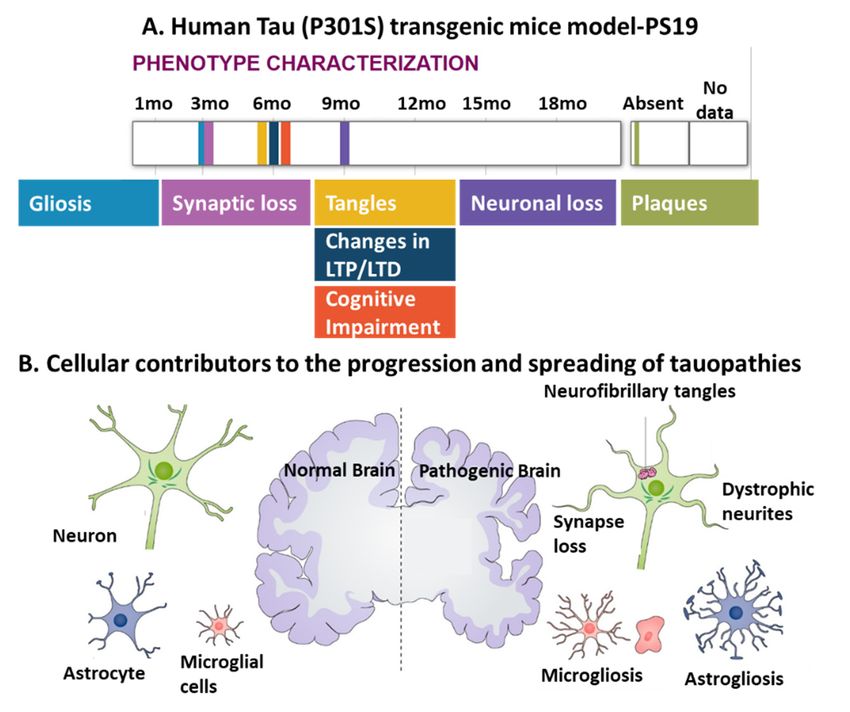
Biomedicines 2021, 9, 190                                                                                             2 of 25

                            cellular uptake and specific receptor-mediated endocytosis, which are highly dependent
                            on the isoform being internalized [8–10].
                                  Extensive experimental data have demonstrated that transcellular propagation of
                            soluble tau species occurs mainly through synaptic connections, leading to neuronal
                            dysfunction characterized by the breakdown of cytoskeletal integrity, abnormal axonal
                            transport, and synapse loss [9,10]. In particular, glial cells, activated microglia, and reactive
                            astrocytes are also involved in the progression of tau pathology by directly affecting
                            the homeostasis of the neuronal microenvironment or indirectly exerting inflammatory
                            effects across multiple tauopathies [11,12]. For example, the degree of glial cell activation
                            correlates with the severity of neurodegeneration in AD, in terms of the degeneration
                            of synapses, neuronal loss, the formation of NFTs, or even cognitive impairment [13].
                            Alternatively, dysfunctional neuron-glial communication has been widely observed in
                            AD patients and has recently developed in vitro tau pathology animal models [14,15].
                            Abnormal neuron-glial crosstalk strongly impairs neuronal homeostasis including neuronal
                            metabolism, synaptogenesis, neurotransmission, and neuromodulation, contributing to
                            the progression of neurodegeneration [14,16]. The investigation of critical molecular
                            and cellular contributors to tau pathology provides a comprehensive understanding of
                            tau pathogenesis that will accelerate the discovery of novel therapeutic targets and the
                            development of drugs for treating tauopathies.
                                  The purpose of this review is to summarize the factors that contribute to the formation
                            of tau aggregates, tau cell-to-cell propagation, and glial contributions in tauopathies,
                            by using the scientific evidence published in the last decade that bring promising insights
                            into the therapeutic development for tau protein pathology. Keywords for this topic, such as
                            tauopathies, Alzheimer’s disease, prion-like propagation, tau self-aggregation, endocytosis,
                            neuron-glial communication, neuroinflammation, and apolipoprotein E were first chosen,
                            and searches conducted in PubMed, Google Scholar, and Web of Science. The results of
                            these searches were then refined and categorized into cellular contributors at the early stage
                            and later stage of neurodegeneration, based on the characterized Braak-like spatiotemporal
                            staging scheme for tau pathology. Lastly, the combined keywords search strategy was used
                            for searching for potential treatments for tauopathies such as using the affected signaling
                            pathway and tau phosphorylation together. The pathological roles of phosphorylation
                            sites, Hsp90 and site-specific mutations in tau aggregation, the roles of CX3CR1/fractalkine
                            signaling in microglia and neurons, the roles of the glutamate-glutamine cycle between
                            astrocyte and neurons in the progression of tau pathologies, and the possible therapeutic
                            role of NLR3 inflammasome in the treatment of tauopathies are the major focus of this
                            review. A list of the abbreviations used in this review is provided in Table 1.

                            Table 1. Table of abbreviations used in this review.

                                Abbreviation                                       Explanation
                                    AD                                      Alzheimer’s disease
                                   MAPT                              Microtubule-associated protein tau
                                   ApoE                                      Apolipoprotein E
                                    NFT                                   Neurofibrillary tangles
                                    PSP                                     Supranuclear palsy
                                    CBD                                  Corticobasal degeneration
                                    PiD                                        Pick’s disease
                                  FTDP-17            Frontotemporal dementia with Parkinsonism linked to chromosome 17
                                    CNS                                   Central nervous system
                                   MBD                                 Microtubule-binding domain
                                    Aβ                                           Amyloid β
                                    PRR                                      Proline-rich region
                                    PHF                                   Paired helical filaments
                                    ISF                                       Interstitial fluid
                                    CSF                                     Cerebrospinal fluid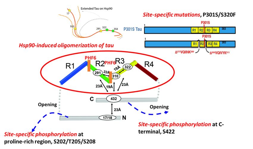
CNS Central nervous system
MBD Microtubule-binding domain
Biomedicines 2021, 9, 190 Aβ Amyloid β 3 of 25
PRR Proline-rich region
PHF Paired helical filaments
ISF Interstitial fluid
Table 1. Cont.
CSF Cerebrospinal fluid
Abbreviation HSPG Heparan sulfate proteoglycans
Explanation
HSPG ALP Heparan Autophagy-lysosomal
sulfate proteoglycans pathway
ALP NMDA N-methyl-D-aspartate
Autophagy-lysosomal pathway
NMDA TFEB N-methyl-D-aspartate
Transcription factor EB
TFEB LTP Transcription factor EBpotentiation
Long-term
LTP Long-term potentiation
LTD
LTD Long-term depression
Long-term depression
2. Factors Involved in the Formation of Tau Seeds
2. Factors Involved in the Formation of Tau Seeds
The formation of NFTs from soluble tau is a multistep process. This process begins
The formation of NFTs from soluble tau is a multistep process. This process begins
with the dimerization of two conformationally altered monomers and is followed by the
with the dimerization of two conformationally altered monomers and is followed by the
formation of intermediate soluble oligomers with varying higher-order conformations
formation of intermediate soluble oligomers with varying higher-order conformations
and degrees of phosphorylation. Tau oligomers have been implicated as toxic ‘tau seeds’
and degrees of phosphorylation. Tau oligomers have been implicated as toxic ‘tau seeds’
capable of seeding new aggregates by recruiting normal monomers. Despite evidence of
capable of seeding new aggregates by recruiting normal monomers. Despite evidence
tau trimers being the minimal unit of spontaneous cellular uptake and intracellular fibril-
of tau trimers being the minimal unit of spontaneous cellular uptake and intracellular
lary structure formation in vivo [17], the folding potency of monomer could be much more
fibrillary structure formation in vivo [17], the folding potency of monomer could be much
critical
more in initiating
critical the the
in initiating early nucleation
early process
nucleation of tau
process aggregation
of tau (Figure
aggregation 1). 1).
(Figure
Figure
Figure1. The molecular
1. The mechanisms
molecular mechanismsinvolved
involvedin in
tautau
aggregation. Molecular
aggregation. Molecularfactors such
factors asas
such site-specific phosphorylation,
site-specific phosphorylation,
site-specific mutations on MAPT, and specific chaperones (Hsp90) are associated with tau aggregation.
site-specific mutations on MAPT, and specific chaperones (Hsp90) are associated with tau aggregation.
2.1. Site-Specific
2.1. Phosphorylation-Mediated
Site-Specific Phosphorylation-Mediated Tau
TauSelf-Aggregation
Self-Aggregation
Previous work details that although tau
Previous work details that although tau itself itself is is
intrinsically disordered,
intrinsically disordered,proteins in in
proteins
solution possess a ‘paperclip-like’ conformation where the N- and C-terminal
solution possess a ‘paperclip-like’ conformation where the N- and C-terminal ends of tau ends of tau
fold over
fold inin
over proximity
proximityto to
thethe
center ofof
center thethe
repeat
repeatdomains
domains [18]. Site-specific
[18]. phosphoryla-
Site-specific phosphoryla-
tion directly influences the conformation of monomeric tau and affects the stability of a
tion directly influences the conformation of monomeric tau and affects the stability of a
folded conformation, contributing to the propensity for tau to aggregate [19]. Two hexapep-
folded conformation, contributing280 to the propensity for tau to aggregate [19]. Two hex-
tides, known as PHF6s, 275 VQIINK , and 306 VQIVYK311 , are located at the beginning
apeptides, known as PHF6s, 275VQIINK280, and 306VQIVYK311, are located at the beginning
of the second and third repeat domains of the MBDs, and appear to drive β-sheet struc-
ture formation during the tau aggregation process. The accessibility of residues in the
two PHF6s defines the structural differences between inert (Mi) and seed-competent (Ms)
tau monomer, meaning that the inert (Mi) tau monomer has less inter-chain accessibility
to these residues compared with that in the seed-competent (Ms) monomer [2]. Phos-
phorylation outside of, but proximal to, these regions is relevant to the formation of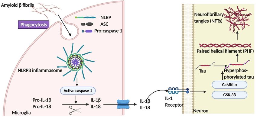
Biomedicines 2021, 9, 190                                                                                       4 of 25

                            NFTs. A previous study systematically investigated the effects of different phosphoryla-
                            tion sites on tau self-aggregation, using a series of in vitro pseudo-phosphorylated tau
                            proteins [20]. Phosphorylation sites T175/T176/T181 within N-terminal, recognized by
                            AT270 antibody, mainly suppress tau aggregation [21]. In addition, phosphorylation at
                            three sites, S202/T205/S208, within the proline-rich region (PRR) is enough to induce
                            tau self-aggregation without any exogenous aggregation inducer [22]. The monoclonal
                            antibody AT8 that specifically recognizes tau phosphorylation at the S202/T205 site has
                            been established as a valid biochemical marker for identifying abnormally phosphorylated
                            tau as well as the paired helical filament form. Moreover, phosphorylation sites near the
                            C-terminus have been found to preferentially promote tau self-aggregation. For example,
                            pseudo-phosphorylated S396, specifically recognized by PHF-1 antibody [21], has led to
                            increased tau aggregation in the presence of metal ion inducer. In particular, the strong
                            effect on aggregation has been seen in pS422 tau protein, which showed increased aggrega-
                            tion in the presence of both metal ions and heparin inducers [20], which may be related
                            to the conversion of tau monomer from inert to seed-competent form, due to increased
                            accessibility of these residues [3], as shown in Figure 1. By performing a comprehensive
                            electrochemiluminescence ELISA assay, Ercan-Herbst et al. [23] found that specific phos-
                            phorylation events (pS198, pS199, and pS416) correlated with increased oligomerization
                            in all brain regions, which implies that phospho-sites regulate tau aggregation during the
                            progression of AD neurodegeneration. Collectively, phosphorylation plays a major role in
                            tau self-aggregation by altering the charge and conformations of physiological tau.

                            2.2. Hsp90-Mediated Tau Aggregation
                                  Tau phosphorylation and aggregation that lead to conformational changes could
                            involve molecular chaperones, which regulate protein folding, degradation, and accumu-
                            lation. The protective effect of Hsp70 and Hsp104 in tauopathies has been described in
                            previous studies [24]. Hsp70 inhibits the aggregation of tau protein by forming a complex
                            with tau oligomer or fibril tau, preventing toxic effects or further seeding of tau aggre-
                            gation [25,26]. Despite the recognition of its disaggregase activity for many aggregates,
                            a distinct mechanism of Hsp104 in preventing tau aggregation is related to its holdase ac-
                            tivity on soluble amyloid tau through the small subdomain of nucleotide-binding domain 2
                            (ssNBD2) [27].
                                  In contrast to the preventative functions of Hsp70 and Hsp104, heat shock protein 90
                            (Hsp90), one of the major tau-binding chaperones, has been found to drive the aggregation
                            of tau species [28]. Although Hsp90 is normally thought to act as cellular protection
                            during stress, Hsp90 binding to tau at the VQIVYK motif facilitates a conformational
                            change that results in its phosphorylation by glycogen synthase kinase 3, which further
                            promotes tau aggregation [28]. Additionally, a recent study found that Hsp90 binding to
                            tau uncovered the repeat domains by conformationally opening the ‘paper-clip’ structure
                            of tau, suggesting that the formation of tau oligomers was caused by the conversion of tau
                            monomers from inert to aggregation-prone forms [29].

                            2.3. Site-Specific Mutations and Tau Aggregation
                                  Abnormal tau mutants related to FTDP-17 possess distinct structures leading to a
                            differential aggregation propensity [30–32]. Recently, Strang and coworkers demonstrated
                            that the susceptibility of FTDP-17-associated mutants to aggregate with seeded, exogenous
                            fibrillar tau depended highly on site-specific mutations and their surrounding amino acid
                            sequences [33]. Robust aggregation with exogenous tau fibril seeds, both homotypic and
                            heterotypic, has been seen in FTDP-17 mutations at sites P301 and S320. In particular,
                            the unique property of the P301L variant in regulating the aggregation propensity of tau
                            has been demonstrated by mutating individual proline residues into leucine residues
                            within conserved PGGG motifs in each of the four MTBDs in tau [33]. Only P301L showed
                            a propensity to aggregate when seeded with exogenous fibrillar tau. In contrast, other
                            FTDP-17-associated variants near the PHF6 site showed no propensity to aggregate when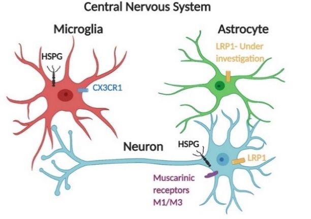
Biomedicines 2021, 9, 190                                                                                         5 of 25

                            seeded. Double mutants at P301L/S320F and P301S/S320F have been shown to facilitate
                            aggregation. For these P301L/S320F and P301S/S320F tau protein variants, robust ag-
                            gregation was observed in vivo without exogenous fibrillar tau seeding [33]. A possible
                            underlying cause of this enhanced aggregation propensity is altered conformation with
                            higher accessibility to PHF6, that converts the inert monomer into aggregation-prone
                            monomer; alternatively, more frequent interactions with chaperones may be required to
                            stabilize a folded conformation for these variants. In either case, further investigation is
                            needed to identify the mechanism.

                            3. Molecular Mechanisms of Tau Cellular Uptake
                                  Transcellular tau propagation has been implicated in tauopathies following a ‘prion-
                            like’ transmission pattern [34], suggesting that the internalization of extracellular tau
                            by recipient cells is mediated mainly by endocytosis. Recent studies showing distinct
                            features of prion-like propagation of tau species under diverse cell and animal models
                            are summarized in Table 2. Endocytosis can be divided generally into clathrin-dependent
                            and -independent internalization, of which the latter can be further divided into caveolin-
                            dependent, -independent endocytosis, and actin-dependent macropinocytosis. Previous
                            studies highlighted cellular internalization pathways associated with tau including bulk
                            endocytosis [35], heparin sulfate proteoglycan (HSPG)-associated macropinocytosis [8],
                            and clathrin-mediated endocytosis [35].
                                  The majority of extracellular tau consists of soluble oligomers and monomers, while a
                            minority of tau species exist in truncated forms cleaved by various proinflammatory
                            cytokines in AD brains [6]. The size and conformation of tau species determine the
                            cellular mechanisms for extracellular tau uptake, which may not be restricted to one
                            particular pathway [8,36]. For instance, smaller sized tau aggregates enter neurons in a
                            dynamin-dependent endocytosis pathway that is independent of actin polymerization [35].
                            For larger tau aggregates, actin-dependent macropinocytosis has been identified as the
                            main pathway for internalization by neuronal cells [37]. However, the cellular entry
                            pathways of monomeric tau are highly dependent on the specific conformation and isoform.
                            A recent study demonstrated that monomeric tau could enter human neurons via both the
                            dynamin-dependent endocytosis process and through actin-dependent macropinocytosis,
                            which could be regulated by HSPGs [35].

                            3.1. The Effects of HSPGs on the Cellular Uptake of Tau Seeds
                                  HSPGs are highly expressed on the cell surface and have been identified as critical
                            cell-surface endocytosis receptors for tau internalization in various studies. Most recent
                            research has focused on understanding the interaction of heparan sulfate (HS) with tau
                            protein at the structural level, which would provide a mechanistic understanding of how
                            tau-HS interaction regulates tau internalization during the progression of tau pathologies.
                            HS-tau interactions appear to be driven mainly by electrostatic forces between negatively
                            charged sulfo groups on HS and positively charged lysines or arginines on tau protein [38].
                            Even though electrostatic interactions between tau and HS are relatively nonspecific, a few
                            studies have also identified the importance of specific HS sulfation patterns on the tau-HS
                            interaction. Prior works demonstrated the crucial role of the 6-O-sulfation of HSPGs in the
                            tau-HS interaction by performing an SPR competition assay [39]. Moreover, 6-O-desulfated
                            heparin showed the weakest competitive effect on tau binding to heparin immobilized on a
                            chip among a variety of HS derivatives tested, including N-desulfated and 2-O-desulfated
                            HS derivatives. NMR mapping showed that HS derivatives bound the second repeat motif
                            (R2) in tau. Consistently, a knockout of 6-O-sulfotransferase also significantly reduced tau
                            uptake by HEK293 cells [40]. Reduced intracellular tau uptake and tau cell surface binding
                            in a 3-O-sulfotransferase knockout cell line compared with the wild-type cells suggest
                            that tau protein is capable of recognizing the less common 3-O-sulfation site of HS [41].
                            The importance of sulfation was further validated in competition assays performed by
                            Zhao and coworkers—3-O-sulfated low molecular weight HS (LMWHS) oligosaccharides
Biomedicines 2021, 9, 190 6 of 25
had higher inhibitory effects on the tau-HS interaction compared with those without
sulfation in an SPR competition assay, further validating the specific role of 3-O-sulfation
in tau-heparin interactions. Furthermore, 3-O-sulfation is rare and minor sulfation is found
on HS chains, which is likely not responsible for any charge effects in HS chains. More
likely, HS interacts with tau via a specific 3-O-sulfation of HS recognized by both the PRR2
and R2 regions of tau instead of non-specific electrostatic interactions. The tau-heparin
interaction has also been found to be chain size-dependent due to enhanced electrostatic
interactions [40]. Knockouts of extension enzymes of the HSPG biosynthetic pathway, such
as extension enzymes exostosin 1 (EXT1), exostosin 2 (EXT2), and exostosin-like 3 (EXTL3)
in HEK293T cells significantly reduced the uptake of tau oligomers [36].
HSPGs can be considered as the natural receptors for the uptake of macromolecules,
such as larger tau fibrils, through the micropinocytosis pathway; nevertheless, the exact
role of HSPGs in the uptake of tau species dominated by the clathrin-mediated path-
way needs further investigation within specific systems. Key questions include whether
HSPGs are part of a multi-receptor complex or merely an initial attachment site during
tau uptake. Moreover, HS-modifying enzyme expression patterns show cell-type-specific
patterns, resulting in enormous HS diversity because of the many different cell types in
the brain. Because of the heterogeneity of HS-expression, the specific role of HSPGs in tau
internalization should be investigated on a cell type-specific basis.Biomedicines 2021, 9, 190 7 of 25
Table 2. A summary of recent tau transmission models.
Forms of Tau Animal/Cell Model Conformation/Characteristics ‘Prion-Like’ Transmission Reference
Internalized by non-neuronal cells, MLECs
2N4R tau monomer Wild-type tau-paperclip like 3-O-sulfation is required for HSPG-mediated tau
Mouse primary hippocampal and cortical monomer internalization
neurons
Spherical structure Dynamin-dependent endocytosis
2N4R tau oligomers Non-neuronal cells including HEK293, Hela,
Diameters ranging from 10 nm to 30 nm Bulk endocytosis
MC17, and MLECs
Helical twist Tau species in endosome transported in neurons
Heparin-induced tau short fibrils (2N4R) anterogradely and retrogradely [41,42]
Varied length, from 40 nm to 250 nm
Internalized by neuronal cells, but not non-neuronal cells
Primary neurons
1-week post-injection, NFTs were detected by
Tau filaments purified from rTg4510 mouse brain Hela cells Fibrillary structure
immunostaining against MC1at the tau aggregates
P301L tau expressing mice
injection site
Primary neurons Helical twist No internalization detected in both neuronal cells and
Heparin-induced tau long fibrils (2N4R)
Hela cells Varied length, from 200 nm to 1600 nm non-neuronal cells
2N4R tau monomer Size averaged at 5.7 nm Internalized by neuronal cells rapidly
HSPG-dependent internalization
Heparin-induced oligomer (2N4R) Human neuroglioma, H4 cells Helical filaments/19 nm Tau monomer interacts with 6-O-sulfation on the HSPGs,
Human SH-SY5Y [36]
Short fibrils (2N4R) Helical fragments/33nm but not N-sulfation
hiPSC-derived neurons
Heparin-induced fibrils (2N4R) Helical filaments/80 nm Less internalization detected in both cell lines
Tau RD monomers Wild-type tau repeat domain No internalization detected
Tau fibrils require HSPGs for neurons
Mouse NPCs, C17.2 6-O- and N-sulfation are required for tau aggregates
Primary neurons internalization [8,40]
Heparin-induced tau fibrils (both RD and WT 2N4R) Fibrillary structure
HSPG mediated intracellular tau seeding
Actin-dependent macropinocytosis
Clathrin- or dynamin-independent endocytosis
Actin-dependent macropinocytosis
Tau P301S monomer Seeding-prone form
Dynamin-dependent endocytosis
hiPSCs-derived cortical neurons [35]
Dynamin-dependent endocytosis
Heparin-induced tau fibrils (Tau P301S) Fibrillary structure
Actin-independent endocytosis
Mouse primary neuronal cells Muscarinic receptor-mediated endocytosis of tau by
Tau oligomers HEK293 Spherical structure neuronal and HEK293 cells, but not by CHO cells
CHO cells Muscarinic receptor highly expressed by neuronal cells [43]
Soluble tau oligomers derived from AD patient Mouse primary neuronal cells Spherical structure Muscarinic receptor-mediated endocytosis of tau
Receptor-mediated tau endocytosis
H4 neuroglioma Spherical structure
2N4R tau monomer, oligomers, and fibrils Knockdown of LRP1 blocks soluble tau uptake including [36]
hiPSCs-derived neuronal cells Filaments
tau monomers/oligomers, but only reduce tau fibrils uptakeBiomedicines 2021, 9, x FOR PEER REVIEW 8 of 25
Biomedicines 2021, 9, 190 8 of 25
3.2. Receptor-Mediated Endocytosis of Tau
3.2. Apart
Receptor-Mediated Endocytosis ofuptake,
from HSPG-dependent Tau cellular internalization of tau is also regulated
by specific
Apart fromreceptor-mediated
HSPG-dependent endocytosis, as suggested
uptake, cellular by several
internalization of tauprevious studies
is also regulated
[35,44]. Rapid
by specific dynamin-dependent
receptor-mediated endocytosis
endocytosis, of tau species
as suggested would
by several typically
previous require
studies one
[35,44].
or more receptors, the identities of which are still under investigation.
Rapid dynamin-dependent endocytosis of tau species would typically require one or Muscarinic recep-
tors
more M1/M3 havethe
receptors, been found to
identities ofregulate
which are monomeric
still undertau internalization
investigation. by neurons
Muscarinic [43].
receptors
Glial
M1/M3 cellshave
including microglia
been found and astrocytes
to regulate monomeric also take
tau up tau efficiently.
internalization by CX3CR1
neurons has [43].
been
Glialdemonstrated
cells including tomicroglia
mediate monomeric
and astrocytes tau also
uptaketakeinup microglia [45]. ForCX3CR1
tau efficiently. astrocytes,has
monomeric tau was internalized
been demonstrated to mediate in a non-HSPG
monomeric taudependent
uptake in pathway
microglia[46];[45].further study is
For astrocytes,
monomeric
still needed totau was internalized
identify in a non-HSPG
specific receptors responsible dependent
for rapid pathway [46]; furtherendo-
dynamin-dependent study
is still needed to identify specific receptors responsible for
cytosis of monomeric tau in astrocytes (Figure 2). Low-density lipoprotein receptor-re- rapid dynamin-dependent
endocytosis
lated protein-1 of(LRP1)
monomeric tau inaastrocytes
represents promiscuous (Figure 2). Low-density
endocytosis receptor lipoprotein receptor-
for macromolecular
related protein-1
ligands, including(LRP1)
ApoE represents
and Aβ, and a promiscuous
delivers these endocytosis
ligands toreceptor for macromolecular
the endosomal/lysosomal
ligands, including
compartments. ApoE andofAβ,
Knockdown LRP1andabolished
delivers these
uptake ligands to theforms
of various endosomal/lysosomal
of tau, including
compartments.
monomers, Knockdown
oligomers, of LRP1
and fibrils in H4abolished
neuroglioma uptake of suggesting
cells, various forms thatofittau,
mayincluding
serve as
monomers, oligomers, and fibrils in H4 neuroglioma cells, suggesting
a master regulator of tau uptake [44]. Additionally, knocking down LRP1 also prevented that it may serve as
a master regulator of tau uptake [44]. Additionally, knocking
tau transmission within human tau transgenic mice. Once associated with specific ligands,down LRP1 also prevented
tau transmission
LRP1 is also involved within human
in the tau transgenic
activation of signalingmice.pathways
Once associated
including with specific
MAPK, byligands,
assist-
LRP1
ing theisassembly
also involved in intracellular
of the the activationprotein
of signaling
complexpathways including
[47]. LRP1 MAPK,
is also by assisting
abundantly ex-
the assembly
pressed of the
by radial intracellular
glia, microglia,protein complex [47].
and astrocytes, andLRP1involvedis also
inabundantly
the clearance expressed
of Aβ
by radial
[47,48]. glia, microglia,
Further studies areand astrocytes,
still needed to and involved
identify in the clearance
whether and howof Aβ [47,48].
LRP1 Further
is involved in
studies are still needed to identify whether and how LRP1 is involved
tau endocytosis by glial cells, and whether the tau-LRP1 interaction alters the immune in tau endocytosis by
glial cells, and whether
response of glial cells. the tau-LRP1 interaction alters the immune response of glial cells.
Receptor-mediatedendocytosis
Figure2.2.Receptor-mediated
Figure endocytosisofoftau
tau species
species is is facilitated
facilitated byby several
several receptors.
receptors. Central
Central
nervous
nervoussystem
systemcells
cellsactively
activelyinternalize
internalizemonomeric
monomerictau tauvia
viareceptor-mediated
receptor-mediated endocytosis
endocytosis in in addi-
addition to the
tion to the HSPG-dependent
HSPG-dependent pathway.
pathway. Monomeric
Monomeric tautau is internalized
is internalized by by muscarinic
muscarinic receptors
receptors M1
M1
andand
M3M3 in neurons
in neurons [43]
[43]. . CX3CR1
CX3CR1 mediates
mediates monomeric
monomeric tautau uptake
uptake in microglia
in microglia [45]
[45]. . For
For astro-
astrocytes,
cytes, monomeric
monomeric taubecan
tau can be internalized
internalized in a non-HSPG
in a non-HSPG dependentdependent pathway
pathway [46]. Further
[46]. Further work be
work should
should be focused on identifying specific receptors of tau endocytosis. Additionally,
focused on identifying specific receptors of tau endocytosis. Additionally, LRP1 has recently been LRP1 has
recently been
identified as identified as a major
a major regulator ofregulator
tau spreadof tau spread
in the brainin[44]; LRP1[44]
the brain ; LRP1 is abundantly
is abundantly expressed by
expressed by microglia, astrocytes, and neuronal
microglia, astrocytes, and neuronal cells [47,48]. cells [47,48] .
4.4.Cellular
CellularContributors
ContributorstotoTau
TauPathology
Pathology
In1991,
In 1991,the
thework
workof of Braak
Braak proposed
proposed thethe sequence
sequence of progression
of progression of Alzheimer’s
of Alzheimer’s dis-
disease neuropathology, demonstrating that soluble hyperphosphorylated
ease neuropathology, demonstrating that soluble hyperphosphorylated tau first appears tau first appears
ininthe
thelocus
locuscoeruleus
coeruleus(LC)
(LC)neurons
neuronsandandsubsequently
subsequentlyappears
appearsalong
alongLC LCaxons
axonsto totheir
their
terminalsin
terminals in the
the entorhinal
entorhinal cortex
cortex(EC)
(EC)[49,50].
[49,50].Transgenic
Transgenicmicemicemodels that
models display
that displayhuman
hu-
tau pathology
man havehave
tau pathology beenbeen
established to recapitulate
established the development
to recapitulate of neurodegeneration
the development of neurodegen-
and diverse
eration pathological
and diverse phenotypes,
pathological including
phenotypes, gliosis,
including synaptic
gliosis, loss, loss,
synaptic tangles, and and
tangles, neu-
ronal loss (Figure 3A). These models also demonstrate the involvement of diverse cellular
contributors, including neuronal cells, microglia, and astrocytes, to the progression and
spread of tauopathies (Figure 3B).Biomedicines 2021, 9, x FOR PEER REVIEW 9 of 25
neuronal loss (Figure 3A). These models also demonstrate the involvement of diverse cel-
Biomedicines 2021, 9, 190 9 of 25
lular contributors, including neuronal cells, microglia, and astrocytes, to the progression
and spread of tauopathies (Figure 3B).
Figure 3.
Figure Theinterplay
3. The interplaybetween
betweendifferent
differentcell
celltypes
typesincluding
including neurons,
neurons, microglia,
microglia, and
and astrocytes,
astrocytes,
in tauopathy
in tauopathy progression.
progression. (A)
(A) AA representative
representative human
human tautau pathology
pathologymodel:
model:PS19PS19transgenic
transgenicmice
mice
express
express mutant human MAPT with aa P301S P301S mutation
mutation and anddisplay
displayaaseries
seriesof
ofpathological
pathologicalfeatures
featuresof
of tauopathies,
tauopathies, such
such asas gliosis,
gliosis, synaptic
synaptic loss,
loss, tangles,
tangles, andand neuronal
neuronal loss
loss overtheir
over theirlifetimes.
lifetimes.Adapted
Adapted
from [51,52]. [51,52]
fromNote . Note
that thatmodel,
in this in thisno
model,
plaquesno plaques were found;
were found; LTP, long-term
LTP, long-term potentia-
potentiation; LTD,
tion; LTD, long-term
long-term depression.
depression. (B) Both (B) Both microgliosis
microgliosis and astrogliosis
and astrogliosis are involved
are involved in the pro- of
in the progression
gression of tauopathies
tauopathies and abnormalandneuronal
abnormal neuronal
activities, asactivities,
indicated as
byindicated
phenotypicbycharacterization
phenotypic characteriza-
of human
tion of human tau pathology models [53] . Copyright ©
tau pathology models [53]. Copyright © 2021, John Wiley and Sons. 2021, John Wiley and Sons.
As
As described
describedinindetail
detailininthethefollowing
following sections,
sections,thethe
development
development of tau-related
of tau-relatedpa-
thologies has been postulated to follow spatiotemporal patterns
pathologies has been postulated to follow spatiotemporal patterns and is characterized and is characterized by
multiple progressive stages, each with pathological features in the form
by multiple progressive stages, each with pathological features in the form of differential of differential cel-
lular behaviors
cellular behaviors and distinguished
and distinguished phenotypes
phenotypes(Figure
(Figure4). 4).At
Atthetheearliest
earlieststage,
stage, tau
tau seeds
formed
formed by byphosphorylated
phosphorylatedtau taudissociate
dissociatefromfrommicrotubules
microtubulesspread spread along
along a transsynaptic
a transsynap-
pathway,
tic pathway, involving
involvingthe release
the releaseof tau
of species in theinsynaptic
tau species cleft, with
the synaptic cleft, subsequent inter-
with subsequent
nalization by post-synaptic
internalization neurons
by post-synaptic [54]. Glial
neurons [54].cells, on cells,
Glial the other
on thehand, adapt
other a neuropro-
hand, adapt a
tective phenotypephenotype
neuroprotective with microglia classicallyclassically
with microglia activated to engulf tau
activated speciestau
to engulf in species
a CX3CR1-in a
dependent way [55], and astrocytes actively involved in clearing tau species with an ex-
CX3CR1-dependent way [55], and astrocytes actively involved in clearing tau species with
an exacerbated
acerbated autophagy-lysosomal
autophagy-lysosomal pathway
pathway (ALP)(ALP) [56].
[56]. As theAsdisease
the disease progresses,
progresses, astro-
astrocytes
cytes display
display a ‘loss-of-function’
a ‘loss-of-function’ phenotype
phenotype by exhibiting
by exhibiting a decreased
a decreased level oflevel of glutamate
glutamate trans-
transporters,
porters, leadingleading to neuronal
to neuronal excitotoxicity
excitotoxicity and upregulated
and upregulated tau release
tau release [57].[57]. Addition-
Additionally,
ally, microglia
microglia develop
develop an alternative
an alternative pro-inflammatory
pro-inflammatory phenotype
phenotype after responding
after responding to diverseto
diverse pro-inflammatory stimuli, including higher concentrations
pro-inflammatory stimuli, including higher concentrations of tau protein and the presence of tau protein and the
presence
of reactiveofoxygen
reactive oxygen
species [11].species
These[11]. Thesemicroglia
activated activatedcontinue
microglia tocontinue
produce to produce
proinflam-
proinflammatory cytokines, such as TNF-α and IL-1β, which are necessary
matory cytokines, such as TNF-α and IL-1β, which are necessary and sufficient to convert and sufficient to
convert inactive
inactive astrocytesastrocytes into reactive
into reactive astrocytes,
astrocytes, resultingresulting
in furtherin further neuroinflammatory
neuroinflammatory cyto-
cytokine
kine release
release [58]. [58].Activation of the NLRP3 inflammasome in microglia has been demonstrated to facil-
itate the progression of tau pathologies, mainly through intensifying neuronal tau hyper-
phosphorylation in an IL-1 receptor-dependent way [59]. At the late stage of disease pro-
gression, dysfunctional synaptic transmission caused by synaptic loss and neuronal death
Biomedicines 2021, 9, 190 10 of 25
leads to microglia-exosomal tau transmission that takes precedence over transsynaptic tau
transmission [60].Finally, neuronal and glial tau plaques are formed, which are the most
important hallmarks of tauopathies [9].
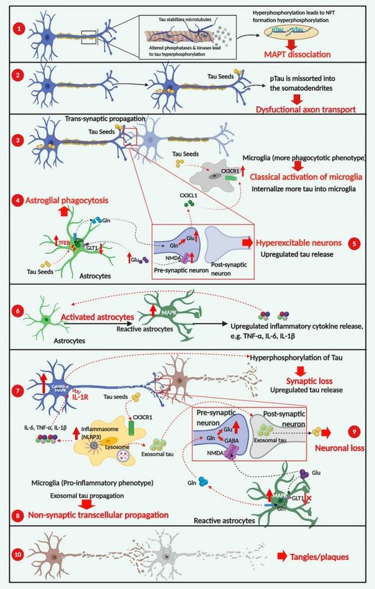
Figure 4. Cellular contributors to tau-dependent degeneration. The development of tau-related
pathogenesis has characteristic stages, starting from the formation of tau species consisting of
phosphorylated tau dissociated from microtubules À. Abbreviation: pTau, phosphorylated tau.
Hyperphosphorylation of tau incorrectly sorts tau into the somatodendritic compartment, which is
linked to dysfunctional axonal transport Á, one of the earliest pathological features of tauopathies.
Glial cells adapt a more neuroprotective phenotype with microglia classically activated  and as-
trocytes actively phagocytosing tau species Ã. As disease progresses, astrocytes transform into a
loss-of-function phenotype via lower-level expression of astrocyte-specific transporters, leading to
neuronal excitotoxicity and upregulated tau release. Additionally, alternatively activated microglia
in a pro-inflammatory phenotype are necessary and sufficient to induce reactive astrocytes with the
capability of releasing neuroinflammatory cytokines Å. At the later stages of disease progression,
the microglial-exosomal pathway acts as the essential tau propagation pathway Ç as an alternative to
transsynaptic transduction, due to extensive synaptic degeneration and neuronal death È. The forma-
tion of neuronal and glial tau plaques is the most important hallmark of tauopathies É. Red arrows
indicate pathological consequences of change. Created in BioRender.com.Biomedicines 2021, 9, 190 11 of 25
Activation of the NLRP3 inflammasome in microglia has been demonstrated to fa-
cilitate the progression of tau pathologies, mainly through intensifying neuronal tau hy-
perphosphorylation in an IL-1 receptor-dependent way [59]. At the late stage of disease
progression, dysfunctional synaptic transmission caused by synaptic loss and neuronal
death leads to microglia-exosomal tau transmission that takes precedence over transsynap-
tic tau transmission [60].Finally, neuronal and glial tau plaques are formed, which are the
most important hallmarks of tauopathies [9].
Overexpression of the ε4 allele of apolipoprotein (ApoE4) in multiple cell types shows
cell-type-specific effects; overexpression in neuronal cells upregulates neurotransmitter
release while enhancing inflammatory signaling of microglia. For astrocytes, ApoE4
overexpression downregulates phagocytosis of pathogenic proteins and disrupts lipid
transport and metabolism. Taken together, ApoE4 serves as a common genetic risk in AD
and primary tauopathies, and can worsen tau pathology, indicating an overlap between
ApoE4 and tau pathogenesis. LRP1, as a major receptor of tau species and ApoE, may play
an intermediate role between ApoE and tau species, which could point to a therapeutic
potential for treating tauopathies via LRP1 interaction.
4.1. The Involvement of Neuronal Activity in the Spreading of Pathogenic Tau
Under physiological conditions, tau is crucial for microtubule stabilization and is
located mainly in axons [61]. Immunoblot analysis with phosphorylation-dependent
antibodies revealed that phosphorylated tau is missorted into the somatodendritic com-
partment during the early stages of AD progression [62]. Missorted tau results in axonal
transport deficits and loss of synaptic functions and is more prone to forming toxic tau
oligomers if seeded [62].
The progression of tau pathology follows a defined hierarchical pattern, starting
from the EC, then advancing into anatomically connected neurons downstream in the
synaptic circuit, such as the dentate gyrus (DG), the hippocampus, and the neocortex,
as demonstrated by tau transgenic animal models [54,63]. Despite the identification of
the physiological role of neurons in regulating synaptic tau release and translocation [5],
the specific neuronal activities resulting in the propagation of tau pathologies are still
under investigation. Amyloid precursor protein (APP) transgenic mouse models show
that endogenous tau in CSF increases during the progression of amyloid plaque forma-
tion, accompanied by hyperexcitable neurons [64,65]. A key question is whether the
hyperexcitable neurons are essential for the release of pathogenic tau, independent of
Aβ. Indeed, tau pathology mouse models combined with novel neuronal stimulation
approaches showed that neuronal hyperexcitability and accelerated synaptic tau release
are critically linked and independent of Aβ toxicity [66]. Using an optogenetic activation
approach, the stimulated side of the hippocampus of the rTg4510 mice line tended to
accumulate more human tau protein, along with increased evidence of neuronal atro-
phy [66]. Additionally, tau pathology spread from the stimulated-EC to the synaptically
connected DG region, suggesting that the propagation of tau pathology accelerates through
synaptic circuits.
Abnormal extracellular glutamate levels have been proposed as one of several mecha-
nisms that account for an excitotoxic microenvironment in AD [67]. Notably, alterations
in synaptic glutamate homeostasis caused by dysfunctional astrocytes can be deleterious
to neuronal cells. To some extent, the activities of reactive astrocytes correlate with the
reduction in astroglial glutamate transporters, which in turn elevates the extracellular
glutamate level. Accumulation of excess glutamate contributes to neuronal excitability
through activating NMDA (N-methyl-D-aspartate) receptors. NMDA receptors, present
in glutamatergic neurons, respond to the glutamate levels via binding to their GluN2
subunit that activates increased calcium flux in the neurons [57]. Sequentially, activation
of extrasynaptic NMDA receptors has been linked to tau-induced neuronal cell death
mediated by calpain I and ERK/MAPK activation [68]. Therefore, alteration of astroglial
glutamate transporters and overstimulation of extrasynaptic NMDA receptors of neuronalBiomedicines 2021, 9, 190 12 of 25
cells may have an overlapping role in neuronal hyperexcitability, and these actions have
been implicated in the progression of tau pathology along with synaptic connections [69].
4.2. Glial Cells Are Involved in the Progression of Tau Pathology
Even though tau is expressed primarily by neurons, most primary tauopathies are
characterized by the presence of both neuronal and glial tau pathologies [12]. Glial cells
adopt immune functions and closely interact with neuronal cells for maintaining brain
development and homeostasis [70]. Most glial tau pathologies have been observed in
astrocytes and oligodendrocytes, and in some cases, tau pathologies have also been seen in
microglia. Moreover, both primary tauopathies and AD are characterized by microgliosis
and astrogliosis, along with a significant increase in the pro-inflammatory cytokines [6,71].
Glial cell dysfunction has also been implicated in the progression of neurodegenerative
diseases [51]. This part of the review aims to highlight the role of dysfunctional neuronal-
glial communication in the spreading and propagation of pathological tau during the
progression of tauopathies.
4.2.1. Microgliosis in Tauopathies
Neuroprotective Effects of CX3CL1/CX3CR1 Signaling
Microglia are the innate immune cells of the CNS and account for 5–20% of total
neural cells in the functional tissue of the brain [72,73]. They have two main CNS func-
tions: immune defense and maintenance and promoting programmed cell death during
development [72,73]. Recently, microglia-induced neuroinflammation has been linked
to tau hyperphosphorylation, suggesting that microglia play an important role in the
progression of tau-related neuropathogenesis [74]. As discussed previously, extensive
studies have demonstrated that tau pathology predominantly spreads along with synaptic
connections. Physiologically, microglia control and regulate synaptic plasticity through
pruning of inactive synapses via phagocytosis during CNS development [75]. Among the
key factors emerging as potential regulators of neuronal-microglial interaction, chemokine
ligand 1 (CX3CL1) secreted by neurons plays an essential role in regulating phagocytic
capability of microglia by binding to CX3CR1 [76], a key receptor that maintains the normal
synaptic pruning ability of microglia [77]. Altered CX3CL1/CX3CR1 signaling has been
demonstrated to regulate the pathological changes in both animal models of tauopathies
and AD patients [78,79]. Single-cell RNA-seq of microglia in AD-transgenic mouse brains
shows that CX3CR1 is upregulated as part of the initial innate immune response [80], which
facilitates the internalization of tau by microglia to enhance the clearance of extracellu-
lar Tau [55].
However, at the later stages of AD, CX3CR1, among many other genes, is down-
regulated [80]. The downregulation of CX3CR1 has also been observed in human brain
tissue from AD patients, showing that CX3CR1 levels decrease as microglial phagocytic
phenotypes are reduced [55]. Microglia have been found to phagocytose extracellular tau
oligomers directly via the tau-CX3CR1 interaction, which is impaired by the loss of CX3CR1
at the later stages of AD. The deletion of CX3CR1 in models of tau pathology has accelerated
tau phosphorylation and exacerbated neurodegeneration [55,81]. This CX3CR1 deficiency
led to elevated levels of tau phosphorylation on the AT8 (pS202), AT180 (pT231), and PHF1
(pS396/S404) epitopes [58], which is mediated by neuronal IL-1 and TLR-4 receptors trig-
gered by the microglial release of proinflammatory cytokines [82]. Indeed, the deletion
of CX3CR1 in the hAPP-transgenic mice model exacerbates microglial inflammation and
neurotoxicity by upregulating the secretion of proinflammatory cytokines [78]. Similarly,
CX3CL1 overexpression in the human tau transgenic mouse model rTg4510 significantly
reduced neurodegeneration and microglial activation [83]. Therefore, the investigation of
CX3CR1-CX3CL1 signaling has provided novel insights for treating tauopathies.Biomedicines 2021, 9, 190 13 of 25
The Role of NLRP3-ASC Inflammasome Activation in Tau Phosphorylation
Extracellular fibrillary Aβ-induced microgliosis has been linked to NOD-like recep-
tor family, pyrin domain containing 3 (NLRP3) inflammasome activation, which further
exacerbates Aβ pathology [84]. The role of microglia and NLRP3-caspase recruitment do-
main (ASC) inflammasome activation has been demonstrated recently in Aβ-independent
tau pathology [59]. Phagocytosis of fibrillar Aβ induced the assembly of the NLRP3
inflammasome consisting of NLRP3, ASC, and pro-caspase 1, which led to the caspase
1-dependent release of pro-inflammatory cytokines such as IL-1β and IL-18 [85]. Stancu
and colleagues [86] demonstrated that aggregated tau was capable of activating the NLRP3
inflammasome, which further exacerbated the tau aggregate seeding and increased the
secretion of proinflammatory cytokines. The importance of NLRP3 on the progression of
tau pathology also was demonstrated in tau transgenic mice models deficient for NLRP3 or
ASC. A significantly lower level of tau phosphorylation was observed in the hippocampal
samples of the transgenic mice deficient for NLRP3 or ASC compared with wild-type
mice [86]. Additionally, templated seeding of tau pathology was reduced in tau transgenic
mice with an ASC deficiency.
Reduced activities of GSK-3β and CaMKII-α, but not p38/MAPK and Cdk5, were cor-
related with the deficiency of NLRP3 or ASC, suggesting the potential role of the NLRP3
inflammasome in regulating tau kinases in neuronal cells [86]. To understand how the
NLRP3 inflammasome regulates tau phosphorylation, conditioned medium collected from
LPS-activated microglia induced an increased level of tau phosphorylation in neuronal
cells, along with the activation of CaMKII-α [59]. However, once the neuronal IL-1 receptor
was inhibited, the effects on CaMKII-α were abolished, suggesting that the activation of the
NLRP3 inflammasome in microglia promotes neuronal tau hyperphosphorylation in an IL-1
receptor-dependent manner via the regulation of multiple tau kinases (Figure 5). Potential
therapeutic interventions targeting the NLRP3 inflammasome have been attempted for
treating AD in mouse models [87]. By increasing ASC and NLRP3 gene expression in
Tau22 transgenic mice, the formation of tau aggregates was attenuated, as determined by
thioflavin T staining and reduced tau phosphorylation at serine 416, due to diminished
CaMKIIα activity [87]. Pharmacological NLRP3 inhibition using the molecular inhibitor
Biomedicines 2021, 9, x FOR PEER REVIEW
MCC50 also significantly decreased tau-seed induced tau aggregates, as determined 14 of 25 by
AT8 detection, in tau transgenic mice [86].
Figure 5. The
Figure rolerole
5. The of the NLRP3-ASC
of the NLRP3-ASCinflammasome
inflammasome in in tau
taupathogenesis.
pathogenesis. Either
Either fibrillary
fibrillary Aβ Aβ or species
or tau tau species
in theinform
the of
form
of monomers
monomersor oroligomers
oligomers are
are sufficient induce the
sufficient to induce the assembly
assemblyofofthetheNLRP3
NLRP3inflammasome,
inflammasome, consisting
consisting of NLRP3,
of NLRP3, ASC,
ASC,
and and pro-caspase
pro-caspase 1, 1, whichfurther
which further leads
leads to
tocaspase
caspase1-dependent
1-dependent release of the
release ofpro-inflammatory
the pro-inflammatorycytokines IL-1β and
cytokines IL-18and
IL-1β
[85]
IL-18 . The
[85]. activation
The activationof the
of NLRP3 inflammasome
the NLRP3 in microglia
inflammasome has been
in microglia hasdemonstrated to promote
been demonstrated neuronal tau
to promote hyper-tau
neuronal
phosphorylation in an IL-1 receptor-dependent manner via the regulation of multiple tau kinases, like GSK-3β and
hyperphosphorylation in an IL-1 receptor-dependent manner via the regulation of multiple tau kinases, like GSK-3β and
CaMKII-α.
CaMKII-α.
Non-Transsynaptic Tau Propagation-Microglial and Exosomal Spreading of Tau Species
as an Alternative Pathway
In addition to the important role of CX3CR1-CX3CL1 signaling and the NLRP3 in-
flammasome in tau pathogenesis via the crosstalk between neurons and microglia, exo-Biomedicines 2021, 9, 190 14 of 25
Even though these studies demonstrate the involvement of neuroinflammation and
altered CX3CR1-CX3CL1 signaling in the spreading of tau pathology, further investigation
is still needed to uncover the interplay between neuroinflammation induced by extracellular
tau aggregates and disrupted phagocytosis caused by impaired CX3CR1-CX3CL1 signaling.
Most likely, the relationship between inflammation and phagocytosis will demonstrate the
crucial role of microglia in the development of tau pathology.
Non-Transsynaptic Tau Propagation-Microglial and Exosomal Spreading of Tau Species as
an Alternative Pathway
In addition to the important role of CX3CR1-CX3CL1 signaling and the NLRP3 inflam-
masome in tau pathogenesis via the crosstalk between neurons and microglia, exosomes
are another key mediator between glial-neuronal communication for both synaptic pruning
in the healthy brain as well as neuroinflammation under pathological conditions [88,89].
Physiologically, neuronal exosomes stimulate microglial phagocytosis under selective
elimination of synaptic connections. Microglia-derived exosomes play a major role in
hierarchical tau transmission [60], despite pathogenic tau readily propagating from neuron
to neuron in the form of free-floating fibrils [34] and interconnected neuronal contacts [90],
as well as neuronal exosomes [4].
Recently, a tau rapid-propagation mouse model was created with adeno-associated vi-
ral (AAV)-tau injection into the EC [60]. This model exhibits rapid tau pathology, as demon-
strated by the spreading of human tau from EC to the DG within 1 month, recapitulating
the perforant diffusion pathway of AD progression in the human brain [91]. Moreover, inhi-
bition of exosome synthesis or depletion of microglia in this AAV-GFP/tau injection mouse
model led to a dramatic reduction of AT8+ tau detected in the DG without changing the
tau expressed in the injection site, indicating the important role of microglia-derived exo-
somes in the spreading of tau pathology. Pharmacologic inhibition of exosome synthesis in
microglia not only dramatically reduced secretion of the tau-containing exosomes but also
decreased the capabilities of exosomes to deliver hTau, as observed in co-cultured primary
neurons [60]. As the synaptic connection becomes less functional throughout disease pro-
gression, the microglial and exosomal transmission pathways become the primary means
of tau propagation [9], suggesting exosomal transmission as a potential therapeutic target.
4.2.2. Astrogliosis in Tauopathies
The concept of astroglial excitability—activation of membrane ion receptors in re-
sponse to stimulation—facilitates the bidirectional communication between neurons and
astrocytes mediated by a ‘tripartite synapse’ [92]. The close physical proximity between
synapses and astrocytes and resulting efficient neurotransmission explain why astrocytes
are key regulators in maintaining essential neuronal functions, including synaptic plasticity
and neurodevelopment [57]. Besides their crucial role in supporting neuronal functions
in the CNS, astrocytes represent the largest group of glial cells that interact closely with
microglia for maintaining efficient immune surveillance of the CNS [92]. Like microglia,
astrocytes also express genes involved in phagocytosis [93], and eliminate synaptic de-
bris [94], and protein aggregates, as seen by the clearance of Aβ [95]. In recent years,
the involvement of astrocytes in the progression of tau pathology has drawn much atten-
tion because of their widely demonstrated role in the progression of neurodegeneration in
tauopathies [11,57]. For example, reactive astrocytes induced by microglial activation have
been observed to precede tangle formation in P301S tau transgenic mice models (PS19) [51].
Reactive Astrocyte Phagocytosis Has a Neuroprotective Effect
Under pathological conditions, astrocytes develop more neurotoxic features by trans-
forming into reactive astrocytes (A1 subtype), induced by activated microglia and neuroin-
flammation in various human neurodegenerative disorders [16]. The phagocytic ability
of reactive astrocytes appears to be enhanced in tau transgenic mouse models [96]. As-
trocyte activation is accompanied by upregulated expression of transcription factor EB
(TFEB), the key regulator of the autophagy-lysosomal pathway (ALP). When comparedYou can also read