Cyclic mismatch binding ligands interact with disease-associated CGG trinucleotide repeats in RNA and suppress their translation
←
→
Page content transcription
If your browser does not render page correctly, please read the page content below
Nucleic Acids Research, 2021 1
                                                                                                                        https://doi.org/10.1093/nar/gkab669

Cyclic mismatch binding ligands interact with
disease-associated CGG trinucleotide repeats in RNA
and suppress their translation
Patryk Konieczny 1,2,† , Sanjukta Mukherjee 3,4,† , Ewa Stepniak-Konieczna 1 ,
Katarzyna Taylor1 , Daria Niewiadomska 1 , Agnieszka Piasecka1 , Agnieszka Walczak1 ,

                                                                                                                                                                  Downloaded from https://academic.oup.com/nar/advance-article/doi/10.1093/nar/gkab669/6343437 by guest on 17 August 2021
Anna Baud1 , Chikara Dohno3 , Kazuhiko Nakatani3 and Krzysztof Sobczak 1,*
1
  Department of Gene Expression, Institute of Molecular Biology and Biotechnology, Adam Mickiewicz University,
Uniwersytetu Poznanskiego 6, 61-614 Poznan, Poland, 2 Institute of Human Biology and Evolution, Adam Mickiewicz
University, Uniwersytetu Poznanskiego 6, 61-614 Poznan, Poland, 3 Department of Regulatory Bioorganic Chemistry,
The Institute of Scientific and Industrial Research, Osaka University, 8-1 Mihogaoka, Ibaraki 567-0047, Japan and
4
  National Centre for Biological Sciences (NCBS), Tata Institute of Fundamental Research (TIFR), Bellary Road,
Bangalore 560065, Karnataka, India

Received September 22, 2020; Revised July 13, 2021; Editorial Decision July 14, 2021; Accepted July 24, 2021

ABSTRACT                                                                                 tion might outweigh adverse effects related to FMRP
                                                                                         depletion.
Fragile X-associated tremor/ataxia syndrome (FX-
TAS) is a late-onset neurodegenerative disorder
caused by a limited expansion of CGG repeats in the
FMR1 gene. Degeneration of neurons in FXTAS cell                                         INTRODUCTION
models can be triggered by accumulation of polyg-                                        Fragile X-associated tremor/ataxia syndrome (FXTAS) is
lycine protein (FMRpolyG), a by-product of transla-                                      a late onset neurodegenerative disorder caused by a limited
tion initiated upstream to the repeats. Specific aims                                    expansion (premutation) of the polymorphic CGG repeat
of our work included testing if naphthyridine-based                                      tract in the 5 untranslated region (5 UTR) of the FMR1
molecules could (i) block FMRpolyG synthesis by                                          gene (1,2). The number of CGG repeats in the mRNA from
binding to CGG repeats in RNA, (ii) reverse patho-                                       premutation alleles ranges from 55 to 200. Typical FXTAS
logical alterations in affected cells and (iii) preserve                                 clinical symptoms manifest in patients older than 50 years
                                                                                         of age and include intention tremor, gait ataxia, parkin-
the content of FMRP, translated from the same FMR1
                                                                                         sonism and cognitive deficits. Magnetic resonance imag-
mRNA. We demonstrate that cyclic mismatch bind-                                          ing and neuropathological examination revealed the un-
ing ligand CMBL4c binds to RNA structure formed by                                       derlying white matter disease and the overall brain atro-
CGG repeats and attenuates translation of FMRpolyG                                       phy (3–6), while further post-mortem brain analyses showed
and formation of nuclear inclusions in cells trans-                                      intra-nuclear eosinophilic and ubiquitin-positive inclusions
fected with vectors expressing RNA with expanded                                         in neurons and astrocytes throughout the cerebrum and
CGG repeats. Moreover, our results indicate that                                         brainstem, predominantly in the hippocampus. Despite the
CMBL4c delivery can reduce FMRpolyG-mediated                                             apparent loss of Purkinje cells, no or rare inclusions were
cytotoxicity and apoptosis. Importantly, its therapeu-                                   observed in the remaining Purkinje cell population (3,7),
tic potential is also observed once the inclusions are                                   raising the question about the toxicity versus protective
already formed. We also show that CMBL4c-driven                                          function of the inclusions. Premutation has relatively high
                                                                                         incidence in humans that ranges from 1:430–850 in men
FMRpolyG loss is accompanied by partial FMRP re-
                                                                                         to 1:150–300 in women. However, the penetrance is in-
duction. As complete loss of FMRP induces FXS in                                         complete, with the current estimate of 40–75% males and
children, future experiments should aim at evalua-                                       16–20% females to develop the full-blown FXTAS symp-
tion of CMBL4c therapeutic intervention in differen-                                     toms (8–10). Additionally, about 20% of the female carriers
tiated tissues, in which FMRpolyG translation inhibi-                                    develop fragile X-associated primary ovarian insufficiency
                                                                                         [FXPOI, (11)].

* To   whom correspondence should be addressed. Tel: +48 61 829 5950; Fax: +48 61 829 5949; Email: ksobczak@amu.edu.pl
†
    The authors wish it to be known that, in their opinion, the first two authors should be regarded as Joint First Authors.

C The Author(s) 2021. Published by Oxford University Press on behalf of Nucleic Acids Research.
This is an Open Access article distributed under the terms of the Creative Commons Attribution-NonCommercial License
(http://creativecommons.org/licenses/by-nc/4.0/), which permits non-commercial re-use, distribution, and reproduction in any medium, provided the original work
is properly cited. For commercial re-use, please contact journals.permissions@oup.com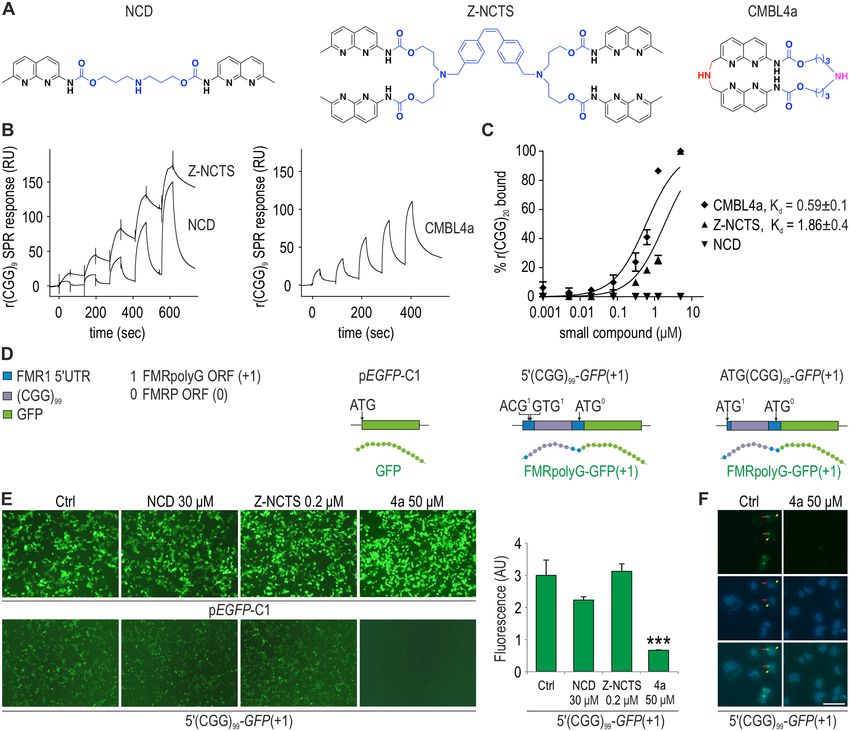
2 Nucleic Acids Research, 2021
While FMR1 mRNA content is elevated in FXTAS pa- transcript degradation. Similarly, in FXTAS they could be
tients, FMRP translated from FMR1 is usually slightly de- used to bind specific sites within FMR1 mRNA from pre-
creased (4,5,7,12–14). Based mainly on the cellular toxicity mutation alleles to induce the transcript degradation or
and accumulation of the RNA from premutation alleles in block FMRpolyG translation (18,25,33–36). Some of these
the nucleus (15–18), a concept was put forward, in which approaches would also result in freeing trapped molecules
neurodegeneration was attributed to sequestration of spe- from the RNA repeats and releasing them to their natural
cific proteins onto the FMR1 mRNA containing expanded environments, additionally contributing to the restoration
CGG repeats. Indeed, over the years many proteins such as of the cellular homeostasis.
hnRNP A2/B1, PUR␣, SAM68, DROSHA and DGCR8 One putative caveat of the strategies based on small
were found to be specifically trapped on expanded CGG re- molecule and antisense oligonucleotide delivery to FXTAS
Downloaded from https://academic.oup.com/nar/advance-article/doi/10.1093/nar/gkab669/6343437 by guest on 17 August 2021
peats (CGGexp ) interacting either directly or indirectly with cells is that both FMRpolyG and FMRP are expressed from
the RNA (16,17,19,20). Counteracting this phenomenon the same transcript and that downregulation of FMRpolyG
with overexpression of some of the proteins was shown to might result in a parallel downregulation of FMRP pro-
amend the apparent toxicity observed in FXTAS cell mod- tein (25,34,36). Specifically, targeted cleavage and removal
els (17,19,20). A second concept of neurodegeneration in of FMR1 mRNA would result in equivalent loss of both
FXTAS is based on the fact that CGGexp within the 5 UTR proteins. This is of particular importance as FMRP loss
of FMR1 transcript give rise to biosynthesis of homopoly- is a hallmark of fragile X syndrome (FXS), an early-onset
meric proteins that can induce cell death (21–23). Transla- disorder characterized by alterations in physical appear-
tion of toxic peptides takes place in FXTAS because the ance, autism and intellectual disabilities (37). In contrast
mRNA from premutation alleles is efficiently transported to to FXTAS-related increases in transcription and the FMR1
the cytoplasm, especially once the repeats are placed within mRNA content, synthesis of FMR1 mRNA in FXS patients
their natural FMR1 context (21,23,24). Competitive trans- is shut down due to CGG triplet cassette expansion to over
lation of FMRP and other peptides and proteins from the 200 repeats and the consequential promoter region hyper-
same FMR1 transcript might also explain slight decreases methylation. The complete absence of FMRP results in den-
of the FMRP content despite the elevated FMR1 mRNA dritic spine changes and alterations in synaptic plasticity as
levels in FXTAS cells (23,25). well as changes in connective tissue due to the FMRP in-
Synthesis of homopolymeric proteins from FMR1 and volvement in site specific translation of proteins in neurons
ASFMR1 antisense transcripts can be carried out by: (i) (37). It is important to note, however, that such severe FXS
repeat-associated non-AUG (RAN) translation initiated di- symptoms might not be observed in established tissues of
rectly from CGGexp (21,22,26), (ii) RAN translation initi- adult patients.
ated with near-cognate start codons located upstream to Targeting RNA structure by small molecules is still chal-
the repeats as a result of 43S preinitiation complex stalling lenging due to the lack of fundamental understanding of
at CGGexp and (iii) repeat expansion-independent transla- how these interactions occur. Yang et al. reported molecules
tion of proteins containing either long or short polyglycine targeting RNA CGG repeat that could regulate RAN trans-
tracts (FMRpolyG) from a near-cognate ACG start codon lation and RNA toxic gain of function (33,34). Their bis-
embedded in the Kozak sequence upstream to the CGG re- benzimidazole derivatives, known as typical minor groove
peats (23,27). The latter is supported by the fact that once a binders of DNA, inhibit synthesis of FMRpolyG without
GFP-stabilizing tag (see for example (28)) is fused to FM- affecting the downstream canonical translation (34). Re-
RpolyG, the protein is readily detectable, even when the cently, a G-quadruplex binder showed an inhibitory ef-
number of CGG repeats is within the normal range (21). fect on FMRpolyG synthesis (35,38), and some different
This indicates that shorter, presumably non-aggregating ho- classes of RAN translation inhibitors, including non-repeat
mopolymeric peptides can be efficiently degraded by the binders, were identified by high-throughput screening (35).
cell without any adverse effect (23). On the contrary, re- Our group has been focusing on a rational design approach
sults from model systems indicate that the stable aggre- to develop mismatch-binding ligands (MBLs) that target
gated form of FMRpolyG translated from the mRNA from specific noncanonical structure of nucleic acids through
premutation alleles is exceptionally toxic (21–23). Partic- complementary hydrogen bonding (39–42). Compounds
ularly, Sellier et al. showed that FMRpolyG interaction used in this study were designed to recognize an array of
with LAP2 triggers nuclear envelope disruption and cell Gs in the 5 -CGG-3 /5 -CGG-3 unit by complementary hy-
death (23). drogen bonding to the Watson-Crick surface (41–43). The
Although the contribution of FMRpolyG to pathology different modes of binding alter the structure of RNA CGG
in FXTAS is still debatable (18,29), there is increasing ev- repeat and its thermodynamic/kinetic stability, which could
idence that its high concentration in different model sys- have significant effects on interactions with RNA binding
tems leads to cell toxicity (21–23). By the same token, proteins and the resulting biological events.
downregulation of FMRpolyG could be a vital therapeu- A series of small molecules designed to recognize an array
tic goal as well as a read-out for successful treatment. Small of Gs have been synthesized, including naphthyridine car-
molecules and modified antisense oligonucleotides target- bamate dimer (NCD; (39)), a naphthyridine tetramer con-
ing RNA with expansion of triplet repeats stand out as taining two NCD molecules (Z-NCTS; (40,44)), and cyclic
prospective therapeutic agents in many genetic diseases (30– mismatch binding ligands (CMBLs) that consist of cy-
32). Importantly, their mode of action could be designed to clophane containing bis(2-amino-1,8-naphthyridine) moi-
either block specific sites on the target mRNA or to induce eties connected by variable linkers (41,42). Importantly,
Nucleic Acids Research, 2021 3
Z-NCTS was shown to bind to 5 -CGG-3 /5 -CGG-3 Filter binding assay (FBA)
DNA sequence in vitro with 1:1 binding stoichiometry and
The r(CGG)20 , r(AGG)20 and r(CUG)20 transcripts were
a dissociation constant (Kd ) of ca. 100 nM (40). We have
gifts from W. Krzyżosiak (Polish Academy of Science).
demonstrated that CMBLs could bind to DNA repeats,
For 5 radiolabeling, 2 pmol of transcripts was incubated
including (TGG)8 , (CCTG)9 and (CAG)9 (41), and CAG
with 2 pmol (12 mCi) of [␥ -P32 ] ATP, 1 U RNasin®Plus
RNA repeats (43). Synthesis of other CMBLs differing in
RNase Inhibitor, 10 U OPTI Kinase (Affymetrix), 1x reac-
molecular composition of the variable linker allowed to ob-
tion buffer (Affymetrix) and ddH2 0 up to 10 l, at 37◦ C for
tain ligands showing selective orthogonal binding relation-
30 min. The labeled RNA was run on a 8% PA gel (29:1)
ships with DNA and RNA repeats. In the present study, we
in 0.5 x TBE, at 100 V for 60 min. The band of RNA was
estimated therapeutic potential of NCD, Z-NCTS and 21
Downloaded from https://academic.oup.com/nar/advance-article/doi/10.1093/nar/gkab669/6343437 by guest on 17 August 2021
visualized on IP through FLA-1500 (FujiFilm), cut out fol-
CMBL compounds. Particularly, we screened their binding
lowed by ethanol precipitation and resuspended in 20 l
affinity to RNA CGG repeats in vitro and tested their abil-
ddH2 O. FBA was carried out by incubating 5 radiolabeled
ity to inhibit FMRpolyG translation in FXTAS cell line
RNA (1 nM) with small compounds of indicated concentra-
models.
tions (ranging from 0 to 20 M). Reactions were performed
in a volume of 30 l, in 1× buffer FBA (50 mM NaCl, 50
mM KCl, 50 mM Tris–HCl, 1 mM MgCl2 , pH 8.0) and in-
MATERIALS AND METHODS
cubated at 37◦ C for 15 min. Next, 25 l of each sample was
Surface plasmon resonance (SPR) assay loaded under vacuum induced pressure followed by a wash
step with 100 ul of buffer FBA onto two membranes, pre-
Surface plasmon resonance (SPR) single cycle kinetics
viously wetted in buffer FBA and placed between the ap-
assay was performed using Biacore T200 platform (GE
paratus frames (dot blotter). Positively charged complexes
Healthcare, Life Science). 5 -Biotin-TEG DNA or RNA
of RNA and small compounds remained on the top nitro-
sequences were immobilized on the Series S sensor chip
cellulose membrane (Protran BA 85, Whatman®) whereas
SA surface using avidin-biotin coupling in HBS-N running
free and negatively charged RNA, which went through the
buffer (HBS-N; 0.01 M, HEPES, 0.15 M NaCl, pH 7.4).
first membrane was subsequently caught on a nylon mem-
Five sensor chips containing immobilized RNA [r(CGG)9 ;
brane (Hybond™ N+, Amersham) placed underneath. The
SA chip-1, -2, -3, -4] or DNA [d(CGG)9 ; SA chip-5] (Sup-
signal from membranes was detected O/N, visualized on IP
plementary Table S1) were used to perform SPR assays for
through FLA-1500 (FujiFilm) and quantified using Multi
NCD, Z-NCTS and 21 CMBL compounds. Another five
Gauge software (FujiFilm). The dissociation constant (Kd )
sensor chips (SA chip-6, -7 -8, -9 and -10) with immobilized
of the RNA/small compounds interaction was calculated in
rSS30 (5 -biotin-TEG-CUUAGUACCAUUAUAGAUU
the GraphPad program using the following equation: one
UACCAUGAUUC-3 ), rDS30 (5 -biotin-TEG-CAGUA
site specific binding curve (Y = Bmax *X/(Kd + X)).
GUAGUAGUUUUUACUACUACUACUG-3 ), or other
repeat DNA or RNA sequences (Supplementary Table S2)
Electrophoretic mobility shift assay (EMSA)
were used for SPR assays for CMBL4c.
Amounts of immobilized RNA and DNA oligomers on 5 radiolabeled r(CGG)20 , r(AGG)20 and r(CUG)20 (see
each sensor surface are summarized in Supplementary Ta- FBA protocol) were first subjected to 1 min denaturation
bles S1 and S2. 5 -Biotinylated oligonucleotides were di- step at 90◦ C followed by 10 min of renaturation on ice.
luted to 0.2 M in 10 mM HEPES-500 mM NaCl and EMSA was carried out by incubating 5 radiolabeled RNA
injected to reach the response of around 500 RU. Blank (1 nM) with small compounds of indicated concentrations
immobilization was performed in the flow cell 1 for refer- (ranging from 0 to 40 M). Reactions were performed in a
ence subtraction. Ligand solution was diluted using HBS- volume of 10 l, in 1× buffer FBA (50 mM NaCl, 50 mM
EP + buffer (0.01 M HEPES, 0.15 M NaCl, 3.0 mM EDTA, KCl, 50 mM Tris–HCl, 1 mM MgCl2 , pH 8.0) and incu-
pH 7.4, 0.005% v/v Surfactant P20). 1.0 mM CMBL stock bated at 37◦ C for 15 min. The samples were run on a native
solutions in DMSO were diluted to obtain the final lig- 8% PA gel in 0.5× TBE at 100 V for 1 h. The gel was sub-
and solutions that contained 5% DMSO. 1xHBS-EP+ (for sequently dried and the signal was detected O/N, and visu-
NCD and Z-NCTS) and 5% DMSO containing 1xHBS- alized on IP through FLA-1500 (FujiFilm). The differenti-
EP+ (for CMBLs) were used as running buffers for bind- ation between free RNA and RNA in complexes with small
ing assays. Sensorgrams were obtained with the concen- compounds was possible due to their differences in mobil-
tration range of 0.5 to 8.0 M for NCD and 0.25 to 4.0 ity. Free and negatively charged RNA migrated faster from
M for Z-NCTS and 21 CMBL analogues. All sensorgrams RNA:small compound complexes which migrated much
were corrected by reference subtraction of blank flow cell slower, most likely due to gained positive charge of tested
response and buffer injection response. SPR sensorgram compounds, size and structural conformation of the com-
obtained for compounds using SA chip-1, -2, -4 and -5 plex. The dissociation constant (Kd) of the RNA/small
were further standardized with respect to the immobiliza- compounds interaction was calculated for a signal of RNA
tion amount (RU538) of (CGG)9 on the SA chip-3. Re- bound to small compounds in the GraphPad program us-
generation of the sensor surface was carried out using 50% ing the following equation: one site specific binding curve
aqueous solution of DMSO and 50 mM aqueous solution (Y = Bmax *X/(Kd + X)). The half-life of RNA/small com-
of NaOH. Apparent binding constants of NCD, Z-NCTS pounds interaction was calculated for a signal of free RNA
and CMBL4a were determined from SPR curves applying using the following equation: one phase decay (Y = (Y0 –
1:1 fitting model (Supplementary Table S3). Plateau)*exp(–K*X) + Plateau)).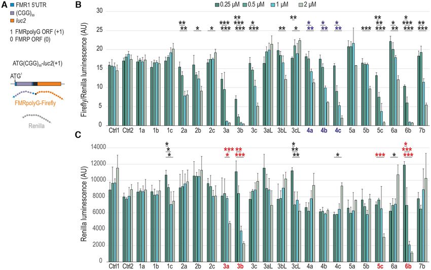
4 Nucleic Acids Research, 2021
Cell culture except for NCD, Z-NCTS and CMBL4a, were dissolved
in dimethyl sulfoxide (DMSO) to 1 mM concentration,
COS7, HeLa, SH-SY5Y and HEK293T cells were grown
aliquoted and stored at 4◦ C. NCD and Z-NCTS were dis-
in a high glucose DMEM medium with L-Glutamine
solved in H2 O and CMBL4a in H2 O or DMSO. No obvi-
(Lonza) supplemented with 10% fetal bovine serum (Sigma)
ous differences in the activity of CMBL4a was observed in
and 1% antibiotic/antimycotic (Sigma). FXTAS (1022-
either solvent. Prior to the experiment, the compounds were
07 (P3), XY, (CGG)81 ; 1044-07 (F3), XY, (CGG)97 ;
brought to the desired concentration in DMEM and added
WC26, XX, (CGG)60 /(CGG)90 ) and control (1028-07 (C4),
to the cells to the total 150 l. Solvent mixed with DMEM
XY, (CGG)22 ; C0603, XY, (CGG)31 ) as well as DM1
was delivered to control wells.
(GM04033; (CTG)1000 ) and control (GM07492) fibrob-
Downloaded from https://academic.oup.com/nar/advance-article/doi/10.1093/nar/gkab669/6343437 by guest on 17 August 2021
lasts were cultured in EMEM medium (Lonza) supple-
mented with 15% fetal bovine serum (Sigma), 1% MEM Fluorescence and confocal microscopy
non-essential amino acids (Thermo Fisher Scientific) and
Images were taken with Axio Observer.Z1 microscope
1% antibiotic/antimycotic (Sigma). All cells were grown at
equipped with AxioCam MRm camera, filter set 09 or
37◦ C in a humidified incubator containing 5% CO2 . FX-
10 (GFP), 49 (Hoechst 33342) and 31 (mCherry), A-Plan
TAS 1022-07 and 1044-07 and control 1028-07 fibroblast
10×/0.25 Ph1 objective (Zeiss), and AxioVs40 module or
lines were a kind gift from P. Hagerman (see also (45)),
a Nikon A1R confocal microscopy (20x objective). Total
while FXTAS WC26 and control C0603 fibroblast lines
fluorescence signal, mCherry-NLS positive cell numbers,
were obtained from A. Bhattacharyya (see also (46)). DM1
inclusion numbers, inclusion areas and nuclear circularity
GM04033 and control GM07492 fibroblasts as well as HD
were calculated using ImageJ. Total fluorescence was esti-
GM04281 fibroblasts were purchased from the Coriell In-
mated based on background subtraction (rolling ball algo-
stitute for Medical Research.
rithm) and mean intensity quantification. Inclusion areas
and numbers were analyzed following image thresholding
Constructs and using ‘Analyze Particles’ function. Nuclei were stained
ATG(CGG)99 -luc2(+1) and 5 (CGG)99 -luc2(0) vectors were with Hoechst 33342 (Invitrogen) per manufacturer’s proto-
described previously (47). To generate 5 (CGG)16 -luc2(0), col. Briefly, cell culture media was removed and cells were
5 (CGG)34 -luc2(0) and 5 (CGG)44 -luc2(0) constructs, washed in PBS. Next, sufficient volume of Hoechst stain-
5 (CGG)99 -GFP(+1) was plated and screened for colonies ing solution (prepared by diluting the Hoechst stock 1:2000
containing shorter number of the repeats. After restric- in PBS) was added to cover the cells. Upon 10-min incuba-
tion digest, the number of the repeats was confirmed by tion in a humidified CO2 incubator at 37◦ C, protected from
Sanger sequencing. The repeats were cut out from the light, staining solution was removed and cell were washed
vector by NheI and pasted into the NheI-digested and once in PBS prior to addition of fresh PBS and fluorescence
dephosphorylated (CIAP, Invitrogen) pmirGLO modified microscopy. For confocal microscopy, COS7 cells were fixed
vector containing the multicloning site in front of the luc2 for 15 min with 4% PFA 48 h post transfection and blocked
sequence (ATG-luc2; (28,47)). 5 (CGG)99 lacking a tag and for 1 h in 1% BSA diluted in PBS-Tween (0.1%; PBS-T). In-
containing the whole FMRpolyG sequence was generated cubation with mouse FMRpolyG 8FM (1:50; (23,49)) and
first by cutting out GFP sequence from the 5 (CGG)99 - rabbit Lamin B1––Nuclear Envelope Marker (1:200; Ab-
GFP(+1) vector with AvrII and EagI and ligating an insert cam, ab16048) primary antibodies was conducted O/N at
constructed from two complementary phosphorylated 4◦ C in the blocking solution. Secondary goat anti-mouse
oligonucleotides (F1/R1, Supplementary Table S4). The FITC-labeled (1:400; Jackson ImmunoResearch Laborato-
obtained vector was then used as a template to amplify a ries) and goat anti-rabbit Alexa Fluor 546 (1:200; Invitro-
PCR product with F2/R2 primers (Supplementary Table gen) secondary antibodies were applied for 1 h at RT in
S4). The product was subsequently digested with XhoI and PBS-T.
EagI and cloned into the digested template vector. The
control plasmid containing 16 CGG repeats (5 (CGG)16 ) Luciferase assay
was generated by cutting out the repeat cassette from the
5 (CGG)16 -luc2(0) plasmid with KasI and XhoI and cloning For luciferase assay, cells were harvested at different time-
it into the digested 5 (CGG)99 vector. points as indicated in figure legends. Following lysis,
cells were transferred to a Nunc F96 MicroWell Black
Polystyrene Plate (137101, Thermo Scientific) and lumines-
Plasmid and small compound delivery
cence of Firefly and Renilla luciferases were measured con-
For average fluorescence and luminescence signal quantifi- secutively using Dual Luciferase Assay System (Promega),
cation, COS7 cells were seeded in 125 l DMEM medium infinite F200 PRO, and i-control 1.8 SP1 microplate reader
on a 96-well plate one day before transfection. 12.5 l software (Tecan).
of either pEGFP-C1, 5 (CGG)99 -GFP(+1), ATG(CGG)99 -
GFP(+1), ATG(CGG)99 -luc2(+1), 5 (CGG)16 , 5 (CGG)99 ,
Cell viability assays
mCherry-NLS (48), 5 (CGG)16 -luc2(0), 5 (CGG)34 -luc2(0),
5 (CGG)44 -luc2(0) or 5 (CGG)99 -luc2(0) constructs were HEK293T and COS7 cells seeded in a 96-well plate (Fig-
delivered with X-tremeGENE HP DNA Transfection ure 7E and Supplementary Figure S16B) were transfected
Reagent (Roche; 125 ng DNA/0.25 l X-tremeGENE HP) with either 5 (CGG)16 or 5 (CGG)99 and treated with indi-
when cells reached 40% confluency. All small compounds, cated amount of CMBL4c or an equal volume of DMSO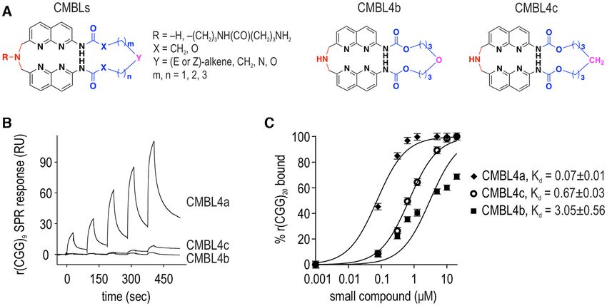
Nucleic Acids Research, 2021 5
(control). Cell viability, toxicity and apoptosis were ana- livery to COS7 cells. Following additional 22 h, RNA was
lyzed at indicated time-points post plasmid delivery using isolated with TRI Reagent (Sigma) according to the man-
ApoToxGlo™ Triplex Assay (Promega) according to the in- ufacturer’s instructions. Total RNA (1 g) was reverse-
structions provided by the manufacturer. Triplicate repeats transcribed with GoScript™ Reverse Transcriptase and
were used for each experiment. Fluorescence at two distinct oligo(dT) primers (Promega). PCR was performed with ei-
wavelength sets (400EX /505EM for viability, 485EX /520EX ther GFP/anchored oligo(dT)20 (Invitrogen) and GAPDH
for cytotoxicity) as well as luminescence (for apoptosis) primers at 50.3◦ C annealing temperature or with FMR1-
were recorded on Tecan Spark microplate reader. and GAPDH-specific primers (Supplementary Table S4).
HeLa cells (5000 cells/well; Supplementary Figure S7) For quantitative PCR (qPCR) and splicing analyses (Fig-
were seeded in a 96-well plate in a final volume of 90 ml ure 6C, Supplementary Figures S10 and S11A), fibroblasts
Downloaded from https://academic.oup.com/nar/advance-article/doi/10.1093/nar/gkab669/6343437 by guest on 17 August 2021
in DMEM containing 10% FBS. After 24 h of incubation, (GM07492 and GM04033) were harvested in TRIzol 48 h
cells were treated with 10 mL of seven different concentra- following delivery of DMSO or CMBLs. Each experiment
tions (0, 0.1, 0.3, 1.0, 3.0, 10, 30, 100 mM) of correspond- was performed in triplicate repeats. RNA was isolated using
ing CMBLs (CMBL1a, 1b, 1c, 3a, 3b, 4a, 4b, 4c and 5c) Total RNA Zol-Out™ kit (A&A Biotechnology) per man-
prepared in DMEM containing 10% FBS and 10% DMSO. ufacturers’ instructions. cDNA was synthesized from 500
Final concentration of 1% DMSO was maintained in each ng total RNA using SuperScript™ IV Reverse Transcriptase
well and each concentration was treated in experimental (Invitrogen) according to manufacturers’ protocol. qPCRs
sextuplicate. After 24 h of incubation, 5 ml of cell prolif- were performed in a QuantStudio™ 7 Flex System (Thermo
eration reagent (WST-8) in DMEM containing 10% FBS Fisher Scientific) using Maxima SYBR Green/ROX qPCR
was added to each well and incubated for 1 h. Readings of Master Mix (Thermo Fisher Scientific) according to the
the absorbance were taken at 450 nm wavelength, after 10 manufactures’ instructions. Targets containing a various
s of shaking time in Spectramax M5. Graphs were plotted number of CGG repeats were amplified with primers listed
using the Graphpad Prism8 version 8.0.2. Each experiment in Supplementary Table S4 at 58–60◦ C annealing tempera-
was performed at least in triplicate. ture. Ct values were normalized against GAPDH. Fold dif-
Cell viability assay in SH-SY5Y cells (Supplementary ferences in expression level were calculated according to the
Figure S8) was performed using the RealTime-Glo MT Cell 2−Ct method (50). Splicing of MBNL1 e1, MBNL1 e5,
Viability Assay according to the manufacturer’s protocol MBNL2 e7, NCOR2 e45, NFIX e7 and PHKA e19 was an-
(Promega). Cells were plated in 20% confluency in a 96-well alyzed following standard PCR reactions using GoTaq®
in 50 l of culture medium. The next day the medium was Flexi DNA polymerase (Promega). Sequences of primers
removed and replaced with 25 l medium containing 2× are listed elsewhere (51,52). PCR images were captured us-
RealTime-Glo reagents. The CMBL titration was prepared ing G:Box EF2 (Syngene) and analyzed using GeneTools
in medium at 2× concentrations and added to the plate at an image analysis software (Syngene). Results were plotted on
equal volume. For time-zero measurements, cells were incu- bar graphs as % exon spliced in (PSI).
bated with RealTime-Glo MT Cell Viability reagent for 1 h
at 37◦ C. Luminescence was then read every 8 for 48 h on
Western blot
Tecan M200. The quantification of the luminescence was
based on four experimental replicas. P-values were calcu- DMSO and CMBL compounds were added to FXTAS
lated using GraphPad Prism software, 8.3.0 (https://www. and non-FXTAS fibroblast lines at ∼80% confluency to 2
graphpad.com/). M (0.2% DMSO) final concentration and incubated for
48 h. Fibroblasts were harvested with trypsin and lysed
Lethality in mice with RIPA buffer. Protein extracts were centrifuged at 10
000 RPM for 10 min at 4◦ C, measured with Pierce™ BCA
The experiments in mice were performed with the per-
Protein Assay Kit (Thermo Fisher Scientific) and heat-
mission of the local ethical committee (NCBS-IAE-
denatured for 10 min at 70◦ C with the addition of Bolt
2017/05(N)). BALB/c mice (4–6-week-old) were injected
LDS buffer (Invitrogen; Figure 8C and Supplementary Fig-
subcutaneously with a gauge needle either with 1% DMSO
ure S17A) or 5 min at 95◦ C following addition of Laemmli
in PBS (n = 4) or one of the five concentrations of CMBL4c
buffer (Supplementary Figure S17B). 20–25 g of protein
(25, 50, 100, 250, 500 mg/kg body weight of mice; n = 6
per well was separated in Bolt™ 4–12% Bis-Tris Plus gel
mice for each treatment group) prepared from the DMSO
(Invitrogen, NW04120BOX) and Bolt™ MES SDS Run-
stock solution and diluted with PBS to obtain the desired
ning Buffer. Proteins were transferred to PVDF transfer
CMBL4c concentration and 1% of DMSO. Mice were mon-
membrane (0.45 M, GE Healthcare) for 1 h at 100 V
itored closely for 48 h. As no signs of morbidity or toxi-
in Leammli buffer with 20% methanol. Following trans-
city were observed during this time period, the treatment
fer, membranes were blocked and incubated with antibodies
was continued at regular intervals of 48 h for additional 12
in SNAP id Protein Detection System (Merck Millipore).
days. No signs of morbidity or toxicity were observed in
Blocking was performed for 20 min with either 0.125%
the course of the two-week experiment in either treatment
non-fat dry milk (NFDM) in TBS with 0.1% Tween 20
group.
(TBS-T; Figure 8C and Supplementary Figure S17A) or
1% BSA in TBS-T (Supplementary Figure S17B). Cor-
RNA isolation and RT-PCR
respondingly, primary antibodies were diluted in either
For semi-quantitative multiplex RT-PCR (Figure 6A and 0.125% NFDM/TBS-T or 1% BSA/TBS-T. The follow-
B), CMBL compounds were added 18 h post plasmid de- ing concentrations and incubation times were used: rabbit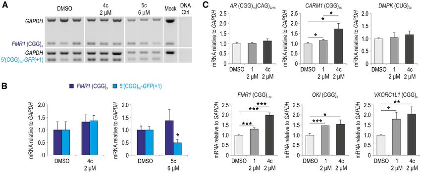
6 Nucleic Acids Research, 2021
anti-FMRP (ab17722, Abcam), 1:500, 1 h 15 min; mouse and CMBL4a were in a nanomolar range (Kd(app) ∼ 300
anti-GAPDH (sc-47724, Santa Cruz), 1:10 000, 20 min; nM and Kd(app) ∼ 800 nM, respectively) characterized by
rabbit anti-SLC40A1 (NBP1-21502, Novus Biologicals), much slower dissociation kinetics of Z-NCTS (Supplemen-
1:1000, 1 h; and rabbit anti-QKI (ab126742, Abcam), tary Table S3). Filter binding assays also revealed the lowest
1:1000, 1 h. Membranes were washed in TBS-T and in- affinity of NCD to r(CGG)20 while CMBL4a and Z-NCTS
cubated with horseradish peroxidase conjugated secondary interacted with the CGG repeats in similar ligand concen-
antibodies: anti-rabbit (A9169, Sigma), 1:20,000 for 15 min trations (Figure 1C).
or anti-mouse (A9044, Sigma), 1:20 000 for 15 min and We hypothesized that binding of small molecules to
washed with TBS. Antibody-antigen complexes were visu- CGG repeat hairpin in vivo can affect efficiency of RAN-
alized by enhanced chemiluminescence (ECL) using Lumi- translation from RNA containing expanded CGG triplets.
Downloaded from https://academic.oup.com/nar/advance-article/doi/10.1093/nar/gkab669/6343437 by guest on 17 August 2021
nata Forte HRP Substrate (Merck Millipore, WBLUF0500) Therefore, we followed the in vitro binding tests with cell cul-
and detected with G:Box System (Syngene). ture experiments, delivering NCD, Z-NCTS and CMBL4a
The western blot analysis for HTT protein (17/68Q to the COS7 cell line transfected with the 5 (CGG)99 -
tract; Supplementary Figure S11B) was performed as pre- GFP(+1) vector, containing the full length of FMR1 5 UTR
viously described (53). Briefly, 25 g of total protein was with 99 CGGs fused with GFP, and from which a fusion
run on a Tris-acetate sodium dodecyl sulphate (SDS)- protein of FMRpolyG and GFP is generated (FMRpolyG-
polyacrylamide gel (1.5 cm, 4% stacking gel/4.5 cm, 5% re- GFP) (Figure 1D-F; (47)). Compound toxicity compar-
solving gel, acrylamide:bis-acrylamide ratio of 35:1) in XT ison revealed increased cell mortality starting with 0.2,
Tricine buffer (Bio-Rad; Hercules, CA, USA) at 140 V in an 30 and 50 M for Z-NCTS, NCD and CMBL4a, respec-
ice-water bath. After electrophoresis, the proteins were wet- tively. Using these subtoxic concentrations, we did not ob-
transferred to a nitrocellulose membrane. The membranes serve any significant inhibitory effect on the FMRpolyG-
were cut according to protein ladder (HiMark) for separate GFP signal upon Z-NCTS and NCD administration 48 h
detection of respective proteins. All steps of immunodetec- post plasmid delivery (Figure 1E). In contrast, addition of
tion were performed in SNAP id Protein Detection System CMBL4a markedly reduced the fluorescence in cells trans-
in buffer containing 0.25% non-fat dry milk in PBS/0.9% fected with 5 (CGG)99 -GFP(+1) vector but not in control
NaCl/0.1% Tween-20. For huntingtin and plectin detec- cells, to which control pEGFP-C1 vector was delivered. In-
tion, the blots were probed with the primary antibodies: terestingly, we observed a variable size of FMRpolyG in-
rabbit anti-huntingtin (1:1000; ab109115 (EPR5526), Ab- clusions in untreated cells and while the larger co-localized
cam) and rabbit anti-plectin-1 (1:1000; #12254 (D6A11), with Hoechst staining (Figure 1F, red arrows), the smaller
Cell Signaling) and then with anti-rabbit HRP-conjugated were often Hoechst-negative (Figure 1F, yellow arrows) in-
secondary antibodies (1:500; 711–035–152, Jackson Im- dicating their cytoplasmic distribution.
munoResearch). The immunoreaction was detected using
ECL Western Blotting Substrate (ThermoScientific).
CMBL4c shows high activity and low toxicity in cell culture
assays and mice
RESULTS
We then focused on CMBL chemistry (Figure 2A) and com-
CMBL4a binds to RNA CGG repeats in vitro and attenuates
pared the effect of 15 previously reported (1a, 1b, 1c, 2a,
FMRpolyG production in cell culture
2b, 2c, 3a, 3b, 3c, 4a, 4c, 5c, 6a, 6b, 7b) and six newly syn-
Based on our previous studies (40–42), we predicted bind- thesized (3aL, 3bL, 3cL, 4b, 5a, 5b) CMBL compounds
ing of naphthyridine ligands to CGG repeats and their use ((41,42); Supplementary Data), differing in molecular com-
as potential therapeutics in FXTAS. CGG repeats in RNA position of the variable linker (Figure 2A, Supplementary
form a thermodynamically stable A-form hairpin structure Figure S1). We first screened CMBLs using SPR assays with
with consecutive structural motifs containing two Watson– immobilized r(CGG)9 repeats (Figure 2B, Supplementary
Crick G–C pairs and structure stabilizing G–G pair with Figure S2). The compounds’ responses differed greatly in
hydrogen bonds between the Watson-Crick and Hoogsteen intensity and kinetic profiles, with CMBL4a showing the
edges (54–56). The most recent study showed that if the highest binding affinity, indicating the significance of the
CGG repeat tract reaches pathogenic length it may also linker structure in the interaction with the CGG repeats
adopt G-quadruplex structure (38). We investigated bind- (Figure 2B, Supplementary Figures S1 and S2). A CMBL4a
ing affinity of NCD, Z-NCTS and CMBL4a that encom- analogue, CMBL4c, also showed a notable SPR response
pass H bonding surface complementary to guanine (39– in concentrations higher than 1 M, with rapid associa-
42,44) in the RNA structure formed by CGG tandem re- tion and very slow dissociation profiles (Figure 2B, Supple-
peats in vitro using surface plasmon resonance (SPR) sin- mentary Figure S2). CMBL5a, CMBL5c and CMBL3aL
gle cycle kinetics and filter binding assays (Figure 1A-C). also exhibited notable SPR responses; however, with faster
All compounds showed apparent binding to r(CGG)9 , and dissociation kinetics than CMBL4c (Supplementary Fig-
NCD had the weakest SPR response among them, con- ure S2). Interactions of selected CMBL compounds with
sidering its two-fold higher concentration used in the as- longer r(CGG)20 as well as control r(AGG)20 and r(CUG)20
say (Figure 1B). Specifically, apparent association (kon ) and RNA hairpin structure were also tested by in vitro filter
dissociation (koff ) constants of NCD applying 1:1 fitting binding and electrophoretic mobility shift assays (Figure
model indicated relatively slow association and rapid disso- 2C, Supplementary Figures S3 and S4). The results showed
ciation kinetics as well as high dissociation constant (Kd(app) that CMBLs bound to r(CUG)20 with several-fold higher
= 15.3 M; Supplementary Table S3). Kd(app) for Z-NCTS Kd than to r(AGG)20 and r(CGG)20 . Particularly, among
Nucleic Acids Research, 2021 7
Downloaded from https://academic.oup.com/nar/advance-article/doi/10.1093/nar/gkab669/6343437 by guest on 17 August 2021
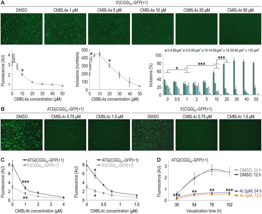
Figure 1. CMBL4a binds with high affinity to rCGG triplet repeats in vitro and attenuates FMRpolyG signal in cell culture. (A) Schematic representations
of NCD, Z-NCTS and CMBL4a structures. (B) SPR single cycle kinetic analysis of NCD, Z-NCTS and CMBL4a to the r(CGG)9 immobilized sensor
surface. NCD was added sequentially to 0.5, 1.0, 2.0, 4.0 and 8.0 M while Z-NCTS and CMBL4a to 0.25, 0.5, 1.0, 2.0 and 4.0 M. (C) Graphical
representation of filter binding assay (FBA) results showing an in vitro interaction of NCD, Z-NCTS and CMBL4a compounds with r(CGG)20 . Mean
values from two experiments ± standard deviation (SD) are shown on the graph. (D) Schematic representation of GFP and GFP-fused vectors used in
the study. (E) Visualization of either GFP or FMRpolyG-GFP signals in COS7 cells 48 h following transfection with pEGFP-C1 or 5 (CGG)99-GFP(+1)
vectors and delivery of NCD, Z-NCTS, CMBL4a or equivalent solvent amounts (H2 O; Ctrl) 18 h post transfection. The graph in the right panel shows
quantification of FMRpolyG-GFP signals. Mean values from at least two photos, each from a different biological replicate, with standard deviation
(SD) are shown on the graph. Statistical significance was determined by unpaired two-tailed Student’s t-test (*** indicates P < 0.001). (F) Representative
FMRpolyG, Hoechst or merged FMRpolyG/Hoechst images of COS7 cells 48 h post transfection with 5 (CGG)99 -GFP(+1) that were treated with either
H2 O or CMBL4a. Hoechst was added to the cells 10 min prior to the analysis. Representative large FMRpolyG inclusions stained with Hoechst and
Hoechst-negative smaller inclusions are marked with red and yellow arrows, respectively. Bar, 50 m.
the all tested CMBLs, we observed the highest affinity of ciferase activity in a concentration-dependent manner (Fig-
CMBL4a to the CGG repeats, in agreement with the SPR ure 3B). Unexpectedly, despite the highest affinity to tan-
data. dem repeats in vitro (Figure 2B and C, Supplementary Fig-
We next compared the effect of 21 CMBL com- ure S4), CMBL4a effect on FMRpolyG-Firefly inhibition
pounds in their capacity to lessen the luciferase signal was moderate (Figure 3B). Particularly, top activities in
of FMRpolyG-Firefly fusion protein in ATG(CGG)99 - COS7 cells were observed for CMBL3a, 3b, 4c, 5c and
luc2(+1)-transfected COS7 cells ((47); Figure 3). CMBL1a- 6b. Most of them displayed associated cellular toxicity in
c, CMBL2c, CMBL3aL, CMBL3cL and CMBL5a showed higher concentrations (marked red in Figure 3C, Supple-
no significant effect on translation of FMRpolyG-Firefly mentary Figures S5–S8). Therefore, for further studies we
whereas other compounds induced a decrease in the lu- chose CMBL4c along with its analogues CMBL4a and8 Nucleic Acids Research, 2021
Downloaded from https://academic.oup.com/nar/advance-article/doi/10.1093/nar/gkab669/6343437 by guest on 17 August 2021
Figure 2. Comparison of CMBL4a, 4b and 4c in vitro binding to CGG repeats. (A) General structure of CMBLs as well as detailed representations of
CMBL4b and 4c used in the study. (B) SPR single cycle kinetic analysis of CMBL4a, 4b and 4c to the r(CGG)9 immobilized sensor surface. CMBLs were
added sequentially to 0.25, 0.5, 1.0, 2.0 and 4.0 M. (C) Comparison of affinities of CMBL4a, 4b and 4c to r(CGG)20 based on FBA. Mean values from
three experiments ± standard deviation (SD) are shown on the graph.
CMBL4b (marked blue in Figure 3B), which showed rela- wards d(CCG)9 , d(CTG)9 , d(GAA)9 , d(G4 C2 )6 , r(CCG)9 ,
tively high activity and low toxicity. We first estimated their r(CUG)9 , r(GAA)9 , r(CCUG)9 , r(G4 C2 )6 , rSS30 and
effect on attenuation of fluorescent signal of FMRpolyG- rDS30. In contrast, d(CCTG)9 showed the highest re-
GFP and inclusion formation (Figure 4). Interestingly, in sponse from all of the tested sequences and the responses
cells transfected with 5 (CGG)99 -GFP(+1), increasing con- of CMBL4c towards d(CGG)9 , d(CAG)9 and r(CAG)9
centrations of CMBL4a (up to 10 M, dashed line in Fig- were comparable to that of r(CGG)9 . Nonetheless, the
ure 4A) were first correlated with higher amounts and then d(CCTG)9 , d(CGG)9 , d(CAG)9 and r(CAG)9 SPR curves
decreasing numbers of FMRpolyG-GFP inclusions, par- revealed relatively fast dissociation kinetics of CMBL4c
ticularly those of larger areas (Figure 4A). In agreement when compared to the r(CGG)9 . This data indicates the
with the luciferase activity data (Figure 3B), CMBL4c com- highest binding affinity of CMBL4c towards r(CGG)9
pared to 4a and 4b showed the highest activity in lower con- among the tested RNA/DNA sequences.
centration range (Figure 4A–C) that was associated with As we observed CMBL4c binding to RNA as well as
FMRpolyG-GFP translation efficiency from 5 (CGG)99 - DNA (CGG)9 , we asked whether the effect of CMBL
GFP(+1) and ATG(CGG)99 -GFP(+1) vectors (Figures 1D, compounds on synthesis of FMRpolyG is related to
4B and C) and the time that the compound was admin- their interaction with tandem repeats in target mRNA
istered following ATG(CGG)99 -GFP(+1) delivery (Figure molecules and/or to transcription inhibition due to bind-
4D). We then tested CMBL4c toxicity in vivo in BALB/c ing to the CGG repeat-rich DNA cassette. Endogenous
mice. Importantly, no signs of toxicity were observed in FMR1 and 5 (CGG)99 -GFP(+1) mRNA levels were quan-
mice treated every 48 h for two weeks with the highest dose tified in COS7 cells following delivery of 5 (CGG)99 -
(500 mg/kg) via subcutaneous injection. GFP(+1) vector and either CMBL4c or 5c (ligand associ-
ated with cellular toxicity; Figure 3C, Supplementary Fig-
ures S7 and S8), relative to GAPDH transcript levels (Fig-
CMBL4c interacts with various repeat sequences but does not ure 6A and B). Importantly, we did not notice any reduc-
lower the level of mRNAs containing CGG, CUG and CAG tion in FMR1 and 5 (CGG)99 -GFP(+1) mRNAs follow-
repeats ing CMBL4c delivery in COS7 cells and rather slightly in-
To evaluate specificity and affinity of CMBL4c towards var- creased amounts of FMR1 mRNA and other short and long
ious disease-associated repeat DNA and RNA sequences, CGG repeat-containing mRNAs, including CARM1, QKI
we performed SPR assays with immobilized d(CGG)9 , and VKORC1L1 in human fibroblasts (Figure 6C, Supple-
d(CCG)9 , d(CAG)9 , d(CTG)9 , d(GAA)9 , d(CCTG)9 , mentary Figure S10). Likewise, addition of CMBL5c did
d(G4 C2 )6 and corresponding RNAs, r(CGG)9 , r(CCG)9 , not change endogenous FMR1 mRNA content in COS7
r(CAG)9 , r(CTG)9 , r(GAA)9 , r(CCTG)9 , r(G4 C2 )6 , as well cells; however, significantly reduced the level of vector-
as random single stranded (rSS30) and double stranded originated mRNA containing CGGexp (Figure 6A and B).
RNAs (rDS30) (Figure 5 and Supplementary Figure S9). The latter presumably pertains to the toxic effect that this
No significant responses were observed for CMBL4c to- compound exerts in higher concentrations.Nucleic Acids Research, 2021 9
Downloaded from https://academic.oup.com/nar/advance-article/doi/10.1093/nar/gkab669/6343437 by guest on 17 August 2021
Figure 3. Comparison of CMBLs’ activities in COS7 cells using luciferase assay. (A) Schematic representation of ATG(CGG)99 -luc2(+1) dual luciferase
vector used in the study. The vector contains 99 CGG repeats fused with luc2, from which a fusion protein of FMRpolyG and Firefly luciferase is generated
(FMRpolyG-Firefly) as well as hRluc-neo sequence, from which Renilla luciferase is produced independently from FMRpolyG-Firefly. (B, C) Graphs
showing luminescence signals from COS7 cells 56 h after transfection with ATG(CGG)99 -luc2(+1) and delivery of CMBL compounds at 0.25, 0.5, 1 or
2 M concentration 6 h post plasmid addition. FMRpolyG-Firefly luciferase signals normalized to Renilla luminescence are shown in B while Renilla
values that represent the content of living cells and toxicity of the compounds in C. Mean values from three experiments with standard deviation (SD) are
shown on the graphs. Statistical significance was determined by unpaired two-tailed Student’s t-test (* indicates P < 0.05, ** indicates P < 0.01 and ***
indicates P < 0.001). CMBL4a, 4b and 4c that inhibit synthesis of FMRpolyG-Firefly without causing cellular toxicity are marked in blue in B. CMBL3a,
3b, 5c, 6b that lower relative FMRpolyG-Firefly contents and affect cell survival are marked red in C.
We also tested whether CMBLs could bind other disease- The effect of CMBL4c on inclusion growth is delivery-time
causing expanded triplet repeats, including CUGexp in DM1 dependent
and CAGexp in Huntington’s disease (HD). In DM1, CM-
We then tested if CMBL4c could be used to reverse the
BLs could potentially act by releasing sequestered MBNL
inclusion deposition in COS7 cells following FMRpolyG-
proteins from expanded CUG repeats in DMPK mRNA
GFP forced translation from 5 (CGG)99 -GFP(+1) (Fig-
(57). As MBNLs play crucial roles in mRNA metabolism,
ure 7A and B, Supplementary Figures S12 and S13) or
particularly splicing regulation (58,59), therapeutic inter-
ATG(CGG)99 -GFP(+1) (Supplementary Figures S14 and
vention would increase the level of available MBNLs and
S15) vectors. We delivered CMBL4c at 2 M concentration
the percentage of correctly spliced mRNAs. In accordance
18 or 30 h post transfection and estimated inclusion sizes
with the in vitro data (Figure 5B, Supplementary Figure S4),
54, 72, 90 and 108 h post transfection. While significant
we observed no significant changes in inclusion of alterna-
time-dependent increases in inclusion areas were observed
tive exons in MBNL-target mRNAs upon CMBL4c deliv-
in untreated cells following delivery of 5 (CGG)99 -GFP(+1)
ery to either non-DM1 or DM1 fibroblasts (Supplemen-
and ATG(CGG)99 -GFP(+1), either no increases (Figure
tary Figure S11A). Likewise, in HD CMBLs could poten-
7B) or slight decreases (Supplementary Figure S14B) were
tially bind to CAGexp in the mutated HTT transcript and
noted once CMBL4c was delivered 18 h post transfection.
block translation of the toxic huntingtin protein containing
In contrast, delivery of CMBL4c in the later time point
long polyglutamine tracts (60,61). We observed; however,
did not have such prominent effect on halting the inclusion
no change in the level of either normal or mutant huntingtin
growth.
in HD fibroblasts upon 48h treatment with 1 M CMBL3a
As our previous results indicated that GFP-tagging in-
(Supplementary Figure S11B). These results indicate that
creases protein stability (28,47), we delivered to COS7 cells
CMBLs, while potentially therapeutic in FXTAS, are not
a construct encoding an untagged version of FMRpolyG
suitable for therapy of DM1 and HD.
containing the full native protein sequence (5 (CGG)99 ;10 Nucleic Acids Research, 2021
Downloaded from https://academic.oup.com/nar/advance-article/doi/10.1093/nar/gkab669/6343437 by guest on 17 August 2021
Figure 4. CMBL4c efficiently reduces FMRpolyG content in COS7 cells. (A) Representative images (upper panel), graphical representations of
FMRpolyG-GFP fluorescence signals (lower left panel), total inclusion numbers (lower middle panel) and percentages of inclusion size areas (lower
right panel) following transfection of COS7 cells with 5 (CGG)99 -GFP(+1) and delivery of increasing concentrations of CMBL4A, as indicated in the
figure. Mean values from two biological replicates ± standard deviation (SD) are shown on the graphs. (B, C) Representative images (B) and graphs (C)
showing GFP fluorescence signals 80 h following delivery of either ATG(CGG)99 -GFP(+1) or 5 (CGG)99 -GFP(+1) plasmids. CMBL4b or 4c compounds
at indicated concentrations were added 6 h after transfection. Mean values from three experiments ± standard deviation (SD) are shown on the graphs.
(D) Fluorescence signals after transfection with ATG(CGG)99 -GFP(+1) and delivery of either DMSO (Ctrl) or CMBL4c at 2 M concentration 12 or 24
h post transfection. Statistical significance was determined by unpaired two-tailed Student’s t-test (fluorescence signals and inclusion numbers in A, C and
D) or by Kruskal–Wallis test (inclusion areas in A). * indicates P < 0.05, ** indicates P < 0.01 and *** indicates P < 0.001.
Figure 7C), including the C-terminal part of the protein and neither addition of CMBL1 nor 4c prevented the cell
shown to exert cellular toxicity (23). Indeed, confocal im- death.
munofluorescence microscopy analysis revealed abnormal- To further assess therapeutic potential of CMBL4c, we
ities in nuclear envelope structure and death of cells con- measured viability, cytotoxicity and apoptosis of HEK293T
taining FMRpolyG aggregates (Figure 7D). To evaluate and COS7 cells following delivery of 5 (CGG)16 or
whether the cell death could be diminished following in- 5 (CGG)99 and either DMSO (as a control) or CMBL4c,
clusion formation, we co-delivered either a long or short at 6 or 24 h post transfection (Figure 7E, Supplemen-
CGG repeat containing construct with a native FMR1 tary Figure S16B). In accordance with the microscopy
5 UTR sequence (5 (CGG)99 or 5 (CGG)16 ; Figure 7C) analysis (Figure 7D, Supplementary Figure S16A), we ob-
along with a marker of transfected cells, mCherry-NLS vec- served decreased viability, high cytotoxicity and apopto-
tor, to COS7 cells and treated them 24 h post transfection sis of cells to which 5 (CGG)99 was delivered. Importantly,
with CMBL1 control compound or CMBL4c (Supplemen- CMBL4c addition early after transfection (6 h) signifi-
tary Figure S16A). Administration of 5 (CGG)99 resulted cantly increased viability of HEK293T cells (Figure 7E).
in significantly reduced numbers of mCherry-positive cells Moreover, CMBL4c lowered cytotoxicity and apoptosis inNucleic Acids Research, 2021 11
Downloaded from https://academic.oup.com/nar/advance-article/doi/10.1093/nar/gkab669/6343437 by guest on 17 August 2021
Figure 5. CMBL4c interacts with selected RNA and DNA repeat sequences. (A, B) SPR single cycle kinetic analysis of CMBL4c interaction with DNA
sequences (A) and RNA sequences (B) as indicated in the figure. CMBL4c was added sequentially to 0.25, 0.5, 1.0, 2.0 and 4.0 M.
Figure 6. CMBL4c does not reduce the level of mRNAs containing CGGexp , CAGexp and CUGexp . (A) Semi-quantitative multiplex RT-PCRs showing
FMR1, 5 (CGG)99 -GFP(+1) and GAPDH mRNA levels in COS7 cells 40 h after transfection with 5 (CGG)99 -GFP(+1) vector and 22 h incubation with
CMBL4c or 5c. Mock represents signals from control untransfected cells and DNA Ctrl lane shows RT-PCR signals from a sample in which 5 (CGG)99 -
GFP(+1) plasmid was used as a template. (B) Quantitative analyses of multiplex RT-PCR signals shown in A. FMR1 and 5 (CGG)99 -GFP(+1) mRNA levels
were normalized to GAPDH content. (C) RT-qPCR analyses showing AR, CARM1, DMPK, FMR1, QKI and VKORC1L1 transcript levels in control
fibroblasts (GM07492) 48 h following delivery of DMSO, CMBL1(b and c) or 4c. Mean values normalized to GAPDH mRNA levels with standard
deviation (SD) are shown on the graphs. Statistical significance was determined by unpaired two-tailed Student’s t-test (* indicates P < 0.05, ** indicates
P < 0.01 and *** indicates P < 0.001).
both COS7 and HEK293T cells, regardless whether it was CMBL4c partially inhibits FMRP translation in FXTAS
provided 6 or 24 h post 5 (CGG)99 administration (Fig- cells
ure 7E, Supplementary Figure S16B). We noticed; how-
As both FMRpolyG and FMRP are translated from the
ever, slightly higher cytotoxicity in cells, to which CMBL4c
same FMR1 transcripts carrying CGGexp , we tested if
was added in the later time point (Figure 7E). Over-
FMRP production is also affected along with FMRpolyG
all, these results indicate that CMBL4c delivery can re-
following delivery of CMBL4c. First, we generated a
duce FMRpolyG-mediated cytotoxicity and apoptosis, in-
number of constructs with varying numbers of CGG
creasing cell viability. Importantly, therapeutic potential of
repeats in the 5 UTR of FMR1 and carrying Firefly
CMBL4c is also observed once the inclusions are already
luciferase sequence (luc2) in the FMRP reading frame
formed.12 Nucleic Acids Research, 2021
Downloaded from https://academic.oup.com/nar/advance-article/doi/10.1093/nar/gkab669/6343437 by guest on 17 August 2021
Figure 7. CMBL4c increases viability and reduces cytotoxicity and apoptosis in an FXTAS HEK293T cell model. (A, B) Visualization (A) and distribution
area plots (B) of FMRpolyG-GFP inclusions after administration of 5 (CGG)99 -GFP(+1) and delivery of either DMSO or CMBL4c 18 or 30 h post
transfection to COS7 cells. (C) Schematic representation of 5 (CGG)16 and 5 (CGG)99 vectors used in the study. (D) Fluorescence microscopy of COS7 cells
immunostained for lamin (red), FMRpolyG (green) and DAPI (blue) 48 h after delivery of 5 (CGG)99 plasmid. (E) Graphs showing viability, cytotoxicity
and apoptosis of HEK293T cells following delivery of 5 (CGG)16 or 5 (CGG)99 and addition of DMSO or CMBL4c, 6 or 24 h post transfection. Mean
values with standard deviation (SD) are shown on the graphs. Statistical significance was determined by Kruskal-Wallis test in B or unpaired two-tailed
Student’s t-test in E. * indicates P < 0.05, ** indicates P < 0.01 and *** indicates P < 0.001.
(Figure 8A). Following delivery of 5 (CGG)16 -luc2(0), and male 1044-07, (CGG)97 ) (Figure 8C, Supplementary
5 (CGG)34 -luc2(0), 5 (CGG)44 -luc2(0) and 5 (CGG)99 - Figure S17A) or DMSO, CMBL1a or 4c to a control line
luc2(0) plasmids and CMBL4c to COS7 cells, we observed (C0603 from male with (CGG)31 ) and an FXTAS line (ho-
the CGG repeat length- and CMBL4c concentration- mozygotic WC26 with (CGG)60 /(CGG)90 ) (Supplementary
associated decrease of Firefly luciferase contents (Figure Figure S17B). Neither DMSO nor CMBL1a affected the
8B), indicative of inhibitory effect of CMBL4c on trans- content of FMRP in either cell line (Figure 8C, Supplemen-
lation of both FMRpolyG and FMRP-initiated open tary Figure S17), while CMBL4a and CMBL4c lowered
reading frame. FMRP in FXTAS fibroblasts in a repeat length-dependent
We then delivered either DMSO, CMBL4a or 4c to one manner, below 50% of control levels in a line containing
control fibroblast cell line (male 1028-07 with (CGG)22 in (CGG)97 (Figure 8C). In contrast, CMBL4c did not affect
FMR1) and two FXTAS cell lines (male 1022-07, (CGG)81 the content of FMRP in non-FXTAS fibroblasts.Nucleic Acids Research, 2021 13
Downloaded from https://academic.oup.com/nar/advance-article/doi/10.1093/nar/gkab669/6343437 by guest on 17 August 2021
Figure 8. CMBL4c attenuates translation of FMRP. (A) Schematic representation of ATG-luc2 and 5 (CGG)n -luc2(0) dual luciferase vectors, differing in
the number of CGG triplet repeats, used in B. (B) Luminescence signals of COS7 cells transfected with a control ATG-luc2 or 5 (CGG)n -luc2(0) plasmids
and treated with CMBL4c at 0.25, 0.5 or 1.5 M concentration. (C) Immunoblotting of control (Ctrl) and FXTAS fibroblast lysates with FMRP, GAPDH
(normalization control), QKI (8 CGG repeats) and SLC40A1 (7 CGG repeats) antibodies. Fibroblasts were treated with DMSO, CMBL4a or 4c (2 M)
for 48 h. Mean values normalized to GAPDH protein levels with standard deviation (SD) are shown on the graphs. Statistical significance was determined
by unpaired two-tailed Student’s t-test. * indicates P < 0.05, ** indicates P < 0.01 and *** indicates P < 0.001.
DISCUSSION TAS cell model. The compounds could be divided into sev-
eral groups, dependent on the molecular composition of the
Based on our previous studies (40–42), we selected three
variable linker (Figure 2A, Supplementary Figure S1). Un-
types of prospective naphthyridine ligands that could in-
expectedly, despite the highest affinity to CGG repeats in
teract with CGGexp and potentially act therapeutically in
vitro (Figure 2B and C) and the ability to penetrate the cell
FXTAS: a naphthyridine carbamate dimer (NCD; (39)), a
membrane in vivo (Figure 8C; the effect on FMRP level),
naphthyridine tetramer containing two NCD molecules (Z-
CMBL4a showed only moderate activity on biosynthesis
NCTS; (40,44)) and a cyclic naphthyridine dimer, CMBL4a
of FMRpolyG in cell culture experiments. Specifically, an
(41,42). Comparison of their activity in vitro and in cell cul-
NH to CH2 group switch between CMBL4a and 4c re-
ture showed that CMBL4a has the highest affinity to CGG
sulted in more than a 5-fold higher reduction of FMR-
repeats in RNA and that it effectively inhibits production of
polyG at 2 M concentration (Figure 3B). Superior activ-
FMRpolyG while being the least toxic. Binding affinity of
ities in cell culture could also be observed for compounds
Z-NCTS to the repeat is higher than NCD and comparable
such as CMBL3b; however, we also noted its concentration-
to CMBL4a in vitro (Kd(app) ∼ 300 nM and Kd = 2 M in
dependent elevated toxicity (Figure 3C, Supplementary Fig-
SPR and filter binding assays, respectively (Supplementary
ure S5) that could stem from the overall higher binding ca-
Table S3, Figure 1C)). Nonetheless, high compound toxi-
pacity to sequences other than CGG triplet repeats (Supple-
city (50% cell viability at 0.3 M concentration in COS7
mentary Figure S4). Importantly, our studies revealed at-
cells; see also (44)) presumably prevented this compound
tenuated FMRpolyG synthesis independently of the trans-
from being effective in COS7 cell culture assays (Figure 1E).
lation initiation start (Figures 4 and 7, Supplementary Fig-
In search for the most optimal CMBL chemistry, we com-
ure S14). This indicates that upon binding to CGGexp ,
pared 21 CMBL analogues in their binding activity in vitro
CMBL4c would block translation of FMRpolyG directly
and capacity to inhibit FMRpolyG synthesis in the FX-You can also read