RNA processing machineries in Archaea: the 5 -3 exoribonuclease aRNase J of the -CASP family is the 3 -5 exoribonucleolytic RNA exosome machinery ...
←
→
Page content transcription
If your browser does not render page correctly, please read the page content below
3832–3847 Nucleic Acids Research, 2020, Vol. 48, No. 7 Published online 7 February 2020
doi: 10.1093/nar/gkaa052
RNA processing machineries in Archaea: the 5 -3
exoribonuclease aRNase J of the -CASP family is
engaged specifically with the helicase ASH-Ski2 and
the 3 -5 exoribonucleolytic RNA exosome machinery
Duy Khanh Phung1,† , Clarisse Etienne1,† , Manon Batista1 , Petra Langendijk-Genevaux1 ,
Downloaded from https://academic.oup.com/nar/article-abstract/48/7/3832/5728875 by IFREMER user on 16 April 2020
Yann Moalic2 , Sébastien Laurent2 , Sophie Liuu3 , Violette Morales1 , Mohamed Jebbar2 ,
Gwennaele Fichant1 , Marie Bouvier1 , Didier Flament2 and Béatrice Clouet-d’Orval 1,*
1
Laboratoire de Microbiologie et de Génétique Moléculaires, UMR5100, Centre de Biologie Intégrative (CBI),
Université de Toulouse, CNRS, Université Paul Sabatier, F-31062 Toulouse, France, 2 Ifremer, Univ Brest, CNRS,
Laboratoire de Microbiologie des Environnements Extrêmes, F-29280 Plouzané, France and 3 Micalis Institute,
PAPPSO, INRA, AgroParisTech, Université Paris-Saclay, 78350, Jouy-en-Josas, France
Received October 24, 2019; Revised January 14, 2020; Editorial Decision January 16, 2020; Accepted January 23, 2020
ABSTRACT INTRODUCTION
A network of RNA helicases, endoribonucleases and Post-transcriptional regulation of gene expression demands
exoribonucleases regulates the quantity and quality accurate and timely RNA processing and decay to ensure
of cellular RNAs. To date, mechanistic studies fo- coordinated cellular behaviours and fate decisions. There-
cussed on bacterial and eukaryal systems due to the fore, understanding RNA metabolic pathways and identi-
challenge of identifying the main drivers of RNA de- fying RNA processing machineries, composed in general
of ribonucleases (RNases) and ancillary enzymes such as
cay and processing in Archaea. Here, our data sup- RNA helicases, are major challenges in RNA biology. Cur-
port that aRNase J, a 5 -3 exoribonuclease of the - rently, the best-understood RNA-dedicated pathways at the
CASP family conserved in Euryarchaeota, engages molecular level are those of Bacteria and Eukarya. In con-
specifically with a Ski2-like helicase and the RNA ex- trast, in Archaea, these molecular processes have been over-
osome to potentially exert control over RNA surveil- looked and are far from being understood.
lance, at the vicinity of the ribosome. Proteomic land- Archaea, micro-organisms with signature sequences re-
scapes and direct protein–protein interaction analy- ported in all terrestrial and in human microbiome (1), have
ses, strengthened by comprehensive phylogenomic attracted considerable attention because of the orthologous
studies demonstrated that aRNase J interplay with relationships existing between their information processing
ASH-Ski2 and a cap exosome subunit. Finally, Ther- machineries and those of eukaryotes (2–8). Regarding RNA
mococcus barophilus whole-cell extract fractiona- machineries, it is worth mentioning that the archaeal 70S
ribosome appears to be a simplified orthologous version
tion experiments provide evidences that an aRNase
of the eukaryal 80S ribosome with a reduced protein num-
J/ASH-Ski2 complex might exist in vivo and hint at an ber (9,10) and that the archaeal RNA polymerase (RNAP)
association of aRNase J with the ribosome that is em- shares several features with eukaryal RNAPII such as sim-
phasised in absence of ASH-Ski2. Whilst aRNase J ilarities in amino acid sequences and of structures (11,12).
homologues are found among bacteria, the RNA exo- Furthermore, most archaeal genomes contain genes encod-
some and the Ski2-like RNA helicase have eukaryotic ing an evolutionary-conserved phosphorolytic 3 -5 RNA-
homologues, underlining the mosaic aspect of ar- degrading machinery, the RNA exosome (13), with the ex-
chaeal RNA machines. Altogether, these results sug- ception of Halophiles and some methanogens that possess
gest a fundamental role of -CASP RNase/helicase homologues of bacterial RNase R (14,15). In addition to
complex in archaeal RNA metabolism. its ribonucleolytic activity, the archaeal RNA exosome pos-
sesses a 3 -end RNA-tailing activity (14). This machinery,
* To whom correspondence should be addressed. Tel: +33 561 335 875; Fax: +33 561 335 886; Email: Beatrice.Clouet-dOrval@ibcg.biotoul.fr
†
The authors wish it to be known that, in their opinion, the first two authors should be regarded as Joint First Authors.
Present address: Duy Khanh Phung, University College London, Gower Street, Darwin Building, London WC1E 6BT, UK.
C The Author(s) 2020. Published by Oxford University Press on behalf of Nucleic Acids Research.
This is an Open Access article distributed under the terms of the Creative Commons Attribution Non-Commercial License
(http://creativecommons.org/licenses/by-nc/4.0/), which permits non-commercial re-use, distribution, and reproduction in any medium, provided the original work
is properly cited. For commercial re-use, please contact journals.permissions@oup.com
Nucleic Acids Research, 2020, Vol. 48, No. 7 3833
which shares high sequence and structure similarity with its pled with phylogenomic analyses, we show that aRNase J
eukaryotic counterpart, is constituted of a central catalytic forms a widely-conserved multi-protein complex with the
core of three dimers of Rrp41-Rrp42 forming an hexamer archaeal specific helicase of the Ski2 family, ASH-Ski2 and
ring with the three Rrp41 subunits carrying the catalytic ac- we present evidences that, in Thermococcales cells, the aR-
tivity (16–18). On the top, an RNA-binding platform com- Nase J cross-talks with the RNA exosome. Moreover, our
posed of a trimer of Rrp4 and/or Csl4 subunits that has mutational analyses pinpoint functionally important do-
high affinity for A-rich RNA sequences and that is required mains involved in the aRNase J/ASH-Ski2 and aRNase
for effective RNA degradation form the RNA exosome cap J/Csl4 protein–protein interactions, respectively. Finally,
(19–22). In addition, a protein annotated as DnaG, com- we observed that a potential aRNase J–ribosome interac-
posed of a primase domain, is part of the RNA exosome tion exists and that could physically link 5 -3 mRNA decay
cap through an interaction with Csl4 (23). To date, the con- to translation in Euryarchaeota.
Downloaded from https://academic.oup.com/nar/article-abstract/48/7/3832/5728875 by IFREMER user on 16 April 2020
tribution of the archaeal RNA exosome to specific biologi- Altogether, our work builds the first blocks of complexes
cal pathways is still unknown and it remains to determine if and networks involved in RNA-metabolic pathways in Eur-
archaeal cells harbour dedicated RNA exosomes, with het- yarchaea as we propose that aRNase J participates in RNA
erogeneous and/or homogenous trimer cap composition in decay routes in the vicinity of the ribosome, in coordina-
vivo. tion with the ASH-Ski2 helicase and/or the RNA exosome,
The -CASP RNases appear as versatile ancient enzymes pointing a close relationship between the RNA exosome
with dual endo- and 5 -3 exo-ribonucleolytic activities that and Ski2-like helicase in Euryarchaea. The mosaic setting
act to control RNA maturation and stability in Bacteria and of players around the 5 -3 exo-RNase aRNase J, which is
Eukarya (24,25). Our previous studies identified and char- orthologous to bacterial RNase J, give a milestone towards
acterized three major groups of archaeal -CASP RNases: the conservation of general principles of RNA-processing
aCPSF1 and aCPSF2 orthologous to the eukaryal Cleav- across the three domains of life since the ASH-Ski2 and
age & Polyadenylation Specific factor CPSF73 and aRNase RNA exosome have homologues found in Eukarya.
J orthologous to bacterial RNase J (26). This composite set-
ting raises the question of the role of each -CASP group in MATERIALS AND METHODS
archaeal RNA homeostasis as well as the evolutionary ori-
gin of this family of enzyme among the three domains of life. Vectors and Oligonucleotides
In recent highlights on this mosaic system, we proposed po- The supplementary Tables S1 and 2 summarize T7-
tential functions within RNA-degradation machineries in promotor-driven pET vectors and oligonucleotides used in
Archaea (15,27). this study. All constructions were obtained by assembling
In Eukarya and Bacteria, as a general common thread, polymerase chain reaction fragments using InFusion®
both mRNA decay and translation are intimately coor- cloning kit (Takara). Using appropriate sets of oligonu-
dinated. These involve, after decapping/deprotection and cleotides, pET vectors were amplified with the PrimeS-
deadenylation, the 5 -3 exoribonucleolytic activity supplied TAR Max DNA polymerase (Takara) and the coding se-
by the eukaryal Xrn1 or bacterial RNase J exo-RNases, quence of Pyrococcus abyssi aRNase J (PAB1751), ASH-
which is complemented by the 3 -5 exoribonucleolytic ac- Ski2 (PAB2313), Hel308 (PAB0592), DnaG (PAB0316),
tivity supplied by the eukaryal RNA exosome or bac- Rrp41 (PAB0420), Rrp4 (PAB0419) and Csl4 (PAB2314)
terial PNPase, respectively (28,29). Conserved 5 -3 exo- were amplified from genomic DNA using the Phusion
ribonucleolytic activities are crucial in mRNA homeosta- High-Fidelity DNA polymerase (ThermoFisherScientific).
sis in all eukaryotic and most bacterial cells. In archaeal The pET vectors expressing the ASH-C124A, Csl4-F121A
cells, the 5 -3 exoribonucleolytic activity is also conserved and Csl4-F129A variants were generated by site-directed
and is carried by aRNase J and aCPSF2. The role of this mutagenesis of their wild-type counterparts with appropri-
activity has been little studied (30,31) and it remains to be ate sets of oligonucleotides using the QuikChange II XL Kit
clarified in which specific RNA decay and processing path- (Stratagene) as recommended.
ways it is involved. aRNase J and aCPSF2 exo-RNases have
been identified being highly processive and having a pref-
erence for mono-phosphorylated RNA substrates in Eur- Thermococcus barophilus strains
yarchaeota and Crenarchaeota, respectively (32,33). In ad- The chromosomal copy of the TERMP 01768 (encoding
dition, a directional 5 -3 RNA decay pathway was pro- Tba-ASH-Ski2) and the TERMP 00146 (encoding Tba-
posed to be at play in the crenarchaeal Sulfolobus solfatari- aRNase J) were deleted by the pop-in/pop-out method
cus cells. In vitro and in vivo studies evidenced cap-like struc- to generate the Thermococcus barophilus ASH-Ski2 and
ture of translation initiation factor (aIF2-␥ ), protecting aRNase J strains, respectively using published protocol
RNA 5 -triphosphorylated ends from a 5 -end-dependent (37,38).
decay (30,33–34).
In this study, we focus on aRNase J as we identified this
Production and purification of bait proteins
enzyme to be encoded in most of the Euryarchaeota phy-
lum (26,32,35–36). Deciphering the physiological functions Escherichia coli BL21-CodonPlus (DE3) cells freshly trans-
and relevance of 5 -3 ribonucleolytic activity of the aR- formed with pET15-aRNase J, pET15-ASH-Ski2, pET21-
Nase J is a mandatory step towards understanding its im- ASH-Ski2, pET15-Csl4 or pET15-Rrp41 vectors (Supple-
pact on cellular RNA homeostasis in euryarchaeal cells. Us- mentary Table S1) were grown in 400 ml of LB medium at
ing combined biochemical, proteomics and genetics cou- 37◦ C. Protein production was induced in exponential phase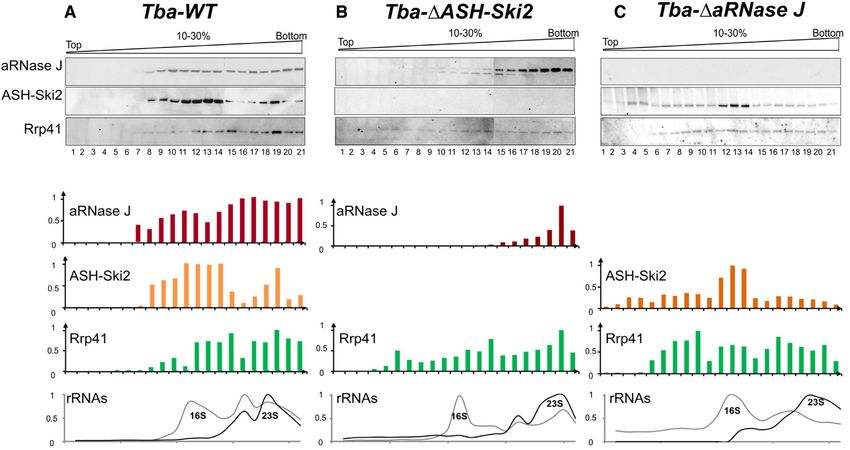
3834 Nucleic Acids Research, 2020, Vol. 48, No. 7
at an OD600nm of 0.6 by the addition of 0.1 mM IPTG (Iso- tial growth phase in bioreactors under physiological condi-
propyl -D-1-thiogalactopyranoside). After 3 h of induc- tions (95◦ C, pH 6.5, anaerobic). The cell extracts were pre-
tion at 30◦ C, the cells were collected, suspended in 10 ml of pared as described in (39). Briefly, P. abyssi pelleted cells
lysis buffer (50 mM NaPhosphate, 300 mM NaCl, 10 mM were re-suspended in 1/3 w/v of 1× phosphate-buffered
Imidazole) supplemented with 1 mg.ml−1 of lysozyme and saline (PBS) buffer (Euromedex), supplemented with 300
a mix of ethylenediaminetetraacetic acid (EDTA)-free pro- mM of NaCl and a mix of EDTA-free protease inhibitor
tease inhibitor (cOmpleteTM, Roche) and lysed by sonica- (cOmpleteTM, Roche). After sonication (VibraCell Biolock
tion (4×[5*10 s], 50% cycle, VibraCell Biolock Scientific). Scientific), the crude extracts were clarified by centrifuga-
When mentioned, the cleared extracts, obtained by cen- tion (20 000 g, 30 min, 4◦ C). Purified His-tagged proteins
trifuging the crude extracts (20 000 g 4◦ C, 20 min), were were used as baits in clarified whole-cell extract of P. abyssi.
treated with a mix of RNase A (20 g.ml−1 ), RNase T1 Briefly, 20 g of bait proteins were immobilized on 0.6
Downloaded from https://academic.oup.com/nar/article-abstract/48/7/3832/5728875 by IFREMER user on 16 April 2020
(1U.l−1 ) and DNase I (20 g.ml−1 ) containing 10 mM of mg of cobalt-coated magnetic beads (Dynabeads, Invitro-
MgCl2 for 30 min at 37◦ C. After a heating step at 70◦ C gen). The complex baits-beads were further incubated with
for 20 min, the extracts were furthered clarified by cen- 2 mg of P. abyssi extract under rotation for 2 h at room
trifugation (20 000 g, 4◦ C, 20 min). The recombinant pro- temperature. The protein complexes formed in vitro were
teins were two-step purified from the soluble fractions to separated on a magnet and washed extensively with PBS
near homogeneity using FPLC (Fast Protein Liquid Chro- buffer (3*4 ml and 2*1 ml). A second round of pull-down
matography, Äkta-purifier10, GE-Healthcare): first, by a assays was performed with an additional step to eliminate
nickel-attached HiTrap chelating column (HisTrap 1 ml, non-specific interaction via DNA or RNA molecules by
GE-Healthcare) with a linear gradient of 100–500 mM im- incubating the protein complexes with an RNase/DNase
idazole; secondly, by a heparin column (HiTrap Heparin mix (10 g.ml−1 of RNase A and DNase I) for 30 min
1 ml, GE-Healthcare) with a linear gradient of 300 mM at room temperature before applying the magnetic force.
to 1 M NaCl. The eluted fractions containing His-tagged A control was also performed under identical conditions
proteins were pooled and dialyzed overnight against 500 ml using cobalt-coated beads alone instead of the baits-beads
of dialysis buffer (20 mM HEPES pH 7.5, 300 mM NaCl, complexes. Purified protein complexes were eluted in 25 l
1 mM DTT (Dithiothréitol), 10% glycerol buffer). of XT sample buffer (BioRad) containing 2 l of 20× re-
ducing buffer at 95◦ C for 10 min and separated by sodium
dodecyl sulphate-polyacrylamide gelelectrophoresis (SDS-
Co-purification by chromatography affinity PAGE) short migration on 12% SDS-PAGE (Criterion XT
Cells expressing His-tagged bait proteins were produced Precast gels–BioRad). After a short migration, the proteins
as described above. Cells expressing untagged prey pro- were extracted by cutting out each protein track into one
teins were obtained after transforming E. coli BL21- piece followed by shotgun proteomic analyses. For western
CodonPlus (DE3) cells with pET11-aRNase J, pET11- blot analyses, protein complexes were separated by SDS-
ASH-Ski2, pET11-Csl4 or pET11-Pab-Rrp41 vectors (Sup- PAGE on 4–20% pre-cast Bis-Tris gel (NuPAGE Life tech-
plementary Table S1) and using the same conditions for nology).
growth and induction. Two cell pellets from 200 ml of cul-
tures expressing either bait or prey protein were mixed upon
lysis (as before). The bait proteins alone or in complex with Proteomic analysis
the prey proteins were purified by nickel affinity chromatog- The mass spectrometry analyses were performed at Paris
raphy using Ni2+ -NTA column matrices (HisTrap 1 ml, GE- Sud Ouest PAPPSO proteomics core facility (http://papso.
Healthcare). The elution was obtained with a linear 100– inra.fr). Briefly, after a short run on a SDS-PAGE gel, bands
500 mM imidazole gradient after washing steps at 10 and of proteins were excised from the gel. In-gel tryptic digestion
50 mM of imidazole. For each couple of bait/prey that were was performed 6 h at 37◦ C with 100 ng of modified trypsine
tested, the co-purification assays were at least performed (Promega) dissolved in 50 mM NH4CO3. Peptides were ex-
twice either in presence or absence of RNase/DNase treat- tracted with 0.1% trifluoroacetic acid (TFA) and 50% ace-
ment (as above). The proteins from recovered fractions were tonitrile (ACN). Extracted peptides were dried and sus-
analysed by Western blotting (4–15% Mini PROTEAN® pended in 30 l of 0.05% HCOOH, and 2% ACN. A total
TGX Stain-Free™ Gels & Trans-blot® Turbo™ Nitrocellu- of 4 l of samples were then loaded on a NanoLC-Ultra sys-
lose Transfer Pack, BioRad). The bait proteins were probed tem (Eksigent). Eluted peptides were analysed on-line with
using a His Tag HRP-conjugated antibody (Ozyme) di- a QExactive mass spectrometer (Thermo Scientific Elec-
luted 5000-fold. The prey proteins were probed using poly- tron) using a nanoelectrospray interface. Peptide ions were
clonal antibodies against Pab-aRNase J, Pab-ASH-Ski2, analysed using Xcalibur 2.1 with data-dependent acquisi-
Pab-Csl4, Pab-Rrp41, Pab-DnaG or Pab-aCPSF1 (custom tion steps. Peptides were identified with X!TandemPipeline
polyclonal antibodies, Eurogentec) diluted 10 000-fold and open source software by spectrum matching approach. The
an anti-rabbit IgG HRP conjugate (Promega) diluted 5000- mass spectrometry proteomics data have been deposited to
fold. the ProteomeXchange Consortium via the PRIDE partner
repository with the dataset identifier PXD015856”.
Pull-down by affinity purification assays The MS data were processed in order to identify spe-
cific interaction signals. Global specific spectra from sam-
The pull-down assays were performed in triplicate with clar- ples were normalized, as described in (40), between repli-
ified whole-cells extract of P. abyssi cultivated on exponen- cate series. We used a cut-off of two normalized spectra as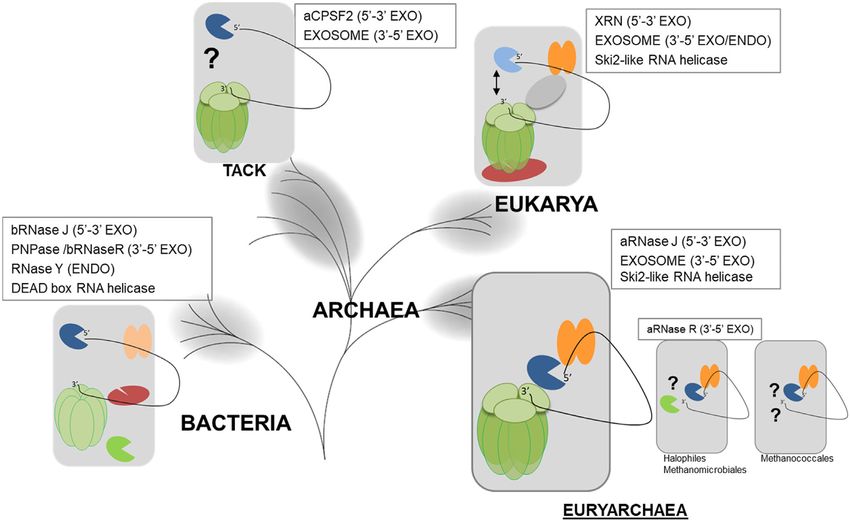
Nucleic Acids Research, 2020, Vol. 48, No. 7 3835
minimum MS signal for network hit validation. Normal- with shape parameter and proportion of invariant sites esti-
ized spectra were then averaged between replicates and ref- mated from the data. The statistical branch support was in-
erenced versus control to calculate the number of ‘Refer- ferred with the parametric bootstrap. The archaeal Ski2-like
enced Spectra’. Calculation of the ‘Specificity Index’ score helicase alignment was built by Mafft incorporating local
is the ratio of the averaged normalized spectra in control pairwise refinement (L-INS-i) up to 2000 iteration (maxiter-
versus assay. The ‘Specificity Index’ varies from 0 to 1 (with ate 2000) (48) and trimmed with trimAL as described above.
a maximum threshold of 1) as the specificity decreases. For
the AP-MS experiments with aRNase J as bait, nuclease The archaeal ASH-Ski2 helicase tree was computed with
and nuclease-free assays were run in triplicate, whereas for the same approach as that of the species tree except that the
ASH-Ski2 the same analyses were performed in 4 replicates. gamma-distributed substitution rate variation was approx-
For aRNase J n = 6 for the controls and for ASH-Ski2 n = imated by four discrete categories. Both trees (species and
8 for the controls. For ASH-Ski2, we took only into consid- Ski2-like helicases) were arbitrarily rooted and were drawn
Downloaded from https://academic.oup.com/nar/article-abstract/48/7/3832/5728875 by IFREMER user on 16 April 2020
eration hit proteins detected independently in both His-Tag with the online version of iTOL (49).
orientations. The effect of nuclease has been identified as:
Spectra No Nuclease − Spectra Nuclease Sucrose gradient fractionation of cell extracts
Nucl. Effect = 100 ×
Spectra No Nuclease
Pyrococcus abyssi and T. barophilus cells were grown at 92◦ C
Diagram networks have been created using Cytoscape and 85◦ C, respectively, in continuous culture in a 5-l Gazlift
3.7.2 software (41). bioreactor in MES medium under anaerobic conditions at
pH 6.8 (50). Cells were maintained in exponential growth
phase between 2 and 4*108 cells per ml. After cell culture
Multiple sequence alignments and phylogenetic tree construc- harvesting at 8◦ C, Cells were pelleted by centrifugation (10
tions 000 g, 20 min at 4◦ C) and washed two times with a sterile
sea salt solution at 30 g.l−1 . Dry cell pellets were stored at
The complete archaeal and bacterial genome entries were
−80◦ C. A total of 200 mg of cell pellet was re-suspended in
retrieved from the EBI (European Bioinformatics Insti-
1/3 w/v of buffer TK buffer (20 mM Tris–HCl, 100 mM
tute). The collection of helicases from super-families SF1
KCl, 10 mM MgCl2 , 1 mM DTT) containing a cocktail
and SF2 were identified by RPS-Blast of Pfam domains
of EDTA-free protease inhibitor (cOmpleteTM, Roche) or
DEAD (PF00270) or Helicase C (PF00271) among a set of
TK-EDTA buffer (TK buffer supplemented with 20 mM
completely sequenced non-redundant genomes composed
EDTA). Whole-cell extracts were prepared by sonication
of 114 archaeal genomes and 1105 bacterial genomes. A
(10*10 s, 50% cycle, VibraCell-Bioblock scientific). Lysates
total of 17 661 helicases were identified and further com-
were cleared at 21 000 g for 30 min. Approximately 10 mg of
pared to each other using the BlastP program. Partition-
whole-cell extract protein were layered on a linear 10–30%
ing of the all-vs-all Blast results was achieved using the
sucrose gradient prepared in TK or TK-EDTA buffers and
Markov Cluster Algorithm (42) and resulted in 84 clus-
centrifuged for 5 h at 35 000 rpm at 4◦ C in Beckman-Avanti
ters of >5 orthologous archaeal helicases. The archaeal se-
XPN-80 SW41 rotor. It was not possible to determine the
quences were collected and corresponded to occurrences
positions of 30S and 50S ribosomal subunits by classical
with COG1202 for ASH-Ski2. We also collected an initial
A254 scanning with the ISCO UA-6 gradient fraction col-
set of proteins using COG profile annotations (COG1096
lector due to the inherent colorimetry of the P. abyssi and
for Csl4 and COG0689 for Rrp41) from complete archaeal
T. barophilus cellular extract. For each gradient, 21 fractions
genomes. Hidden Markov model (hmm) profiles were built
of 500 l were collected. A total of 250 l of each fraction
for each family and used with hmmsearch (HMMER pack-
were precipitated with of 45 l of 100% trichloacetic acid
age, (43)) to identify homologues in our archaeal proteome
(TCA). After 30min at −20◦ C, samples were centrifuged 30
sample. In addition, to identify potential unannotated genes
min at 16 000 g at 4◦ C. The protein pellets were washed with
and pseudogenes, we performed tblastn searches.
500 l of glacial acetone (16 000 g, 15 min at 4◦ C), dried
The archaeal phylogenetic tree was inferred from a con-
and re-suspended in 20 l of 20 mM Tris–HCL pH 8. After
catenated dataset of 81 protein families obtained from COG
adding 4 l of 6× loading buffer (375 mM Tris–HCL pH
annotation. If multiple copies occurred in a genome, all par-
6.8, 12% SDS, 30% -mercaptoethanol, 0.6% Bromophe-
alogues were removed. The sequence alignments for each
nol Blue, 36% glycerol), samples were heated at 95◦ C for 10
family were created using the MUSCLE program (44) with
min before proceeding to SDS-PAGE.
the default parameters. We used the trimAl program (45)
to remove spurious sequences and poorly aligned positions
Relative quantification of ribosomal RNAs by Slot Blot
and to analyse the quality of the alignments according to
gap numbers and residue conservation in the columns of A total of 20l of each fraction collected from sucrose gra-
the alignments. These parsed alignments were concatenated dient, equally diluted beforehand to set the fraction with
to produce a single alignment of 17 827 residues. When the highest nucleic acid content at a concentration of 2.5
a species did not have a record for a family, the miss- ng.l−1 , was added to 55 l of denaturation buffer (2.2 M
ing sequence was replaced by gaps in the alignment. The formaldehyde, 50% deionized formamide, 0.5 mM EDTA,
maximum-likelihood tree was computed with PhyML (46) 10 mM MOPS, 4 mM NaCl) and incubated at 65◦ C for
and the optimal combinations of parameters was selected 5 min. Each 75 l-sample was spotted on a nylon mem-
using ProtTest3 program (47). The LG model of sequence brane (Amersham Hybond-XL; GE Healthcare) by vac-
evolution was used and the gamma-distributed substitution uum filtration (PR648-HoeferTM Slot Blot). After UV-
rate variation was approximated by eight discrete categories crosslinking, blots were hybridized overnight at 42◦ C with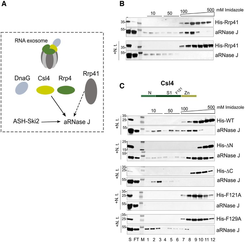
3836 Nucleic Acids Research, 2020, Vol. 48, No. 7

5 -end P32 -labelled oligonucleotides in Roti-Hybrid-Quick     teraction between RPA41 and the RNA polymerase has
buffer (Roth) and washed three times at 42◦ C for 15 min        been previously demonstrated in P. abyssi (39). It was re-
with SSC buffer (SSC 20×: 3M NaCl, 300 mM sodium cit-           vealed that the RPA complex enhance transcription rate
rate, pH 7) containing 0.1% of SDS (5×, 1× and 0.1×, re-        in vitro, probably via RPA41 interacting with the non-
spectively). Sequences of antisense oligonucleotides used to    template strand of the elongating complex. In this light, the
detect T. barophilus 16S and 23S rRNAs are listed in Supple-    presence of RPA subunit in the interaction network of aR-
mentary Table S2. Radioactive signals were visualized on a      Nase J could be considered as a further evidence for the in-
phoshorImager (Typhoon Trio-Amersham-Bioscience) and            volvement of aRNase J in RNA metabolism.
quantified using MultiGauge software (Fujifilm).                   Finally, to spotlight protein complexes of aRNase J net-
                                                                work, we built a protein interaction map of aRNase-His
                                                                based on the spectral index of Supplementary Table S3 (Fig-

                                                                                                                                Downloaded from https://academic.oup.com/nar/article-abstract/48/7/3832/5728875 by IFREMER user on 16 April 2020
RESULTS                                                         ure 1B). In light of this network, we propose that the 5 -3
                                                                exonuclease aRNase J cross-talks with RNA machineries of
The protein–protein interaction network of aRNase J in-
                                                                the cell.
cludes key components of RNA metabolism
To explore the protein interaction network of the eur-
yarchaeal aRNase J, we carried out affinity purification cou-
                                                                The P. abyssi ASH-Ski2 helicase shares a common network
pled to mass spectrometry (AP-MS) analyses in P. abyssi
                                                                with P. abyssi aRNase J RNase and the RNA exosome
cell extracts, as already described (39). Recombinant aR-
Nase J from P. abyssi, with C-terminal (His)6 -tag (aRNase      Interestingly, in the network of aRNase J, we retrieved
J-His), was produced, purified and used as baits for AP-        PAB2313, a putative RNA helicase, that appears to be a
MS experiments. In order to discriminate specific protein       member of the Ski2-like helicase family (53) renowned for
interaction from column background, we performed mock           their crucial role in RNA decay in eukaryotic cells (54) and
AP-MS analyses with P. abyssi cell extracts in absence of       we named it ASH-Ski2. To gain further experimental evi-
protein bait. In addition, to determine if some interactions    dence of a coordinated action of aRNase J and ASH-Ski2,
are mediated through RNA or DNA, we implemented a nu-           we performed new rounds of AP-MS analyses in duplicate
clease treatment after incubating the cell extract with the     with recombinant C- and N-terminal (His)6 -tagged versions
bait. The co-purified partners were identified by bottom-       of P. abyssi ASH-Ski2 as baits, respectively, and obtained
up proteomic techniques coupled with mass spectrome-            an exhaustive list of partners (Supplementary Table S4).
try. We used an algorithm that prioritizes the specificity      The ASH-Ski2 partner pie chart shows that a large major-
of the interaction to classify partners identified in tripli-   ity (73%) of identified proteins are annotated as players of
cate AP-MS. The list from the most to the least specific        RNA metabolism and translation. Indeed, out of 34 in to-
is displayed on Supplementary Table S3. Remarkably, we          tal, 12 ribosomal proteins and four subunits of the RNA
found that aRNase J is at the centre of a protein interac-      exosome are identified as specific partners (Figure 1A, right
tion network that includes proteins with central functions      panel and Supplementary Table S4). Notably, aRNase J is
in physiological processes. The pie chart, shown on Figure      ranked as the first candidate of the ASH-Ski2 partner list of
1A (left panel), categorizes these processes. A large major-    Supplementary Table S4. aRNase J is detected, both with a
ity (64%) of the protein partners of aRNase J-His are an-       high-specific score and reference spectra in assays treated
notated as involved in RNA metabolism from transcrip-           or not with nucleases. Furthermore, 21 protein partners
tion (RNA polymerase subunits), RNA modification (C/D           are common to both aRNase J and ASH-Ski2 networks
guide RNP components) and RNA decay (RNA exosome)               (Figure 1B). This highlights a tight cross-talk of aRNase
to translation (ribosomal proteins and translation initiation   J and ASH-Ski2 in P. abyssi cells. It should be noted that
factors). Amongst protein partners, 14 were detected with a     several partners of ASH-Ski2 are affected by the nuclease-
high-specific score in the aRNase J pull-downs whereas no       treatment when compared to aRNase J, which includes the
spectra signal were sensed in the controls for these candi-     RNA polymerase subunits but also the RNA exosome sub-
dates (values of 0 in Supplementary Table S3). Remarkably,      units.
the Rrp41 and Rrp42 catalytic core subunits and the Rrp4           To ensure that aRNase J and ASH-Ski2 are part of the
cap subunit of the RNA exosome were retrieved among the         protein interaction network of the RNA exosome, we per-
foremost of the fourteen highly significant protein partners    formed additional pull-down assays, in duplicate, using a
(Supplementary Table S3). Both subunits of the RNA poly-        purified N-tagged His-Rrp41 of P. abyssi as bait. Amongst
merase (RpoA1/A2) were also detected with highly signif-        the pull-downed proteins of the Rrp41 subunit, by west-
icant scores. Ribosomal proteins and components of the          ern blotting, we specifically detected endogenous Csl4 and
C/D box guide RNP complex are also part of the aRNase           DnaG cap subunits, aRNase J and ASH-Ski2, but not the
J network (51,52). Remarkably, these exploratory analyses       endo-RNase aCPSF1 (Figure 2). Altogether with the lim-
reveal PAB2313, a putative ATP-dependent RNA helicase,          itation that the pull-down was performed with the sole
as a potential partner of aRNase J-His.                         Rrp41 subunit and not with a re-formed complex with the
   In addition, proteins known to be involved in DNA main-      Rrp42 core and Csl4/Rrp4 cap subunits, these results sup-
tenance processes, like RPA41, were also recovered as sig-      port a potential interplay between aRNase J, ASH-Ski2 and
nificant partners (Supplementary Table S3). RPA41 cor-          components of the archaeal RNA exosome in P. abyssi cells.
responds to the large subunit of the RPA complex that           This is thoroughly consistent with the results of the above
binds to single-stranded DNA. Interestingly, a physical in-     AP-MS analysis set.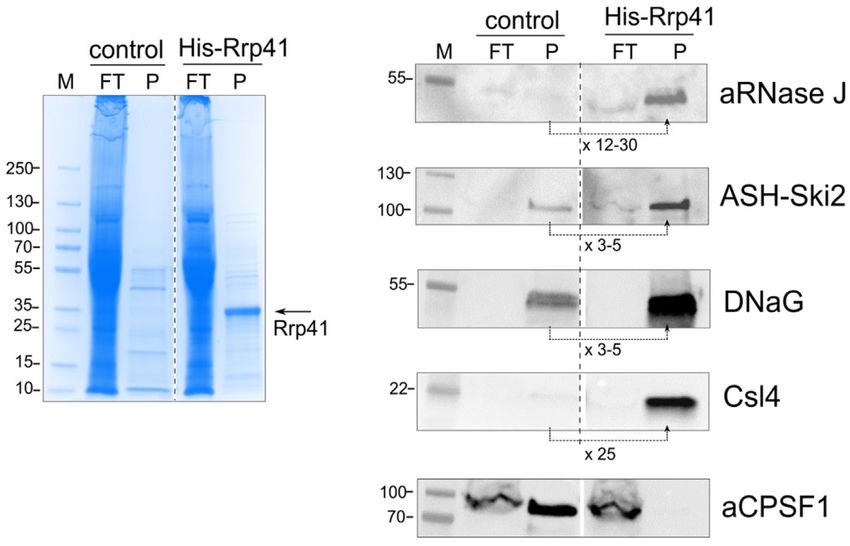
Nucleic Acids Research, 2020, Vol. 48, No. 7 3837
Downloaded from https://academic.oup.com/nar/article-abstract/48/7/3832/5728875 by IFREMER user on 16 April 2020
Figure 1. (A) Functional classification pie charts of proteins identified by pulldown-MS/MS approach using recombinant aRNase J and ASH-Ski2 as baits,
respectively. (B) Network of protein partners for aRNase J and ASH-Ski2. The aRNase J and ASH-Ski2 baits are labelled in yellow. Network backbone
is represented in black lines except for ASH-Ski2 and aRNase J for which reciprocal interactions are in red. Common partners of ASH-Ski2 and aRNase
J are indicated in pink whereas specific partners are indicated in blue. Bolded circle indicate highly specific hits, absence in all control runs, with a ‘spectra
index’ of 0 (see Supplementary Tables S3 and 4).
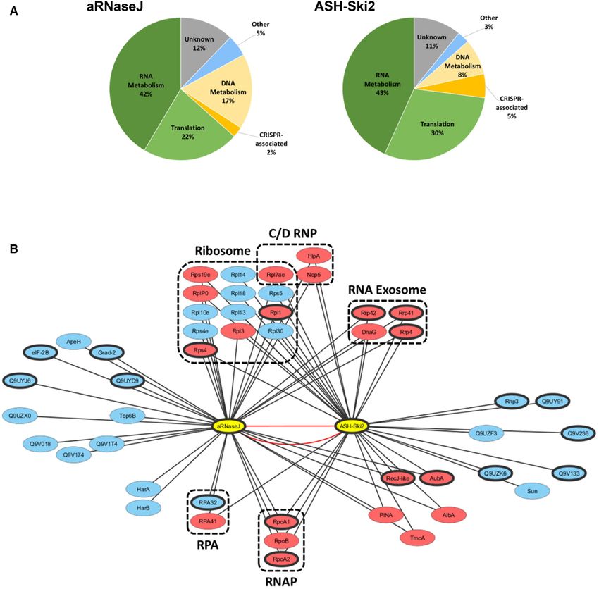
The archaeal ASH-Ski2 members share identical taxonomic genomes. In contrast to the role of eukaryotic Ski2-like
distribution as aRNase J members RNA helicases in RNA homeostasis, reports on the func-
tions of archaeal Ski2-like helicase family members are still
To gain insights into the relationship that exists between ar-
scarce. As a common theme throughout SF2 helicase fam-
chaeal aRNase J and ASH-Ski2 members as well as with
ilies, the unique characteristics of Ski2-like family mem-
the RNA exosome subunits, we compared their taxonomic
bers are mainly derived from accessory domains that dec-
distributions amongst the archaeal phylogeny. To do so,
orate the helicase core and provide additional functionali-
we first upgraded a phylogenomic study of the superfam-
ties (55,56). Ski2-like helicase structures demonstrate that
ily of the archaeal SF2 helicase (53) in order to accurately
the molecular ‘core’ of all Ski2-like helicases is a ring-like
identify the Ski2 family of helicases encoded in archaeal3838 Nucleic Acids Research, 2020, Vol. 48, No. 7
Downloaded from https://academic.oup.com/nar/article-abstract/48/7/3832/5728875 by IFREMER user on 16 April 2020
Figure 2. Pull down assays using the recombinant (His)-Rrp41 protein as bait in whole-cell extracts of Pyrococcus abyssi; FT corresponds to the flow
through, P to the pull-downed fraction and M to the protein ladder. Each fraction was analysed by Coomassie blue staining of SDS-PAGE (left panel)
and by western blotting using specific antibodies for aRNase J, ASH-Ski2, DnaG, Csl4 and aCPSF1 (right panel). To highlight the pull-downed proteins,
the relative signals were average from two independent assays as indicated below each panel.
four domain assembly of two RecA domains, a winged he- with equivalent activity could be, so far, detected by com-
lix domain and a ratchet domain (54,57). By performing parative genomics.
RPS Blast searches, we retrieved all archaeal Ski2-like he-
licase member sequences from 147 non-redundant com-
aRNase J forms a stable complex with the helicase-like ASH-
pletely sequenced archaeal genomes. In addition to Hel308
Ski2, in vitro
members, characterized by the COG 1204 and documented
with enzymatic features of DNA helicases (54,58–61), we To investigate a direct interaction between aRNase J and
also retrieved the ASH-Ski2 group, characterized by the ASH-Ski2, we used an in vitro co-purification assay on
COG 1202 (53). Interestingly ASH-Ski2 members hold an nickel affinity chromatography to test pairwise interactions.
additional N-terminal domain in which four strictly con- A recombinant (His)6 -tagged bait (His-ASH-Ski2) and an
served cysteines have the propensity to form a zinc-finger- untagged prey (aRNase J) from P. abyssi were expressed in-
like motif (62) (Figure 3A). ASH-Ski2 members are spe- dependently in E. coli cells using the pET15b and pET11b
cific of euryarchaeal phylogenetic groups with the Lokiar- expression vectors, respectively (Supplementary Table S1).
chaeum as an exception (Figure 3B). Remarkably aRNase J Cells over-expressing the recombinant (His)6 -tagged bait or
and ASH-Ski2 members present similar occurrence, which the untagged prey proteins were mixed in a 1:1 ratio prior
is restricted to most euryarchaeal groups, with the excep- the lysis step. After nucleic acids removal with a cocktail of
tion of the archeoglobales and thermoplasmatales. Fur- nucleases, most of the proteins from E. coli were eliminated
thermore, the co-evolution of aRNase J and ASH-Ski2 at 70◦ C for a specific enrichment of the thermo-resistant
members shown by the comprehensive congruence of aR- proteins from P. abyssi. The clarified cellular extracts were
Nase J and ASH-Ski2 phylogenetic trees, with only few ob- then injected on a nickel column to retain the bait His-ASH-
served leaf displacements within taxonomic groups, rein- Ski2 protein and its potential interacting partners. Washing
forces their potential functional interplay (Supplementary steps with low imidazole concentrations served at remov-
Figure S1). Altogether, protein networks combined with ing weakly bound contaminants. The bait and the retained
our phylogenomics analyses argue for a coordinated action interacting proteins were eluted with a linear gradient of
of aRNase J and ASH-Ski2 in specific cellular processes in imidazole. The protein content of each recovered fractions
Euryarchaeota. was analysed by SDS-PAGE. The specific presence of aR-
Subsequently, we compared the taxonomic distribution Nase J and His-ASH-Ski2 was revealed by western blot-
of aRNase J, ASH-Ski2 and the RNA exosome Rrp41 and ting using specific antibodies as shown in Figure 4A. As
Csl4 subunits along the archaeal phylogeny (Figure 3B). expected, aRNase J co-elutes with His-ASH-Ski2 even af-
Like in our previous studies (15,27), we showed that the ter nucleic acids removal. This establishes that, at high tem-
RNA exosome is not present throughout the whole archaeal perature and ionic strength (300 mM NaCl), aRNase J and
taxonomy and that aRNase J, as well as ASH-Ski2, do His-ASH-Ski2 associate to form a stable complex that is
not follow the taxonomic distribution of RNA exosome not mediated through RNA or DNA molecules (Figure 4A,
components. Indeed, it is absent from the Halobacteria top panel). As a control, to demonstrate that protein co-
and Methanomicrobiales members for which the 3 -5 exo- purification is a consequence of complex formation rather
ribonucleolytic activity is carried by another enzyme, the than non-specific interactions with the column matrices, we
aRNase R (14,15). In the Methanococci group, no enzyme controlled that untagged versions of aRNase J or ASH-Ski2Nucleic Acids Research, 2020, Vol. 48, No. 7 3839
Downloaded from https://academic.oup.com/nar/article-abstract/48/7/3832/5728875 by IFREMER user on 16 April 2020
Figure 3. (A) Schematic protein domain architecture of the ASH-Ski2 group. The common SF2 core is composed of the RecA1 and RecA2 subdomains
(Pfam DEAD & Helicase C) in orange, of the C-terminal domain in green and of the N-terminal domain in grey. The strictly conserved cysteine residues
of the N-terminal domain are represented. (B) Taxonomic distributions of ASH-Ski2, aRNase J and Rrp41/Csl4 from the RNA exosome among archaeal
genomes. Their occurrence is plotted and juxtaposed to the archaeal specie tree. Taxonomic orders and Pfam domains are colour coded. Darker and lighter
shades indicate multiple copies and pseudogenes, respectively.
are not intrinsically retained on Ni2+ -NTA column matri- nificant proportion of aRNase J is shifted to fractions of
ces (Supplementary Figure S2A). Despite the robustness different molecular weight. This shift is most likely due to
of these data, we cannot completely exclude a stabilization an interaction with ASH-Ski2 that is found in the same frac-
of an ASH-Ski2 /aRNase J complex by remaining E. coli tions as aRNase J.
proteins like ribosomal proteins that are highly abundant. To uncover which protein domain of ASH-Ski2 is specif-
However, we have additional evidence supporting an inter- ically involved in the formation of a complex with aRNase
action between aRNase J and ASH-Ski2. By comparing the J, variants of His-ASH-Ski2 were used as baits for the un-
sedimentation profiles (5–25% sucrose gradient) of each in- tagged prey aRNase J. Pairwise interactions were tested as
dividual purified protein to the one obtained with a mixture previously described performing in vitro co-purification as-
of both (Supplementary Figure S2B), we showed that the says (Figure 4A, lower panels). Our results indicate that the
sedimentation profile of aRNase J is affected by the pres- accessory C-terminal domain of ASH-Ski2 (His-C) is not
ence of ASH-Ski2. Indeed, whilst the sedimentation profile required in establishing an in vitro interaction with aRNase
of aRNase J alone most likely corresponds to its homote- J as the elution profile is comparable to the one with full-
trameric form as observed for Methanolobus psychrophilus length ASH-Ski2. In contrast, the accessory N-terminal do-
aRNase J in solution (63), in presence of ASH-Ski2, a sig- main of ASH-Ski2 (His-N) seems to be critical for the in-3840 Nucleic Acids Research, 2020, Vol. 48, No. 7
Downloaded from https://academic.oup.com/nar/article-abstract/48/7/3832/5728875 by IFREMER user on 16 April 2020
Figure 4. In vitro co-purification assay of aRNase J challenged by His-ASH-Ski2 and His-Hel308. (A) Schematic representation of the protein domain
architecture of ASH-Ski2 from Pyrococcus abyssi and affinity co-purification assays on nickel chromatography column of recombinant untagged aRNase
J with His-ASH-Ski2 and its variants. The full-length protein (His-WT), deleted for the C-terminal (His-C) and N-terminal (His-N) domains or
harbouring a punctual substitution of the conserved cysteine residue into alanine (His-C124A) were used as baits. The fractions were analysed by western
blotting using an His-Tag or a specific antibody against aRNase J. S: supernatant loaded on the affinity Nickel column; FT: flow through; 10 mM (lanes
1–3) and 50 mM (lanes 4–6) imidazole washing steps; Elution with an Imidazole gradient from 100 to 500 mM (lanes 7–12). Experiments performed with
nuclease treatment (+Nt) are indicated on the left of each panel. (B) Assays as performed in (A) with the wild-type N-terminal domain DomN (His-WT)
or containing the C124A substitution (His-C124A) of ASH-Ski2. (C) Assays as carried in (A) but with His-Hel308 as the protein bait.
teraction since aRNase J does not co-elute with the N aRNase J forms a complex with the exosome cap-subunit
variant of ASH-Ski2. Finally, we showed that the acces- Csl4 in vitro
sory N-terminal domain of ASH-Ski2 expressed alone (His-
We also tested the pairwise interactions between aRNase J
DomN) is sufficient to establish an in vitro interaction with
or ASH-Ski2 and RNA exosome subunits that are among
aRNase J (Figure 4B, top panel).
the most retrieved proteins with aRNase J and ASH-Ski2
The sequence alignment of the N-terminal domains of
in the AP-MS analyses (Figure 1B; Supplementary Tables
archaeal ASH-Ski2 highlights the presence of four strictly
S3 and 4). The results of the in vitro co-purification assays
conserved cysteines with the propensity to form a zinc
performed as described above are summarized in Figure
finger-like motif (Figure 4A). To test whether this motif
5A. First, we challenged the catalytic subunit His-Rrp41
is critical for the interaction between ASH-Ski2 and aR-
as bait with aRNase J or ASH-Ski2 as preys. Only aR-
Nase J, we substituted the C124 residue of full-length His-
Nase J could be recovered in the elution fractions together
ASH-Ski2 and His-DomN by an alanine (A) and we chal-
with His-Rrp41. The Rrp41/aRNase J complex is nucleic
lenged their capacity to retain aRNase J on Ni2+ -NTA col-
acid-dependent since its formation is sensitive to nuclease
umn as before. Our results show that the conserved C124
treatment (Figure 5B). Then, we challenged the exosome
residue is critical in establishing or in stabilizing an inter-
cap subunits His-Rrp4, DnaG-His or His-Csl4 as baits with
action between the full-length or DomN of ASH-Ski2 and
aRNase J or ASH-Ski2 as preys. In our conditions, nei-
aRNase J (Figure 4A and B, lower panels). Indeed, the
ther Rrp4 nor DnaG were found to interact with aRNase
single C124A substitution is sufficient in destabilizing this
J (Supplementary Figure S2C) or Rrp4, DnaG nor Csl4
interaction.
with ASH-Ski2 (Supplementary Figure S2D). In contrast,
Finally, to confirm the specificity of the interaction be-
the exosome cap-subunit Csl4 is able to form a complex
tween aRNase J and ASH-Ski2, we performed similar in
with aRNase J that is not sensitive to nuclease treatment
vitro co-purification assays using aRNase J as a prey and
and therefore is not mediated by nucleic acids (Figure 5C).
His-Hel308, another Ski2-like helicase encoded in the P.
To go further in the characterization of this association,
abyssi genome, as bait. In this configuration, aRNase J is
we then searched for the protein domains of Csl4 that are
not retained on the Ni2+ -NTA column matrices (Figure 4C)
involved in the formation of the Csl4/aRNase J complex.
meaning that aRNase J specifically interacts with ASH-
Whilst the N-terminal domain of Csl4 (N) is not required,
Ski2. This result is not surprising as members of the Hel308
its C-terminal domain (C) is critical for the formation of
family do not possess the N-terminal zinc finger-like motif
the Csl4/aRNase J complex (Figure 5C).
extension.Nucleic Acids Research, 2020, Vol. 48, No. 7 3841
Downloaded from https://academic.oup.com/nar/article-abstract/48/7/3832/5728875 by IFREMER user on 16 April 2020
Figure 5. In vitro interaction of ASH-Ski2 and aRNase J with the RNA exosome subunits. (A) Schematic representation of the RNA exosome machine and
of the pairwise interactions detected in this study. Plain arrows indicate interactions resistant to nuclease treatment. Dotted arrows indicate interactions
sensitive to nuclease treatment. The in vitro co-purification assays of ASH-Ski2 or aRNase J challenged by His-Csl4 are shown in (B) and (C), respectively.
The schematic representation of the protein domain architecture of Csl4 is shown (see also Supplementary Figure S3B). Wild-type Csl4 (His-WT) and
variants with a deletion of the C-terminal (His-C) or N-terminal (His-N) or a punctual substitution of phenylalanine 121 in alanine in the S1 domain
(His-F121A) were used as baits. Legend details are as in Figure 4.
To highlight specifically conserved residues in Csl4 se- der protein complexes in archaeal cells. To this end, we dis-
quences that are co-distributed with aRNase J, we derived criminated the sedimentation profile behaviours of endoge-
weblogos from multiple alignments of Csl4 co-distributed nous aRNase J, ASH-Ski2 and Rrp41 in two Thermococ-
or not with aRNase J. Several positions (including F/Y121 cales organisms, T. barophilus (Tba) and P. abyssi (Pab) by
and F129, numbering according to the P. abyssi sequence) doing ultracentrifugation of whole-cell extracts on contin-
in the S1 domain of Csl4 could be identified as highly uous 10–30% sucrose density gradient (Figure 6A and Sup-
conserved in Csl4 sequences in the context of the co- plementary Figure S4A, respectively). Since to this date,
distribution with aRNase J (Supplementary Figure S3A). P. abyssi cannot be genetically modified, the T. barophilus
Strikingly, the phenylalanine residues, F121 and F129, lo- strain was used as a genetically tractable model (37,38) to
cated on the central S1 domain of Csl4 and solvent-exposed assess the sedimentation profile of endogenous aRNase J
in the P. abyssi Csl4 structural model (Supplementary Fig- in absence of ASH-Ski2 and vice-versa. Moreover, note
ure S3B) are also key for the association with aRNase J that the antibodies produced to detect our proteins of in-
(Figure 5C). terest from P. abyssi by immunoblotting cross-react with
their counterparts from T. barophilus whilst preserving their
specificity. Unexpectedly, the vast majority of these proteins
aRNase J and ASH-Ski2 and the RNA exosome co-sediment
co-sediment in heavy fractions with barely any signal in
with high molecular weight complexes in sucrose gradient
light fractions. This is not the case for the -CASP endo-
fractionation of whole-cell extract from P. abyssi and Ther-
RNase Pab-aCPSF1, which was not identified in any of the
mococcus barophilus
protein interaction networks that we uncovered in this study
Altogether, our results propose a tight relationship between (Supplementary Figure S4). It is interesting to note that
aRNase J, ASH-Ski2 and the RNA exosome. Here, we Pab/Tba-aRNase J and Pab/Tba-ASH-Ski2 are similarly
asked whether these protein partners could form higher or- distributed through the gradient (Figure 6A and Supple-3842 Nucleic Acids Research, 2020, Vol. 48, No. 7
Downloaded from https://academic.oup.com/nar/article-abstract/48/7/3832/5728875 by IFREMER user on 16 April 2020
Figure 6. Sedimentation profiles of endogenous aRNase J, ASH-Ski2 and Rrp41 of Thermococcus barophilus wild-type (WT) in (A), ASH-Ski2 in (B) and
aRNase J in (C), strains. Clarified whole-cell extracts were fractionated on 10–30% density sucrose gradients by ultracentrifugation. Equal volumes of
fraction, precipitated with TCA, were monitored by western blotting using specific antibodies. The digital images and bar charts representing the relative
amounts of protein are given. The ribosomal RNAs were also profiled by slot blotting equal volume of denatured fractions with probes complementary to
16S and 23S rRNAs. The relative quantification of 16S and 23S ribosomal RNAs is plotted in grey and black, respectively.
mentary Figure S4). This distribution pattern is also highly nesium concentration (TK-EDTA buffer) (Supplementary
similar to the one of the RNA exosome subunit Pab/Tba- Figure S5). In these conditions, the assembled ribosomes
Rrp41 (Figure 6A and Supplementary Figure S4). In con- dissociate into individual subunits (66). In the wild-type
clusion, our data are indicative of a co-migration of the strain, endogenous Tba-aRNase J and Tba-ASH-Ski2 co-
Pab/Tba-aRNase J, the Pab/Tba-ASH-Ski2 and the Rrp41 sediment with the 16S and 23S rRNAs from the small and
RNA exosome subunit in high molecular weight fractions. large subunits, respectively (Supplementary Figure S5A).
To go further, we fractionated whole-cell extracts from T. Again, we observe a perfect overlay of the sedimentation
barophilus strains enclosing a deletion of the gene encod- profiles of Tba-aRNase J and Tba-ASH-Ski2 as previously
ing Tba-ASH-Ski2 (Tba-ΔASH-Ski2, Figure 6B) or Tba- seen at a higher concentration of magnesium (Figure 6A
aRNase J (Tba-ΔaRNase J, Figure 6C). On one hand, when and Supplementary Figure S5A). Similar results were ob-
Tba-ASH-Ski2 is missing, most of the endogenous Tba- tained with extracts from P. abyssi cells (Supplementary
aRNase J protein is shifted towards the bottom of the gradi- Figure S4B). More interestingly, in absence of crosstalk with
ent. On the other hand, when Tba-aRNase J is not present, ASH-Ski2, a flagrant alteration of the sedimentation be-
the distribution profile of the endogenous Tba-ASH-Ski2 is haviour of Tba-aRNase J was also detected. Indeed, in the
also clearly altered; but in this case, Tba-ASH-Ski2 is more context of the Tba-ΔASH-Ski2 strain, at low magnesium
evenly spread out along the gradient. Note that the pro- concentration, Tba-aRNase J is shifted from the bottom
file of the core RNA exosome subunit, Rrp41, was barely of the gradient to fractions overlapping the profiles of the
affected in both genomics contexts. Overall, these results 16S and 23S rRNAs (Supplementary Figure S5B). In ab-
strongly suggest that the aRNase J & ASH-Ski2 are part of sence of Tba-aRNase J, we observe overlay sedimentation
higher-order complexes in T. barophilus cells. In addition, profiles of Tba-ASH-Ski2 and Tba-Rrp41 (Supplementary
we observe that the 16S and 23S ribosomal RNA profiles Figure S5C). From these results, we argue for an association
overlap the ones of aRNase J, ASH-Ski2 and Rrp41. In ab- of aRNase J with ribosome/polysome that is emphasised in
sence of ASH-Ski2, aRNase J is shifted towards the ribo- absence of ASH-Ski2 in addition to an interplay between
some. From these results and in light of data reported for aRNase J, ASH-Ski2 and Rrp41 RNA exosome subunit.
bacterial RNase J and eukaryal Ski2 helicases (64,65), we
propose that these two enzymes cooperate with the ribo- DISCUSSION
some in Thermococcale cells.
We also challenged the physical association of Tba- Elucidating the RNA processing machineries is of a criti-
aRNase J and Tba-ASH-Ski2 in higher-order complexes, cal importance to understand the molecular basis of RNA
by performing similar fractionation assays but at low mag- decay and processing in Euryarchaeota. In this study, toNucleic Acids Research, 2020, Vol. 48, No. 7 3843
Downloaded from https://academic.oup.com/nar/article-abstract/48/7/3832/5728875 by IFREMER user on 16 April 2020
Figure 7. Schematic representation of the interplay between enzymes and machineries in charge of 5 -3 and 3 -5 RNA decay and surveillance in the three
domains of life. The life tree model is as in (84). The 3 end RNA trimming activity is carried by the RNA exosome machinery (barrel in green and cap in
light green) in most Archaea (15) and Eukarya (76) that is similar to bacterial PNPase (barrel in light green). In Halobacteria and Methanomicrobiales the
aRNase R, orthologous to bRNase R, (green packman) carries the 3 -5 exo activity. In Methanoccocci, no 3 -5 exo activity has yet been described (15).
The 5 -3 exoribonucleolytic activities (blue packman) are carried by XRN RNases in Eurkarya (85), bRNase J and aRNase J in Bacteria and Euryarchaea
and aCPSF2 in the TACK group. RNA helicase activities shown to be involved in RNA surveillance belong to the Ski2-like family in Eukarya (42) and
Euryarchaea (this study) and to the DEAD box family in Bacteria (86). The interplay between 5 -3 and 3 -5 exoribonucleolytic activities are reported to
take place in the RNA-degradosome-like complex in Bacteria that also involves endoRNases (red packman) (87) and through the 5 -3 communication
by a ‘closed-loop’ architecture of mRNA in Eukarya (88). In most Euryarchaea, we propose that aRNase J/ASH-Ski2 complex interplays with the RNA
exosome. In the TACK group and the Halophiles, Methanomicrobiales and Methanoccocales, such an interplay remains to be characterized (question
marks).
our knowledge, we provide the first experimental evidences be essential in T. barophilus in optimal growth conditions.
hinting at the existence of high-order -CASP protein com- However, we could not obtain a strain that combined both
plex(es) in Thermococcales that would carry 5 -3 & 3 -5 ex- gene deletions. This synthetic lethality as demonstrated in
oribonucleolytic and helicase-like activities and that could yeast for genes involved in same RNA degradation path-
play pivotal roles in archaeal RNA surveillance. First, we ways (67,68) strongly hints at a functional relationship be-
identified the interaction landscape of aRNase J from P. tween aRNase J and ASH-Ski2 in Thermococcales.
abyssi, a 5 to 3 -CASP exoRNase (32). Candidate part- In conjunction with our previous demonstration that -
ners include, notably, a Ski2-like helicase ASH-Ski2, the ar- CASP RNases, ubiquitous in Archaea, must be key play-
chaeal RNA exosome that exhibits 3 to 5 exoribonucle- ers in archaeal RNA metabolism (26,27), our current data
olytic and 3 end-tailing activities (17) and ribosomal pro- support a model whereby the -CASP RNase, aRNase J,
teins. Of high significance, we also found these proteins to the ASH-Ski2-like helicase and the RNA exosome inter-
be candidate partners in the interaction landscape of ASH- play in most euryarchaeal cells (Figure 7). It remains to
Ski2 and in the pull-down of the core exosome subunit determine how this interplay could impact the archaeal
Rrp41 (Figures 1B and 2). In addition, we showed that aR- RNA metabolism. Since our data imply that ASH-Ski2 has
Nase J is able to interact, in vitro, with ASH-Ski2 and the an impact on aRNase J and vice versa, we favour a sce-
exosome cap subunit Csl4 (Figures 4 and 5). Our in vitro nario where, at least, ASH-Ski2 and aRNase J have a func-
data are strengthened by our phylogenomic studies showing tional connection. This is all the more so given that aR-
that, with only one exception, ASH-Ski2 is only encoded in Nase J and ASH-Ski2 show a taxonomic co-distribution
archaeal genomes that carry a gene for aRNase J and that amongst the archaeal phylogeny and nearly perfect phylo-
amino acids of Csl4, involved in the in vitro interaction with genetic tree congruence suggesting a co-evolution and po-
aRNase J, are only conserved in archaeal genomes where tentially an ancient origin (Figure 3B and Supplementary
their encoding genes are co-distributed (Figure 3). Finally, Figure S1, respectively). Based on what is known about the
our T. barophilus whole-cell extract fractionation experi- role of these factors in Eukarya and Bacteria, we discuss
ments provide evidences that an aRNase J/ASH-Ski2 com- what we believe to be the most probable scenarios. Evi-
plex might exist in vivo as their sequential deletions modify dently, further work will be needed to determine how these
the sedimentation profile of their protein partner (Figure proteins alone or in complex shape the euryarchaeal RNA
6). Individually, aRNase J and ASH-Ski2 do not appear to metabolism.3844 Nucleic Acids Research, 2020, Vol. 48, No. 7
First, recent published work proposes that ASH-Ski2, bonucleolytic and polyadenylation activities reminiscent to
also named Eta for euryarchaeal termination activity, in- the archaeal RNA exosome (79) (Figure 7). Although, in
fluences transcription termination by disrupting archaeal some organisms, the bRNase J degradosome-like complex
transcription elongation complex (69). However, as its he- appears to be transient and dynamic, limiting its validation
licase activity is rather slow and cannot keep up with wild- in vitro, this is consistent with interplay between 5 -3 and 3 -
type RNAP elongation rates, Eta was recently proposed to 5 RNA surveillance decay in Bacteria. Therefore, it emerges
be more likely involved in the rescue of transcriptional ar- that, in the three domains of life, RNA surveillance could
rest (70). In our study, the presence of RPA and RpoB sub- be taken care of by evolutionary conserved RNase-based
units in the interaction network of ASH-Ski2 could be a fur- RNA-degradation machines with RNA helicase and with
ther evidence for the involvement of ASH-Ski2 in the tran- 5 -3 and 3 -5 exoribonucleolytic activities (Figure 7).
scription. In addition, transcriptional termination and res- From cellular fractionation assays, we observed that both
Downloaded from https://academic.oup.com/nar/article-abstract/48/7/3832/5728875 by IFREMER user on 16 April 2020
cue pathways also involve proteins with ribonucleolytic ac- aRNase J and ASH-Ski2 could potentially associate with
tivities. In Eukarya, the 5 -3 exoribonucleolytic activity of the ribosome. Interestingly, it was also shown that bRNase
Rat1p/Xrn2p was proposed to be associated in a transcrip- J can associate with translating ribosomes (64) and the crys-
tion termination pathway called ‘torpedo model’ (71). In tal structure of the eukaryotic 80S ribosome-Xrn1 complex
chloroplast, RNase J was shown to play a critical role in re- showed that the 5 -3 Xrn1 nuclease binds at the mRNA
moving antisense RNAs that are generated by read-through exit site of the ribosome (80). Since the sedimentation pro-
transcription (72). Nevertheless, chloroplast RNase J ex- files of T. barophilus and P. abyssi aRNase J resemble to
hibits a DNA binding domain found in transcription fac- the one reported for the eukaryal 5 -3 exo-RNase Xrn1,
tors of plants that is not present in archaeal RNase J and we speculate that the archaeal aRNase J is also associated
that could be essential to fulfil this function (73). Finally, with polysomes in Thermococcales cells. This is reminiscent
co-factors of the nuclear RNA exosome were shown to con- of what is described in eukaryal mRNA surveillance path-
nect transcription termination to RNA processing by guid- ways (28) in which mRNA decay is initiated by an endonu-
ing terminated transcripts to the appropriate exonuclease cleolytic cleavage of ribosome-associated mRNAs (81,82),
(74). In Archaea, it is still not known if a ribonucleolytic followed by rapid degradation in the 3 -5 direction by the
activity is critical since, until now, the complete picture of RNA exosome or in the 5 -3 direction by Xrn1 (83). Re-
transcription termination is far from being understood (70). cently, direct experimental evidence showed that the cyto-
It remains to identify if aRNase J, ASH-Ski2 and the RNA plasmic RNA exosome is able to interact with the ribo-
exosome are involved together or alone in transcription ter- some through a direct interaction mediated by the SKI com-
mination or rescue pathways which have been shown to be plex (composed of Ski2-Ski3-Ski8 factors). More precisely,
key in influencing the fate of a transcribed RNA. it was shown that ribosome binding displaces the auto-
Moreover, based on the roles of the Ski2-like helicases inhibitory domain of the Ski2 RNA helicase (65).
in Eukarya (75), the idea that the euryarchaeal ASH- On a side note, it is important to notice that the RNA
Ski2 group is closely connected to RNA-related pathways exosome is not present throughout the whole archaeal tax-
emerges (Figure 7). Indeed, eukaryal Ski2, Mtr4 and Brr2 onomy (15,27) and that aRNase J does not accurately fol-
which are described as RNA helicases that process RNAs in low the taxonomic distribution of RNA exosome compo-
a 3 -5 direction were shown to play critical roles in RNA de- nents. In the Halophiles that do not encode the exosome
cay, surveillance and splicing, respectively (54,75). Notably, subunits, the 3 -5 exo-ribonucleolytic activity is carried by
Ski2 and Mtr4 are essential co-factors of the cytoplasmic another enzyme, the aRNase R (14,15). It would be in-
and nuclear RNA exosomes, respectively, by being involved teresting to know whether the halophilic and methanomi-
in the recruitment of RNA substrates (13). Likewise, our crobiale aRNase J, alone or in complex with ASH–Ski2
work provides several evidences of interplay between the complex, interplays with aRNase R. In the Methanococci
archaeal RNA exosome and ASH-Ski2. Our data also sug- group, no 3 -5 exo-RNase could be detected by compar-
gest that this interplay is done through aRNase J that in- ative genomics. In this case, aRNase J might connect to
teracts in vitro with Csl4 from the RNA exosome cap and specific archaeal RNases that remain to be identified (Fig-
with ASH-Ski2. Altogether, our results converge towards a ure 7). In conclusion, this work establishes a solid base in
cross-talk between the 5 -3 and 3 -5 exo-ribonucleolytic ac- the largely overlooked but extremely important field of ar-
tivities carried by aRNase J and the RNA exosome, respec- chaeal RNA metabolism. We identified two phylogenetical
tively. This is consistent with what is observed in Eukarya, conserved factors in Euryarchaea, the 5 -3 exo-RNase aR-
where mounting evidences show a functional interplay be- Nase J and the Ski2-like helicase ASH-Ski2, which have
tween surveillance pathways taking place at the 5 and 3 - homologues in Bacteria and Eukarya, respectively (Figure
ends of RNAs (76) (Figure 7). 7). Therefore, a mosaic system in Euryarchaeota could be
Finally, it must be remembered that aRNase J is the at the centre of networks interplaying with the archaeal
homologue of bacterial RNase J (bRNase J) which was RNA exosome and the ribosome. This is an example show-
shown to be critical in mRNA decay and rRNA matura- ing that Archaea use composite systems and thereby eluci-
tion pathways and to be part of a degradosome-like com- dating mechanism in archaeal organisms could give valu-
plex (29,77–78). bRNase J which can carry 5 -3 exo- and able insights into understanding function and evolutionary
endo-RNase activities was shown to associate with pro- routes of protein networks or/and high-order complexes.
teins identified as, among others, helicase-like proteins and Analogous to eukaryal and bacterial systems, we propose
a polynucleotide phosphorylase (PNPase). Bacterial PN- that these factors act in partnership in major archaeal RNA
Pase form an RNA-degradation machine with 3 -5 exori- decay or surveillance pathways. Deeper insights need to beYou can also read