Epitranscriptomics: A New Layer of microRNA Regulation in Cancer - MDPI
←
→
Page content transcription
If your browser does not render page correctly, please read the page content below
cancers
Review
Epitranscriptomics: A New Layer of microRNA Regulation
in Cancer
Veronica De Paolis † , Elisa Lorefice † , Elisa Orecchini, Claudia Carissimi * , Ilaria Laudadio *                                and Valerio Fulci

                                          Dipartimento di Medicina Molecolare, Sapienza Università di Roma, 00161 Rome, Italy;
                                          veronica.depaolis@uniroma1.it (V.D.P.); elisa.lorefice@uniroma1.it (E.L.); elisa.orecchini@uniroma1.it (E.O.);
                                          valerio.fulci@uniroma1.it (V.F.)
                                          * Correspondence: claudia.carissimi@uniroma1.it (C.C.); ilaria.laudadio@uniroma1.it (I.L.)
                                          † These authors contributed equally to this work.

                                          Simple Summary: MicroRNAs are small non-coding RNAs, acting as post-transcriptional regulators
                                          of gene expression. In the last two decades, their role in cancer as oncogenes (oncomir), as well as
                                          tumor suppressors, has been extensively demonstrated. Recently, epitranscriptomics, namely the
                                          study of RNA modifications, has emerged as a new field of great interest, being an additional layer
                                          in the regulation of gene expression. Almost all classes of eukaryotic RNAs, including miRNAs,
                                          undergo epitranscriptomic modifications. Alterations of RNA modification pathways have been
                                          described for many diseases—in particular, in the context of malignancies. Here, we reviewed
                                          the current knowledge on the potential link between epitranscriptomic modifications of miRNAs
                                          and cancer.

                                          Abstract: MicroRNAs are pervasive regulators of gene expression at the post-transcriptional level in

                                   metazoan, playing key roles in several physiological and pathological processes. Accordingly, these
Citation: De Paolis, V.; Lorefice, E.;    small non-coding RNAs are also involved in cancer development and progression. Furthermore,
Orecchini, E.; Carissimi, C.; Laudadio,   miRNAs represent valuable diagnostic and prognostic biomarkers in malignancies. In the last twenty
I.; Fulci, V. Epitranscriptomics: A       years, the role of RNA modifications in fine-tuning gene expressions at several levels has been
New Layer of microRNA Regulation          unraveled. All RNA species may undergo post-transcriptional modifications, collectively referred
in Cancer. Cancers 2021, 13, 3372.        to as epitranscriptomic modifications, which, in many instances, affect RNA molecule properties.
https://doi.org/10.3390/                  miRNAs are not an exception, in this respect, and they have been shown to undergo several post-
cancers13133372                           transcriptional modifications. In this review, we will summarize the recent findings concerning
                                          miRNA epitranscriptomic modifications, focusing on their potential role in cancer development
Academic Editor: Nicola Amodio
                                          and progression.

Received: 3 June 2021
                                          Keywords: microRNA; cancer; epitranscriptomics; m6A; m5C; A-to-I editing; m7G
Accepted: 30 June 2021
Published: 5 July 2021

Publisher’s Note: MDPI stays neutral
                                          1. Introduction
with regard to jurisdictional claims in
published maps and institutional affil-        MicroRNAs (miRNAs) are a class of short, non-coding RNAs that control gene expression
iations.                                  at the post-transcriptional level via either translational repression or mRNA degradation.
                                               Since miRNAs act as pervasive regulators of gene expression, it is not surprising
                                          that they were involved in normal animal development and in a variety of biological
                                          processes [1,2]. The aberrant expression of miRNAs is also associated with many human
Copyright: © 2021 by the authors.         diseases [3,4].
Licensee MDPI, Basel, Switzerland.             A large amount of literature documents the wide involvement of miRNAs in cancer
This article is an open access article    as key players in the development and progression of different malignancies (reviewed
distributed under the terms and           in reference [5]), as diagnostic/prognostic biomarkers (reviewed in reference [6]) and as
conditions of the Creative Commons        potential therapeutic targets [7].
Attribution (CC BY) license (https://          One hundred and seventy-two post-transcriptional modifications of RNAs have been
creativecommons.org/licenses/by/          reported thus far [8], collectively known as the “epitranscriptome” [9]. Some of these
4.0/).

Cancers 2021, 13, 3372. https://doi.org/10.3390/cancers13133372                                                 https://www.mdpi.com/journal/cancers
Cancers 2021, 13, 3372 2 of 20
epitranscriptomic modifications have been thoroughly investigated, unraveling their contri-
bution to RNA stability and/or activity [10–12]. The most common and best-characterized
epitranscriptomic modifications include N6-methyl-Adenosine (m6A) [13], pseudoUridine
(Ψ) [14], Adenosine-to-Inosine (A-to-I) editing [15] and 5-methyl-Cytidine (m5C) [16].
In epigenetics, a widely exploited paradigm postulates that DNA methylation and
histone modifications are installed by “writer” enzymes, recruit “reader” proteins and
are removed by “eraser” enzymes [17]. Although it has been proposed that the same
general view may hold true for epitranscriptomic modifications, the intrinsic features of
RNA imply that “readers” and “erasers” may be dispensable for some modifications [18].
“Writer” enzymes have been identified for all major RNA modifications [19–24]. Otherwise,
“reader” proteins have been described only for m6A [25] and m5C [26]. Several RNA
modifications directly affect the RNA structure and/or base pairing, thus requiring no
“reader” proteins to exert their functions. This is obvious for A-to-I editing, which changes
the identity of a base, and it has also been demonstrated for Ψ [27,28]. Furthermore, while
it has been suggested that m6A can be removed from modified RNA molecules [29,30],
most epitranscriptomic modifications are apparently not dynamic. On the one hand,
because of the very short half-life of most eukaryotic RNAs, specific “eraser” enzymes
might be dispensable at least for some epitranscriptomic modifications that may actually
be removed through the rapid turnover of modified RNA molecules. On the other hand,
epitranscriptomic modifications on more stable RNA molecules (e.g., rRNAs) may lack any
“eraser” enzymes simply because reverting such modifications is not beneficial to the cell.
Accordingly, no “eraser” enzyme has been identified yet for m5C, Ψ, A-to-I editing and
many other epitranscriptomic modifications [31,32].
The first evidence of an epitranscriptomic modification in miRNAs was reported in
2004 [33]. From that moment on, the role of the epitranscriptomics of miRNAs in cancer
promotion and progression started to be elucidated. Notably, epitranscriptomic modifi-
cations have also been described in RNAs targeted by miRNAs, positively or negatively
affecting miRNA:target interactions.
In this review, we will focus on the current evidence supporting the role in cancer of
epitranscriptomic modifications of miRNAs and of miRNA-targeted RNAs.
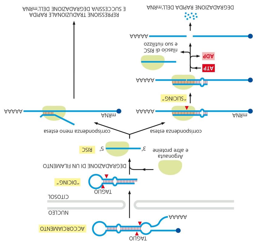
2. miRNAs: Biogenesis and Functions
miRNAs are a class of small (18–24 nt) non-coding RNAs that are processed from long
primary miRNAs (pri-miRNAs) generally transcribed by RNA Polymerase II [34–36] and har-
bor one or more hairpin structure [37]. Pri-miRNA processing starts in the nucleus, where
the Microprocessor complex, formed by the RNase III enzyme DROSHA, the RNA-binding
protein Di George Syndrome Critical Region Gene 8 (DGCR8) and other proteins [38,39]
catalyzes the endonucleolytic cleavage of the pri-miRNA to yield a ~70-nt-long hairpin
pre-miRNA [40].
Pre-miRNAs are then exported to the cytoplasm by Exportin-5 [41–43]. In the cyto-
plasm, pre-miRNAs undergo further cleavage by DICER, which removes the terminal loop
of the hairpin to yield a duplex consisting of the mature miRNA (guide strand) base-paired
to the passenger strand [44,45].
Mature miRNAs interact with the RNA-binding proteins belonging to the Argonaute
family (AGO), thus becoming integral components of the RNA-Induced Silencing Complex
(RISC) (reviewed in references [46,47]). The mature miRNA within RISC recruits the
complex onto target RNA molecules by base-pairing between a “seed” region (nt 2–7) at the
50 end of the miRNA and the 30 UTR of the target RNA [48–50], leading to gene silencing
through translation repression and mRNA decay.
MiRNA expression is tightly controlled in cells by mechanisms acting at both the
transcriptional and post-transcriptional levels (reviewed in references [51–53]). The titration
of miRNAs by competing endogenous RNAs (ceRNAs) adds a further layer of regulation
of miRNA activity [54–57]. These long RNA molecules (which can be mRNAs, lncRNAs,
Cancers 2021, 13, 3372                                                                                         3 of 20

                         pseudogene-encoded RNAs or circRNAs) sequester miRNAs, thereby preventing their
                         interaction with other targets, whose repression is therefore relieved.
                               MiRNAs participate in gene regulatory networks that control diverse biological pro-
                         cesses in multicellular organisms, such as animal development (reviewed in reference [1]),
                         cell fate specification and differentiation [58], the immune response [59] and inflamma-
                         tion [60]. Changes in the miRNA expression levels have been associated with a wide range
                         of human diseases, including diabetes, cardiovascular and kidney disease and cancer [3,4].
                         A huge number of miRNAs are downregulated or upregulated in human cancers, where
                         they exert oncogenic or tumor suppressor functions, depending on the cellular context.
                         Alterations of miRNAs in different malignancies have been linked to genetic deletion or am-
                         plification, as well as to DNA methylation of the miRNA genomic loci, to the modulation of
                         the pri-mRNA transcription level by transcription factors or to the dysregulation of one or
                         more steps in miRNA biogenesis (reviewed in reference [5]). Recently, epitranscriptomics
                         is emerging as an additional layer of the regulation of the miRNA function in cancer.

                         3. Epitranscriptomic Modifications of miRNA in Cancer
                         3.1. N6-Methyl-Adenosine (m6A)
                               m6A was first reported in the 1970s in mammalian RNAs [61–63]. A full com-
                         prehension of the role of this modification took several decades. In 1997, the protein
                         Methyltransferase-like (METTL) 3 was identified as the first “writer” of m6A in mam-
                         malian cells [23]. Further investigations have shown that m6A is installed by a nuclear
                         complex comprised of METTL3, METTL14 and WT1-Associated Protein (WTAP) [22].
                         Further components of this complex include KIAA1429, RNA Binding Motif Protein 15
                         (RBM15) and Zinc Finger CCCH-Type Containing 13 (ZC3H13) [64–66].
                               Several members belonging to the YTH (YT521-B homology) family, such as human
                         YT521-B (also known as YTHDC1), YTHDC2, YTHDF1, YTHDF2 and YTHDF3, have been
                         identified as m6A-binding or “reader” proteins [18,25]. The members belonging to the
                         DF family likely confine m6A-modified RNAs in specific cytoplasmic liquid–liquid phase
                         separation compartments [67].
                               Several other “reader” proteins have been shown to bind m6A-modified RNAs thanks
                         to a so-called “m6A switch” [68]. Indeed, m6A installation may trigger a conformational
                         switch that allows the binding of these “reader” proteins, which, in fact, do not directly
                         bind to the m6A residue itself [13]. This mechanism is exploited by several members of
                         the hnRNP (heterogeneous nuclear ribonucleoprotein) family. Finally, insulin-like growth
                         factor 2 mRNA-binding proteins (IGF2BP) were also reported to bind m6A-modified RNAs,
                         promoting their stability [69].
                               Although two enzymes able to “erase” m6A from mammalian RNAs have been
                         reported, i.e., FTO Alpha-Ketoglutarate-Dependent Dioxygenase (FTO) and AlkB Homolog
                         5, RNA Demethylase (ALKBH5) [29,30], the specificity and the relevance of these enzymes
                         in physiological conditions are still a matter of debate [70].
                               About 0.1–0.4% of all adenosines in global cellular RNAs are modified as m6A, and
                         this modification accounts for ~50% of all methylated ribonucleotides [61]. m6A was found
                         in all classes of cellular RNAs: mRNAs (in particular, in long internal exons, locations
                         upstream of stop codons and the 30 -UTR regions) [25,71,72]; ribosomal RNAs; transfer
                         RNAs and various non-coding RNAs [73–75].
                               The first report of m6A modification in miRNAs dates back to 2014, when Yuan and
                         colleagues [76] reported this epitranscriptomic modification in miR-125b. Surprisingly,
                         the authors identified NOP2/Sun RNA Methyltransferase 2 (Nsun2), a well-characterized
                         m5C “writer” [21,77], as the “writer” enzyme of this modification. Furthermore, their data
                         suggested that a m6A modification may prevent pri-miR-125b-2 processing into mature
                         miR-125b [76].
                               In 2015, two pivotal contributions by Alarcón and colleagues [78,79] provided evidence
                         that a m6A modification by METTL3 globally enhances miRNA processing in mammalian
                         cells. Mechanistically, a novel nuclear m6A “reader”, namely hnRNPA2B1, binds to
Cancers 2021, 13, 3372 4 of 20
Cancers 2021, 13, x 4 of 20
m6A-modified pri-miRNAs and interacts with the Microprocessor complex, promoting
miRNA
miRNA processing
processing in ainMETTL3-dependent
a METTL3-dependent manner.
manner. Interestingly
Interestingly in Arabidopsis
in Arabidopsis thaliana,
thaliana,
an ortholog of METTL3 has been recently shown to mediate m6A
an ortholog of METTL3 has been recently shown to mediate m6A installation on pri-miR- installation on pri-
miRNAs, thus promoting an interaction with the Microprocessor complex,
NAs, thus promoting an interaction with the Microprocessor complex, suggesting that the suggesting that
the regulation of miRNA processing by m6A is a widely conserved
regulation of miRNA processing by m6A is a widely conserved mechanism [80]. mechanism [80].
Neither
Neither FTOFTOnornor ALKBH5
ALKBH5 werewere proven
proven yetyet
to to
be be
ableable
to to catalyze
catalyze thethe demethylation
demethylation
of miRNAs. However, it is worth mentioning that DEAD-box
of miRNAs. However, it is worth mentioning that DEAD-box RNA helicase 3 (DDX3) RNA helicase 3 (DDX3)
controls the methylation status of microRNAs and interacts with
controls the methylation status of microRNAs and interacts with both the AGO2 and both the AGO2 andthethe
ALKBH5 enzymes [81], suggesting a possible role of ALKBH5 in miRNA demethylation.
ALKBH5 enzymes [81], suggesting a possible role of ALKBH5 in miRNA demethylation.
In cancer, the relevancy of m6A in miRNA maturation was first unveiled for miR-126
In cancer, the relevancy of m6A in miRNA maturation was first unveiled for miR-126
in hepatocellular carcinoma (HCC) [82]. The authors showed that a METTL14-dependent
in hepatocellular carcinoma (HCC) [82]. The authors showed that a METTL14-dependent
m6A modification on pri-miR-126 is required for its processing by the Microprocessor
m6A modification on pri-miR-126 is required for its processing by the Microprocessor
complex. Indeed, in hepatocellular carcinoma, METTL14 is downregulated, thus decreasing
complex. Indeed, in hepatocellular carcinoma, METTL14 is downregulated, thus decreas-
m6A on pri-miR-126 and reducing the miR-126 abundance. Furthermore, their findings
ing m6A on pri-miR-126 and reducing the miR-126 abundance. Furthermore, their find-
support the hypothesis that the miR-126 modification by METTL-14 is instrumental to
ings support the hypothesis that the miR-126 modification by METTL-14 is instrumental
prevent cell invasion, as assessed by in vitro assays.
to prevent cell invasion, as assessed by in vitro assays.
From that moment on, increasing evidence has disclosed the relevance of the m6A
From that moment on, increasing evidence has disclosed the relevance of the m6A
modification of miRNA in cancer progression. Most of the literature confirms that m6A
modification of miRNA
mainly promotes in cancer
pri-miRNA progression.
processing Most
and that theofderegulation
the literatureof confirms
the enzymesthatinvolved
m6A
mainly promotes pri-miRNA processing and that the deregulation
in writing or reading m6A is correlated with tumor onset. Notably, alteration of of the enzymes in-the
volved
m6Ain writing oron
deposition reading
miRNAs m6A is correlated
is not with tumor
only a common onset.
feature Notably,tumors
of different alteration
but of
also
theparticipates
m6A deposition on miRNAs is not only a common
in tumorigenesis processes (Figure 1 and Table 1).feature of different tumors but also
participates in tumorigenesis processes (Figure 1 and Table 1).
Nucleus RNAPII
miRNA gene
Pri-miRNA
Pri-miRNA processing
Microprocessor
complex
Pre-miRNA
m6A
Exportin5
A-to-I editing
Cytoplasm
Dicer cleavage
m7G
Pre-miRNA m5C
Dicer
miRNA duplex
RISC
lex
Mature miRNA comp RISC loading
Target recognition
mRNA RISC
Figure 1. Epitranscriptomic
Figure 1. Epitranscriptomicmodification impacts
modification on on
impacts miRNA processing
miRNA andand
processing activity. m6A,
activity. A-to-I
m6A, editing,
A-to-I m5C
editing, andand
m5C m7Gm7G
can affect different steps of miRNA biogenesis, including Microprocessor cleavage, Dicer cleavage and RISC loading or
can affect different steps of miRNA biogenesis, including Microprocessor cleavage, Dicer cleavage and RISC loading or alter
alter target recognition and binding.
target recognition and binding.
Colorectal cancer
Colorectal (CRC)
cancer (CRC)displays
displaysaalow
lowexpression
expression of
of METTL14, and this
METTL14, and thisisisassociated
associ-
atedwith
with an impaired m6A modification and processing of pri-miR-375. Low levels
an impaired m6A modification and processing of pri-miR-375. Low levels of miR-375 of
miR-375 result in the overexpression of its targets YAP1 (Yes-associated protein 1) and SP1,
thus increasing cell proliferation, migration and invasion [83].Cancers 2021, 13, 3372 5 of 20
result in the overexpression of its targets YAP1 (Yes-associated protein 1) and SP1, thus
increasing cell proliferation, migration and invasion [83].
The overexpression of METTL3 in bladder, colorectal, lung, ovarian and gallbladder
cancers promotes the processing of several oncomirs, such as miR-221/222 [84], miR-
1246 [85], miR-143 [86], miR-126 [87] and miRNA-92 [88]. These miRNAs have tumor
suppressors as targets; therefore, the m6A-dependent accumulation of miRNAs results
in promoting tumor progression. The same mechanism was described by Zhang and
colleagues [89], who recently showed that, in response to cigarette smoke condensate in
pancreatic ductal adenocarcinoma, there was an accumulation of mature miR-25-3p, caused
by the overexpression of METTL3, which enhanced the m6A modification of pri-miR-25-3p.
Increased miR-25-3p resulted in the repression of its target PH Domain and Leucine-Rich
Repeat Protein Phosphatase 2 (PHLPP2), thus triggering AKT activation. Interestingly, a
novel m6A reader, namely NF-κB-Associated Protein (NKAP), facilitated the interaction of
pri-miR-25 with DGCR8, thus promoting the maturation of miR-25-3p [89].
A recent report identified a further putative m6A “reader” protein, RALY (also known
as hnRNPCL2), which interacts with miR-483, miR-676 and miR-877 in CRC and is required
for their m6A-dependent processing. Mechanistically, RALY interacts with DROSHA and
DGCR8 to enhance pri-miRNA processing [90].
Recently, it has been proposed that a m6A modification reduces the ability of miRNAs
to suppress target mRNA translation. Indeed, Konno and colleagues [91] showed that
the m6A modification of let-7a-5p and miR-17-5p caused a large structural change in
the RISC complex, which affected the target RNA recognition. In pancreatic and CRC
tissues, the m6A levels on let-7a-5p and miR-17-5p increased without affecting the miRNA
expression level.
3.2. A-to-I Editing
A-to-I editing is catalyzed by enzymes highly conserved in vertebrates, called Adeno-
sine Deaminases Acting on RNA (ADAR) [92]. Mammalian genomes encode for three
members of the ADAR family: ADAR1, ADAR2 and ADAR3 [93].
ADAR enzymes bind double-stranded (ds) regions of coding and noncoding RNAs [94];
in RNAs forming imperfect dsRNA structures, A-to-I editing involves only one or two
adenosines (site selective editing), while, in the case of long perfect dsRNA regions, the
random modification of several A residues is observed (hyper-editing) [95–97].
Inosine is recognized by the cellular machinery as guanosine, causing a change in
the RNA sequence. As a consequence, depending on the modification site, this type of
RNA editing can influence the RNA stability [98–100], splicing [101–103], localization and
translation, as well as redefine its interactions with specific factors [104,105]. In mRNAs,
the modification of A-to-I can lead to a codon change, thus affecting the primary structure
of the encoded protein [106,107].
A-to-I editing mainly targets noncoding regions of RNA, such as introns and UTRs,
containing repetitive Alu elements and Long Interspersed Elements (LINEs) that fold into
dsRNA structures recognized by ADARs [108].
In most types of cancer, the activity of ADAR enzymes is significantly decreased, as
witnessed by the extensive hypoediting of Alu RNAs, as well as by the reduced expression
of ADAR enzymes [109].
The first evidence of the editing of a miRNA was shown in 2004 by Luciano and
colleagues [33], who reported A-to-I conversion within the miR-22 precursor in Homo
sapiens and Mus musculus. Soon after, it was shown that the A-to-I editing of pri-miR-142
prevents processing by DROSHA [110]. ADAR enzymes have a degree of specificity for
different miRNA precursors, depending on their secondary structure [111]. The ADAR1
interaction with DICER was associated with enhanced miRNA processing in oral squamous
cells carcinoma [112] and in melanoma [113], although, in both cases, the authors did not
assess the editing of the miRNA precursors. Furthermore, ADAR editing has been shownCancers 2021, 13, 3372 6 of 20
to affect the DICER-dependent processing of viral miRNAs [114]. Of note, ADARs can also
alter miRNA metabolism independently from their editing activity [115–117].
Several examples showed that the A-to-I editing of miRNA precursors inhibits the
biogenesis of mature miRNAs (Figure 1 and Table 2). The deregulation of ADAR1 and/or
ADAR2 in glioblastoma and in chordoma affects the expression levels of miR-21, miR-
221 and miR-222 [118] and of miR-10a and miR-125a [119], respectively. Furthermore,
the impairment of let-7 biogenesis by means of ADAR1-mediated A-to-I editing drives
leukemia stem cells renewal [120].
Table 1. Effects of m6A modification of miRNAs in cancer.
Effects on miRNA
m6A-Modified Increase/
Cancer Type Process- Effects on Tumor Progression Reference
miRNA(s) Decrease 1
ing/Function
Up-regulation of mature of miR-1246
results in the reduction of SPRED2,
miR-1246 ↑ processing [85]
thus activating the
RAF/MEK/ERK pathway
Down-regulation of mature miR-375
increases the expression of its targets
Colorectal miR-375 ↓ processing YAP1 and SP1 thus increasing [83]
Cancer proliferation, and migration
and invasion
miR-483, miR-676 and miR-877
miR-483,
modulate mitochondrial metabolism
miR-676 n.d. processing [90]
by targeting electron transport
miR-877
chain genes
miR-17-5p
↑ Binding to targets n.d. [91]
let-7a-5p
Up-regulation of mature miR-25-3p
miR-25-3p ↑ processing results in the reduction of PHLPP2, [89]
Pancreatic
leading to AKT activation.
cancer
miR-17-5p
↑ Binding to targets n.d. [91]
let-7a-5p
Hepatocellular Down-regulation of mature miR-126
miR-126 ↓ processing [82]
Carcinoma which acts as a tumor suppressor
Up-regulation of mature
Bladder
miR-221/222 ↑ processing miR-221/222 results in the reduction [84]
cancer
of PTEN, leading to proliferation
Up-regulation of mature miRNA-92
Gallbladder
miRNA-92 ↑ processing results in the reduction of PTEN, thus [88]
cancer
activating PI3K/AKT signaling
Up-regulation of mature miR-126-5p
Ovarian results in the reduction of PTEN, thus
miR-126 ↑ processing [87]
cancer activating the
PI3K/Akt/mTOR pathway
Gastric miR-17-5p
↑ Binding to targets n.d. [91]
cancer let-7a-5p
Up-regulation of mature miR-143-3p
Lung cancer promotes the metastatic potential of
(brain miR-143-3p ↑ processing lung cancer via regulation of [86]
metastasis) angiogenesis and microtubules
through VASH1
1 increase (↑) or decrease (↓) of the epitranscriptomic modification (n.d., not detected; SPRED2, Sprouty Related EVH1 Domain Containing
2; YAP1, yes-associated protein 1; SP1, Sp1 Transcription Factor; PHLPP2, PH Domain And Leucine Rich Repeat Protein; PTEN, Phosphatase
2 Phosphatase And Tensin Homolog; VASH1, Vasohibin 1.Cancers 2021, 13, 3372 7 of 20
A further mechanism by which A-to-I editing alters miRNA functions is the remod-
ulation of potential targets of mature miRNAs [121]. Indeed, A-to-I editing in the “seed”
sequence causes a loss-of-function when no more targets are recognized [122] or a gain-
of-function when a new target is recognized by the edited miRNA [123,124]. Therefore,
changes in the relative abundance of the edited and unedited forms of the miRNA lead, in
turn, to altered gene expression profiles.
In this context, in 2012, it has been shown that the loss of mir-376a-5p editing results
in the increased invasiveness of glioblastoma multiforme (GBM). Mechanistically, unedited
miR-376a-5p promotes aggressive glioma growth by its ability to target Ras-Related Protein
Rap-2a (RAP2A), a member of the RAS oncogene family, and the concomitant inability
to target Autocrine Motility Factor Receptor (AMFR) [125]. These findings were further
corroborated by the discovery that miRNA hypoediting is widespread in GBM [126].
Similarly, the impairment of ADAR-mediated editing of miR-455-5p enhances the
progression in melanoma. Indeed, unedited miR-455-5p targets the tumor suppressor Cy-
toplasmic Polyadenylation Element-Binding Protein 1 (CPEB1), thus promoting metastasis,
while edited miR-455-5p exerts the opposite effect [127]. In a follow-up of their work, the
authors showed that, in melanoma, the editing of miR-378a-3p allows the targeting of
the oncogene Parvin Alpha (PARVA). Hence, the loss of miR-378a-3p editing promotes
melanoma progression [128].
In the brain, ADAR2 edits the seed sequence of miR-589-3p. In glioblastoma, the
editing of miR-589-3p decreases. Higher levels of the unedited version of miR-589-3p
promote proliferation and invasion by targeting the tumor suppressor Protocadherin 9
(PCDH9). On the contrary, editing within miR-589-3p retargets the miRNA to the ADAM
Metallopeptidase Domain 12 (ADAM12) to contrast the progression of the tumor [129].
In different contexts, miRNA A-to-I editing stimulates the progression of the tumor
by altering the selection of miRNA targets. In thyroid cancer, the slight overexpression
of ADAR1 corresponds to a higher expression of ZEB1, a master regulator of Epithelial–
Mesenchymal Transition (EMT). It has been demonstrated that editing of the seed sequence
of miR-200b by ADAR1 impairs its ability to inhibit ZEB1 expression, favoring the progres-
sion of the cancer [130,131].
Another target of A-to-I editing is miR-381, a microRNA involved in stemness and
chemoresistance [132,133] that is overedited in non-small cell lung carcinoma (NSCLC)
cell lines harboring the genomic amplification of ADAR1. Edited miR-381 promotes cell
viability [134].
Besides these examples on specific miRNAs, a global analysis of miRNA sequenc-
ing data from healthy and cancerous tissues unveiled that miRNA editing is frequently
dysregulated in cancer [130,135–139].
3.3. 5-Methylcytosine (m5C)
m5C is one of the most representative post-transcriptional RNA modifications [140],
and it has long been known to be present in all three kingdoms of life [141,142].
m5C was originally reported in tRNAs, rRNAs [62] and coding RNAs [143]; later, it
was identified in other noncoding RNAs, thanks to technologies such as bisulfite treatment
and Next-Generation Sequencing (NGS) [144–146].
The synthesis of m5C is catalyzed by the seven members of the NOL1/NOP2/SUN do-
main (NSUN) family of methyltransferases [147] or by DNA methyltransferase-2 (DNMT2) [148].
These enzymes are responsibles for the methylation of rRNAs, tRNAs [149–153], mR-
NAs [154–156], lncRNAs [157], vault-RNAs [158], enhancer-RNAs [145], mitochondrial
tRNAMet [159] and mitochondrial 12S rRNA [160].
In vitro and in vivo studies have demonstrated that aly/REF nuclear factor (ALYREF)
is a putative “reader” of m5C sites on mRNAs and that, following the knockdown of
NSUN2, ALYREF loses its RNA-binding ability and is retained in the nucleus, suggesting a
role for m5C in mRNA exports from the nucleus [26]. A further m5C “reader” is Y-Box-Cancers 2021, 13, 3372 8 of 20
Binding Protein 1 (YBX1) that recognizes and binds m5C-modified mRNAs and stabilizes
their target mRNAs by recruiting ELAV-like Protein 1 (ELAVL1) [161,162].
Table 2. Effects of A-to-I editing of miRNAs in cancer.
Effects on miRNA
A-to-I-Modified Increase/
Cancer Process- Effects on Tumor Progression Reference
miRNA(s) Decrease 1
ing/Function
Unedited miR-376a-5p promotes
aggressive glioma growth, by its
mir-376a-5p ↓ Binding to targets [125]
ability to target RAP2A and
Glioma
concomitant inability to target AMFR
Up-regulation of mature
miR-221/222 and miR-21 results in
miR-221/222 the repression of its targets p27Kip1
↓ processing [118]
miR-21 and PDCD4, thus increasing
proliferation and migration of
glioblastoma
Editing within miR-589–3p retargets
the miRNA from the protocadherin
miR-589-3p ↓ Binding to targets PCDH9 to the metalloprotease [129]
ADAM12, which is involved in
glioblastoma cell invasion.
Unedited miR-455-5p but not the
edited form targets the tumor
miR-455-5p ↓ Binding to targets suppressor gene CPEB1, thus [127]
Melanoma
promoting tumor growth
and metastasis
Edited miR-378a-3p but not the
unedited form specifically targets the
miR-378a-3p ↓ Binding to targets PARVA oncogene, thus preventing [128]
the progression of melanoma towards
the malignant phenotype
Down-regulation of miR-10a and
miR-10a
Chordoma ↑ processing miR-125a expression and upregulates [119]
miR-125a
expression of their target genes
Down-regulation of mature let-7
Chronic
results in increased LIN28B
myeloid let-7 ↑ processing [120]
expression and enhanced
leukemia
self-renewal
Edited miR-200b has weakened
Thyroid activity against its target gene ZEB1,
miR-200b ↑ Binding to targets [131]
cancer an epithelial–mesenchymal transition
(EMT) marker
Edited miR-381 enhances the growth
Lung cancer miR-381 ↑ n.d. of non-small-cell lung cancer cells as [134]
compared to the unedited form
1increase (↑) or decrease (↓) of the epitranscriptomic modification (n.d., not detected; RAP2A, Ras-Related Protein Rap-2a; AMFR, Autocrine
Motility Factor Receptor; PDCD4, Programmed Cell Death 4; PCDH9, Protocadherin 9; ADAM12, ADAM Metallopeptidase Domain 12;
CPEB1, Cytoplasmic Polyadenylation Element-Binding Protein 1; PARVA, Parvin Alpha).
m5C “writers” and “readers” are primarily implicated in fundamental cancer-related
processes such as cell differentiation, motility [163,164], proliferation [165,166], cell cycle
progression [167] and senescence [155].
In particular, NSUN2 is aberrantly expressed and plays important roles in the devel-
opment and pathogenesis of different types of tumors, such as breast, colorectal, lung, skin,
ovarian and bladder cancers [168].Cancers 2021, 13, 3372 9 of 20
The distribution of m5C in small RNAs is poorly understood so far; nevertheless, this
modification has been recently highlighted in vault RNAs (vtRNAs) [158], piwi-associated
RNAs (piRNAs) [169] and miRNAs [91,170,171]. m5C deposition regulates the processing
of vault ncRNAs into small vault RNAs (svRNAs) [158,172].
m5C has been only recently characterized in miRNAs. Interestingly, methylation,
but not an abundance of miR-200c-3p and miR-21-3p, was increased in pancreatic and
colorectal cancer tissues, as well as in serum samples from pancreatic and colorectal cancer
patients [91] (Figure 1 and Table 3).
Table 3. Effects of m5C and m7G modifications of miRNAs in cancer.
Effects on miRNA
Modified Increase/
Cancer Process- Effects on Tumor Progression Reference
miRNA(s) Decrease 1
ing/Function
Cytosine-methylated miRNA-181a-5p
miRNA-181a-5p
Glioma ↑ Binding to targets loses its ability to target the mRNA of [169]
(m5C)
the pro-apoptotic protein BIM
Colorectal
cancer;
miR-200c-3p
gastric
miR-21-3p ↑ Binding to targets n.d. [88]
cancer;
(m5C)
pancreatic
cancer
let-7 family m7G methylation within miRNAs
Lung cancer n.d. processing [173]
(m7G) regulates cell migration
Down-regulation of mature let-7e
let-7e results in the activation of its targets
Colon cancer ↓ processing [174]
(m7G) HMGA2 thus stimulating colon
cancer cell viability and mobility
1 Increase (↑) or decrease (↓) of epitranscriptomic modifications (n.d., not detected; HMGA2 High Mobility Group AT-hook 2).
Cheray and colleagues [170] proposed that the DNMT3A/AGO4 complex promotes
the methylation of cytosine residues of miRNAs at CG dinucleotides. In glioblastoma-
derived cell lines and glioblastoma tumor samples, the methylation of mature miR-181a-5p
by the DNMT3A/AGO4 complex inhibits the recognition of its target mRNA BIM, a
proapoptotic gene, also known as B-cell chronic lymphocytic leukemia/lymphoma (Bcl-2)-
like 11 (BCL2L11) [170]. This preliminary evidence highlights that the profiling in tumor
samples of m5C in miRNAs deserves further investigation.
Recently, we described that m5C is widely spread in human miRNAs in various
sequence contexts by taking advantage of a novel NGS analysis of bisulfite-treated small
RNAs (BS-miRNA-seq) [171].
Finally, 5mC is oxidized by the Ten-eleven translocation (TET) enzymes both in
DNA and RNA. TET enzymes are Fe(II)- and 2-oxoglutarate-dependent dioxygenases that
mediate the conversion of 5mC to 5hmC, then to 5-formylcytosine (5-fC) [175] and, finally,
to 5-carboxylcytosine (5-caC) [18,176,177]. In DNA, these subsequent conversions have the
purpose of demethylating 5mC [178], but it is still not clear if this mechanism is conserved
in RNA.
hm5C has been detected in RNA isolated from different mouse and human tissues,
including the brain, heart, pancreas and spleen [179]. Transcriptome-wide analyses of
hm5C in mouse and in Drosophila RNAs have revealed the presence of 5hmC on hundreds
of messenger RNAs, mainly in UC-rich motifs [180,181]. The deposition of hm5C in mRNAs
has been associated with the differentiation of murine embryonic stem cells and brain
development in Drosophila via controlling the mRNA stability or translation, respectively.
To date, decreased levels of hm5C in RNA have been shown in tumor tissues, such as
CRC and hepatocellular carcinoma [176].Cancers 2021, 13, 3372 10 of 20
Interestingly, we recently unraveled not only the presence of m5C but, also, of hm5C
on several miRNAs in human cancer cell lines [171]. However, no evidence of the role of
hm5C modification in miRNAs in tumors has yet been reported.
3.4. N7-Methylguanosine (m7G)
m7G is a positively charged modification installed cotranscriptionally at the 5’ Caps
of eukaryotic mRNAs [182]. This modification protects and stabilizes transcripts from
exonucleolytic degradation [183] and influences all the events responsible for the processing
of the mRNA molecules, from transcript elongation to translation [184,185].
Notably, the presence of internal m7G sites was found not only in tRNA and rRNA
molecules [186–188] but also in mammalian mRNAs [188]. Internal m7G could affect
mRNA translation, and this modification typically occurs near the start and stop codons in
a GA-enriched motif [189].
The enzyme responsible for this internal m7G modification is METTL1, which coop-
erates with the cofactor WD Repeat Domain 4 (WDR4) [189,190]. Interestingly, METTL1
has been linked to tumor vascular invasion and poor prognosis in hepatocellular carci-
noma [173,191].
Recently, by high-throughput screening, several miRNAs were identified as harboring
internal m7G sites [192]. In particular, METTL1-dependent m7G was discovered in a subset
of tumor-suppressor miRNAs involved in the inhibition of cell migration, including the
let-7 family. METTL1-mediated m7G occurs on pri-miRNA within G-rich regions that
display the propensity to form G-quadruplexes, i.e., structures known to be inhibitory to
miRNA processing [174,193,194] (Figure 1 and Table 3).
Indeed, m7G in the let-7 family affects G-quadruplex formations, thus facilitating
the formation of a canonical stem-loop structure and miRNA processing [192]. In line
with this study, Liu and colleagues showed that, in colon cancer, the downregulation of
METTL1 leads to a decrease in the let-7e levels. The alteration of let-7e expression affects
its downstream target High Mobility Group AT-hook 2 (HMGA2), thus promoting cell
proliferation, invasion and EMT [195].
4. Epitranscriptomic Modifications of miRNA Targets
4.1. m6A in miRNA Targets
The installation of m6A in miRNA targets may affect the pairing with miRNAs, thus
affecting miRNA functions. Such a mechanism was first suggested in 2015, when Ke and
colleagues [72] reported that the majority of m6A sites on mRNAs were in the last exon. The
authors also reported a significant overlap between the m6A residues and AGO-binding
sites. This finding was further corroborated by a later study [196].
Importantly, m6A modification may positively or negatively affect miRNA–mRNA
pairing by several distinct mechanisms. On the one hand, m6A modifications within
miRNA-binding sites may directly affect the miRNA:mRNA duplex stability. On the other
hand, m6A modifications nearby AGO-binding sites may alter the mRNA secondary struc-
ture and/or recruit other RNA-binding proteins, thus modifying the mRNA accessibility
to miRNA. Thus far, a few examples have been reported.
In the liver, YAP is a target of miR-582-3p, which binds at residues 313-321 of YAP 30
UTR. Such binding is enhanced by m6A modifications of YAP 30 UTR at residue 355. In
HCC, the m6A modification at residue 355 of YAP 30 UTR is impaired, resulting in the loss
of YAP repression by miR-582-3p [197].
In cancer cell lines, m6A modification promotes the recruitment of IGF2BP1 onto
Serum Response Factor (SRF) mRNA 30 UTR. Interestingly, IGF2BP1 acts as a m6A “reader”,
and its recruitment is instrumental to reduce miRNA-mediated AGO binding to SRF. There-
fore, m6A deposition on the 30 UTR of SRF mRNA relieves the negative post-transcriptional
regulation by miRNAs [198]. Furthermore, a genome-wide analysis of m6A in glioma
stem cells highlighted that m6A deposition on the 30 UTR of several mRNAs may affect
their targeting by different miRNAs [199]. On the other hand, Cheng and colleagues [200]Cancers 2021, 13, 3372 11 of 20
showed an effect at odds with this. Indeed, in neuroblastoma, m6A modification in N-Myc
30 UTR near a miR-98-binding site is necessary to promote the miR-98-mediated post-
transcriptional repression of N-Myc. Overall, these examples highlight that the effect of
m6A within mRNA 30 UTRs on miRNA targeting is ambiguous.
m6A not only affects miRNA:mRNA interactions but, also, miRNA:ceRNA interac-
tions. Yang and colleagues [201] showed that the m6A modification of linc-RNA-1281 is
critical for its interaction with miRNAs belonging to the let-7 family. In nasopharyngeal
carcinoma, lncRNA FAM225A acts as a ceRNA, sequestering miR-590-3p and miR-1275,
thus activating FAK/PI3K/Akt signaling; in this scenario, m6A modification contributes to
this mechanism by increasing the lncRNA FAM225A stability [202]. Finally, LINC00958, a
ceRNA acting to sponge miR-3619-5p in HCC, is also stabilized through m6A modification
by METTL3 [203].
4.2. A-to-I Editing in miRNA Targets
A-to-I editing can influence microRNA targeting. Indeed, when A-to-I editing oc-
curs within the miRNA-binding site in the 30 UTR of a mRNA, this can remodulate the
interactions between miRNA and mRNA in different ways.
The editing of the 30 UTR of Rho GTPase Activating Protein 26 (ARHGAP26) by
ADAR1 blocks the interaction with miR-30b-3p and miR-573 to favor the expression of
the protein [204]. On the contrary, in HCC cells, for instance, the RNA editing catalyzed
by ADAR1 of 30 UTR of Aryl hydrocarbon Receptor (AhR) creates a miR-378-binding site
to negatively regulate the expression of this protein [205]. Through a similar mechanism,
editing of the 30 UTR of the mRNA encoding for the tumor suppressor Phosphatase and
Actin Regulator 4 (PHACTR4) mediated by ADAR1 prevents the binding of miR-196a-3p.
Accordingly, the decreased activity of ADAR1 in gastric cancer results in the repression of
PHACTR4 by miR-196-3p [206].
Interestingly, a pan-cancer RNA editing study highlighted that the 30 UTR of the
Mouse double minute 2 homolog (MDM2) oncogene underwent A-to I editing in 11 out
of the 14 cancer types investigated within a region of the 30 UTR complementary to the
miR-200 “seed” region. This editing impaired MDM2 repression by miR-200 [207].
The above-mentioned examples suggest that A-to-I editing within regions of 30 UTR
pairing with miRNA “seed” affects miRNA binding. However, a genome-wide report
pinpointed that the editing of A residues lying outside of the miRNA:mRNA pairing
region affects the mRNA structure, thus modulating the accessibility to AGO2-miRNA
complexes [208].
4.3. m5C in miRNA Targets
To date, little is known about the mechanisms through which m5C can regulate the
function of miRNAs, but, interestingly, a possible role for m5C in miRNA targeting was
suggested by the overlap between the AGO2-binding sites and m5C positions reported in
the 30 UTR of human mRNAs [209].
5. Methodological Challenges and the Potential Limits of Current Knowledge
Our understanding of the mechanisms underlying the epitranscriptomic regulation of
miRNAs is still potentially biased by the relatively small number of modifications which
have been widely investigated in miRNAs, with most reports focused on m6A and A-to-I
editing. Indeed, further mechanisms through which epitranscriptomic modifications may
affect miRNA function are conceivable. For example, the recent report by Konno and
colleagues suggested that m5C modification in position 9 of miR-200-3p might affect the
interaction with AGO proteins [91]. However, further investigation will be required to
assess whether this may represent a general paradigm. Epitranscriptomic modifications
might also modulate miRNA half-life, or control miRNA subcellular localization (either
in membrane enclosed organelles, or by liquid-liquid phase separation) or secretion of
modified miRNAs. Secreted miRNAs have been widely reported as potential biomarkersCancers 2021, 13, 3372 12 of 20
for a variety of diseases and they also serve as signaling molecules to mediate cell-cell
communications [210,211].
Our current knowledge of the modifications installed on miRNAs is likely incomplete
because of technical limits which still prevent application of several high-throughput NGS
methods for the quantification and characterization of epitranscriptomic modification to
miRNAs. Indeed, most epitranscriptomic modifications are investigated through NGS
methods which exploit the block of reverse transcription at the modified nucleobase
of interest by means of different biochemical treatments (i.e., antibody cross-linking or
chemical modifications). These protocols result in reads truncated at the modified positions,
thus allowing the genome-wide mapping of the epitranscriptomic modification at single-
nucleotide resolution [212,213]. Those methods cannot be applied to mature miRNAs, as
premature reverse trascriptase termination would yield reads too short to be effectively
mapped on the genome. Currently, single-nucleotide resolution analysis in miRNAs is
only possible for those modifications assessed through methods based on mismatched
nucleotides introduced during the reverse transcription step. Furthermore, techniques
relying on unmodified nucleobases conversion (e.g., bisulfite treatment) require dedicated
pipelines for data analysis as alignment of short reads is severely affected by complexity
reduction associated with those techniques [171].
Finally, emerging methods to assess epitranscriptomic modifications through third
generation sequencing are best-suited for long RNA molecules and cannot be easily adapted
to the study of miRNAs [214].
6. Conclusions
In this review, we summarized the current knowledge on the epitranscriptomic modi-
fications of miRNAs that play a role in cancer development and/or progression. In most
cases, epitranscriptomic modifications exert their effect on miRNAs by affecting either their
biogenesis or their binding to target mRNAs.
The current lack of mature, high-throughput technologies to profile epitranscriptomic
modifications of miRNAs, with the notable exception of A-to-I editing, is likely one of
the main reasons why the prognostic/diagnostic use of the “miRNA epitranscriptome”
has been poorly explored thus far. Nevertheless, a plethora of studies focused on the
mechanistic role of specific modifications in cancer development and/or progression point
out how promising the miRNA epitranscriptome is.
Adaptation of the existing NGS methods to yield techniques allowing cheap, high-
throughput and quantitative assessments of epitranscriptomic modifications in miRNAs
could potentially propel research in this field by allowing mechanistic investigations of fur-
ther modifications. Furthermore, such methodologies would be fundamental to investigate
the potential prognostic role of miRNAs, including miRNA secreted into extracellular fluids.
Author Contributions: V.F. is responsible for the ideation; V.D.P., E.L., E.O., C.C., I.L., V.F. performed
the literature search V.D.P., E.L., E.O., C.C., I.L., V.F. drafted the work and I.L., C.C., V.F. revised the
manuscript. All authors have read and agreed to the published version of the manuscript.
Funding: The APC was funded by a Sapienza Università di Roma grant (“Ateneo Medi 2019
#RM11916B7A048AA0”) to C.C.
Conflicts of Interest: The authors declare no conflict of interest.
References
1. Alberti, C.; Cochella, L. A Framework for Understanding the Roles of MiRNAs in Animal Development. Development 2017, 144,
2548–2559. [CrossRef]
2. Tüfekci, K.U.; Meuwissen, R.L.J.; Genç, Ş. The Role of MicroRNAs in Biological Processes. In miRNomics: MicroRNA Biology
and Computational Analysis; Yousef, M., Allmer, J., Eds.; Methods in Molecular Biology; Humana Press: Totowa, NJ, USA, 2014;
pp. 15–31, ISBN 978-1-62703-748-8.
3. Paul, P.; Chakraborty, A.; Sarkar, D.; Langthasa, M.; Rahman, M.; Bari, M.; Singha, R.S.; Malakar, A.K.; Chakraborty, S. Interplay
between MiRNAs and Human Diseases. J. Cell. Physiol. 2018, 233, 2007–2018. [CrossRef]Cancers 2021, 13, 3372 13 of 20
4. Huang, Z.; Shi, J.; Gao, Y.; Cui, C.; Zhang, S.; Li, J.; Zhou, Y.; Cui, Q. HMDD v3.0: A Database for Experimentally Supported
Human MicroRNA-Disease Associations. Nucleic Acids Res. 2019, 47, D1013–D1017. [CrossRef]
5. Ali Syeda, Z.; Langden, S.S.S.; Munkhzul, C.; Lee, M.; Song, S.J. Regulatory Mechanism of MicroRNA Expression in Cancer. Int. J.
Mol. Sci. 2020, 21, 1723. [CrossRef]
6. Wu, Y.; Li, Q.; Zhang, R.; Dai, X.; Chen, W.; Xing, D. Circulating MicroRNAs: Biomarkers of Disease. Clin. Chim. Acta 2021, 516,
46–54. [CrossRef]
7. Forterre, A.; Komuro, H.; Aminova, S.; Harada, M. A Comprehensive Review of Cancer MicroRNA Therapeutic Delivery
Strategies. Cancers 2020, 12, 1852. [CrossRef] [PubMed]
8. Boccaletto, P.; Machnicka, M.A.; Purta, E.; Piatkowski,
˛ P.; Bagiński, B.; Wirecki, T.K.; de Crécy-Lagard, V.; Ross, R.; Limbach, P.A.;
Kotter, A.; et al. MODOMICS: A Database of RNA Modification Pathways. 2017 Update. Nucleic Acids Res. 2018, 46, D303–D307.
[CrossRef]
9. Saletore, Y.; Meyer, K.; Korlach, J.; Vilfan, I.D.; Jaffrey, S.; Mason, C.E. The Birth of the Epitranscriptome: Deciphering the Function
of RNA Modifications. Genome Biol. 2012, 13, 175. [CrossRef] [PubMed]
10. Peer, E.; Rechavi, G.; Dominissini, D. Epitranscriptomics: Regulation of MRNA Metabolism through Modifications. Curr. Opin.
Chem. Biol. 2017, 41, 93–98. [CrossRef]
11. Wiener, D.; Schwartz, S. The Epitranscriptome beyond M6A. Nat. Rev. Genet. 2020. [CrossRef] [PubMed]
12. Nachtergaele, S.; He, C. The Emerging Biology of RNA Post-Transcriptional Modifications. RNA Biol. 2017, 14, 156–163. [CrossRef]
[PubMed]
13. Zaccara, S.; Ries, R.J.; Jaffrey, S.R. Reading, Writing and Erasing MRNA Methylation. Nat. Rev. Mol. Cell Biol. 2019, 20, 608–624.
[CrossRef]
14. Li, X.; Ma, S.; Yi, C. Pseudouridine: The Fifth RNA Nucleotide with Renewed Interests. Curr. Opin. Chem. Biol. 2016, 33, 108–116.
[CrossRef] [PubMed]
15. Eisenberg, E.; Levanon, E.Y. A-to-I RNA Editing—Immune Protector and Transcriptome Diversifier. Nat. Rev. Genet. 2018, 19,
473–490. [CrossRef]
16. Trixl, L.; Lusser, A. The Dynamic RNA Modification 5-Methylcytosine and Its Emerging Role as an Epitranscriptomic Mark.
Wiley Interdiscip. Rev. RNA 2019, 10, e1510. [CrossRef]
17. Janzen, W.P.; Wigle, T.J.; Jin, J.; Frye, S.V. Epigenetics: Tools and Technologies. Drug Discov. Today Technol. 2010, 7, e59–e65.
[CrossRef]
18. Fu, Y.; Dominissini, D.; Rechavi, G.; He, C. Gene Expression Regulation Mediated through Reversible M6A RNA Methylation.
Nat. Rev. Genet. 2014, 15, 293–306. [CrossRef]
19. Cortese, R.; Kammen, H.O.; Spengler, S.J.; Ames, B.N. Biosynthesis of Pseudouridine in Transfer Ribonucleic Acid. J. Biol. Chem.
1974, 249, 1103–1108. [CrossRef]
20. Koonin, E.V. Pseudouridine Synthases: Four Families of Enzymes Containing a Putative Uridine-Binding Motif Also Conserved
in DUTPases and DCTP Deaminases. Nucleic Acids Res. 1996, 24, 2411–2415. [CrossRef]
21. Bohnsack, K.E.; Höbartner, C.; Bohnsack, M.T. Eukaryotic 5-Methylcytosine (M5C) RNA Methyltransferases: Mechanisms,
Cellular Functions, and Links to Disease. Genes 2019, 10, 102. [CrossRef]
22. Liu, J.; Yue, Y.; Han, D.; Wang, X.; Fu, Y.; Zhang, L.; Jia, G.; Yu, M.; Lu, Z.; Deng, X.; et al. A METTL3–METTL14 Complex Mediates
Mammalian Nuclear RNA N 6 -Adenosine Methylation. Nat. Chem. Biol. 2014, 10, 93–95. [CrossRef]
23. Bokar, J.A.; Shambaugh, M.E.; Polayes, D.; Matera, A.G.; Rottman, F.M. Purification and CDNA Cloning of the AdoMet-Binding
Subunit of the Human MRNA (N6-Adenosine)-Methyltransferase. RNA 1997, 3, 1233–1247.
24. Bass, B.L.; Weintraub, H. An Unwinding Activity That Covalently Modifies Its Double-Stranded RNA Substrate. Cell 1988, 55,
1089–1098. [CrossRef]
25. Dominissini, D.; Moshitch-Moshkovitz, S.; Schwartz, S.; Salmon-Divon, M.; Ungar, L.; Osenberg, S.; Cesarkas, K.; Jacob-Hirsch, J.;
Amariglio, N.; Kupiec, M.; et al. Topology of the Human and Mouse m 6 A RNA Methylomes Revealed by m 6 A-Seq. Nature
2012, 485, 201–206. [CrossRef] [PubMed]
26. Yang, X.; Yang, Y.; Sun, B.-F.; Chen, Y.-S.; Xu, J.-W.; Lai, W.-Y.; Li, A.; Wang, X.; Bhattarai, D.P.; Xiao, W.; et al. 5-Methylcytosine
Promotes MRNA Export—NSUN2 as the Methyltransferase and ALYREF as an M5C Reader. Cell Res. 2017, 27, 606–625.
[CrossRef] [PubMed]
27. Newby, M.I.; Greenbaum, N.L. Sculpting of the Spliceosomal Branch Site Recognition Motif by a Conserved Pseudouridine.
Nat. Struct. Biol. 2002, 9, 958–965. [CrossRef] [PubMed]
28. Kierzek, E.; Malgowska, M.; Lisowiec, J.; Turner, D.H.; Gdaniec, Z.; Kierzek, R. The Contribution of Pseudouridine to Stabilities
and Structure of RNAs. Nucleic Acids Res. 2014, 42, 3492–3501. [CrossRef] [PubMed]
29. Jia, G.; Fu, Y.; Zhao, X.; Dai, Q.; Zheng, G.; Yang, Y.; Yi, C.; Lindahl, T.; Pan, T.; Yang, Y.-G.; et al. N 6-Methyladenosine in Nuclear
RNA Is a Major Substrate of the Obesity-Associated FTO. Nat. Chem. Biol. 2011, 7, 885–887. [CrossRef]
30. Zheng, G.; Dahl, J.A.; Niu, Y.; Fedorcsak, P.; Huang, C.-M.; Li, C.J.; Vågbø, C.B.; Shi, Y.; Wang, W.-L.; Song, S.-H.; et al. ALKBH5 Is
a Mammalian RNA Demethylase That Impacts RNA Metabolism and Mouse Fertility. Mol. Cell 2013, 49, 18–29. [CrossRef]
31. Barbieri, I.; Kouzarides, T. Role of RNA Modifications in Cancer. Nat. Rev. Cancer 2020, 20, 303–322. [CrossRef]
32. Haruehanroengra, P.; Zheng, Y.Y.; Zhou, Y.; Huang, Y.; Sheng, J. RNA Modifications and Cancer. RNA Biol. 2020, 17, 1560–1575.
[CrossRef] [PubMed]Cancers 2021, 13, 3372 14 of 20
33. Luciano, D.J.; Mirsky, H.; Vendetti, N.J.; Maas, S. RNA Editing of a MiRNA Precursor. RNA 2004, 10, 1174–1177. [CrossRef]
[PubMed]
34. Lee, Y.; Kim, M.; Han, J.; Yeom, K.-H.; Lee, S.; Baek, S.H.; Kim, V.N. MicroRNA Genes Are Transcribed by RNA Polymerase II.
EMBO J. 2004, 23, 4051–4060. [CrossRef] [PubMed]
35. Rodriguez, A.; Griffiths-Jones, S.; Ashurst, J.L.; Bradley, A. Identification of Mammalian MicroRNA Host Genes and Transcription
Units. Genome Res. 2004, 14, 1902–1910. [CrossRef]
36. Kim, Y.-K.; Kim, V.N. Processing of Intronic MicroRNAs. EMBO J. 2007, 26, 775–783. [CrossRef]
37. Lagos-Quintana, M.; Rauhut, R.; Lendeckel, W.; Tuschl, T. Identification of Novel Genes Coding for Small Expressed RNAs.
Science 2001, 294, 853–858. [CrossRef]
38. Denli, A.M.; Tops, B.B.J.; Plasterk, R.H.A.; Ketting, R.F.; Hannon, G.J. Processing of Primary MicroRNAs by the Microprocessor
Complex. Nature 2004, 432, 231–235. [CrossRef]
39. Gregory, R.I.; Yan, K.; Amuthan, G.; Chendrimada, T.; Doratotaj, B.; Cooch, N.; Shiekhattar, R. The Microprocessor Complex
Mediates the Genesis of MicroRNAs. Nature 2004, 432, 235–240. [CrossRef]
40. Lee, Y.; Ahn, C.; Han, J.; Choi, H.; Kim, J.; Yim, J.; Lee, J.; Provost, P.; Rådmark, O.; Kim, S.; et al. The Nuclear RNase III Drosha
Initiates MicroRNA Processing. Nature 2003, 425, 415–419. [CrossRef]
41. Yi, R.; Qin, Y.; Macara, I.G.; Cullen, B.R. Exportin-5 Mediates the Nuclear Export of Pre-MicroRNAs and Short Hairpin RNAs.
Genes Dev. 2003, 17, 3011–3016. [CrossRef]
42. Bohnsack, M.T.; Czaplinski, K.; Görlich, D. Exportin 5 Is a RanGTP-Dependent DsRNA-Binding Protein That Mediates Nuclear
Export of Pre-MiRNAs. RNA 2004, 10, 185–191. [CrossRef]
43. Lund, E.; Güttinger, S.; Calado, A.; Dahlberg, J.E.; Kutay, U. Nuclear Export of MicroRNA Precursors. Science 2004, 303, 95–98.
[CrossRef] [PubMed]
44. Ketting, R.F.; Fischer, S.E.; Bernstein, E.; Sijen, T.; Hannon, G.J.; Plasterk, R.H. Dicer Functions in RNA Interference and in
Synthesis of Small RNA Involved in Developmental Timing in C. Elegans. Genes Dev. 2001, 15, 2654–2659. [CrossRef]
45. Hutvágner, G.; McLachlan, J.; Pasquinelli, A.E.; Bálint, É.; Tuschl, T.; Zamore, P.D. A Cellular Function for the RNA-Interference
Enzyme Dicer in the Maturation of the Let-7 Small Temporal RNA. Science 2001, 293, 834–838. [CrossRef]
46. Gebert, L.F.R.; MacRae, I.J. Regulation of MicroRNA Function in Animals. Nat. Rev. Mol. Cell Biol. 2019, 20, 21–37. [CrossRef]
47. Yoda, M.; Kawamata, T.; Paroo, Z.; Ye, X.; Iwasaki, S.; Liu, Q.; Tomari, Y. ATP-Dependent Human RISC Assembly Pathways.
Nat. Struct. Mol. Biol. 2010, 17, 17–23. [CrossRef] [PubMed]
48. Krek, A.; Grün, D.; Poy, M.N.; Wolf, R.; Rosenberg, L.; Epstein, E.J.; MacMenamin, P.; da Piedade, I.; Gunsalus, K.C.; Stoffel, M.;
et al. Combinatorial MicroRNA Target Predictions. Nat. Genet. 2005, 37, 495–500. [CrossRef] [PubMed]
49. Lewis, B.P.; Burge, C.B.; Bartel, D.P. Conserved Seed Pairing, Often Flanked by Adenosines, Indicates That Thousands of Human
Genes Are MicroRNA Targets. Cell 2005, 120, 15–20. [CrossRef]
50. Chipman, L.B.; Pasquinelli, A.E. MiRNA Targeting: Growing beyond the Seed. Trends Genet. TIG 2019, 35, 215–222. [CrossRef]
51. Schanen, B.C.; Li, X. Transcriptional Regulation of Mammalian MiRNA Genes. Genomics 2011, 97, 1–6. [CrossRef]
52. Saj, A.; Lai, E.C. Control of MicroRNA Biogenesis and Transcription by Cell Signaling Pathways. Curr. Opin. Genet. Dev. 2011, 21,
504–510. [CrossRef]
53. Michlewski, G.; Cáceres, J.F. Post-Transcriptional Control of MiRNA Biogenesis. RNA 2019, 25, 1–16. [CrossRef]
54. Karreth, F.A.; Tay, Y.; Perna, D.; Ala, U.; Tan, S.M.; Rust, A.G.; DeNicola, G.; Webster, K.A.; Weiss, D.; Perez-Mancera, P.A.; et al. In
Vivo Identification of Tumor- Suppressive PTEN CeRNAs in an Oncogenic BRAF-Induced Mouse Model of Melanoma. Cell 2011,
147, 382–395. [CrossRef] [PubMed]
55. Tay, Y.; Kats, L.; Salmena, L.; Weiss, D.; Tan, S.M.; Ala, U.; Karreth, F.; Poliseno, L.; Provero, P.; Di Cunto, F.; et al. Coding-
Independent Regulation of the Tumor Suppressor PTEN by Competing Endogenous MRNAs. Cell 2011, 147, 344–357. [CrossRef]
56. Sumazin, P.; Yang, X.; Chiu, H.-S.; Chung, W.-J.; Iyer, A.; Llobet-Navas, D.; Rajbhandari, P.; Bansal, M.; Guarnieri, P.; Silva, J.;
et al. An Extensive MicroRNA-Mediated Network of RNA-RNA Interactions Regulates Established Oncogenic Pathways in
Glioblastoma. Cell 2011, 147, 370–381. [CrossRef]
57. Cesana, M.; Cacchiarelli, D.; Legnini, I.; Santini, T.; Sthandier, O.; Chinappi, M.; Tramontano, A.; Bozzoni, I. A Long Noncoding
RNA Controls Muscle Differentiation by Functioning as a Competing Endogenous RNA. Cell 2011, 147, 358–369. [CrossRef]
[PubMed]
58. Galagali, H.; Kim, J.K. The Multifaceted Roles of MicroRNAs in Differentiation. Curr. Opin. Cell Biol. 2020, 67, 118–140. [CrossRef]
59. Mehta, A.; Baltimore, D. MicroRNAs as Regulatory Elements in Immune System Logic. Nat. Rev. Immunol. 2016, 16, 279–294.
[CrossRef] [PubMed]
60. Tahamtan, A.; Teymoori-Rad, M.; Nakstad, B.; Salimi, V. Anti-Inflammatory MicroRNAs and Their Potential for Inflammatory
Diseases Treatment. Front. Immunol. 2018, 9. [CrossRef]
61. Wei, C.M.; Gershowitz, A.; Moss, B. Methylated Nucleotides Block 50 Terminus of HeLa Cell Messenger RNA. Cell 1975, 4,
379–386. [CrossRef]
62. Desrosiers, R.; Friderici, K.; Rottman, F. Identification of Methylated Nucleosides in Messenger RNA from Novikoff Hepatoma
Cells. Proc. Natl. Acad. Sci. USA 1974, 71, 3971–3975. [CrossRef]
63. Perry, R.P.; Kelley, D.E. Existence of Methylated Messenger RNA in Mouse L Cells. Cell 1974, 1, 37–42. [CrossRef]You can also read