Emerging Functions of Actins and Actin Binding Proteins in Trypanosomatids - Frontiers
←
→
Page content transcription
If your browser does not render page correctly, please read the page content below
REVIEW
                                                                                                                                            published: 09 October 2020
                                                                                                                                        doi: 10.3389/fcell.2020.587685

                                            Emerging Functions of Actins and
                                            Actin Binding Proteins in
                                            Trypanosomatids
                                            Chhitar M. Gupta 1* , Bindu Ambaru 1,2 and Rani Bajaj 1,2
                                            1
                                              Institute of Bioinformatics and Applied Biotechnology, Bengaluru, India, 2 Manipal Academy of Higher Education, Manipal,
                                            India

                                            Actin is the major protein constituent of the cytoskeleton that performs wide range of
                                            cellular functions. It exists in monomeric and filamentous forms, dynamics of which
                                            is regulated by a large repertoire of actin binding proteins. However, not much was
                                            known about existence of these proteins in trypanosomatids, till the genome sequence
                                            data of three important organisms of this class, viz. Trypanosoma brucei, Trypanosoma
                                            cruzi and Leishmania major, became available. Here, we have reviewed most of the
                          Edited by:
                                            findings reported to date on the intracellular distribution, structure and functions of
                    Michael Schnoor,        these proteins and based on them, we have hypothesized some of their functions.
         National Polytechnic Institute     The major findings are as follows: (1) All the three organisms encode at least a set
      of Mexico (CINVESTAV), Mexico
                                            of ten actin binding proteins (profilin, twinfilin, ADF/cofilin, CAP/srv2, CAPz, coronin, two
                        Reviewed by:
                   Ulrike Kemmerling,       myosins, two formins) and one isoform of actin, except that T. cruzi encodes for three
              University of Chile, Chile    formins and several myosins along with four actins. (2) Actin 1 and a few actin binding
     Rebeca Georgina Manning-Cela,
         National Polytechnic Institute
                                            proteins (ADF/cofilin, profilin, twinfilin, coronin and myosin13 in L. donovani; ADF/cofilin,
      of Mexico (CINVESTAV), Mexico         profilin and myosin1 in T. brucei; profilin and myosin-F in T.cruzi) have been identified
                *Correspondence:            and characterized. (3) In all the three organisms, actin cytoskeleton has been shown
                   Chhitar M. Gupta         to regulate endocytosis and intracellular trafficking. (4) Leishmania actin1 has been the
              cm44gupta@gmail.com
                                            most characterized protein among trypanosomatid actins. (5) This protein is localized
                    Specialty section:      to the cytoplasm as well as in the flagellum, nucleus and kinetoplast, and in vitro, it
         This article was submitted to
                                            binds to DNA and displays scDNA relaxing and kDNA nicking activities. (6) The pure
         Cell Adhesion and Migration,
                a section of the journal    protein prefers to form bundles instead of thin filaments, and does not bind DNase1 or
  Frontiers in Cell and Developmental       phalloidin. (7) Myosin13, myosin1 and myosin-F regulate endocytosis and intracellular
                                Biology
                                            trafficking, respectively, in Leishmania, T. brucei and T. cruzi. (8) Actin-dependent
            Received: 27 July 2020
      Accepted: 22 September 2020
                                            myosin13 motor is involved in dynamics and assembly of Leishmania flagellum. (9)
        Published: 09 October 2020          Leishmania twinfilin localizes mostly to the nucleolus and coordinates karyokinesis by
                             Citation:      effecting splindle elongation and DNA synthesis. (10) Leishmania coronin binds and
   Gupta CM, Ambaru B and Bajaj R
                                            promotes actin filament formation and exists in tetrameric form rather than trimeric
 (2020) Emerging Functions of Actins
           and Actin Binding Proteins       form, like other coronins. (11) Trypanosomatid profilins are essential for survival of all
                 in Trypanosomatids.        the three parasites.
     Front. Cell Dev. Biol. 8:587685.
     doi: 10.3389/fcell.2020.587685         Keywords: tryanosomatids, actin, actin binding proteins, intracellular distribution, structure, functions

Frontiers in Cell and Developmental Biology | www.frontiersin.org                  1                                          October 2020 | Volume 8 | Article 587685
Gupta et al.                                                                                                  Actin Binding Proteins in Trypanosomatids

INTRODUCTION                                                                 have been identified to encode for close homologues of
                                                                             eukaryotic actin (Braun et al., 2015; Spang et al., 2015; Zaremba-
Eukaryotic cell cytoskeleton is a dynamic structure comprised                Niedzwiedzka et al., 2017).
of three components, viz. microfilaments, microtubules and                       The ancient actin gene during early evolution crossed over
intermediate filaments. Actin is the major protein constituent               several times with genes of other species, giving rise to genes
of the microfilaments, which regulates a variety of cell                     for actin-related proteins, called “Arps.” These genes further
functions, such as motility (Theriot and Mitchison, 1991;                    diversified into several families exhibiting differing functions,
Pollard and Borisy, 2003), cell division (Pollard, 2008),                    when the common progenitor of animals, fungi, and amoebas
endocytosis, intracellular trafficking (Girao et al., 2008; Khaitlina,       diverged from the large clade of organisms, including algae,
2014), chromatin remodeling, DNA repair and regulation of                    plants, and a variety of other single-celled organisms (Muller
transcription (Bettinger et al., 2004; Miralles and Visa, 2006;              et al., 2005). Arps share 17–52% amino acid sequence identity
Percipalle and Visa, 2006; Chen and Shen, 2007; Hurst et al.,                with actin and depending on their divergence from actin, these
2019). The dynamics (assembly and disassembly) of actin                      proteins have been numbered from Arp1 to Arp11, wherein Arp1
microfilaments is regulated by a large array of actin binding                has the maximum and Arp 11 the minimum closeness to actin
proteins (dos Remedios et al., 2003; Pollard, 2016), activities              (Muller et al., 2005). These proteins have been further divided,
of which in turn are controlled by specific signaling pathways               depending on their presence in the cells, into two groups–
(Mackay and Hall, 1998).                                                     cytoplasmic and nuclear Arps. Whereas Arp 1-3, Arp10 and
   Trypanosomatids are protozoan parasites that infect                       Arp11 have been classified into cytoplasmic group of Arps, Arp4-
invertebrate hosts. But some of them also infect humans                      Arp9 constitute the nuclear group (Muller et al., 2005), which
and animals, where the invertebrate host serves as the vector                participate in chromatin remodeling and other related nuclear
that facilitates their transmission. As microtubules constitute              functions (Oma and Harata, 2011; Maruyama et al., 2012).
most of the cytoskeleton network in trypanosomatids and there                    Actin is a 375 amino acids long globular polypeptide that
was no convincing evidence until the year 2004 on the role                   primarily exists in monomeric (G-actin) and filamentous (F-
of actin and its network proteins in these organisms, it was                 actin) forms. In cells, monomeric actin is almost exclusively
believed that actin cytoskeleton perhaps has no major role in                found in the ATP-bound state, whereas in filamentous form,
their cellular activities (Gull, 1999). Nevertheless, the genome             it largely exists in the ADP-bound form. Actin polymerization
analysis data of trypanosomatids, especially Trypanosoma brucei,             comprises three steps, which in the first step involves a slow
Trypanosoma cruzi and Leishmania major, revealed that their                  association (called “lag phase”) of two actin monomers to form a
genomes contained genes that putatively encode for actin and                 dimer that has high tendency to revert back to monomers rather
several actin binding proteins (Berriman et al., 2005; El-Sayed              than to assemble further. This is followed by the formation of a
et al., 2005; Ivens et al., 2005), some of which have now been               stable trimer that serves as the nucleus for further polymerization,
characterized and in few instances, their functions have been                and finally the “elongation phase,” where filament assembly takes
unraveled. Here, we have critically reviewed all the findings that           place faster due to rapid association of ATP-bound G-actin to the
have been reported to date on actin and actin binding proteins               polar (or barbed) end of growing filament (dos Remedios et al.,
in trypanosomatids, and based on the available knowledge, we                 2003). The polymerization process is promoted by the presence of
have hypothesized their potential roles in cellular activities of            divalent cations under physiological conditions. The ATP bound
these organisms.                                                             to actin in filaments is hydrolyzed into ADP and phosphate, and
                                                                             as filament matures, the phosphate is released with concomitant
                                                                             dissociation of ADP-actin from the pointed end. The ADP bound
BRIEF OVERVIEW OF CONVENTIONAL                                               G-actin released from the filament, after undergoing exchange of
ACTINS                                                                       ADP for ATP, can then undergo fresh round of polymerization. In
                                                                             a steady state, a dynamic equilibrium is reached where the length
Actin is an ancient and highly conserved protein present in                  of the actin filaments remains constant, with actin monomers
all eukaryotic cells, which shares a high amino acid sequence                continually associating to and dissociating from the ends. This
identity among widely diverse groups of eukaryotes, for example              process is referred to as “actin treadmilling.”
Caenorhabditis elegans and Homo sapiens, sharing about 90%                       The inherent tendency of actin to polymerize into filaments
sequence identity in their actins. Even in bacterial cells, the              hindered for decades the growth of actin crystals, suitable for
presence of distant homologues of actin, such as MreB, FtsA                  determining its 3-d structure. Eventually, in the 1990s high
and ParM, have been identified (Graumann, 2007; Pogliano,                    resolution 3-d structure was resolved separately for co-crystals
2008). While MreB protein functions in regulating the bacterial              of actin with DNase I (Kabsch et al., 1990) and profilin (Schutt
cell shape and cell wall synthesis, FtsA and ParM participate,               et al., 1993), and also for free G (monomer)-actin (Chik et al.,
respectively, in the bacterial cell division and plasmid segregation         1996). Afterward, several actin structures have been reported
(Graumann, 2007; Pogliano, 2008). Additionally, more than                    (Dominguez and Holmes, 2011) and in all, the conformation
30 genes encoding for actin-like proteins (ALPs) have been                   of actin monomer remained fundamentally the same. The actin
characterized in bacteria which are mainly present on plasmids               polypeptide folds into one large and one small domain. These
and bacteriophage genomes (Derman et al., 2009). Further,                    domains further have two sub-domains each. While the small
lineages of archaea which are closely related to eukaryotes                  domain comprises subdomain 1 and subdomain 2, the large

Frontiers in Cell and Developmental Biology | www.frontiersin.org        2                                    October 2020 | Volume 8 | Article 587685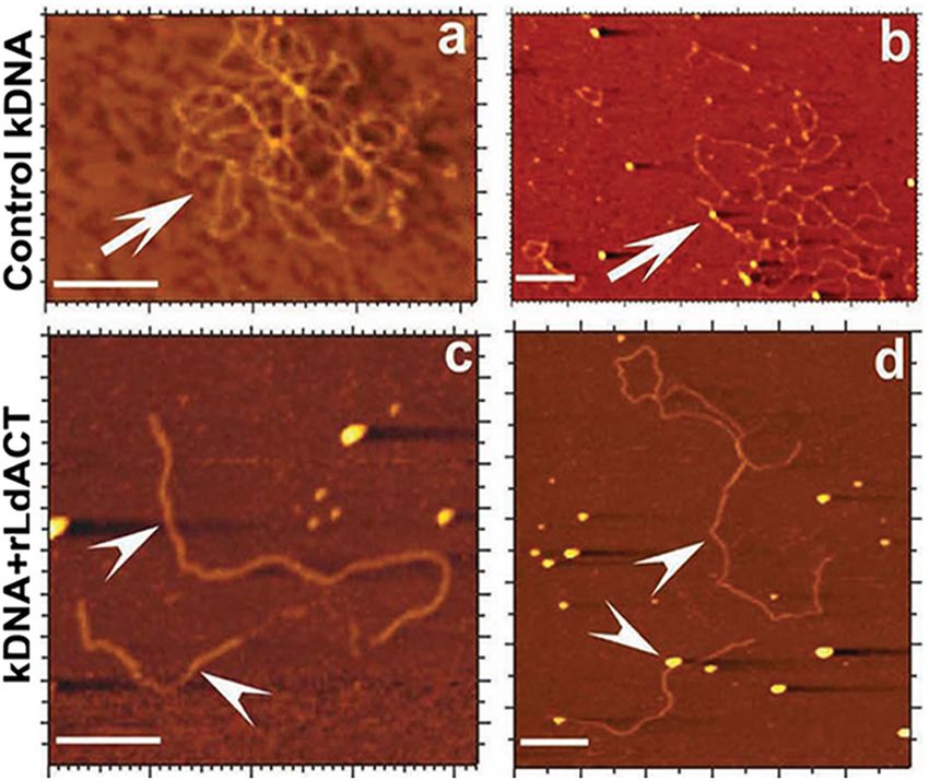
Gupta et al.                                                                                                                  Actin Binding Proteins in Trypanosomatids

domain comprises subdomain 3 and subdomain 4. Between these                            the DNase I binding site. A loop of eleven aa residues (aa 40–50
sub-domains two clefts are formed – “nucleotide binding cleft,”                        of subdomain 2) that also include a four aa residues hydrophobic
that binds to ATP or ADP with a divalent cation and the other                          plug then stabilizes the filament. This loop inserts into the
one is the “target binding cleft,” which is hydrophobic and defines                    hydrophobic pocket formed by subdomains 2 and 3 of adjacent
the region where most actin binding proteins (ABPs) and small                          monomers on the opposing strands (Figure 1). Based on the
molecules interact with actin and also where actin subunits make                       X-ray diffraction pattern of oriented F-actin gels, Holmes et al.
contacts in the filament (Figure 1). Depending on the state of                         (1990) proposed the first structural model of actin filament,
bound nucleotide in the “nucleotide binding cleft,” structure of                       which was further modified by Oda et al. (2009). The modified
actin monomer changes, which in turn modulates the binding                             model illustrated that actin monomers are arranged in a two-start
affinities of “target binding cleft” for ABPs and also alters the                      filament of 7–10 nm thickness having a half pitch of 37 nm and
strength of actin monomer interaction in the filament. Further,                        a rise of 2.75 nm per monomer. A large number of proteins
the DNase I binding site is formed from amino acid residues                            associate with and effect the functions of actin by remodeling
(aa) 39–46 and 60–64 of subdomain 2 and aa 202–204 and 207                             its network in cells (dos Remedios et al., 2003; Pollard et al.,
of subdomain 4 of which aa 40–50 of subdomain 2 are highly                             2016; Merino et al., 2020). These proteins are mostly conserved
disordered, that form the DNase I binding loop.                                        in a wide variety of eukaryotes. Some members belonging to
    Assembly of actin filaments involves association of                                these proteins by virtue of their actin monomer sequestering
subdomains 2 and 4 of one G-actin molecule with subdomains                             activity affect availability of the polymerizable pool of free
1 and 3 of the other molecule. A part of amino acid sequences                          actin monomers, while there are others that control filament
that are contributed by subdomain 2 in this process constitute                         formation and stability through their nucleating, elongating,

  FIGURE 1 | (A) Ribbon diagram of the actin molecule with space filling ATP (protein data bank [PDB]: 1ATN). N, amino terminus; C, carboxyl terminus. Numbers 1,
  2, 3, and 4 label the four subdomains (re-printed from Pollard et al. (2016) with copyright permission from Elsevier Publishers). (B) Model of actin protofilaments
  derived from linear polymers along a single strand of F-actin.

Frontiers in Cell and Developmental Biology | www.frontiersin.org                  3                                          October 2020 | Volume 8 | Article 587685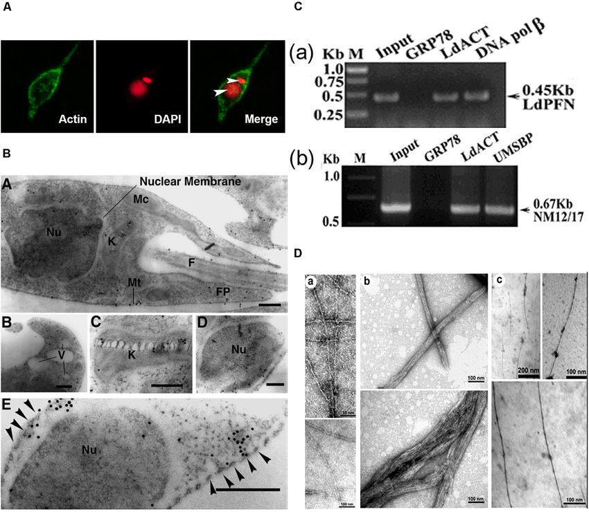
Gupta et al.                                                                                                                   Actin Binding Proteins in Trypanosomatids

depolymerizing, severing, capping, crosslinking and bundling                            functions, such as morphogenesis, intracellular trafficking and
activities (Winder and Ayscough, 2005).                                                 cytokinesis (Paredez et al., 2011, 2014).
    The main features that define conventional (or canonical)
actins are based on their following properties: (1) they form
long and stable filaments having width between 7 and 10 nm                              CLASSIFICATION OF PROTOZOAN
in the presence of a divalent cation as Mg+2 , with or without                          ORGANISMS
ATP; (2) they bind DNase I and inhibit its activity; (3) their
filaments are stabilized by phallotoxins and destabilized by                            Protozoans are single-celled microscopic eukaryotic organisms
cytochalasins or latrunculins (Reisler, 1993; Wakatsuki et al.,                         of a group of phyla of the kingdom Protista. In the widely
2001); and (4) their filament dynamics is regulated by a set of                         used 1980 classification based on locomotion (Levine et al.,
about 20 core ABPs that include actin depolymerizing factor                             1980), the protozoan subkingdom was classified into seven
(ADF)/cofilins, twinfilin, profilin, gelsolin, CAP/Srv2, formin,                        phyla which included the Sarcomastigophora (combination
Arp2/3 complex, β-thymosin, troponin, filamin, fimbrin, villin,                         of Mastigophora and Sarcodina), Apicomplexa, Microspora,
actinin, plastin, spectrin and CapZ. However, lower eukaryotic                          Myxozoa and Ciliophora. The most recent classifications
organisms such as Plasmodium, Toxoplasma, Trypanosoma,                                  recognized 13 phyla, of which seven contain important
Leishmania, Giardia, Amoebae and Ciliate group of protozoans                            parasites (Figure 2): Metamonada (intestinal flagellates, e.g.,
contain actins which display highly unusual characteristics                             Giardia); Parabasalia (intestinal and related flagellates, e.g.,
(Villalobo et al., 2001; Gupta et al., 2015). While some of                             Trichomonas); Percolozoa (flagellated amoebae, e.g., Naegleria);
these organisms express actins and ABPs that exhibit unusual                            Euglenozoa (kinetoplastid flagellates, e.g., Trypanosoma,
biochemical and functional characteristics, there are others, such                      Leishmania); Amoebozoa (amoebae, e.g., Entamoeba); Sporozoa
as Giardia lamblia, which express single copy of highly divergent                       (sporozoans, e.g., Toxoplasma, Plasmodium) and Ciliophora
actin (Drouin et al., 1995), and their genome lacks genes that                          (ciliates, e.g., Tetrahymena) (Levine et al., 1980; Cavalier-
encode the core ABPs, which are essentially required to regulate                        Smith, 2002; Cox, 2002). Further, kinetoplastids comprise
actin dynamics in higher eukaryotic organisms (Morrison et al.,                         five orders: Trypanosomatida, Eubodonida, Parabodonida,
2007; Pollard, 2016). Yet these organisms utilize their actin                           Neobodonida and Prokinetoplastida (Moreira et al., 2004).
similar to other eukaryotic actins in their all vital cellular                          Within kinetoplastids, the most studied family is the

  FIGURE 2 | Seven important phyla of subkingdom protozoa with schematic representations. Metamonada (intestinal flagellates, e.g., Giardia); Parabasalia (intestinal
  and related flagellates, e.g., Trichomonas); Percolozoa (flagellated amoebae, e.g., Naegleria); Euglenozoa (kinetoplastid flagellates, e.g., Trypanosoma, Leishmania);
  Amoebozoa (amoebae, e.g., Entamoeba); Sporozoa (sporozoans, e.g., Toxoplasma, Plasmodium) and Ciliophora (ciliates, e.g., Tetrahymena). Schematic images of
  the protozoan parasites.

Frontiers in Cell and Developmental Biology | www.frontiersin.org                   4                                           October 2020 | Volume 8 | Article 587685
Gupta et al.                                                                                                        Actin Binding Proteins in Trypanosomatids

Trypanosomatidae, which comprises mainly of monoxenous                              subdomain 2 (aa33– 69), subdomain 3 (aa145–180 and aa270–
parasite species that infect invertebrates (Leptomonas) and                         337) and subdomain 4 (aa181–269) have higher divergence
of dixenous species that can be pathogenic to plants,                               (30–40%) from the corresponding subdomains of human actin,
animals and/or humans (Phytomonas, Trypanosoma and                                  compared with subdomain 1 of Leishmania actin (aa1–32, 70–
Leishmania) (Kaufer et al., 2017). Trypanosomatidae got                             144 and aa338–375). These diverged amino acid sequences partly
much of their fame because of the two genera, Trypanosoma                           included the sites that are engaged in actin self-association and
and Leishmania, which cause African sleeping sickness                               DNase I binding.
(Trypanosoma brucei), Chagas disease (Trypanosoma cruzi),                               The presence of actin gene in trypanosomatid organisms
visceral Leishmaniasis (Leishmania donovani, Leishmania                             was established about 30 years ago (Ben Amar et al., 1988;
infantum, Leishmania chagasi), cutaneous Leishmaniasis                              de Arruda and Matsudaira, 1994), but all earlier attempts
(L. major, L. panamensis, L. tropicana) and mucocutaneous                           to isolate and characterize actin protein, using conventional
Leishmaniasis (L. braziliensis). In the following sections, we                      methods, met with failure, primarily due to lack of these proteins
will revisit all the findings reported to date on characterization,                 to bind DNase I (Mortara, 1989). Also, fluorescently labeled
intracellular localization and functions of trypanosomatid                          phalloidin staining failed to identify the presence of filament-
actins and actin binding proteins, and based on the available                       like structures in trypanosomatids, especially Leishmania cells
knowledge, we will attempt to stipulate their functions in                          (Mortara, 1989). Further, by using electron microscopy, no
these organisms.                                                                    filament-like structures corresponding to mammalian actin
                                                                                    filaments (5–7 nm diameter) could be seen in these cells (Gull,
                                                                                    1999), suggesting that actin may not have any major role in
TRYPANOSOMATID ACTINS AND ACTIN                                                     cellular functions of trypanosomatids (Gull, 1999). However,
BINDING PROTEINS                                                                    genomic analysis of three major pathogenic organisms of
                                                                                    trypanosomatid family, viz. T. brucei, T. cruzi and Leishmania
Trypanosomatid Actins                                                               spp., identified genes that putatively encode at least for one
Trypanosomatid actins possessed approximately 70% aa identity                       copy of conventional actin (Act1), similar to other eukaryotic
to human or yeast actin (Gupta et al., 2015). The major                             actins, and numerous actin-like, actin-related and actin binding
differences in the aa sequence were confined to the aa1–9,                          proteins (Tables 1 and 2), revealing the presence of a dynamic
aa 40–53, aa194–200, aa 229–240, aa 266–281 and aa307–                              actin network in trypanosomatids. Compared with T. brucei
315, most of which were located on the surface of yeast or                          and L. major, T. cruzi appears to have more complex actin
mammalian actin (Figure 3). Domain-wise analysis revealed that                      cytoskeleton, as its genome encodes for multiple copies of actin
                                                                                    and an expanded set of actin binding proteins (Berriman et al.,
                                                                                    2005). T. cruzi has as many as four actin genes of which TcAct1
                                                                                    and TcAct2 have been characterized (De Melo et al., 2008;
                                                                                    Vizcaíno-Castillo et al., 2019). However, actin 2 and 3 are absent
                                                                                    in T. brucei, L. donovani, L. major and L. braziliensis. The fourth
                                                                                    actin is encoded by T. cruzi and L. major, but not by T. brucei
                                                                                    (Cevallos et al., 2011; Vizcaíno-Castillo et al., 2019). This actin
                                                                                    isoform is also present in L. donovani and L. braziliensis, but its
                                                                                    annotation has been given as actin-like protein. Besides actins, a
                                                                                    variable number of actin-like and actin related proteins are also
                                                                                    encoded by the trypanosamatid genomes (recently reviewed in
                                                                                    Vizcaíno-Castillo et al., 2020).
                                                                                        The presence and intracellular distribution of Act1 in all the
                                                                                    trypanosomatids studied so far have been analyzed by employing
                                                                                    polyclonal antibodies against recombinant version of Act1, as
                                                                                    a probe. This technique enabled to identify differences in the
                                                                                    subcellular distribution of Act1 not only in different species, but
                                                                                    also in the different developmental stages. TbAct1 was equally
                                                                                    expressed in both the blood stream and procyclic stages of
                                                                                    T. brucei, but in blood stream stage, it was more enriched at
  FIGURE 3 | An average molecular dynamics simulated homology model of              the posterior end and colocalized with the endocytic pathway
  LdAct showing colored stretches of diverged amino acid residues (aa 1–9 of
  subdomain 1, aa 40–53 of subdomain 2, aa 266–281 and aa 307–315 of
                                                                                    (García-Salcedo et al., 2004). However, in procyclic form, it
  subdomain 3, aa 194–200 and 229–240 of subdomain 4), brown ball and               was distributed throughout the cytoplasm (García-Salcedo et al.,
  stick residues in the DNase-I binding loop are the diverged replacements in       2004). Unlike T. brucei, there have been numerous contradictory
  LdAct that are known to make strong interactions with DNase-I in the              reports on the intracellular distribution in the various forms
  actin-DNase-I complex crystal structure, whereas green ball and stick
                                                                                    of T. cruzi. Presence of TcAct1 in T. cruzi epimastigotes was
  residues are conserved amino acid residues that are known to make weak
  interactions with DNase-I (taken from Kapoor et al., 2008 with permission).
                                                                                    first reported by de Souza et al. (1983), using polyclonal anti-
                                                                                    rabbit muscle actin antisera, wherein they claimed that this

Frontiers in Cell and Developmental Biology | www.frontiersin.org               5                                    October 2020 | Volume 8 | Article 587685
Gupta et al.                                                                                                               Actin Binding Proteins in Trypanosomatids

TABLE 1 | Presence of actin and actin-like proteins in five main disease causing trypanosomatids.

                          Trypanosoma cruzi                Trypanosoma brucei              Leishmania donovani           Leishmania major Leishmania
                                                                                                                                          braziliensis

                          Chagas disease                   Sleeping sickness               Visceral Leishmaniasis        Cutaneous              Mucocutaneous
                                                                                                                         Leishmaniasis          Leishmaniasis

Actin                     TcAct1*                          ActinA*                         LdAct (LDBPK_041250)          LmjF.04.1230           LbrM.04.1250
                          (TcCLB.510571.30)                (Tb927.9.8850)
                          (TcCLB.510127.79)                ActinB (Tb927.9.8880)
Actin 2                   TcAct2 (TcCLB.507129.10)         Absent                          Absent                        Absent                 Absent
Actin, putative (actin 3rd) TcCLB.510945.30                Absent                          Absent                        Absent                 Absent
Actin, putative (actin 4th) TcCLB.503841.40                Absent                          LdBPK_350810.1 (actin-like    LmjF.35.0790           LbrM.34.0780
                                                                                           protein)                      (actin-like protein)   (actin-like protein)
Actin -like protein 1     TcCLB.508277.330                 Tb927.9.5440                    LdBPK_151350.1                LmjF.15.1330           LbrM.15.1280
Actin -like protein 2     TcCLB.506405.30                  Tb927.4.980                     LdBPK_343560.1                LmjF.34.3760           LbrM.20.3360
Actin -like protein 3     TcCLB.506733.50                  Tb927.11.3880                   LdBPK_130840.1                LmjF.13.0950#          LbrM.13.0760
Actin -like protein 4     TcCLB.510719.110                 Tb927.11.10110                  LdBPK_363470.1                LmjF.36.3310           LbrM.35.3540
Actin -like protein 5     TcCLB.506695.10                  Tb927.3.3020                    LdBPK_292850.1                LmjF.29.2740           LbrM.29.2800
Member of ARP6 family     TcCLB.508951.29                  Tb927.10.2000                   LdBPK_210290.1                LmjF.21.0230           LbrM.21.0300
(Actin like protein)

The data was downloaded from the TriTrypDB version 48 database (www.tritrypdb.org). IDs of strains used: T. cruzi CL Brener Esmeraldo-like; T. brucei brucei TREU927;
L. donovani BPK282A1; L. major strain Friedlin; L. braziliensis MHOM/BR/75/M2904.
*This actin locus contains two copies of the gene on one allele and one copy on the second allele (Ben Amar et al., 1988; Cevallos et al., 2003).
# This has been referred as actin-like protein 3 in Cevallos et al. (2011) and as ARP1 in Singh et al. (2014).

protein was sparsely distributed throughout the cell body and                          (Vizcaíno-Castillo et al., 2019). Because of differences in cellular
the paraxial structure of the flagellum. Subsequent studies using                      localization of TcAct1 and TcAct2, it may be envisaged that these
polyclonal antibodies against the conserved N-terminal region                          proteins possibly have non-redundant functions in T. cruzi cells.
of TcAct1 showed that TcAct1 was distributed in patch-like                                L. donovani Act1 (LdAct1) is the most characterized protein
structures throughout the cytoplasm in epimastigote, amastigote                        amongst all trypanosomatid actins (Sahasrabuddhe et al., 2004;
and bloodstream trypomastigote forms of T. cruzi (De Melo                              Kapoor et al., 2008, 2010). This protein was abundantly
et al., 2008). This distribution was further confirmed using                           expressed in both the promastigote and amastigote stages of
polyclonal anti-recombinant TcAct1 antibodies (Cevallos et al.,                        L. donovani (Sahasrabuddhe et al., 2004). LdAct1 in Leishmania
2011). Further analysis revealed that these antibodies recognized                      promastigotes was present as granules, patches, and filament-
a single band in one-dimensional electrophoresis, but in two-                          like structures throughout the cell body, including the flagellum,
dimensional electrophoresis, it identified five isoforms of TcAct1                     the nucleus and the kinetoplast (Sahasrabuddhe et al., 2004;
in all stages of the parasite development (Cevallos et al.,                            Kapoor et al., 2008). These LdAct1 structures could not be
2011). Immunofluorescence analysis, using anti-recombinant                             stained with fluorescently labeled phalloidin nor could they
TcAct1 antibodies, showed that TcAct1 was faintly distributed                          be disrupted by treatment with cytochalasin D (Sahasrabuddhe
throughout the cell with intense staining at the base of the                           et al., 2004). In the nucleus and the kinetoplast, LdAct1 was
flagellum near the flagellar pocket area and along the flagellum in                    found to associate, respectively, with the chromatin and kDNA
epimastigotes (Cevallos et al., 2011). In trypomastigotes, TcAct1                      (Figures 4A–C). Besides this, recombinant LdAct1 (rLdAct1)
was uniformly distributed with a low level of staining (Cevallos                       polymerized in vitro to form bundles instead of thin filaments,
et al., 2011), and in the cell-derived amastigotes, a heterogeneous                    only between pH 7.0 to pH 8.0 (Figure 4D), and its critical
TcAct1 localization with sometimes no apparent expression                              concentration of polymerization was 3–4 times lower than of
was observed (Cevallos et al., 2011). Similar protein expression                       rabbit muscle actin (Kapoor et al., 2008). In addition, it did
levels and intracellular TcAct1 distributions in epimastigotes,                        not bind DNase I or phalloidin and during polymerization, it
amastigotes and metacyclic trypomastigotes were also observed                          displayed significantly higher ATPase activity, compared with
by using mouse polyclonal anti-recombinant TcAct1 antibodies                           muscle actin (Kapoor et al., 2008). This apart, unlike any other
(Souza et al., 2013). In addition to TcAct1, TcAct2 has also been                      eukaryotic actin, rLdAct bound to DNA primarily through
characterized. This protein was expressed throughout the life                          electrostatic interactions involving its unique DNase-1-binding
cycle of T. cruzi with several variants (Vizcaíno-Castillo et al.,                     region and the DNA major groove (Figure 5) and relaxed
2019). In all stages, TcAct2 did not co-localize withTcAct1, and                       negatively supercoiled DNA and nicked the kDNA, which
had a diffused distribution throughout the cell body and in the                        converted kDNA minicircles into their open form (Figure 6),
flagellum, with a fine granular pattern (Vizcaíno-Castillo et al.,                     a unique property which no other eukaryotic actin has been
2019). Further, detergent fractionation of epimastigotes revealed                      found to have till date (Kapoor et al., 2010). The DNA nicking
that TcAct2 was a cytoplasmic rather than a cytoskeletal protein                       activity was largely confined to the DNase-1 binding loop,

Frontiers in Cell and Developmental Biology | www.frontiersin.org                  6                                        October 2020 | Volume 8 | Article 587685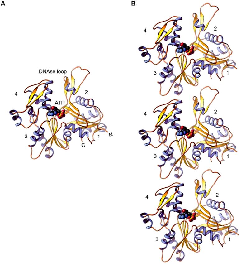
Gupta et al.                                                                                                                 Actin Binding Proteins in Trypanosomatids

TABLE 2 | Presence of actin binding proteins in five main diseases causing trypanosomatids, compared to higher eukaryotes.

                       Higher eukaryotes             Trypanosoma cruzi         Trypanosoma brucei        Leishmania donovani       Leishmania         Leishmania
                                                                                                                                   major              braziliensis

Disease caused         NA                            Chagas disease            Sleeping sickness         Visceral Leishmaniasis    Cutaneous          Mucocutaneous
                                                                                                                                   Leishmaniasis      Leishmaniasis
Actin monomer          Profilin                      TcCLB.510911.10           Tb927.11.13780            LdBPK_320550.1            LmjF32.0520        LbrM.32.0570
binding
                       Thymosin B4                   Absent                    Absent                    Absent                    Absent             Absent
                       ADF/Cofilin                   TcCLB.510145.20           Tb927.3.5180              LdBPK_290520.1            LmjF29.0510        LbrM.29.0450
                       Gelsolin                      Absent                    Absent                    Absent                    Absent             Absent
                       Twinfilin                     TcCLB.506559.300          Tb927.4.2350              LdBPK_342060.1            LmjF.34.2290       LbrM.20.1790
                       CAP/Srv2                      TcCLB.504137.80           Tb927.10.9250             LdBPK_365830.1            LmjF36.5590        LbrM.35.5860
Filament binding       Myosin                        TcCLB.511527.70 (myosin   Tb927.11.16310            LdBPK_324020.1            LmjF.32.3870       LbrM.32.4110
                                                     13)                       (Unconventional myosin)   (myosin XXI)              (myosin XXI)       (myosin XXI)
                                                     TcCLB.507739.110 (1B      Tb927.4.3380              LdBPK_341070.1            LmjF.34.1000       LbrM.20.0970
                                                     heavy chain)              (1B heavy chain)          (1B heavy chain)          (1B heavy chain)   (1B heavy chain)
                                                     TcCLB.504867.120          Tb927.9.1340
                                                     MyoA                      (myosin like protein 2)
                                                     TcCLB.506779.190          Tb927.11.330
                                                     MyoB                      (myosin like protein 1)
                                                     TcCLB.504103.30
                                                     MyoC
                                                     TcCLB.503905.10
                                                     MyoD
                                                     TcCLB.503905.10
                                                     MyoE
                                                     TcCLB.507445.50
                                                     MyoF
                                                     TcCLB.507093.210
                                                     MyoG
                       Coronin                       TcCLB.510515.100          Tb927.8.3100              LdBPK_231400.1            LmjF.23.1165       LbrM.23.1260
                       CAPz                          TcCLB.506181.90           Absent                    Absent                    Absent             Absent
                                                     TcCLB.506363.60
Nucleating             Arp2/3 complex
                       (7 subunits)                  TcCLB.511361.40           Tb927.10.15800            LdBPK_191190.1            LmjF19.1200        LbrM.19.1370
                       Arp2
                       Arp3                          TcCLB.508277.260          Tb927.9.5350              LdBPK_151410.1            LmjF.15.1360       LbrM.15.1360
                       ARPC1                         TcCLB.504215.40           Tb927.10.13190            LdBPK_180920.1            LmjF.18.0920       LbrM.18.0980
                       ARPC2                         TcCLB.506865.10           Tb927.8.4410              Absent                    Absent             Absent
                       ARPC3                         TcCLB.510963.70           Tb927.10.4540             Absent                    Absent             Absent
                       ARPC4                         TcCLB.509127.104          Tb927.2.2900              LdBPK_020570.1            LmjF.02.0600       LbrM.02.0580
                       ARPC5                         TcCLB.442297.10           Tb927.10.10600            LdBPK_050290.1            LmjF.05.0285       LbrM.05.0280
                       ARPC-like                     Absent                    Absent                    LdBPK_101080.1            LmjF.10.1000       LbrM.10.1100
                       Formin                        TcCLB.511313.30           Tb927.5.2300              LdBPK_171040.1            LmjF24.1110        LbrM.17.0950
                                                     TcCLB.506203.80           Tb927.11.5740             LdBPK_241130.1            LmjF17.0930        LbrM.24.1120
                                                     TcCLB.511393.30
Crosslinking proteins Fimbrin, villin, α- actinin,   Absent                    Absent                    Absent                    Absent             Absent
                      plastin, spectrin, filamin

The data was downloaded from the TriTrypDB version 48 database (www.tritrypdb.org). IDs of strains used: T. cruzi CL Brener Esmeraldo-like; T. brucei brucei TREU927;
L. donovani BPK282A1; L. major strain Friedlin; L. braziliensis MHOM/BR/75/M2904.

as treatment of LdAct1 with subtilisin, which was known to                                 Trypanosomatid actins, similar to conventional actins,
selectively cleave the DNase I binding loop without altering much                       participate in the process of endocytosis. This process in T. brucei
the Act1 structure (Schwyter et al., 1989), significantly reduced                       and Leishmania primarily occurs through the flagellar pocket
its DNA nicking activity (Kapoor et al., 2010). Further, rLdAct1                        (Morgan et al., 2002a), which is a well-defined structure formed
inhibited the kDNA decatenation activity of bacterial type II                           from a lateral cell membrane depression that is continuous
topoisomerase (Kapoor et al., 2010), suggesting that LdAct1                             with the flagellar membrane. However, in T. cruzi, it mainly
may play an important role in remodeling of the chromatin                               takes place through an additional entry site, called “cytostome”
and kDNA in trypanosomatids (Liu et al., 2005; Kapoor et al.,                           that represents a round opening at the plasma membrane near
2010).                                                                                  the flagellar pocket, which is absent in both T. brucei and

Frontiers in Cell and Developmental Biology | www.frontiersin.org                   7                                         October 2020 | Volume 8 | Article 587685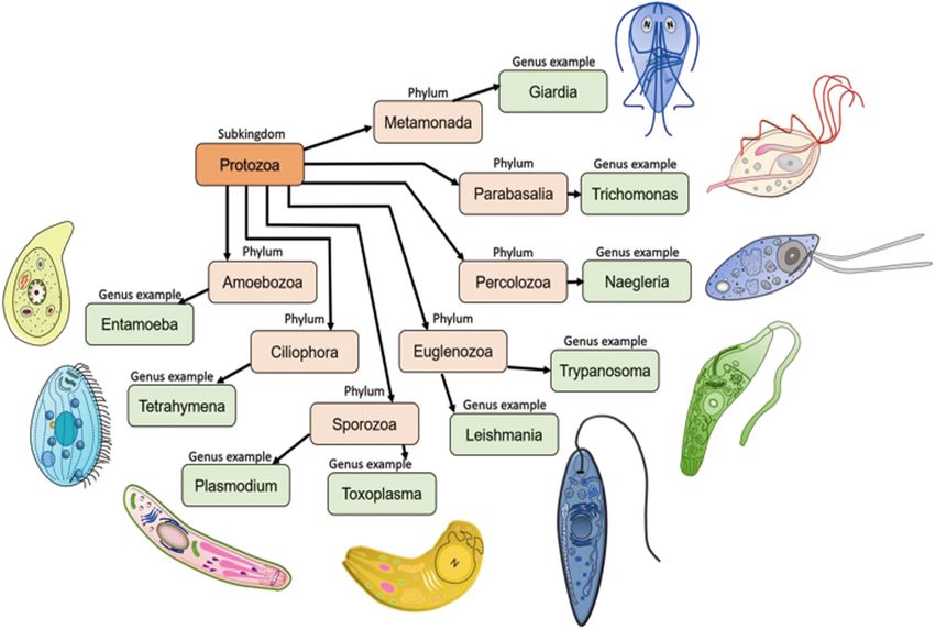
Gupta et al.                                                                                                                   Actin Binding Proteins in Trypanosomatids

  FIGURE 4 | (A) Immunofluorescence micrograph of Leishmania promastigotes after treating them with 0. 5% NP-40 and staining with anti-LdAct antibodies and
  DAPI showing the presence of LdAct in the nucleus and kinetoplast and its association with nuclear DNA and kDNA (adapted from Sahasrabuddhe et al., 2004 with
  permission). (B) Electron micrographs of immunogold-labeled actin showing the presence (panel a) of LdAct in the nucleus (Nu), the kinetoplast (K), the flagellum (F),
  and the flagellar pocket (FP). In addition, the presence of LdAct on membranes of vacuoles (V) may also be noticed in panel (b), and its associations with kDNA
  network, nuclear membrane and subpellicular microtubules may clearly be seen in panels (c–e), respectively. The arrowheads in panel (e) mark the microtubules.
  Bar, 200 nm (Adapted from Sahasrabuddhe et al., 2004 with permission). (C) Chromatin Immuno-precipitation (ChIP) analysis using anti-LdAct antibodies showing
  the in vivo association of LdAct with chromatin (a) and kDNA network (b). Panels (a,b) are the agarose gels of PCR products after ChIP assay. Lanes are marked on
  the top with their respective antibodies used in the ChIP assay and arrows indicated the genes amplified after pull down. An irrelevant, non-DNA associating
  antibody, GRP78, was used as a negative control, whereas antibodies against DNA polβ, and UMSBP (universal minicircle sequence-binding protein), were used as
  positive controls for nuclear DNA and kDNA respectively. LdPfn, Leishmania profilin; NM12/17, specific minicircle primers (this was originally published in Nucleic
  Acids Research, Kapoor et al., 2010© Oxford University Press). (D,a) Negatively stained transmission electron micrograph of in vitro reconstituted rabbit muscle actin
  (RbAct) filaments in F-buffer (100 mM KCl, 2 mM MgCl2 and 2 mMATP; pH 8.0; 25◦ C) and (b) LdAct at 2 µM protein concentration, unlike RbAct, formed bundles
  rather thin filaments, under identical conditions. (c) LdAct forms very thin filaments at 0.2 µM G-LdAct concentration in F-buffer, pH7.0 at 25◦ C. RbAct under these
  conditions failed to form filaments (taken from Kapoor et al., 2008 with permission).

Leishmania (Soares and de Souza, 1991; Porto-Carreiro et al.,                           possessed high endocytic activity (Bogitsh et al., 1995; Corrêa
2000). The endocytic activity in all these organisms depended                           et al., 2008), whereas in trypomastigotes, a stage that lacks
on the stage of their life cycle. While T. brucei bloodstream                           cytostome structure, this activity was low (Soares and de Souza,
form displayed high rates of endocytic activity, this activity                          1991; Figueiredo et al., 2004). That TcAct1 is involved in this
was absent or significantly reduced in procyclic form (Morgan                           process has been demonstrated in T. cruzi epimastigotes by
et al., 2002a,b). Involvement of TbAct1 during this process has                         observing inhibition of endocytosis of peroxidase, LDL and gold
been shown by down regulating TbAct1 expression in blood                                particles after disrupting actin cytoskeleton by treatment with
stream stage of T. brucei, using RNAi, and then observing                               cytochalasin B and latrunculin B (Soares and de Souza, 1991;
significantly reduced receptor-mediated uptake of transferrin                           Bogitsh et al., 1995; Corrêa et al., 2008). Furthermore, endocytosis
(García-Salcedo et al., 2004). Further, T. cruzi epimastigotes                          was downregulated in L. mexicana promastigotes, as compared

Frontiers in Cell and Developmental Biology | www.frontiersin.org                   8                                           October 2020 | Volume 8 | Article 587685
Gupta et al.                                                                                                               Actin Binding Proteins in Trypanosomatids

  FIGURE 5 | Computational docking of average simulated model of LdAct with DNA showing the interaction of the diverged DB-loop of LdAct with the major groove
  of DNA. (A) Sequence alignment of LdAct with other actins showing the presence of nuclear export signals (NES-1 and NES-2) in the LdAct aa sequence and the
  diverged DB-loop predicted to be involved in the DNA binding, by DP-Bind server. (B) Energy minimized average simulated model of LdAct showing positions of
  NES-1, NES-2 (red) and the diverged stretches of amino acid sequences (yellow) including the sequence that fall in DB loop (blue). (C) Docking of LdAct (orange)
  with DNA (green) using HADDOCK protocols. (D) Amino acid residues of the DB loop of LdAct (yellow) showing hydrogen bonding with the nucleotides (green) of
  DNA. DB, DNase I binding; NES, nuclear export signal (This was originally published in Nucleic Acids Research, Kapoor et al., 2010© Oxford University Press).

to both metacyclic promastigotes and amastigotes (Ali et al.,                        distribution has also been observed earlier by overexpressing
2012). Role of actin during endocytosis in Leishmania has been                       fluorescently tagged version of ALP3 in T. brucei (Ersfeld and
established by observing significantly reduced uptake of the                         Gull, 2001). However, in L. donovani, ALP3 (earlier classified
fluorescent dye FM4-64 after inhibiting the LdCof-driven LdAct1                      as Arp1) was predominantly localized to the mitochondrion,
dynamics in L. donovani promastigotes (Tammana et al., 2010).                        besides localizing to the cytoplasm and the flagellum (Singh
   Together with actins, trypanosomatids encode for at least five                    et al., 2014). And depletion of its intracellular levels resulted
actin-like proteins (ALPs). Out of which, three proteins, viz.                       in decreased mitochondrion membrane potential and the ATP
ALP1, ALP3 and ALP4, have been characterized in T. brucei                            content, and also in shortening of the flagella length. These
and Leishmania. These proteins were first identified as a part of                    effects were, however, reversed by episomal complementation
the flagellar proteome of T. brucei and L. mexicana (Broadhead                       of LdALP3 gene (Singh et al., 2014), suggesting that ALP3
et al., 2006; Beneke et al., 2019) and thereafter, their localization                regulates mitochondrion potential, ATP synthesis and flagellum
to the flagellum was confirmed by fluorescent tagging during                         length in Leishmania promastigotes. The difference observed
the genome wide search to assign their location within the                           between intracellular distributions of TbALP3 and LdALP3 may
T. brucei cells (Dean et al., 2017; Halliday et al., 2019). Similar                  perhaps be attributed to the larger size of LdALP3 (483 amino

Frontiers in Cell and Developmental Biology | www.frontiersin.org                9                                          October 2020 | Volume 8 | Article 587685
Gupta et al.                                                                                                            Actin Binding Proteins in Trypanosomatids

  FIGURE 6 | Atomic force micrographs of kDNA after its incubation in the presence and absence (control) of LdAct, showing decatenation of kDNA with LdAct.
  Panels (a,b) control kDNA, arrows indicate catenated kDNA. Panels (c,d) kDNA with rLdAct, arrowheads indicate decatenated nicked kDNA (scale bar: 500 nm)
  (This figure was originally published in Nucleic Acids Research, Kapoor et al., 2010© Oxford University Press).

acids), compared with TbALP3 (433 amino acids), which is                            trypanosomatids. Out of the limited set of ten core ABPs (profilin,
perhaps caused by insertions that confer distinctive properties                     twinfilin, ADF/cofilin, CAP/srv2, CAPz, coronin, two myosins,
to this protein.                                                                    two formins), only a few ABPs have so far been characterized.

Actin Binding Proteins (ABPs) in                                                    Actin Filament Nucleating Proteins
Trypanosomatids                                                                     Actin polymerization in itself is an energetically unfavorable
A large number of proteins (>150) bind actin to regulate                            process till three actin monomers associate together to form
its functions in higher eukaryotic cells. However, because of                       a stable nucleus for further polymerization, and this stage is
their limited functions, lower eukaryotic organisms, such as                        referred to as the “lag phase.” The lag phase is, however, removed
trypanosomatids, express only a small repertoire of ABPs that are                   in vivo due to participation of ABPs, viz. the Arp2/3 complex
sufficient to meet their requirement. Analysis of genomic data of                   and formins, which ensure rapid nucleation of actin monomers
trypanosomatids revealed that T. brucei, T. cruzi and Leishmania                    and thus significantly accelerate actin polymerization. On one
spp., encode at least one copy each of profilin, ADF/cofilin,                       hand, the Arp2/3 complex promotes the growth of new filaments
twinfilin, CAP/Srv2 and coronin, whereas variable number of                         by the side of the existing filaments, which is important in
formins and myosins are encoded in these organisms (Table 2).                       dendritic branching found at the leading edge of a lamellipodium
Further, T. brucei and Leishmania spp. encode two copies each                       of motile cells (Pollard and Borisy, 2003). On other hand,
of formins and myosins, while T. cruzi encodes many myosins                         formin proteins promote actin assembly by directing rapid
and three formins. Besides this, T. cruzi encodes for two copies                    nucleation and elongation of unbranched actin filaments. Besides
of CAPz, which is absent in both T. brucei and Leishmania spp.                      this, these proteins also assist formation of a variety of actin-
This apart, all the seven subunits of the Arp2/3 complex (viz.                      structures, including stress fibers, filopodia, and lamellipodia, and
Arp2, Arp3, ARPC1, ARPC2, ARPC3, ARPC4 and ARPC5) are                               modulate the stability and organization of microtubules (Pollard,
encoded by T. cruzi and T. brucei, but only four to five subunits                   2016). This dual activity of formins helps them to coordinate
of this complex appeared to be encoded in Leishmania spp..                          the activities of these two cytoskeleton networks, which allows
Other proteins such as thymosin β4, gelsolin, fimbrin, villin,                      them to regulate various cellular processes, such as assembly of
α-actinin, plastin, spectrin and filamin are completely absent in                   contractile ring, centrosome assembly, centriole duplication, and

Frontiers in Cell and Developmental Biology | www.frontiersin.org              10                                       October 2020 | Volume 8 | Article 587685
Gupta et al. Actin Binding Proteins in Trypanosomatids
centrosome positioning (Breitsprecher and Goode, 2013). The ADF/cofilin (LdCof) bound to both monomeric and filamentous
majority of trypanosomatids encode for two formins, but to date, LdAct1 and displayed filament-depolymerizing and severing
none of these proteins has been characterized. activities (Tammana et al., 2008; Kumar et al., 2012), whereas
T. brucei ADF/cofilin (TbCof) bound to only monomeric actin,
Actin Filament Elongating Proteins but similar to LdCof, it possessed filament-depolymerizing and
After nucleation, actin filaments can grow rapidly upon addition severing activities. Further, both the proteins were co-distributed
of actin monomers to their barbed ends. Filament length with actin throughout the cell body, including the flagellum
is controlled by capping proteins. While gelsolin and tensin (Tammana et al., 2008; Dai et al., 2013). In addition, both the
cap the barbed ends of growing actin filaments by blocking proteins had similar structures which consisted of a conserved
addition of new monomers at this end, the pointed end ADF/cofilin fold with a central mixed β-sheet formed of six
cappers reduce loss of actin monomers from the pointed β-strands, which was surrounded by five α-helices (Pathak et al.,
end and thereby promote rapid extension of the filament. 2010; Dai et al., 2013). These proteins possessed conserved G/F-
Besides serving as barbed end capper, gelsolin also displays actin binding site that included the characteristic long kinked
filaments severing activity, which accelerates actin dynamics. α-helix (α3).
The best characterized proteins that drive depolymerization are ADF/cofilin-driven actin dynamics regulates a number of
the actin depolymerizing factor (ADF) and the cofilin family important cellular activities, such as motility, endocytosis,
members (ADF/cofilin). ADF/cofilin family of proteins are vesicular trafficking, cell division etc. (Pollard and Borisy,
ubiquitous highly conserved, low molecular-weight ABPs that 2003). Similar to other eukayotic ADF/cofilins, trypanosomatid
depolymerize F-actin into actin monomers and consequently ADF/cofilin, especially LdCof, regulates the cell morphology,
increase the turnover of actin filaments (dos Remedios et al., motility, endocytosis, vesicular trafficking and early phase of
2003; Ono, 2007). In addition, these proteins exhibit actin cell division in Leishmania promastigotes, as revealed by the
filament-severing activity that generates new barbed ends, which reverse genetic experiments (Tammana et al., 2008, 2010). The
accelerates the filament assembly (Ono, 2007). By virtue of heterozygous and homozygous LdCof mutants prepared through
their ability to increase the rate of actin turnover at the steady targeted LdCof gene replacement by the selective marker gene,
state, ADF/cofilin family of proteins have been implicated lost not only their motility, but their flagella were completely
in the treadmilling process (Figure 7; Ono, 2007). Although devoid of the paraflagellar rod (PFR) and length of their flagellum
gelsolin and tensin are completely absent in trypanosomatids, was significantly shortened (Tammana et al., 2008). Additionally,
one copy of ADF/cofilin is encoded by all these organisms. these cells were short and stumpy that contained vesicle-like
Amongst these, T. brucei and L. donovani ADF/ cofilins have structures throughout the length of their flagellum (Figure 8).
been structurally and functionally characterized. Leishmania However, all these changes were restored to normal by episomal
FIGURE 7 | Picture cartoon of actin treadmilling, showing that the rate of treadmilling is regulated by ADF/cofilins and profilin, which results in an increase and
decrease in the size of actin filaments, respectively. It further shows that Arp2/3 complex nucleate new filaments by its binding with actin monomers and the side of
actin filaments, while formins nucleate new filaments by binding actin monomers and through cooperation of profilin.
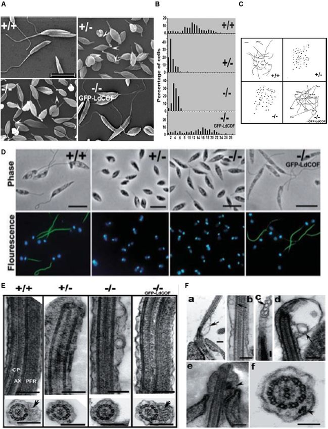
Frontiers in Cell and Developmental Biology | www.frontiersin.org 11 October 2020 | Volume 8 | Article 587685Gupta et al. Actin Binding Proteins in Trypanosomatids FIGURE 8 | (A) Scanning electron micrographs, showing short and stumpy cell body with significantly shortened flagella of heterozygous (+/-) and homozygous (-/-) LdCof mutants, compared with wild type (+/+) cells. Episomal complementation of LdCof- / - cells with LdCof gene (-/- comp) restored the wild-type morphology and flagellar length. Bar, 10 µm. Arrowheads indicate the “blob-like” structures seen at the tip of the flagella of mutant cells (taken from Tammana et al., 2008 with permission). (B) Histogram, showing flagellar lengths of LdCof+/+ , LdCof+/ - , LdCof- / - and LdCof−/−comp cells (taken from Tammana et al., 2008 with permission). (C) Motility analysis of LdCof mutants by time lapse microscopy. Traces of paths of live, individual cells in the movies indicate that LdCof+/ - and LdCof- / - cells are completely immotile. However, upon episomal complementation of LdCof- / - cells the motility is restored back to normal. Origin of the path is indicated by solid dots. Bar, 50 µm (taken from Tammana et al., 2008 with permission). (D) Immuno -flourescence micrographs, showing loss of paraflagellar rod proteins, PFR1 and PFR2, after staining the LdCof +/ - and LdCof - / - mutants with mAb2E10 antibodies and their restoration after complimenting LdCof gene in the null mutants. Bar, 5 µm (taken form Tammana et al., 2008 with permission). (E) Transmission electron micrographs of thin sections of flagellum from chemically fixed whole cells showing the absence of PFR in LdCof +/ - and LdCof - / - cells and its restoration upon episomal complementation. Longitudinal sections of the flagellum showing the axoneme (AX) with the central pair microtubules (CP) and PFR confined between the axoneme and the flagellar membrane in wild type cells and GFP–LdCof complemented mutants. Bar, 200 nm. Cross sections of the flagellum, showing the PFR in LdCof +/+ (marked by arrow) and GFP–LdCof-complemented mutant cells, and complete absence of this structure in the cross sections of LdCof+/ - and LdCof- / - cells. Bar, 200 nm (taken from Tammana et al., 2008 with permission). (F) (a–d) Longitudinal sections of chemically fixed whole cells of LdCof- / - mutants, showing accumulation of membrane-bound vesicles at the base (a), along the length (b,c) and tip (d) of the flagellum. Arrows indicate the membrane-bound vesicles. Longitudinal section (e) and cross section (f) of chemically fixed whole cells of LdCof- / - mutant, showing IFT-like particles along the length of the flagellum. Arrowheads indicate IFT-like particles. Bar, for panels (a–e) 200 nm and for panel (f) 100 nm. IFT, intraflagellar transport (taken from Tammana et al., 2008 with permission). Frontiers in Cell and Developmental Biology | www.frontiersin.org 12 October 2020 | Volume 8 | Article 587685
Gupta et al.                                                                                               Actin Binding Proteins in Trypanosomatids

complementation of the LdCof gene (Tammana et al., 2008,                  promoted actin polymerization, whereas at high concentrations,
2010). Further studies are, however, required to evaluate the             it strongly inhibited the polymerization process by sequestering
functions of ADF/cofilin-driven actin dynamics in Leishmania              actin monomers. This was in accordance with the earlier
amastigotes and in other trypanosomatids.                                 studies which have shown that in protozoan organisms, such
                                                                          as Acanthamoeba, Chlamydomonas and Toxoplasma, profilins
Actin Monomer Binding Proteins                                            mainly function as actin sequestering proteins (Reichstein and
In motile cells, a rapid growth and reorganization of actin               Korn, 1979; Tseng and Pollard, 1982; Kovar et al., 2001;
filaments, in response to both intracellular and extracellular            Skillman et al., 2012). Besides actin, LdPfn also bound to PLP
stimuli, is required, which is dependent on the availability of           motifs and polyphosphoinositides in vitro. However, among
polymerizable pool of actin monomers. Although there are a                phosphoinositides, it bound more efficiently to PI (3,5) P2,
large number of actin monomer binding proteins, only six major            which is found on early or late endosomes and lysosomes
classes of proteins are found in most eukaryotic organisms                (Wallroth and Haucke, 2018), as compared to PI (4,5) P2
(Winder and Ayscough, 2005). The monomer-binding proteins,                and PI (3,4,5) P3 (Ambaru et al., 2020). Further, LdPfn
on one hand, are involved in binding ADP-actin soon after its             heterozygous mutants, prepared through targeted replacement
release from filament ends (e.g., twinfilin, ADF/cofilin), while          of LdPfn gene by selective marker gene, grew at much slower
on the other, they facilitate the exchange of ADP for ATP (e.g.,          pace, compared to wild type cells, in culture, and displayed slower
profilin and CAP) and then deliver ATP-bound actin monomer                intracellular trafficking activity (Ambaru et al., 2020). These
to the barbed ends to facilitate new rounds of polymerization             defects were, however, reversed upon episomal complementation
(e.g., twinfilin, Srv2/CAP, profilin, verprolin/WIP and WASP).            of LdPfn gene, indicating that profilin plays an important role in
All trypanosomatids examined to date encode for only four                 intracellular trafficking. Furthermore, the slower growth of the
actin monomer binding proteins, viz., profilin, ADF/cofilin,              heterozygous mutants could perhaps be due to aberrations in
twinfilin and CAP/Srv2, that are involved in actin turnover.              the cell division cycle of these cells, which needs to be further
Amongst these, LdCof, profilin (LdPfn) and twinfilin (LdTwf)              explored. Unlike LdPfn, TbPfn and TcPfn have been partially
in L donovani, TbCof and profilin (TbPfn) in T. brucei and only           characterized. Expression of TcPfn in different developmental
profilin (TcPfn) in T. cruzi have so far been characterized.              stages of T. cruzi (Osorio-Méndez et al., 2016) has been
    Profilins are low molecular weight actin monomer binding              determined, and the protein ligands that might interact with
proteins (Theriot and Mitchison, 1993) that regulate actin                this protein in T. cruzi epimastigotes were analyzed by mass
dynamics in eukaryotic cells. These proteins are involved in              spectrometry. TcPfn was expressed in all the developmental
a variety of actin-driven cellular processes, such as motility,           stages of the parasite and possibly interacted with a large number
vesicular trafficking, chromatin remodeling, nuclear actin export,        of potential ligands, including actin, microtubule components
membrane signaling, etc. (Wilkes and Otto, 2003). Profilins,              and elongation factor 1α (Osorio-Méndez et al., 2016). However,
on one hand, display actin monomer sequestering activity,                 role of these interactions of TcPfn in cellular functions needs
while on the other, they catalyze nucleotide exchange on actin            to be determined. Further, profilin expression in T. brucei has
monomers and also recycle ATP-bound actin monomers to                     been demonstrated only at the mRNA level, and the gene
the barbed end (+ end), thereby significantly promote the                 encoding for this protein has been shown to complement a yeast
polymerization process (Witke, 2004; Carlier and Pantaloni,               mutant lacking profilin (Wilson and Seebeck, 1997). Further
2007; Krishnan and Moens, 2009). Besides the actin-binding                studies on these proteins are, however, required to evaluate their
site, profilins also contain two additional binding sites-one for         biochemical and functional properties.
polyphosphoinositides and the other for poly-L-proline (PLP)                  Another actin monomer binding protein of ADF/cofilin
motives (Sohn and Goldschmidt-Clermont, 1994; Jockusch et al.,            family, twinfilin, has also been characterized in L. donovani,
2007). The PLP binding domain in profilins is comprised of                but not in other trypanosomatids. Leishmania twinfilin (LdTwf),
their N- and C-terminal helices that form PLP binding cleft               unlike other eukayotic twinfilins (Goode et al., 1998), was mainly
(Metzler et al., 1994; Mahoney et al., 1999). It is through the           localized to the nucleolus and only to a small extent, it distributed
PLP binding domain that profilins bind a large number of                  in the basal body region in the promastigotes. However, in the
proteins. While a number of such binding proteins help profilin           dividing cells, it redistributed to the mitotic spindle (Figure 9)
in regulation of actin dynamics, other proteins partner with              and stayed there partly associated with the spindle microtubules
profilin in regulating endocytosis, nuclear export, and Rac/Rho           (Kumar et al., 2016). In addition, the growth of heterozygous
effector protein signaling (Witke, 2004; Jockusch et al., 2007).          LdTwf mutants, prepared by targeted LdTwf gene replacement
Besides this, binding of profilin to actin (Lassing and Lindberg,         by the selective marker gene, was considerably decreased due
1985) as well as to PLP has been shown to be regulated through            the delayed nuclear DNA synthesis and altered mitotic spindle
its binding to PI (4,5) P2 (Lambrechts et al., 1997).                     length and architecture, suggesting that twinfilin harmonizes
    In trypanosomatid profilins, LdPfn is the most characterized          karyokinesis in Leishmania promastigotes (Kumar et al., 2016).
protein (Ambaru et al., 2020). This protein besides localizing to         Although all twinfilins characterized to date have been shown
the cytoplasm, it was also localized to the flagellum, the nucleus        to interact with monomeric actin, no such interaction of LdTwf
and the kinetoplast. Under in vitro and in vivo conditions,               with LdAct1 could be demonstrated in vivo or in vitro in this
LdPfn bound to monomeric actin and in vitro it catalyzed                  study (Kumar et al., 2016), suggesting that LdTwf function in
nucleotide exchange on G-actin. At its low concentrations, LdPfn          the nucleus could be independent of LdAct1. The other class of

Frontiers in Cell and Developmental Biology | www.frontiersin.org    13                                     October 2020 | Volume 8 | Article 587685
Gupta et al. Actin Binding Proteins in Trypanosomatids FIGURE 9 | Immunofluorescence micrographs (A–F) after staining the cells with anti-LdTwf antibodies, showing movement of twinfilin (Twf) from the nucleolus to origin of the mitotic spindle where it completely localized on the extending spindle microtubules and finally redistributed to the spindle poles. Arrow heads mark distribution patterns of TWF on the spindle, showing the presence of residual TWF on the spindle microtubules while the larger TWF bulk migrated to the poles in the later stages of karyokinesis. Mitotic spindle has been marked by anti α-tubulin (aTub) antibody. Bar, 5 mm (taken from Kumar et al., 2016 with permission). proteins that make a free pool of actin monomers available in to bottom, to effectively cap at both barbed and pointed ends and motile cells is of actin sequestering proteins, such as the thymosin thus prevent its incorporation into filaments (Hertzog et al., 2004; family of proteins. These proteins act by clamping ATP actin top Irobi et al., 2004). Appropriate signals at the cell cortex can then Frontiers in Cell and Developmental Biology | www.frontiersin.org 14 October 2020 | Volume 8 | Article 587685
You can also read