Prediction and mitigation of mutation threats to COVID-19 vaccines and antibody therapies
←
→
Page content transcription
If your browser does not render page correctly, please read the page content below
Prediction and mitigation of mutation threats to COVID-19
                                                               vaccines and antibody therapies

                                                               Jiahui Chen1 , Kaifu Gao1,† , Rui Wang1,† , and Guo-Wei Wei1,2,3∗
                                                                                1
                                                                                  Department of Mathematics,
arXiv:2010.06357v2 [q-bio.BM] 9 Mar 2021

                                                                         Michigan State University, MI 48824, USA.
                                                                   2
                                                                      Department of Electrical and Computer Engineering,
                                                                         Michigan State University, MI 48824, USA.
                                                                    3
                                                                      Department of Biochemistry and Molecular Biology,
                                                                         Michigan State University, MI 48824, USA.
                                                                          †
                                                                            First three authors contributed equally.

                                                                                              March 10, 2021

                                                                                                 Abstract
                                                    Antibody therapeutics and vaccines are among our last resort to end the raging COVID-19 pandemic.
                                                They, however, are prone to over 5,000 mutations on the spike (S) protein uncovered by a Mutation
                                                Tracker based on over 200,000 genome isolates. It is imperative to understand how mutations would
                                                impact vaccines and antibodies in the development. In this work, we first study the mechanism, frequency,
                                                and ratio of mutations on the S protein that is the common target of most COVID-19 vaccines and
                                                antibody therapies. Additionally, we build a library of 56 antibody structures and analyze their 2D and
                                                3D characteristics. Moreover, we predict the mutation-induced binding free energy (BFE) changes for
                                                the complexes of S protein and antibodies or ACE2. By integrating genetics, biophysics, deep learning,
                                                and algebraic topology, we reveal that most of 462 mutations on the receptor-binding domain (RBD)
                                                will weaken the binding of S protein and antibodies and disrupt the efficacy and reliability of antibody
                                                therapies and vaccines. A list of 31 vaccine escape mutants is identified, while many other disruptive
                                                mutations are detailed as well. We also unveil that about 65% existing RBD mutations, including those
                                                variants recently found in the United Kingdom (UK) and South Africa, will strengthen the binding
                                                between the S protein and human angiotensin-converting enzyme 2 (ACE2), resulting in more infectious
                                                COVID-19 variants. We discover the disparity between the extreme values of RBD mutation-induced
                                                BFE strengthening and weakening of the bindings with antibodies and angiotensin-converting enzyme 2
                                                (ACE2), suggesting that SARS-CoV-2 is at an advanced stage of evolution for human infection, while the
                                                human immune system is able to produce optimized antibodies. This discovery, unfortunately, implies the
                                                vulnerability of current vaccines and antibody drugs to new mutations. Our predictions were validated
                                                by comparison with more than 1,400 deep mutations on the S protein RBD. Our results show the urgent
                                                need to develop new mutation-resistant vaccines and antibodies and to prepare for seasonal vaccinations.

                                              Key words: vaccine, antibody, mutation, protein-protein interaction, binding free energy change, algebraic
                                           topology, deep learning.

                                             ∗ Corresponding   author. E-mail: weig@msu.edu

                                                                                                    i
1    Introduction
The expeditious spread of coronavirus disease 2019 (COVID-19) pandemic caused by severe acute respiratory
syndrome coronavirus 2 (SARS-CoV-2) has led to 95,932,739 confirmed cases and 2,054,853 fatalities as
of January 20, 2021. In the 21st century, three major outbreaks of deadly pneumonia are caused by β-
coronaviruses: SARS-CoV (2002), Middle East respiratory syndrome coronavirus (MERS-CoV) (2012), and
SARS-CoV-2 (2019) [1]. Similar to SARS-CoV and MERS-CoV, SARS-CoV-2 causes respiratory infections,
and the transmission of viruses occurs among family members or in healthcare settings at the early stages of
the outbreak. However, SARS-CoV-2 has an unprecedented scale of infection. Considering the high infection
rate, high prevalence rate, long incubation period [2], asymptomatic transmission [3,4], and potential seasonal
pattern [5] of COVID-19, the development of specific antiviral drugs, antibody therapies, and effective
vaccines is of paramount importance. Traditional drug discovery takes more than ten years, on average,
to bring a new drug to the market [6]. However, developing potent SARS-CoV-2 specified antibodies and
vaccines is a relatively more efficient and less time-consuming strategy to combat COVID-19 for the ongoing
pandemic [7]. Antibody therapies and vaccines depend on the host immune system. Recently studies have
been working on the host-pathogen interaction, host immune responses, and the pathogen immune evasion
strategies [8–13], which provide insight into understanding the mechanism of antibody therapies and vaccine
development.
    The immune system is a host defense system that protects the host from pathogenic microbes, eliminates
toxic or allergenic substances, and responds to an invading pathogen [14]. It has innate immune system
and adaptive immune system as two major subsystems. The innate system provides an immediate but
non-specific response, whereas the adaptive immune system provides a highly specific and effective immune
response. Once the pathogen breaches the first physical barriers, such as epithelial cell layers, secreted mucus
layer, mucous membranes, the innate system will be triggered to identify pathogens by pattern recognition
receptors (PRRs), which is expressed on dendritic cells, macrophages, or neutrophils [15]. Specifically, PPRs
identify pathogen-associated molecular patterns (PAMPs) located on pathogens and then activate complex
signaling pathways that introduce inflammatory responses mediated by various cytokines and chemokines,
which promote the eradication of the pathogen [16, 17]. Notably, the transmission of SARS-CoV-2 even
occurs in asymptomatic infected individuals, which may delay the early response of the innate immune
response [8]. Another important line of host defense is the adaptive immune system. B lymphocytes (B
cells) and T lymphocytes (T cells) are special types of leukocytes that are the acknowledged cellular pillars
of the adaptive immune system [18]. Two major subtypes of T cells are involved in the cell-mediated
immune response: killer T cells (CD8+ T cells) and helper T cells (CD4+ cells). The killer T cells eradicate
cells invaded by pathogens with the help of major histocompatibility complex (MHC) class I. MHC class
I molecules are expressed on the surface of all nucleated cells [19]. The nucleated cells will firstly degrade
foreign proteins via antigen processing when viruses infect them. Then, the peptide fragments will be
presented by MHC Class I, which will activate killer T cells to eliminate these infected cells by releasing
cytotoxins [20]. Similarly, helper T cells cooperate with MHC Class II, a type of MHC molecules that are
constitutively expressed on antigen-presenting cells, such as macrophages, dendritic cells, monocytes, and
B cells [21]. Helper T cells express T cell receptors (TCR) to recognize antigen bound to MHC class II
molecules. However, helper T cells do not have cytotoxic activity. Therefore, they can not kill infected cells
directly. Instead, the activated helper T cells will release cytokines to enhance the microbicidal function
of macrophages and the activity of killer T cells [22]. Notably, an unbalanced response can result in a
“cytokine storm,” which is the main cause of the fatality of COVID-19 patients [23]. Correspondingly, a
B cell involves in humoral immune response and identifies pathogens by binding to foreign antigens with
its B cell receptors (BCRs) located on its surface. The antigens that are recognized by antibodies will be
degraded to peptides in B cells and displayed by MHC class II molecules. As mentioned above, helper
T cells can recognize the signal provided by MHC class II and upregulate the expression of CD40 ligand,
which provides extra stimulation signals to activate antibody-producing B cells [24], rendering millions of

                                                       1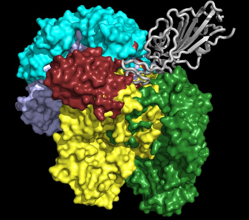
copies of antibodies (Ab) that recognize the specific antigen. Additionally, when the antigen first enters the
body, the T cells and B cells will be activated, and some of them will be differentiated to long-lived memory
cells, such as memory T cells and memory B cells. These long-lived memory cells will play a role in quickly
and specifically recognizing and eliminating a specific antigen that encountered the host and initiated a
corresponding immune response in the future [25]. The vaccination mechanism is to stimulate the primary
immune response of the human body, which will activate T cells and B cells to generate the antibodies and
long-lived memory cells that prevent infectious diseases, which is one of the most effective and economical
means for combating with COVID-19 at this stage.
    As mentioned above, secreted by B cells of the adaptive immune system, antibodies can recognize and
bind to specific antigens. Conventional antibodies (immunoglobulins) are Y-shaped molecules that have two
light chains and two heavy chains [26]. Each light chain is connected to the heavy chain via a disulfide bond,
and heavy chains are connected through two disulfide bonds in the mid-region known as the hinge region.
Each light and heavy chain contain two distinct regions: constant regions (stem of the Y) and variable regions
(“arms” of the Y) [27]. An antibody binds the antigenic determinant (also called epitope) through the variable
regions in the tips of heavy and light chains. There is an enormous amount of diversity in the variable regions.
Therefore, different antibodies can recognize many different types of antigenic epitopes. To be specific, there
are three complementarity determining regions (CDRs) that are arranged non-consecutively in the tips of each
variable region. CDRs generate most of the diversities between antibodies, which determine the specificity
of individuals of antibodies. In addition to conventional antibodies, camelids also produce heavy-chain-only
antibodies (HCAbs). HCAbs, also referred to as nanobodies, or VHHs, contain a single variable domain
(VHH) that makes up the equivalent antigen-binding fragment (Fab) of conventional immunoglobulin G
(IgG) antibodies [28]. This single variable domain typically can acquire affinity and specificity for antigens
comparable to conventional antibodies. Nanobodies can easily be constructed into multivalent formats
and have higher thermal stability and chemostability than most antibodies do [29]. Another advantage of
nanobodies is that they are less susceptible to steric hindrances than large conventional antibodies [30].
    Considering the broad specificity of antibodies, seeking potential antibody therapies has become one
of the most feasible strategies to fight against SARS-CoV-2. In general, antibody therapy is a form of
immunotherapy that uses monoclonal antibodies (mAb) to target pathogenic proteins. The binding of
antibody and pathogenic antigen can facilitate immune response, direct neutralization, radioactive treatment,
the release of toxic agents, and cytokine storm inhibition (aka immune checkpoint therapy). The SARS-
CoV-2 entry of a human cell facilitated by the process of a series of interactions between its spike (S)
protein and the host receptor angiotensin-converting enzyme 2 (ACE2), primed by host transmembrane
protease, serine 2 (TMPRSS2) [31]. As such, most COVID-19 antibody therapeutic developments focus on
the SARS-CoV-2 spike protein antibodies that were initially generated from patient immune response and
T-cell pathway inhibitors that block T-cell responses. A large number of antibody therapeutic drugs are
in clinical trials. Fifty-five S protein antibody structures are available in the Protein Data Bank (PDB),
offering a great resource for mechanistic analysis and biophysical studies.

                                                       2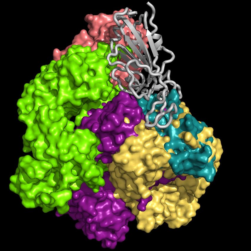
Virus                                              Viral-vector
                            vaccine                                               vaccine
                                                    or                                                       or

                                             Body                                                     Body

                                              Antigen-presenting cell           Cell                  Antigen-presenting cell
                         Cell

                                      Virus                                                   Virus
                                    replicates                                              replicates
                                                               Virus                                                   Virus
                                                              peptide                                                 peptide
                                                     Immune response                                         Immune response

                          DNA vaccine             RNA vaccine                 Protein-based
                                                                                 vaccine
                                                                                                     or
      Electroporation

                                                                                          Spike protein
                                                                                                                  VLP

                                           Body                                                     Body

                                              Antigen-presenting cell                          Antigen-presenting cell

                        Nucleus
                                    mRNA
                                Cell
                                                                                                                         Virus
                                                               Virus                                                    peptide
                                                              peptide
                                                     Immune response                                      Immune response
                          Figure 1: Illustration of four types of COVID-19 vaccines that are currently in the development.

    Currently, most antibody therapy developments focus on the use of antibodies isolated from patient con-
valescent plasma to directly neutralize SARS-CoV-2 [32–34], although there are efforts to alleviate cytokine
storm. A more effective and economical means to fight against SARS-CoV-2 is the vaccine [35], which is the
most anticipated approach for preventing the COVID-19 pandemic. A vaccine is designed to stimulate effec-
tive host immune responses and provide active acquired immunity by exploiting the body’s immune system,
including the production of antibodies, which is made of an antigenic agent that resembles a disease-causing
microorganism, or surface protein, or genetic material that is needed to generate the surface protein. For
SARS-CoV-2, the first choice of surface proteins is the spike protein. There are four types of COVID-19
vaccines, as shown in Figure 1. 1) Virus vaccines use the virus itself in a weakened or inactivated form. 2)
Viral-vector vaccines are designed to genetically engineer a weakened virus, such as measles or adenovirus,
to produce coronavirus S proteins in the body. Both replicating and non-replicating viral-vector vaccines
are being studied now. 3) Nucleic-acid vaccines use DNA or mRNA to produce SASR-CoV-2 S proteins
inside host cells to stimulate the immune response. 4) Protein-based vaccines are designed to directly inject
coronavirus proteins, such as S protein or membrane (M) protein, or their fragments, into the body. Both

                                                                         3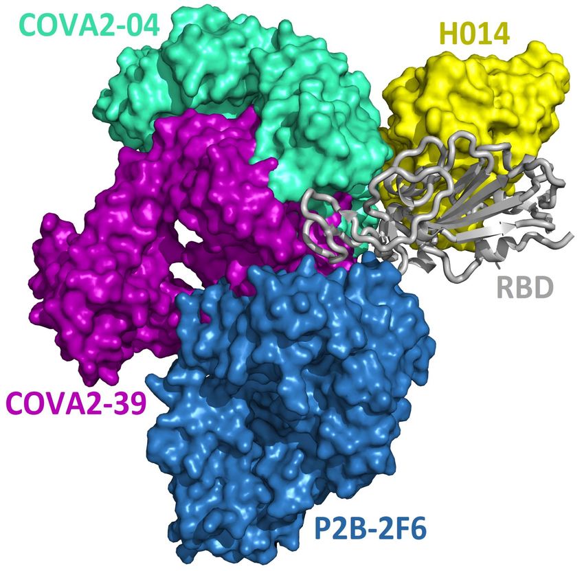
protein subunits and viral-like particles (VLPs) are under development for COVID-19 [36]. Among these
technologies, nucleic-acid vaccines are safe and relatively easy to develop [36]. However, they have not been
approved for any human usage before.
    However, the general population’s safety concerns are the major factors that hinder the rapid approval
of vaccines and antibody therapies. A major potential challenge is an antibody-dependent enhancement, in
which the binding of a virus to suboptimal antibodies enhances its entry into host cells. All vaccine and
antibody therapeutic developments are currently based on the reference viral genome reported on January
5, 2020 [37]. SARS-CoV-2 belongs to the coronaviridae family and the Nidovirales order, which has been
shown to have a genetic proofreading mechanism regulated by non-structure protein 14 (NSP14) in synergy
with NSP12, i.e., RNA-dependent RNA polymerase (RdRp) [38, 39]. Therefore, SARS-CoV-2 has a higher
fidelity in its transcription and replication process than other single-stranded RNA viruses, such as the
flu virus and HIV. Even though the S protein of SARS-CoV-2 has been undergoing many mutations, as
reported in [40, 41]. As of January 20, 2021, a total of 5,003 unique mutations on the S protein has
been detected on 203,346 complete SARS-CoV-2 genome sequences. Among them, 462 mutations were on
the receptor-binding domain (RBD), the most popular targets for antibodies and vaccines. Therefore, it
is of paramount importance to establish a reliable paradigm to predict and mitigate the impact of SARS-
CoV-2 mutations on vaccines and antibody therapies. Moreover, the efficacy of a given COVID-19 vaccine
depends on many factors, including SARS-CoV-2 biological properties associated with the vaccine, mutation
impacts, vaccination schedule (dose and frequency), idiosyncratic response, assorted factors such as ethnicity,
age, gender, or genetic predisposition. The effect of COVID-19 vaccination also depends on the fraction of
the population who accept vaccines. It is essentially unknown at this moment how these factors will unfold
for COVID-19 vaccines.
    It is no doubt that any preparation that leads to an improvement in the COVID-19 vaccination effect will
be of tremendous significance to human health and the world economy. Therefore, in this work, we integrate
genetic analysis and computational biophysics, including artificial intelligence (AI), as well as additional
enhancement from advanced mathematics to predict and mitigate mutation threats to COVID-19 vaccines
and antibody therapies. We perform single nucleotide polymorphism (SNP) calling [41] to identify SARS-
CoV-2 mutations. For mutations on the S protein, we analyze their mechanism [42], frequency, ratio, and
secondary structural traits. We construct a library of 56 existing antibody structures by January 1, 2021 from
the PDB and analyze their two-dimensional (2D) and three-dimensional (3D) characteristics. We further
predict the mutation-induced binding free energy (BFE) changes of antibody and S protein complexes using
a topology-based network tree (TopNetTree) [43], which is a state-of-the-art model that integrates deep
learning and algebraic topology [44–46]. In this work, TopNetTree is trained with newly available deep
mutation datasets on the S protein, ACE2, and some antibodies and its predictions are validated with
thousands of experimental data points. Our studies indicate that most mutations will significantly disrupt
the binding of essentially all known antibodies to the S protein. Therefore, vaccines and antibody drugs
that were developed based on the early SARS-CoV-2 genome will be seriously compromised by mutations.
Additionally, we show that most known mutations will strengthen the binding between the S protein and
ACE2, which gives rise to more infectious variants. Our studies also reveal that SARS-CoV-2 is at an
advanced stage of evolution with respect to its ability to infect human. Although the human immune system
is able to produce antibodies that are optimized respect to a pathogen, the antibodies, once produced, are
very vulnerable to the attack of mutants.

2    Mutations on the spike protein
As a fundamental biological process, mutagenesis changes the organism’s genetic information and servers as a
primary source for many kinds of cancer and heritable diseases, which is a driving force for evolution [47,48].
Generally speaking, virus mutations are introduced by natural selection, replication mechanism, cellular en-

                                                      4
vironment, polymerase fidelity, gene editing, random genetic drift, gene editing, recent epidemiology features,
host immune responses, etc [49, 50]. Notably, understanding how mutations have changed the SARS-CoV-2
structure, function, infectivity, activity, and virulence is of great importance for coming up with life-saving
strategies in virus control, containment, prevention, and medication, especially in the antibodies and vaccines
development. Genome sequencing, SNP calling, and phenotyping provide an efficient means to parse muta-
tions from a large number of viral samples [40](see the Supporting material (S1)). In this work, we retrieved
more than 200,000 complete SARS-CoV-2 genome sequences from the GISAID database [51] and created a
real-time interactive SARS-CoV-2 Mutation Tracker to report more than 26,000 unique single mutations
along with its mutation frequency on SARS-CoV-2 as of January 20, 2021. Figure 2 is a screenshot of
our online Mutation Tracker. It describes the distribution of mutations on the complete coding region of
SARS-CoV-2. The y-axis shows the natural log frequency for each mutation at a specific position. A reader
can download the detailed mutation SNP information from our Mutation Tracker website.

Figure 2: The distribution of genome-wide SARS-CoV-2 mutations on 26 proteins. The y-axis represents the natural log
frequency for each mutation on a specific position of the complete SARS-CoV-2 genome. While only a few landmark positions
are labeled with gene (protein) names, the relative positions of other genes(proteins) can be found in our Mutation Tracker
MutationTracker( https://users.math.msu.edu/users/weig/SARS-CoV-2 Mutation Tracker.html)

    As mentioned before, the S protein has become the first choice for antibody and vaccine development.
Among 203,346 complete genome sequences, 5,003 unique single mutations are detected on the S protein.
The number of unique mutations (NU ) is determined by counting the same type of mutations in different
genome isolates only once, whereas the number of non-unique mutations (NNU , i.e., frequency) is calculated
by counting the same type of mutations in different genome isolates repeatedly. Table 1 lists the distribution
of 12 SNP types among unique and non-unique mutations on the S protein of SARS-CoV-2 worldwide. It
can be seen that C>T and A>G are the two dominated SNP types, which may be due to the innate host
immune response via APOBEC and ADAR gene editing [42].
   Moreover, 144 non-degenerated mutations occurred on the S protein RBD, which are relevant to the
binding of SARS-CoV-2 S protein and most antibodies as well as ACE2. Additionally, 218 mutations that
occurred on the S protein NTD (residue id: 14 to 226) are relevant to the binding of another two antibodies
(4A8, FC05) and SARS-CoV-2 S protein.
    Furthermore, since antibody CDRs are random coils, the complementary antigen-binding domains must
involve random coils as well. Table 2 lists the statistics of non-degenerate mutations on the secondary
structures of SARS-CoV-2 S protein. Here, the secondary structures are mostly extracted from the crystal
structure of 7C2L [52], and the missing residues are predicted by RaptorX-Property [53]. We can see that
for both unique and non-unique cases, the average mutation rates on the random coils of the S protein have

                                                            5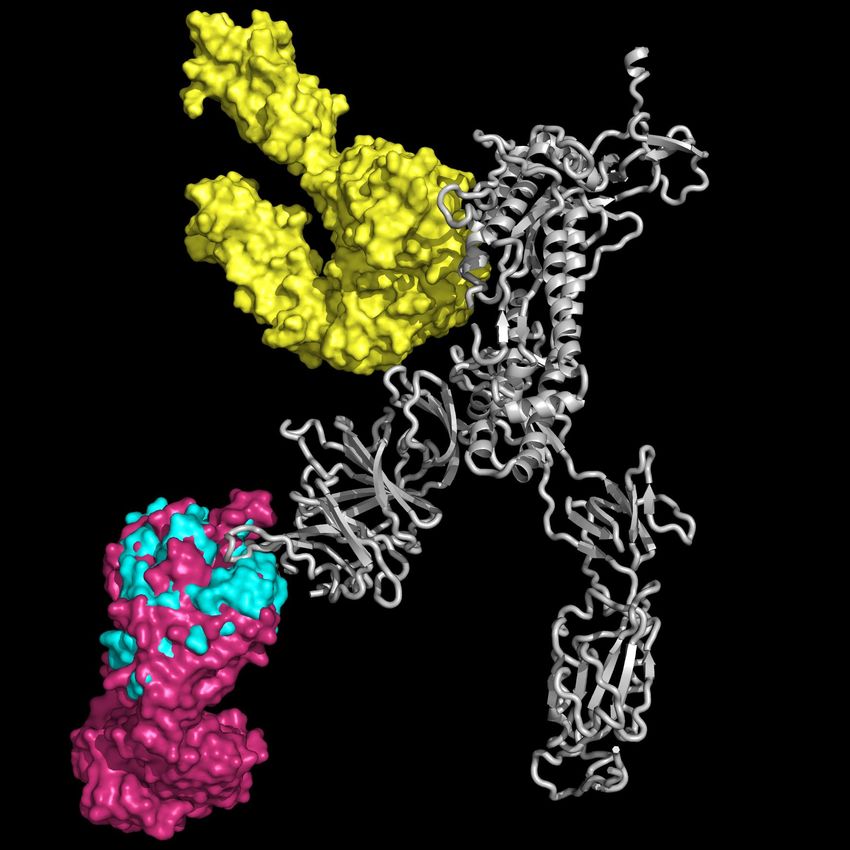
Table 1: The distribution of 12 SNP types among 5,003 unique mutations and 467,604 non-unique mutations on the S gene
of SARS-CoV-2 worldwide. NU is the number of unique mutations and NNU is the number of non-unique mutations. RU and
RNU represent the ratios of 12 SNP types among unique and non-unique mutations.

SNP Type Mutation Type NU NNU                  RU       RNU SNP Type Mutation Type NU NNU                      RU       RNU
    A>T      Transversion      454 5236 9.07% 1.12%                C>T       Transition        542 158898 10.83% 33.98%
    A>C      Transversion      341 2571 6.82% 0.55%                C>A       Transversion      313 10301 6.26% 2.20%
    A>G      Transition        700 199015 13.99% 42.56%            C>G       Transversion      156 968 3.12% 0.21%
    T>A      Transversion      356 1614 7.12% 0.35%                G>T       Transversion      435 34421 8.69% 7.36%
    T>C      Transition        779 19313 15.57% 4.13%              G>C       Transversion      225 6090 4.50% 1.30%
    T>G      Transversion      277 1940 5.54% 0.41%                G>A       Transition        425 27237 8.49% 5.82%

the highest values. Particularly, the 23403A>G-(D614G) mutation on the random coils has the highest
frequency of 192284. If we do not consider the 23403A>G-(D614G) mutations, then the unique and non-
unique average rates on the random coils of S protein still have the highest values (2.81 and 212.01),
indicating that mutations are more likely to occur on the random coils. Consequently, the natural selection
of mutations may tend to disrupt antibodies.
Table 2: The statistics of non-degenerate mutations on the secondary structure of SARS-CoV-2 S protein. The unique and
non-unique mutations are considered in the calculation. NU , NNU , ARU , ARNU represent the number of unique mutations,
the number of non-unique mutations, the average rate of unique mutations, and the average rate of non-unique mutations on
the secondary structure of S protein, respectively. Here, the secondary structure is mostly extracted from the crystal structure
of 7C2L, the missing residues are predicted by RaptorX-Property.

                 Secondary structure            Length         NU          NNU          ARU          ARNU
                        Helix                     249          516          9535        2.07         38.29
                        Sheet                     276          711         20422        2.58         73.99
                     Random coils                 748         2100        350659        2.81        468.80
                     Whole Spike                 1273         3327        380616        2.61        298.99

3     SARS-CoV-2 antibodies
In this work, we consider 56 3D structures available from the PDB ( https://www.rcsb.org) before January
1, 2021. These 56 structures include 51 structures of antibodies binding to S protein RBD, 4 structures of
antibodies having binding domains outside the S protein RBD, and an ACE2-S protein complex. Among
four structures having binding domains outside the RBD, there are three distinct antibodies not binding to
the RBD, namely 4A8 [52], FC05 [54], and 2G12 [55]. This is because that FC05 has two sets of structures
(PBD IDs 7CWU and 7CWS) that differ from each other by their components on the RBD (i.e., H014 and
P17). Some antibodies are given as combinations of other unique ones. Among 51 antibodies on the RBD,
there are only 42 unique ones, including MR17-K99Y as a mutant of MR17 [56].

3.1    3D antibody structure alignment on the S protein
We present the 3D alignment of 45 structures of SARS-CoV-2 S protein with ACE2 and antibodies (excluding
the mutant MR17-K99Y of MR17) in Figure 3. ACE2 in Figure 3 (a) is a reference. Figures 3 (a)-(j) list 42
single antibodies binding to the RBD, and Figure 3 (k) includes the other 3 alignments of 4A8, FC05, and
2G12 whose binding domains are outside the RBD. Figure 3 (m) presents a 3D structure of a single chain
of S protein. The PDB IDs of these complexes can be found in Figure 4.
   Figure 3 reveals, except for Fab 52 [62], S309 [57], CR3022 [63], EY6A [67], 4A8 [52], FC05 [54], and
2G12 [55], all the other 38 antibodies have their binding sites spatially clashing with that of ACE2. Notably,
the paratope of H014 [69] and S304 [60] do not overlap with that of ACE2 directly, but in terms of 3D

                                                               6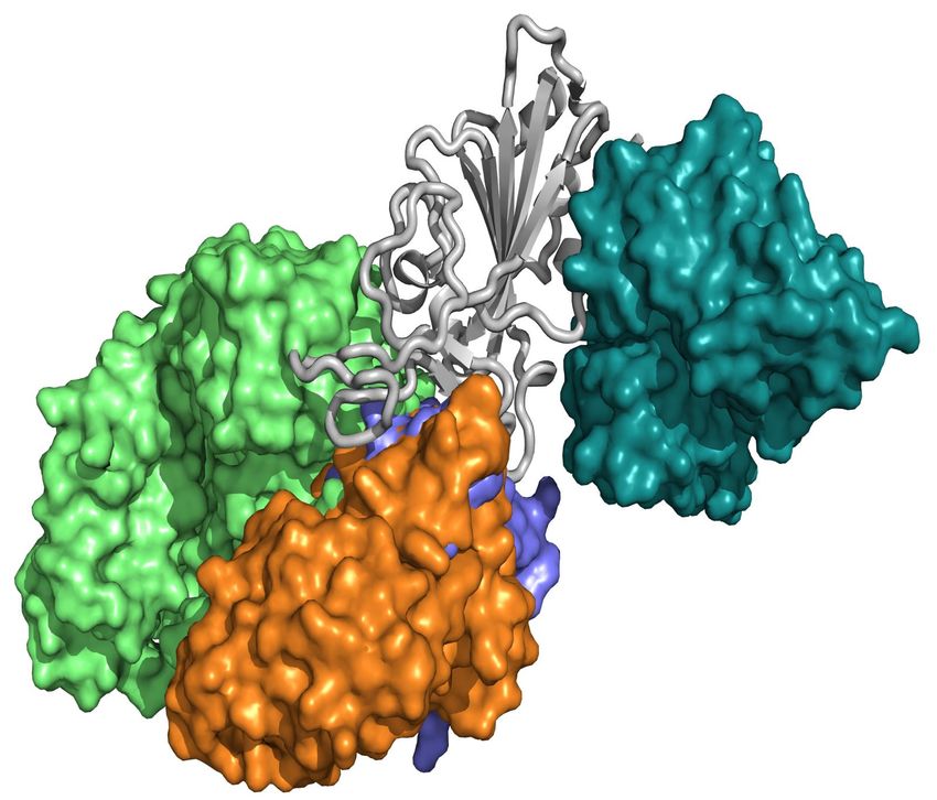
BD-629                                                                            BD-604
                                                                     CR3022
                                             RBD           CC12.3
                                                                                                                                   RBD

                                                                                                       MR17                 Fab 2-4
                       ACE2                                                          RBD
                                       H11-H4
             (a)                                     (b)               B38                      (c)
             S304                                                                                                           EY6A
                              RBD                                    RBD                                      CC12.1
                                           Fab 52                                              BD-236
                                                             CV30

            CB6
                                     H11-D4                                             S309
                                                                                                                                   RBD
                                                                               SR4                                     Nb
                                   S2H13
                        Fab 298                                              BD23       BD-368-2
             (d)                                     (e)                                        (f)
                                                                    A fab            RBD
            COVA2-04                    H014                                S2E12                       S2A4                RBD

                                                                                               COVA1
                                                                                               -16                               VH
                                                                                                                               binder

                                              RBD
                                                     S2H14
            COVA2-39

                                                                                                P2C-1F11
                                  P2B-2F6                                                                                   CV07-250
             (g)                                     (h)      P2C-1A3        CV07-270            (i)

                          STE90-C11                          2G12
                                            RBD

                                                               NTD                                     NTD
                                              Sb23
              C1A
                      S2M11                           FC05                              RBD
                                       P17
                                                                                                                       RBD
             (j)                                     (k)             4A8                         (m)
Figure 3: Aligned structures of 46 complexes of the S protein and ACE2 and single antibodies. (a)-(j) The 3D alignment of
the available unique 3D structures of SARS-CoV-2 S protein RBD in binding complexes with 42 antibodies (MR17-K99Y
is excluded because its binding mode is the same as that of MR17). (k) The 3D alignment of the three antibodies binding
outside RBD. (m) The 3D structure of S protein RBD. The red, green, and blue colors represent for helix, sheet, and random
coils of RBD, respectively. The darker color represents the higher mutation frequency on a specific residue. The structures
are (a) ACE2 (6M0J) [57], BD-629 (7CH5), H11-H4 (6ZBP); (b) CC12.3 (6XC4) [58], B38 (7BZ5) [59], CR3022 (6XC3) [58];
(c) BD-604 (7CH4), MR17 (7C8W) [56], Fab 2-4 (6XEY) [56]; (d) S304 (7JW0) [60], CB6 (7C01) [61], Fab 52 (7K9Z) [62],
S2H13 (7JV6) [60], H11-D4 (6YZ5) [63], Fab 298 (7K9Z) [62]; (e) CV30 (6XE1) [64], BD23 (7BYR) [65], SR4 (7C8V) [56], S309
(6WPS) [66]; (f) CC12.1 (6XC2) [58], EY6A (6ZCZ) [67], BD-236 and nanobody (Nb) (7CHE) [68], BD-368-2 (7CHH) [68]; (g)
H014 (7CAH) [69], COVA2-04 (7JMO) [70], COVA2-39 (7JMP) [70], P2B-2F6 (7BWJ) [71]; (h) P2C-1A3 (7CDJ), CV07-270
(6XKP) [72], S2H14 (7JX3) [60], A fab (7CJF), S2E12 (7K45) [73]; (i) CV07-250 (6XKQ) [72], P2C-1F11 (7CDI), VH binder
(7JWB) [74], S2A4 (7JVA) [60], COVA1-16 (7JMW) [75], (j) C1A (7KFV) [76], STE90-C11 (7B3O) [77], Sb23 (7A29) [78],
S2M11 (7K43) [73], P17 (7CWM) [79]; (k) 4A8 (7C2L) [52], FC05 (7CWU) [54], and 2G12 (7L06) [55].

                                                                       7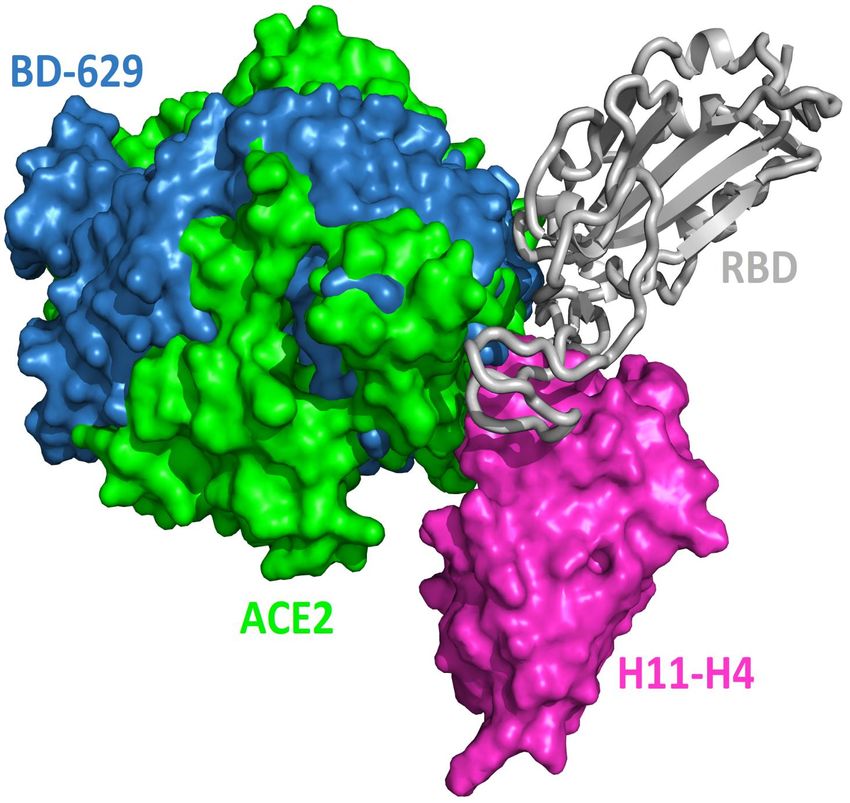
structures, their binding sites still overlap. This suggests that the bindings of 39 antibodies are in direct
competition with that of ACE2. Theoretically, this direct competition reduces the viral infection rate. For
such an antibody with strong binding ability, it will directly neutralize SARS-CoV-2 without the need for
antibody-dependent cell cytotoxicity (ADCC), antibody-dependent cellular phagocytosis (ADCP), or other
immune mechanisms.
    The paratopes of S309, Fab 52, CR3022, and EY6A on the RBD are away from that of ACE2, leading
to the absence of binding competition [66, 67, 80]. One study shows that the ADCC and ADCP mechanisms
contribute to the viral control conducted by S309 in infected individuals [66]. For Fab 52, it was suggested
that its mechanism could involve S protein destabilization [62]. For CR3022, one research indicates that it
neutralizes the virus in a synergistic fashion [81]. For EY6A, the hypothesis is that glycosylation of ACE2
accounts for at least part of the observed crosstalk between ACE2 and EY6A. [67]. More radical examples
are 4A8, FC05, and 2G12. 4A8 binds to NTD of the S protein (Figure 3(h)), which is quite far from the
RBD. It is speculated that 4A8 may neutralize SARS-CoV-2 by restraining the conformational changes of
the S protein, which is very important for the SARS-CoV-2 cell entry [52]. FC05 is combined with P17
or H014 to form a cocktail [54]. 2G12 binds to the S protein S2 domain [55]. Any antibody or drug that
can inhibit serine protease TMPRSS2 priming of the S protein priming can effectively stop the viral cell
entry [31].

3.2    2D residue contacts between antibodies and the S protein RBD
Figure 3 provides a visual illustration of antibody and ACE2 competitions. It remains to know in the
residue detail what has happened to these competitions. To better understand the antibody and S protein
interactions, we study the residue contacts between antibodies and the S protein. We include the ACE2 as a
reference but excluding antibodies MR17-K99Y as well as 4A8, FC05, and 2G12 that bind to other domains.
   In Figure 4, the paratopes of 42 individual antibodies (excluding MR17-K99Y) and ACE2 were aligned
on the S protein RBD 2D sequence, and their contact regions are highlighted. From the figure, one can
see that, except for Fab 52, S309, CR3022, EY6A, H014, and S304, all the other 36 antibodies have
their antigenic epitopes overlapping with the ACE2, especially on the residues from 486 to 505 of the RBD.
Although, the paratope of H014 and S304 do not overlap with that of ACE2 directly, their binding sites still
overlap in 3D structures. Therefore, these 38 antibodies competitively bind against ACE2 as revealed in
Figure 3.

3.3    Antibody sequence alignment and similarity analysis
The next question is whether there is any connection or similarity between the antibody paratopes in our
library, particularly for those antibodies that share the same binding sites. To better understand this
perspective, we carry out multiple sequence alignment (MSA) to further study the similarity and difference
among existing antibodies. Many antibodies are very similar to each other and can be classified into several
clusters by CD-HIT suite [82]. The first and largest cluster includes COVA2-04, CC12.1, BD-236, BD-604,
B38, EY6A, S304, P2C-1A3, A fab, C1A, STE90-C11, and CB6. Their identity scores to CB6 are 90.48 %,
94.74 %, 93.59 %, 93.35 %, 94.77 %, 92.52 %, 90.62 %, 90.51 %, 91.18 %, 94.08 %, and 93.00 %, respectively.
The second cluster contains BD-629, CC12.3, P2C-1F11, and CV30. Their identity scores to CV30 are 95.41
%, 96.32 %, and 97.68 %, respectively. The third cluster has CV07-270 and COVA2-39, the pairwise identity
score is 90.18 %. The fourth cluster is composed of H11-H4, H11-D4, and Nb, and their identity scores
to Nb are 99.25 % and 95.52 %, respectively. They are all nanobodies. The fifth cluster has Fab 298 and
COVA1-16, the pairwise identity score is 90.80 %. Their alignment plots are in the Supporting material
(Figures S1-S5).
   The above similarity indicates that the adaptive immune systems of individuals have a common way to

                                                     8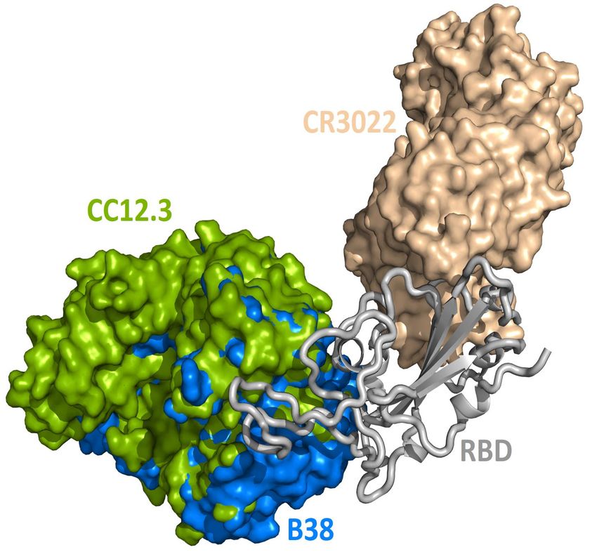
Figure 4: Illustration of the contact positions of antibody and ACE2 paratope with SARS-CoV-2 S protein RBDs on RBD 2D
sequences. The corresponding PDB IDs are given in parentheses.

generate antibodies. On the other hand, the existence of five distinct clusters, as well as antibodies 4A8,
FC05, and 2G12 suggests the diversity in the immune response. Note that we have also included ACE2 in
our MSA as a reference, but none of the existing antibodies is similar to ACE2 because they were created
from entirely different mechanisms.

4     Mutation impacts on SARS-CoV-2 antibodies
To investigate the influences of existing S protein mutations on the binding free energy (BFE) of S protein
and antibodies, we consider 462 mutations that occurred on the S protein RBD, which are relevant to the
binding of SARS-CoV-2 S protein and antibodies as well as ACE2. Additionally, 540 mutations occurred on

                                                          9
the NTD of the S protein (residue id: 14 to 226) which are relevant to the binding of SARS-COV-2 S protein
and antibody 4A8 (PDB: 7C2L). We predict the free energy changes following existing mutations using our
TopNetTree model [43]. The mutations on the RBD are considered for the predictions of BFE changes. Our
predictions are built from the X-ray crystal structure of SARS-CoV-2 S protein and ACE2 (PDB 6M0J) [57],
and various antibodies (PDBs 6WPS [66], 6XC2 [58], 6XC3 [58], 6XC4 [58], 6XC7 [58], 6XE1 [64], 6XEY [83],
6XKP [72], 6XKQ [72], 6YLA [63], 6YZ5, 6Z2M, 6ZBP, 6ZCZ [67], 6ZER [67], 7A29 [78], 7B3O, 7BWJ [71],
7BYR [65], 7BZ5 [59], 7C01 [61], 7C2L [52], 7C8V [56], 7C8W [56], 7CAH [69], 7CAH [69], 7CAN [56], 7CDI,
7CDJ, 7CH4 [68], 7CH5 [68], 7CHB [68], 7CHE [68], 7CHF [68], 7CHH [68], 7CJF, 7CWM [79], 7CWN [79]
7JMO [70], 7JMP [70], 7JMW [75], 7JV6 [60], 7JVA [60], 7JVC [60], 7JW0 [60], 7JWB [74], 7JX3 [60],
7K43 [73], 7K45 [73], 7K9Z [62], 7KFV [76], 7KFW [76], 7KFX [76], and 7KFY [76]). The BFE change
following mutation (∆∆G) is defined as the subtraction of the BFE of the mutant type from the BFE of the
wild type, ∆∆G = ∆GW − ∆GM where ∆GW is the BFE of the wild type and ∆GM is the BFE of mutant.
Therefore, a negative BFE change means that the mutation decreases affinities, making the protein-protein
interaction less stable.
Four antibody-S protein complexes are examined in in this section. Next, we present a library of mutation-
induced BFE changes for all mutations and 51 antibodies, as well as ACE2. The statistical analysis of
mutation impacts on antibodies is discussed.
4.1 Single antibody-S protein complex analysis
4A8
0.0 W152C
BFE changes (kcal/mol)
104
R102I
0.5
Frequency
103
1.0
1.5 102
2.0 PDB: 7C2L
101 A222V
R102I
R102S
D111N
S112L
L118F
I119V
A123V
N125S
V127F
V127A
E132Q
N137S
D138H
D138Y
L141V
L141F
G142S
G142D
G142V
V143F
Y144H
Y144F
Y145H
H146Y
N149Y
S151I
W152L
W152C
M153V
M153T
M153I
E154Q
S155G
S155I
E156Q
E156A
E156D
F157L
V159I
S162I
A163V
N164T
V171F
Q173K
P174S
L176F
M177T
M177I
D178N
D178H
D178G
L179F
E180G
E180V
G181R
G181V
K182R
Q183R
Q183H
G184S
G184V
N185S
F186S
L189F
R190K
R190M
R190S
V193L
I197V
D198G
H207R
H207Q
T208M
V213L
V213A
R214H
R214L
D215H
D215Y
D215G
D215V
L216F
Q218E
Q218H
S221L
A222S
A222V
E224Q
V227L
L229F
I233V
R237K
Q239K
T240I
L242F
L242P
A243S
A243V
H245Y
R246T
R246I
S247G
S247N
Y248H
T250N
P251S
P251H
P251L
G252V
D253N
D253Y
D253G
S254F
S255F
S256P
S256L
G257S
G257D
G257V
W258R
W258L
T259I
A260S
G261S
G261R
G261D
G261V
A262S
A262V
A263T
A263S
A263V
A264V
Q271R
P272H
P272L
E281Q
E281V
T286I
V289I
V289L
Figure 5: Illustration of SARS-CoV-2 mutation-induced binding free energy changes for the complexes of S protein and 4A8
(PDB: 7C2L). The blue color in the structure plot indicates a positive BFE change while the red color indicate a negative
BFE change, and toning indicate the strength. Here, mutations R102I, W152C, W152L, S247N, and Y248H could potentially
disrupt the binding of antibody 4A8 and S protein.
For four antibody-S protein complexes, since there are too many mutations, we only consider those
mutations whose frequencies are greater than 10. We first present the BFE changes (∆∆G) of SARS-CoV-2
S protein binding domain with antibody 4A8 in Figure 5, which is one of the three complexes that are not on
the RBD in our collections of S protein and antibody complexes. A total 141 of 540 mutations on residues
ID from 14 to 226 have frequencies larger than 10. Most mutations have small BFE changes (from −0.5
kcal/mol to 0.5 kcal/mol) in their binding free energies, while 28 mutations have negative BFE changes less
than −0.5 kcal/mol. Notably, 53 out of 141 mutations on the binding domain have positive BFE changes,
which means that the mutations increase affinities and would make the S protein-4A8 interactions more
stable. However, the majority (63%) of mutations have negative BFE changes, including high-frequency
mutations, R102I, and W152C with frequencies of 89 and 356, respectively. Since the largest positive and
negative BFE changes are 0.37 and -2.06 (-3.1 if low frequency mutations are counted) kcal/mol, respectively,
the prediction indicates that antibody 4A8 isolated from 10 convalescent patients at the early stage of the
pandemic [52] is an optimized product of the human immune system with respect to the original S protein.
It is also noted that many mutations on the binding domain, such as W152L, S247N, and Y248H, have
significant negative free energy changes. The mutations on the binding domain with large negative BFE
changes reveal that the binding of antibody 4A8 and S protein will be potentially disrupted.
100.5
104 N501Y
0.0
BFE changes (kcal/mol)
0.5
Frequency
103
1.0
1.5 F490S
102
Fab 2-4
2.0 E484K
E484Q
2.5 PDB: 6XEY
101 F486L
L335F
G339D
E340A
V341I
A344S
N354D
R346K
A348S
A352S
N354K
K356R
R357K
S359N
V367I
V367F
N370S
S373L
V382L
T376I
P384S
P384L
T385I
D389E
I402V
D427G
V445A
R403K
E406Q
R408I
I410V
A411S
Q414K
Q414R
K417N
D427Y
N440K
K444R
K444N
G446V
L452M
L452R
Y453F
L455F
S459Y
S459F
P463S
I468V
I468T
E471Q
A475S
A475V
G476S
S477N
V503F
S477I
S477R
T478K
T478R
T478I
P479S
P479L
V483F
V483A
E484K
E484Q
F486L
F490S
S494P
S494L
N501Y
N501T
Y508H
S514F
E516Q
L517F
A520S
P521S
P521R
A522S
A522V
S477N
Figure 6: Illustration of SARS-CoV-2 mutation-induced binding free energy changes for the complexes of S protein and Fab
2-4 (PDB: 6XEY). The blue color in the structure plot indicates a positive BFE change while the red color indicate a negative
BFE change, and toning indicate the strength. Here, mutations E484K, E484Q, F486L, and F490S could potentially disrupt
the binding of antibody Fab 2-4 and the S protein.
Next, we study the BFE changes (∆∆G) induced by 80 mutations on the SARS-CoV-2 S protein RBD
for the antibody Fab 2-4 (PDB: 6XEY) in Figure 6. Antibody Fab 2-4 shares a similar binding domain
with ACE2 and thus is a potential candidate for the direct neutralization of SARS-CoV-2. Most mutations
induce small changes in the binding free energies, while mutations, E484K, E484Q, F486L, and F490S, have
large negative BFE changes. Overall, 38 out of 80 mutations on the RBD lead to negative BFE changes,
which means 48% of mutations will potentially weaken the binding between antibody Fab 2-4 and S protein.
For positive BFE changes, the largest value is only 0.55 kcal/mol and the average of positive BFE changes
is 0.16 kcal/mol. However, many mutations with negative BFE changes have a very large magnitude,
indicating that antibody Fab 2-4 was an immune product optimized with respect to the original un-mutated
S protein. In general, the mutations on S protein weaken the Fab 2-4 binding with S protein and make it
less competitive with ACE2 as most mutations strengthen the S protein and ACE2 binding. It is interesting
to note that mutation E484K is the so-called South Africa variant. It indeed has a strong vaccine-escape
effect.
0.25
104
0.00 S494L
BFE changes (kcal/mol)
0.25
Frequency
0.50 103
0.75
1.00 L452R
102 MR17
1.25 F490S
F486L
1.50
PDB: 7C8W E484K
101
S477N
L335F
G339D
E340A
V341I
A344S
V367F
V382L
A475V
V483A
R346K
A348S
A352S
N354D
N354K
K356R
R357K
S359N
V367I
N370S
S373L
T376I
P384S
P384L
T385I
D389E
I402V
R403K
E406Q
D427Y
D427G
V445A
R408I
I410V
A411S
Q414K
Q414R
K417N
N440K
K444R
K444N
G446V
L452M
L452R
Y453F
L455F
S459Y
S459F
P463S
I468V
I468T
E471Q
A475S
G476S
S477N
S477I
S477R
T478K
T478R
T478I
P479S
P479L
V483F
E484K
E484Q
F486L
F490S
S494P
S494L
N501Y
N501T
V503F
Y508H
S514F
E516Q
L517F
A520S
P521S
P521R
A522S
A522V
Figure 7: Illustration of SARS-CoV-2 mutation-induced binding free energy changes for the complexes of S protein and MR17
(PDB: 7C8W). Blue in the structure plot indicates a positive BFE change while red indicate a negative BFE change, and
toning indicate the strength. Here, mutations L452R, E484K, F486L, F490S, and S494L could potentially disrupt the binding
of antibody MR17 and the S protein.
In Figure 7, we illustrate the mutation-induced BFE changes for antibody MR17 (PDB: 7C8W), which
shares the binding domain with ACE2 as well. One can notice that four mutations, L452R, E484K, F486L,
F490S, and S494L, have BFE changes less than −1 kcal/mol as well as high frequencies. The rest mutations
have a small magnitude of changes. Twenty seven out of 80 mutations have positive BFE changes with
the largest value less than 0.25 kcal/mol. Our results indicate that antibody MR37 is likely to be isolated
from patients at the early stage and thus, it was optimized based on an early version of SARS-CoV-2 virus.
Mutations L452R, E484K, F486L, F490S, and S494L will reduce its competitiveness with ACE2.
Finally, we consider the BFE change predictions for antibody S309 and S protein complex, whose receptor
110.75
104 E340A S309
0.50
BFE changes (kcal/mol)
0.25
Frequency
103
0.00
N354D
0.25
102
0.50
0.75
R403K
PDB: 6WPS
101
L335F
G339D
E340A
V341I
A344S
V382L
R346K
A348S
A352S
N354D
N354K
K356R
R357K
S359N
V367I
V367F
N370S
S373L
T376I
P384S
P384L
V445A
T385I
D389E
I402V
R403K
E406Q
R408I
I410V
A411S
Q414K
Q414R
K417N
D427Y
D427G
N440K
K444R
K444N
G446V
L452M
L452R
Y453F
L455F
S459Y
S459F
P463S
I468V
I468T
E471Q
A475S
A475V
G476S
S477N
V483F
V483A
S477I
S477R
T478K
T478R
T478I
P479S
P479L
E484K
E484Q
F486L
F490S
S494P
S494L
N501Y
N501T
V503F
Y508H
S514F
E516Q
L517F
A520S
P521S
P521R
A522S
A522V
Figure 8: Illustration of SARS-CoV-2 mutation-induced binding free energy changes for the complexes of S protein and S309
(PDB: 6WPS). The blue color in the structure plot indicates a positive BFE change while the red color indicate a negative BFE
change, and toning indicate the strength. Here, mutations E340A, N354D, and K356R could potentially weaken the binding of
antibody S309 and the S protein.
binding motif (RBM) does not overlap with the RBM of ACE2 (see Fig. 3(e)). The BFE changes induced by
80 mutations are predicted. Among them, 38 changes are positive. Similar to the aforementioned antibodies,
most of the mutations lead to small changes in their binding affinity magnitude but three mutations, E340A,
N354D, and K356R, induce moderate negative changes. Interestingly, all the 80 RBD mutations do not
have a major impact on S309. Although mutation R403K might disrupt S309, it does weaken many other
antibody bindings with the S protein. While antibodies play a variety of functions in the human immune
system, such as neutralization of infection, phagocytosis, antibody-dependent cellular cytotoxicity, etc., their
binding with antigens is crucial for these functions. Our analysis of BFE changes following mutations on the
S protein suggests that some antibodies will be less affected by mutations, which is important for developing
vaccine and antibody therapies.
4.2 Mutation impact library
In this section, we build a library of mutation-induced BFE changes for all mutations and all antibodies as
well as ACE2. In principle, we could create a library of all possible mutations for all antibodies, as we did
for ACE2 [84]. Here, we limit our effort to all existing mutations. Antibody 4A8 on the NTD has been
discussed above. We consider antibodies on the RBD.
Based on our earlier analysis, three types of SARS-CoV-2 S protein secondary structural residues have
different mutation rates. Among them, the random coils are major components of the RDB and the NTD,
as shown in Fig. 3. Most RBD mutations (287 of 462) occur on the residues whose secondary structures are
coil, while 93 out of 462 mutations are on the helix, and 82 out of 462 mutations are on the sheet. Therefore,
mutations on the RBD are split into three categories based on their locations in secondary structures helix,
sheet, and coil. In Figure 9, we present the BFE changes for the complexes of the S protein and antibodies
or ACE2 induced by mutations on the helix residues of the S protein RBD. The frequency for each mutation
is also presented. Most mutations on helix residues lead to negative BFE changes (pink squares), which
weaken the bindings, whereas some mutations induce positive BFE changes (green squares). It is noted that
most mutations lead to the strengthening of the S protein and ACE2 binding, which is consistent with the
natural selection rule. Mutations N406G, I418N, N422K, D442H, Y505S, and Y505C give rise to a strong
weakening effect on most antibodies. The N439K mutation having the highest frequency, shows a positive
BFE change on ACE2 but negative changes on most antibodies. Mutation D405Y appears to strengthen
most antibodies.
In Figure 10, we present the BFE changes for the S protein and antibody (ACE2) complexes following
sheet residue mutations of the S protein RBD. Like the last case, most mutations lead to positive BFE changes
for ACE2, indicating infectivity strengthening. There are many disruptive mutations, such as R355W, F401I,
F401C, I402F, C432G, I434K, A435P, O493P, V510E, V512G, and L513P, that will weaken most antibodies
12G339D
D405G
D420G
G504D
Q409H
D442H
N370D
N439D
N440D
N343H
G504C
G339R
R408G
G339A
G404A
G504A
D405V
D420V
E340Q
E406Q
G339S
G504S
E340G
E406G
N370K
K386N
N388K
K417N
N422K
N439K
N440K
E340D
D405E
D420E
N370S
N388S
N422S
N439S
N440S
Y365D
D405Y
Y421D
V367A
Y365H
Y505H
T385N
N440Y
N440T
P384H
K386R
R408K
K417R
Q409L
R408S
A419S
E340A
Y365C
Y369C
Y505C
R408T
T385A
E340K
P384A
F338C
V367F
V407F
V503F
Y365S
Y369S
S371T
T385S
Y421S
Y505S
V367L
S366P
S371P
P384S
S366F
S371F
L387S
P384L
F338L
F342L
L441F
I418N
R408I
V341I
V367I
V407I
I418V
T385I
L387I
L441I
ACE2 2
CR3022
B38
S309
CB6 1
H11-D4
CR3022 H11-D4
P2B-2F6
BFE changes (kcal/mol)
BD23
EY6Z Nb 0
EY6Z
SR4
MR17
MR17-K99Y
CV30 1
CC12.1
CC12.1 CR3022
CC12.3
CC12.3 CR3022 2
Fab 2-4
H11-H4
H014
COVA2-04
COVA2-39 3
BD-604
BD-629
BD-236
BD-236 BD-368-2 4
BD-604 BD-368-2
BD-368-2
VH binder
S2M11
S2E12
S2H13
S2A4
S304
S309 S2H14 S304
CV07-270
CV07-250
COVA1-16
Sb23
Fabs 298 52
A fab
P2C-1F11
P2C-1A3
C1A-B12
C1A-B3
C1A-F10
C1A-C2
STE90-C11
P17
P17 H014
Frequency
0 101 102 103 104
Frequency
Figure 9: Illustration of SARS-CoV-2 helix-residue mutation induced BFE changes for the complexes of S protein and 51
antibodies or ACE2. Positive changes strengthen the binding while negative changes weaken the binding. Mutation frequency
is given for each mutation. Grey color indicates PDB structures does not include residues induced by those mutations.
and S protein complexes. On the other hand, most mutations strengthen certain antibodies but weaken other
ones, which allows the effectiveness of antibody cocktails for a better protection. The binding of antibody
H014 and the S protein is strengthened by many mutations, particularly S375F, K378O, R403K, and Y453F.
Among them, Y453F is an infectivity-strengthening mutation with a relatively high frequency.
Figures 11, 12, and 13 present the BFE changes for the S protein and antibody (ACE2) complexes
following coil residue mutations of the S protein RBD. Overall, most mutations on coil residues lead to mild
negative BFE changes. However, mutations V350F, W353R, I401N, G416V, G431V, Y449D, Y449S, C480R,
P491R, P491L, Y495C, and O506P will weaken most antibody bindings to the S protein. Some residues, like
A348, N460, and P521, can produce many binding-strengthening mutations for most antibodies and ACE2.
For the high-frequency mutation S447N in Figure 13, the BFE changes are mild on ACE2 and antibodies.
Additionally, the N501Y mutation, one of the typical mutations in the UK B.1.1.7 variants, strengthens the
infectivity but induces a mixed reactions to antibodies as shown in Figure 13 .
4.3 Statistical analysis of mutation impacts on COVID-19 antibodies
First, we perform a statistical analysis of all mutation-induced BFE changes studied in the last section.
Most mutations induce binding-weakening BFE changes. The total rate of negative BFE changes is 71%
(i.e., 16661 out of 23512), for coil residues, 67% BFE changes are negative, while for helix or sheet residues,
72% and 80% BFE changes are negative, respective. However, for ACE2, 300 out of 462 mutations (i.e.,
65%) on the REB produce positive or binding-strengthening BFE changes, showing the effect of the natural
selection of mutations. In contrast, at most 200 of 462 mutations on the RBD give rise to negative BFE
changes for antibodies. More specifically, 11 antibodies have less than 100 positive BFE changes while 41
antibodies have less than 200 positive BFE changes. Interestingly, in our prediction, 4 out of the 43 single
antibodies have less than 100 positive BFE changes, while 7 out of the 9 antibody cocktails have less than
100 positive BFE changes. Although antibody cocktails have mild negative BFE changes, it turns out that
13R355W
W436L
Q493H
N354D
K356M
K378M
N354H
C432G
R357G
V512G
D398V
K378Q
L452M
E516Q
E516G
N354K
K356N
K378N
D398E
Q493P
N354S
N394S
N437S
V395A
Y508H
T376N
N394Y
N437T
R355K
K356R
R357K
R403K
R509K
Q493L
C379S
R403S
A397S
A435S
S494A
V510E
Y396C
C432Y
A435P
F377C
C379F
F400C
F515C
V433F
L452R
S375T
T376S
S399T
Y508S
S514T
S514Y
V395L
V401L
V510L
S494P
P507S
T393P
S375F
S514F
S399L
S494L
Y396F
Y453F
L513P
I358M
L513F
N354I
I358V
V395I
V401I
I402V
V433I
I434V
V512I
I434K
T376I
F400I
I402F
ACE2 2
CR3022
B38
S309
CB6 1
H11-D4
CR3022 H11-D4
P2B-2F6
BD23
BFE changes (kcal/mol)
0
EY6Z Nb
EY6Z
SR4
MR17
MR17-K99Y 1
CV30
CC12.1
CC12.1 CR3022
CC12.3 2
CC12.3 CR3022
Fab 2-4
H11-H4
H014 3
COVA2-04
COVA2-39
BD-604
BD-629
BD-236 4
BD-236 BD-368-2
BD-604 BD-368-2
BD-368-2
VH binder
S2M11
S2E12
S2H13
S2A4
S304
S309 S2H14 S304
CV07-270
CV07-250
COVA1-16
Sb23
Fabs 298 52
A fab
P2C-1F11
P2C-1A3
C1A-B12
C1A-B3
C1A-F10
C1A-C2
STE90-C11
P17
P17 H014
Frequency
0 101 102 103 104
Frequency
Figure 10: Illustration of SARS-CoV-2 sheet-residue mutation induced BFE changes for the complexes of S protein and 51
antibodies or ACE2. Positive changes strengthen the binding while negative changes weaken the binding. Mutation frequency
is presented for each mutation. Grey color indicates PDB structures does not include residues induced by those mutations.
they have high affinities to S protein and the BFE changes are mild for positive ones as well.
Figure 14 indicates the BFE changes extreme values (maximal in cyan and minimal in pink) and average
values (positive in blue and negative in red) of the complexes of S protein ACE2 or antibodies following
mutations. The maximal BFE changes of the helix, sheet, and coil residues are 1.44 kcal/mol, 1.94
kcal/mol, and 1.00 kcal/mol, respectively, while the minimal BFE changes are -3.87 kcal/mol, -3.9 kcal/mol,
and -4.38 kcal/mol, respectively. The disparity in their maximal and minimal values indicates that relatively
optimal nature of the S protein and antibody binding complexes. It means that the human immune system
has the ability to produce optimized antibodies for a given antigen. However, antibodies, once generated,
are prone to the infection of new mutants. The disparity shown in Figure 14 also means that the SARS-
CoV-2 was at an advanced stage of evolution with respect to human infection. There is not much room for
SARS-CoV-2 to improve its infectivity by single-site mutations.
Many antibody cocktails, such as CR3022/H11-D4, CC12.1/CR3022, BD-236/BD368-2, BD604/BD368-
2, S309/S2H14/S304, and Fabs 298/52, are relatively less sensitive to the current S protein mutations.
However, some other antibodies, such as H11-D4, CV30, CC12.3, and S2H13, can be dramatically affected
by SARS-CoV-2 mutations. Importantly, ACE2 is also impacted by mutations and has the largest positive
BFE change on average.
5 Mutation impacts on COVID-19 vaccines
The increasing number of infection cases and deaths, the global spread situation, and the lack of prophylactics
and therapeutics give rise to the urgent demand for the prevention of COVID-19. Vaccination is the most
effective and economical means to control pandemics [35]. Currently, 248 vaccines are in various clinical
14G413W
W353R
D427G
D364H
D427H
D364N
D389N
D427N
Q414R
G413R
G381V
V382G
G416V
A352D
D389V
D427A
D427V
Q414K
K424Q
Q414E
G413E
N334K
N360K
K424N
D389E
C361R
Q414P
S359N
D364Y
D427Y
A344V
A348V
V350A
A352V
A372V
Y380N
T415N
P337H
R346K
S359C
C361S
C391S
R346S
S359R
A344S
A348S
S349A
A352S
S373A
A411S
A348E
C361Y
R346T
P337R
A344T
A348T
A363T
A372T
A411T
T415A
A348P
A372P
V350F
V362F
L390R
T345S
S349Y
S359T
Y380S
S383Y
T415S
V382L
L425V
P337S
S349P
S373P
P426S
P337T
P412T
P426T
L335S
S373L
Y351F
Y380F
P337L
P412L
P426L
L335F
F347L
F374L
L390F
I410N
R346I
I410V
S359I
L390I
2
ACE2
CR3022
B38 1
S309
CB6
H11-D4
CR3022 H11-D4
0
BFE changes (kcal/mol)
P2B-2F6
BD23
EY6Z Nb
EY6Z
SR4 1
MR17
MR17-K99Y
CV30
CC12.1
CC12.1 CR3022 2
CC12.3
CC12.3 CR3022
Fab 2-4
H11-H4 3
H014
COVA2-04
COVA2-39
BD-604
BD-629 4
BD-236
BD-236 BD-368-2
BD-604 BD-368-2
BD-368-2
VH binder
S2M11
S2E12
S2H13
S2A4
S304
S309 S2H14 S304
CV07-270
CV07-250
COVA1-16
Sb23
Fabs 298 52
A fab
P2C-1F11
P2C-1A3
C1A-B12
C1A-B3
C1A-F10
C1A-C2
STE90-C11
P17
P17 H014
Frequency
0 101 102 103 104
Frequency
Figure 11: Illustration of SARS-CoV-2 coil-residue mutation induced BFE changes for the complexes of S protein and 51
antibodies or ACE2. Positive changes strengthen the binding while negative changes weaken the binding. Mutation frequency
is presented for each mutation. Grey color indicates PDB structures does not include residues induced by those mutations.
trial stages, as reported in an online COVID-19 Treatment And Vaccine Tracker ( https://covid-19track
er.milkeninstitute.org/#vaccines intro). Broadly speaking, there are four types of coronavirus vaccines in
progress: virus vaccines, viral-vector vaccines, nucleic-acid vaccines, and protein-based vaccines, as shown
in Figure 1. The first type of vaccine is the virus vaccine, which injects weakened or inactivate viruses into
the human body. A virus is conventionally weakened by altering its genetic code to reduce its virulence
and elicit a stronger immune response. A biotechnology company Codagenix is currently working on a
“codon optimization” technology to weaken viruses, and it has weakened virus vaccine is in progress [85].
Unlike a weakened virus, the inactivated virus cannot replicate in the host cell. A virus is inactivated
by heating or using chemicals, which induces neutralizing antibody titers and has been proven to have its
safety [86]. At this stage, both Sinopharm, which works with the Beijing Institute of Biological Products and
Wuhan Institute of Biological Products, and Sinovac, which works with Institute Butantan and Bio Farma
is developing inactive SARS-CoV-2 vaccines that are in Phase III clinical trials.
The second type of vaccine is the viral-vector vaccine, which is genetically engineered so that it can
produce coronavirus surface proteins in the human body without causing diseases. There are two subtypes
of viral-vector vaccines: the non-replicating viral vector and the replicating viral vector. On February
25, 2021, the World Health Organization (WHO) granted an emergency use listing (EUL) for the vaccine
developed by AstraZeneca and the University of Oxford, which is a non-replicating viral vector vaccine.
Moreover, there are 3 non-replicating viral vector vaccines in Phase III trials as well. It works by taking a
chimpanzee virus and coating it with the S proteins of SARS-CoV-2. The chimp virus causes a harmless
infection in humans, but the spike proteins will activate the immune system to recognize signs of a future
SARS-CoV-2 invasion. Notably, booster shots can be needed to keep long-lasting immunity. Furthermore,
at this stage, only one replicating viral-vector vaccine is in Phase II. The University of Hong Kong, in
cooperating with the Xiamen University and Wantai Biological Pharmacy, is developing such a replicating
viral vaccine, which tends to be safe and provoke a strong immune response.
The third type of vaccines is nucleic acid vaccines, including two subtypes: DNA-based vaccines and
15D428G
G446D
G476D
Q474H
K444M
N450D
D467N
G446C
G447C
G476C
G431V
G446V
G447V
G476A
G476V
D428A
D467A
D467V
K458Q
E465Q
E471Q
G431S
G446S
G476S
S477G
E471G
K444N
N450K
K458N
N460K
E465D
E471D
Y449D
V445A
A475V
Y451H
N460T
T470N
K444R
R457K
K458R
R466K
S438C
S438A
S443A
A475S
E471A
E471V
L461H
T470A
A475T
K462E
K462T
V445F
T430S
Y449S
S459Y
P463S
S469P
S438F
S443F
S459F
F464S
F429Y
Y449F
F456Y
Y473F
I468M
I472M
F429L
L455F
F456L
N460I
V445I
I468V
I472V
K462I
I468S
I472S
T430I
I468T
T470I
I472T
F464I
I468F
I472F
ACE2 2
CR3022
B38
S309
CB6 1
H11-D4
CR3022 H11-D4
P2B-2F6
BFE changes (kcal/mol)
BD23
EY6Z Nb 0
EY6Z
SR4
MR17
MR17-K99Y 1
CV30
CC12.1
CC12.1 CR3022
CC12.3
CC12.3 CR3022 2
Fab 2-4
H11-H4
H014
COVA2-04 3
COVA2-39
BD-604
BD-629
BD-236
BD-236 BD-368-2 4
BD-604 BD-368-2
BD-368-2
VH binder
S2M11
S2E12
S2H13
S2A4
S304
S309 S2H14 S304
CV07-270
CV07-250
COVA1-16
Sb23
Fabs 298 52
A fab
P2C-1F11
P2C-1A3
C1A-B12
C1A-B3
C1A-F10
C1A-C2
STE90-C11
P17
P17 H014
Frequency
0 101 102 103 104
Frequency
Figure 12: Illustration of SARS-CoV-2 coil-residue mutation induced BFE changes for the complexes of S protein and 51
antibodies or ACE2 (continued from Fig. 11). Positive changes strengthen the binding while negative changes weaken the
binding. Mutation frequency is presented for each mutation. Grey color indicates PDB structures does not include residues
induced by those mutations.
C525W
G496D
Q506H
H519Q
N481D
N487D
N481H
Q498R
G485C
G502C
G485R
G482A
G482V
G485V
G502V
A522G
G526V
V524D
Q506K
E484Q
Q506E
G482S
G485S
N481K
E484D
N487K
N501K
K529N
C480R
Q506P
S477N
N481S
N501S
V483A
A520V
A522V
Y489H
H519Y
N481Y
N501Y
N501T
P479H
P499H
H519P
K528R
K529R
L518Q
C488S
C525S
S477R
A520S
A522S
E484A
A520E
A522E
Y495C
T478R
P491R
P499R
P521R
T478A
A520T
T523A
E484K
P491A
P499A
A522P
K529E
F486C
C525F
T478K
V483F
F486V
F490V
S477T
T478S
T500S
T523S
V483L
L517V
P479S
P521S
P527S
P479T
P521T
F490S
F490Y
P479L
P491L
L517P
P521L
F486L
F490L
F497L
L517F
N501I
V483I
K528I
S477I
T478I
T500I
T523I
F490I
L517I
L518I
ACE2 2
CR3022
B38
S309
CB6
H11-D4 1
CR3022 H11-D4
P2B-2F6
BFE changes (kcal/mol)
BD23
EY6Z Nb
EY6Z 0
SR4
MR17
MR17-K99Y
CV30
CC12.1 1
CC12.1 CR3022
CC12.3
CC12.3 CR3022
Fab 2-4
H11-H4 2
H014
COVA2-04
COVA2-39
BD-604
BD-629 3
BD-236
BD-236 BD-368-2
BD-604 BD-368-2
BD-368-2
VH binder 4
S2M11
S2E12
S2H13
S2A4
S304
S309 S2H14 S304
CV07-270
CV07-250
COVA1-16
Sb23
Fabs 298 52
A fab
P2C-1F11
P2C-1A3
C1A-B12
C1A-B3
C1A-F10
C1A-C2
STE90-C11
P17
P17 H014
Frequency
0 101 102 103 104
Frequency
Figure 13: Illustration of SARS-CoV-2 coil-residue mutation induced BFE changes for the complexes of S protein and 51
antibodies or ACE2 (continued from Fig. 12). Positive changes strengthen the binding while negative changes weaken the
binding. Mutation frequency is presented for each mutation. Grey color indicates PDB structures does not include residues
induced by those mutations.
16You can also read


