The Hsp70-Chaperone Machines in Bacteria - Frontiers
←
→
Page content transcription
If your browser does not render page correctly, please read the page content below
REVIEW
                                                                                                                                         published: 07 June 2021
                                                                                                                                 doi: 10.3389/fmolb.2021.694012

                                           The Hsp70-Chaperone Machines in
                                           Bacteria
                                           Matthias P. Mayer *
                                           Center for Molecular Biology of Heidelberg University (ZMBH), DKFZ-ZMBH-Alliance, Heidelberg, Germany

                                           The ATP-dependent Hsp70s are evolutionary conserved molecular chaperones that
                                           constitute central hubs of the cellular protein quality surveillance network. None of the
                                           other main chaperone families (Tig, GroELS, HtpG, IbpA/B, ClpB) have been assigned with
                                           a comparable range of functions. Through a multitude of functions Hsp70s are involved in
                                           many cellular control circuits for maintaining protein homeostasis and have been
                                           recognized as key factors for cell survival. Three mechanistic properties of Hsp70s are
                                           the basis for their high versatility. First, Hsp70s bind to short degenerate sequence motifs
                                           within their client proteins. Second, Hsp70 chaperones switch in a nucleotide-controlled
                                           manner between a state of low affinity for client proteins and a state of high affinity for
                                           clients. Third, Hsp70s are targeted to their clients by a large number of cochaperones of
                                           the J-domain protein (JDP) family and the lifetime of the Hsp70-client complex is regulated
                         Edited by:        by nucleotide exchange factors (NEF). In this review I will discuss advances in the
                  Pierre Genevaux,         understanding of the molecular mechanism of the Hsp70 chaperone machinery
FR3743 Centre de Biologie Intégrative
                      (CBI), France
                                           focusing mostly on the bacterial Hsp70 DnaK and will compare the two other
                        Reviewed by:
                                           prokaryotic Hsp70s HscA and HscC with DnaK.
                    Pierre Goloubinoff,
                                           Keywords: molecular chaperone, Hsp70, HscA, HscC, allostery, protein folding, stress response
  University of Lausanne, Switzerland
                     Rina Rosenzweig,
 Weizmann Institute of Science, Israel
                   Paolo De Los Rios,      INTRODUCTION
    École Polytechnique Fédérale de
                Lausanne, Switzerland      The ATP-dependent 70 kDa heat shock proteins (Hsp70s) are without doubt the most versatile of all
             *Correspondence:              chaperones and involved in many diverse folding processes in the cell (Meimaridou et al., 2009;
               Matthias P. Mayer           Clerico et al., 2015). To name just a few of their functions in bacteria, Hsp70s assist de-novo-folding
   M.Mayer@zmbh.uni-heidelberg.de          of proteins interacting with nascent chains already at the ribosome (Deuerling et al., 1999; Calloni
                                           et al., 2012), prevent aggregation of stress denatured proteins (Mogk et al., 1999), and solubilize
                   Specialty section:      protein aggregates (Goloubinoff et al., 1999) (Figure 1A). They disassemble native protein
        This article was submitted to      complexes like, for example, the λO-λP-DnaB complex during replication of bacteriophage λ
      Protein Folding, Misfolding and      (Zylicz et al., 1989), the homodimeric replication initiation proteins RepA of P1 phages
                         Degradation,
                                           (Wickner et al., 1991) and RepE of the mini-F plasmids (Ishiai et al., 1994), and the dimeric
               a section of the journal
   Frontiers in Molecular Biosciences
                                           RctB replication initiator of chromosome 2 in Vibrio cholerae (Jha et al., 2017). Hsp70s are important
                                           for the insertion of tail-anchored proteins into the plasma membrane (Peschke et al., 2018). Hsp70s
            Received: 12 April 2021
                                           prevent formation of amyloids in the cytoplasm and assist secretion of the functional amyloid curli
            Accepted: 20 May 2021
           Published: 07 June 2021         that is necessary for biofilm formation and cell adhesion (Evans et al., 2011; Sugimoto et al., 2018).
                                           Hsp70s are also involved in virulence of many pathogenic bacteria [for review see (Ghazaei, 2017)].
                             Citation:
       Mayer MP (2021) The Hsp70-
                                           For example, swimming, swarming, and twitching motility, cell adherence, expression of virulence
    Chaperone Machines in Bacteria.        factors and their injection into host cells, engulfment of the pathogen into phagocytosomes, and
        Front. Mol. Biosci. 8:694012.      survival in endosomes were shown to depend on Hsp70s (Köhler et al., 1996; Hanawa et al., 2002;
    doi: 10.3389/fmolb.2021.694012         Singh et al., 2007; Okuda et al., 2017; Collet et al., 2018). Most importantly, Hsp70s are involved in

Frontiers in Molecular Biosciences | www.frontiersin.org                        1                                           June 2021 | Volume 8 | Article 694012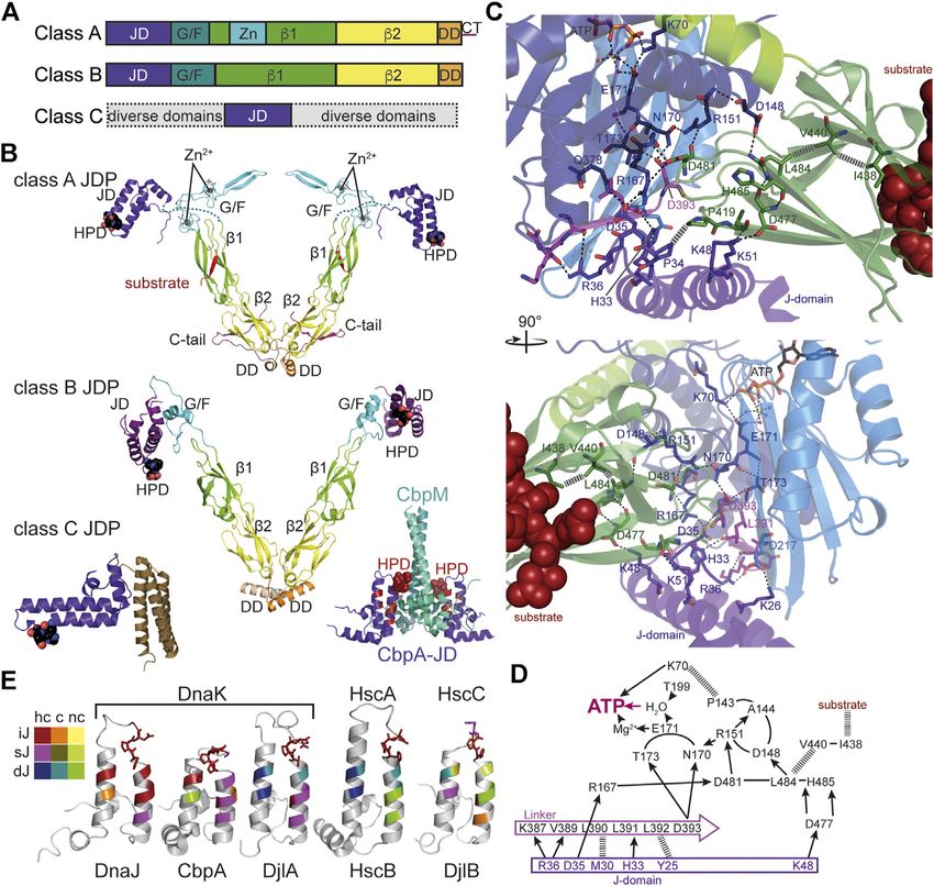
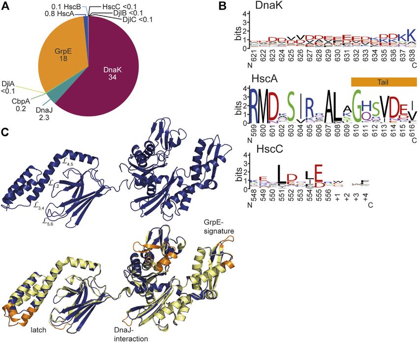
Mayer                                                                                                                               The Hsp70-Chaperone Machines in Bacteria

  FIGURE 1 | Diversity of Hsp70s and their functions in prokaryotic cells. (A), Diversity of functions of Hsp70s under optimal growth conditions (middle to left) and
  upon exposure to environmental and physiological stress (middle to right). Hsp70/DnaK (70) assists de-novo-folding of proteins, interacting with nascent chains already
  at the ribosome (1) and with folding intermediates after release from the ribosome (2). Folding intermediates and even native proteins may misfold, in particular under
  stress conditions, and become aggregation prone. Hsp70 prevents aggregation (3) and refolds the misfolded protein by unfolding (4) or target it for degradation (5).
  Under severe stress conditions protein aggregates are formed by coaggregation with sHSPs. Hsp70 targets ClpB (B) to the aggregates (6). ClpB solubilizes the
  aggregated proteins that are subsequently refolded by Hsp70 (7). Hsp70 disassembles homo-and heterooligomeric protein complexes like RepE-dimers and the
  λO·λP·DnaB complex (8). Proteins destined for insertion into the plasma membrane (e.g., DjlC) (9), or secretion into the periplasmic space (e.g., PhoA or curli) (10) are
  guided by Hsp70 and prevented from forming aggregates or amyloid fibrils (curli) (11) in the cytoplasm. Hsp70 also interacts with some native proteins like the heat shock
  transcription factor σ32 to keep them in an alter-native inactive conformation (12) and target them to degradation (13). (B), Domain organization of the three types of
  Hsp70s that exist in prokaryotes, DnaK, HscA and HscC. NBD, nucleotide binding domain (blue); CL, conserved linker (magenta); SBDβ, β-sandwich domain (dark
  green; light green: insertion in HscCs); SBDα, α-helical lid domain (chartreuse); black lines, C-terminal intrinsically disordered tails (for HscA also N-terminal extension);
  white bars, larger deletions in NBD and SBD of HscA and HscC as compared to DnaK. (C), Phylogenetic tree of different prokaryotic clades that contain organisms which
  have all three Hsp70s. ECOLI, Escherichia coli (γ-Proteobacteria); SALEN, Salmonella enteritidis (γ-Proteobacteria); PSEFL, Pseudomonas fluorescence
  (γ-Proteobacteria); RALSO, Ralstonia solanacaearum (β-proteobacteria); 9BURK, Paraburkholderia fungorum (β-proteobacteria); 9BACT, Acidobacteria bacterium
  (unclassified Acidobacteria); PLABA, Planctomycetes bacterium (unclassified Planctomycetes); HSPA1A, human Hsp70; HSPA8, human Hsc70 (for comparison). A
  more extensive phylogenetic tree can be found in Barriot et al. (2020).

the regulation of the heat shock response in many proteobacteria                             regulated by an intricate allosteric mechanism through ATP
(Matsui et al., 2008; Kobayashi et al., 2011; Schumann, 2016;                                binding and hydrolysis in their nucleotide binding domain
Schramm et al., 2017).                                                                       (NBD). Third, Hsp70s are targeted to client proteins by
   This enormous versatility of Hsp70s is based in three basic                               cochaperones of the J-domain protein (JDP) family, for
principles. First, with their tweezer-like polypeptide substrate                             example DnaJ, the prototype JDP, and for generalist Hsp70s
binding domain (SBD) Hsp70s bind short degenerative                                          the lifetime of the Hsp70-client complex is regulated by the
sequence motifs found in most proteins with high frequency.                                  nucleotide exchange factor (NEF) GrpE. In addition, Hsp70
Thus, the actions of Hsp70s are not limited by size or                                       cooperate with other families of chaperones, like the small
conformation of their clients, as long as the sequence motif is                              heat shock proteins (sHSPs, inclusion body binding proteins,
accessible. Second, binding of Hsp70s to client proteins is                                  IbpA, IbpB) (Veinger et al., 1998; Żwirowski et al., 2017), the

Frontiers in Molecular Biosciences | www.frontiersin.org                                 2                                                June 2021 | Volume 8 | Article 694012
Mayer                                                                                                  The Hsp70-Chaperone Machines in Bacteria

oxidative stress activated Hsp33 (Winter et al., 2005), the              35°C and cells exhibit a filamentous phenotype (Paek and Walker,
chaperonin (GroEL-GroES) (Langer et al., 1992), the Hsp90                1987). The ΔdnaK strain tends to accumulate a second site
(Genest et al., 2011; Morán Luengo et al., 2018), and the                suppressor mutation in the rpoH gene down-regulating
Hsp100/ClpB (Goloubinoff et al., 1999) chaperones and take               amount or activity of the heat shock transcription factor σ32,
over clients from them or relay clients to them.                         indicating that unchecked σ32 leads to a detrimental imbalance in
    Despite their involvement in such a large number of protein-         transcription (Bukau and Walker, 1990). Cells with the second
folding processes, Hsp70s are not strictly essential in many             site suppressor are still temperature sensitive but are not anymore
bacteria and two free-living bacterial species of the Aquificales         filamentous at 30°C. Similar observations were made for the
order, Desulfobacterium thermolithotrophum and Thermovibrio              α-proteobacterium Caulobacter crescentus (Schramm et al.,
ammonificans, have been described that do not encode for any              2017). Deletion of hscA increased the doubling time of E. coli
Hsp70, nor any of its JDP cochaperones or GrpE, and have                 by twofold in rich medium but not in minimal medium and
apparently lost these genes in the course of evolution (Warnecke,        combined deletion of hscA and dnaK increased the doubling time
2012). These strictly anaerobic, chemolithotrophic organisms             threefold as compared to wild type E. coli (Hesterkamp and
have a growth temperature optimum of 70 and 75°C,                        Bukau, 1998). However, plating efficiency was not altered.
respectively, and have a significantly reduced genome size that           Deletion of hscC did not decrease viability of E. coli at 30 and
is only about one third the size of the Escherichia coli genome.         37°C in rich medium and the deletion of either hscA or hscC or
Apparently, proteins can evolve to fold efficiently even at high          both together do not aggravate the temperature sensitivity
temperatures without the assistance of the Hsp70 chaperone               phenotype of a ΔdnaK strain (Kluck et al., 2002). Neither hscA
system. Consistently, the Hsp70 system is also absent in                 nor hscC could complement the temperature sensitivity
hyperthermophilic archaea, whereas it is present in their                phenotype of a ΔdnaK strain when overexpressed and
mesophilic relatives. However, the absence of the Hsp70                  overexpression of either hscA or dnaK in a ΔhscC strain does
system also comes with a price. Like Hsp90s (Rutherford and              not alleviate increased Cd2+ sensitivity, clearly showing the
Lindquist, 1998; Queitsch et al., 2002) and Hsp60s (Maisnier-            distinction between the different Hsp70s in E. coli (Kluck
Patin et al., 2005), Hsp70s buffer the accumulation of mutations         et al., 2002).
in the genome and therefore increase the evolvability of the                 Since DnaK is not only physiologically more important in
organism (Aguilar-Rodríguez et al., 2016; Kadibalban et al.,             E. coli, more widespread in the prokaryotic kingdom, and more
2016). In fact, proteins that depend strongly on Hsp70, as               closely related to human Hsp70, it has been for many years the
defined by Calloni and colleagues (Calloni et al., 2012), evolve          paradigm for Hsp70s and its molecular mechanism was
faster than proteins that do not depend on Hsp70 for folding             investigated in great detail. In the following I will mainly focus
(Aguilar-Rodríguez et al., 2016; Kadibalban et al., 2016).               on E. coli DnaK. Insights into structure and mechanism of
    The model organism Escherichia coli harbors three structurally       Hsp70s gained through studies on yeast and mammalian
and functionally distinct Hsp70s: DnaK that is found in all              Hsp70 are included when there is reason to believe that these
prokaryotes, with the exceptions mentioned above, and that is            features are also valid for the prokaryotic Hsp70 systems or to
the best-studied of all Hsp70s; HscA, an Hsp70 that is not found         point out particular distinctions.
in many bacteria and that is specialized to assist the assembly of
iron sulfur clusters (Vickery and Cupp-Vickery, 2007); and HscC,
a specialized Hsp70 that confers resistance to Cd2+-ions and UV          HSP70 DOMAIN STRUCTURE AND
irradiation through an unknown mechanism (Kluck et al., 2002)            FUNCTIONAL CYCLE
(Figure 1B). The differences in sequence and structure between
the three Hsp70s is quite remarkable including some deletions            Structure of DnaK-Like Hsp70s
and insertions in otherwise highly conserved regions (Figure 1B).        Bona fide Hsp70s like DnaK consist of an N-terminal nucleotide
In fact, E. coli DnaK shares more sequence identity with human           binding domain (NBD) of 385 amino acids connected via a
Hsp70 (48.4/61.9% identity/similarity), than with E. coli HscA           conserved linker to a polypeptide substrate binding domain
(39.3/56.6%) or E. coli HscC (27.8/46.8%), and HscA and HscC             (SBD) of around 240 residues (Figure 2A). The NBD is built
are also only distantly related to each other (28.9/46.7%). This         up of four subdomains (IA, IB, IIA, IIB) arranged in two lobes
becomes even more apparent in a phylogenetic tree where DnaK,            that are separated by a deep cleft at the bottom of which the
HscA and HscC segregate in clearly independent branches                  nucleotide binds with nanomolar affinity (Flaherty et al., 1990).
(Figure 1C) [see (Barriot et al., 2020) for a more extensive             ATP binding and hydrolysis involves rotation of the lobes relative
phylogenetic analysis]. This sequence divergence may have                to each other (Kityk et al., 2012). The SBD is subdivided in a
significant mechanistic distinctions but have only been                   β-sandwich subdomain (SBDβ) of around 110 residues, an
investigated to a limited extent. HscA and HscC are not found            α-helical subdomain (SBDα) of approximately 100 residues
outside the prokaryotic kingdom, though, in some fungi, Hsp70s           and a C-terminal intrinsically disordered region of some 30
that are specialized for iron sulfur cluster assembly emerged            residues. The polypeptide binding cleft is formed by the two
through convergent evolution (Schilke et al., 2006; Kleczewska           twisted four-stranded β-sheets of the SBDβ and two concentric
et al., 2020).                                                           pairs of upward protruding loops (Zhu et al., 1996). In the high
    Deletion of dnaK in E. coli leads to cold and heat sensitivity       affinity conformation, the SBDα docks onto two faces of the
with a very restricted growth temperature range between 20 and           SBDβ, stabilizing the inner loops (L1,2, L4,5) and forms a latch of

Frontiers in Molecular Biosciences | www.frontiersin.org             3                                      June 2021 | Volume 8 | Article 694012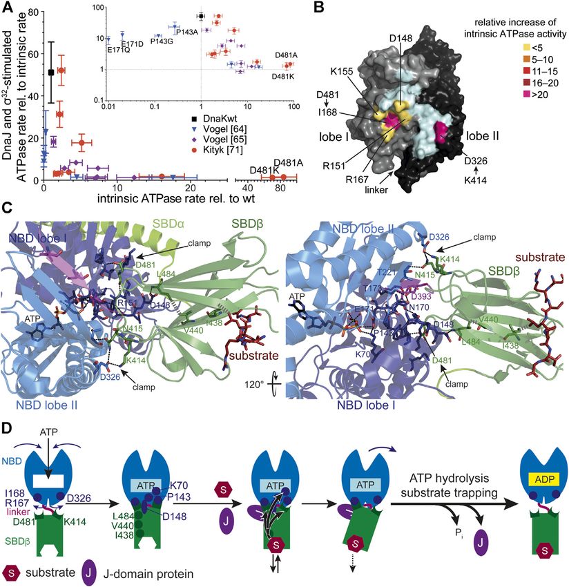
Mayer                                                                                                                               The Hsp70-Chaperone Machines in Bacteria

  FIGURE 2 | Structure and functional cycle of Hsp70s. (A), Cartoon representation of DnaK in the ADP·Pi·Mg2+-bound, SBD-closed and domain-undocked
  conformation (upper panel; PDB ID 2KHO (Bertelsen et al., 2009)) and ATP·Mg2+-bound, SBD-open, domain-docked conformation [lower panels in two orientations;
  4B9Q (Kityk et al., 2012)]. NBD lobe I (subdomains IA and IB), dark blue; NBD lobe II (subdomains IIA and IIB), marine blue; conserved linker, magenta; SBDβ, dark green;
  SBDα, chartreuse; ADP and ATP in space-filling representation colored according to atoms with carbon, black, oxygen, red, nitrogen blue and phosphorus,
  orange, Mg2+, green; substrate peptide, dark red in space-filling representation. (B), Overlay of the structures of the SBD of the ADP-bound, closed [SBDβ, dark green
  and SBDα, chartreuse; 1DKX (Zhu et al., 1996)] and the ATP-bound, open conformation [SBDβ, dark red and SBDα, orange, cut for space reasons; 4B9Q (Kityk et al.,
  2012)]. Substrate enclosing loops L1,2, L3,4, L4,5, and L5,6 are labeled. (C), space-filling representation of the crystal structure of the SBDβ in the closed, substrate-bound
  conformation (upper panel, dark green), and the open conformation in the ATP-bound state (lower panel, dark red); arch forming residues M404 and A429 are indicated.
  (D), ATPase cycle of Hsp70s. Partially folded or misfolded substrate polypeptides associate with and dissociate from Hsp70 with high rates in the ATP-bound open
  conformation. Substrates may also interact with the J-domain protein (JDP) co-chaperone. Substrate and JDP synergistically trigger ATP hydrolysis and transition to the
  closed, domain-undocked conformation. During this process substrate unfolding may occur. Alternatively or in addition, Hsp70 may select the more unfolded species
  from a equilibrium of different conformations. At physiological ATP concentrations nucleotide exchange is rate-limiting for substrate release. Nucleotide exchange factors
  (NEF) catalyze ADP release, and ATP rebinding stimulates substrate release that subsequently might fold into the native state or might rebind to Hsp70 for another folding
  cycle. Dark red indicate Hsp70 binding site. KD values for typical high-affinity binding peptides to ADP and ATP bound states are indicated. Association of the substrate to
  the ATP-bound state with subsequent ATP hydrolysis creates a non-equilibrium situation called ultra-affinity (De Los Rios and Barducci, 2014).

hydrogen bonds and a salt bridge with the outer loops (L3,4, L5,6).                          dissociates from the SBDβ and both subdomains dock onto
Therefore, the SBDα acts like a lid over the substrate binding                               different faces of the NBD resulting in a scissors like opening
groove and restricts substrate association and dissociation (Mayer                           of the β-sandwich and peptide enclosing loops (Figures 2B,C),
et al., 2000; Moro et al., 2004). This arrangement allows for the                            increasing the peptide association and dissociation rates by 100
tweezer-like binding to short, extended polypeptide segments of                              and 1,000-fold, respectively, decreasing the affinity for peptide
around five residues with a central hydrophobic sidechain                                     substrates by 10–50-fold (Schmid et al., 1994; Mayer et al., 2000;
inserting into a deep hydrophobic pocket that seems to be                                    Kityk et al., 2012; Qi et al., 2013). ATP binding and hydrolysis,
tailored for leucine. Upon ATP binding to the NBD, the SBDα                                  thus, allosterically regulate the affinity of Hsp70s for peptide and

Frontiers in Molecular Biosciences | www.frontiersin.org                                 4                                                June 2021 | Volume 8 | Article 694012
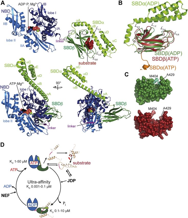
Mayer                                                                                                                             The Hsp70-Chaperone Machines in Bacteria

  FIGURE 3 | Allostery in Hsp70s. (A), Amino acid replacements outside the catalytic pocket that impair interdomain communication increase the intrinsic ATPase
  rate. A signature for allosteric proficiency of DnaK variants is the synergistic stimulation of DnaK’s ATPase rate by DnaJ and its protein client σ32. Thus, single turnover
  ATPase rates of wild type and mutant DnaK proteins in the presence of 50 nM DnaJ and 1 µM σ32 is plotted vs. their intrinsic ATPase rate. Defects in allostery reduce the
  DnaJ-σ32-stimulated ATPase rate. Inset, same data on logarithmic scales. Data taken from (Vogel et al., 2006a; Vogel et al., 2006b; Kityk et al., 2015). (B), Surface
  representation of the NBD of DnaK in the ATP-bound state (4B9Q) with lobe I and lobe II colored in gray and black, respectively, and the interface to which the SBDβ
  docks in light cyan, except for the indicated residues known to be involved in allostery themselves or contacted by residues of the SBDβ known to be involved in allostery.
  These are colored according to the relative increase of intrinsic ATPase activity when these residues are replaced themselves by alanine or if their pendant in the SBDβ is
  replaced by alanine (D481A) or isoleucine (K414I) [modified from (Mayer, 2018)]. (C), Intramolecular pathways of allostery. Polar (black dashed lines) and non-polar (gray
  hatched lines) interactions from the substrate to the catalytic center for ATP hydrolysis. Indicated are contacts (D481→I168 and K414→D326, N415→T221) that fix the
  NBD lobes in the rotated, ATP hydrolysis-incompetent state (clamp) (Kityk et al., 2015). Right panel rotate by 120° as compared to the left panel as indicated. The central
  leucin of the substrate peptide forms hydrophobic contacts with I438 on β-strand 4. This interaction is transmitted to V440 on strand four and further, through
  hydrophobic interactions, to L484 on β-strand 6. L484 forms hydrogen bond interactions with D148 that is connected through a rigid loop with P143. P143 contacts K70
  that forms a hydrogen bond with the γ-phosphate of ATP and stabilizes the transition state of hydrolysis. In this way binding of substrates is directly transmitted into the
  catalytic center. (D), Cartoon of ATP induced docking of SBDβ and NBD and substrate induced ATP hydrolysis and transition to the high affinity conformation of the
  SBDβ. SBDα is omitted for clarity. Indicated are ATP induced rotation of the NBD lobes and residues (D481→R167/I168; K414→D326) that form the clamp to prevent
  back rotation of the NBD lobes, as well as residues (I438, V440, L484, D148, P143, K70) that are important for transmission of the substrate binding signal to the catalytic
  center for γ-phosphate cleavage. The J-domain is important for tight coupling of substrate binding and signal transmission (more detailed in Figure 4).

protein substrates (Figure 2D). It is important to note that,                               concentration and 30°C) to compete efficiently with the
although the ability to prevent aggregation of a misfolded                                  aggregation reaction and in the ATP bound state the affinity
protein was the original definition of a molecular chaperone,                                for binding sites is too low (1–50 µM for good binders) to reduce
Hsp70s alone are generally not particularly apt to do so: In the                            the free concentration of aggregation prone species enough to
ADP-bound or nucleotide-free state the association rates to                                 prevent the concentration dependent oligomerization process of
binding segments are too low (ca. 104 M−1s−1 corresponding to                               misfolded client proteins. Therefore, Hsp70s need to encounter
a half-life for complex formation of ca. 1–2 min at 1 µM                                    their misfolded protein clients in the ATP bound low-affinity

Frontiers in Molecular Biosciences | www.frontiersin.org                                5                                                June 2021 | Volume 8 | Article 694012
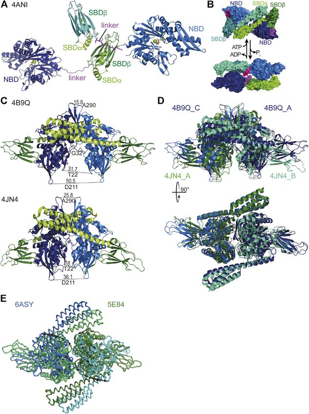
Mayer                                                                                                                                 The Hsp70-Chaperone Machines in Bacteria

  FIGURE 4 | Structure and function of J-domain proteins (JDPs). (A), Domain structure of the three classes of JDPs; JD, J-domain; G/F, glycine-phenylalanine rich
  region; β1/2, β-sandwich domain 1 and 2; Zn, Zn2+-finger domain; DD, dimerization domain; CT, C-terminal tail. (B), Cartoon representation of the crystal structures of
  JDPs; domains colored as in A; HPD motif in space-filling representation. Top panel, structure of the S. cerevisiae class A JDP Ydj1 since no structure for a class A JDP of
  prokaryotic origin has been solved to my knowledge. Composite of crystal structures 1NLT (Li et al., 2003) and 1XAO (Wu et al., 2005) and the NMR structure of the
  J-domain 5VSO (Schilke et al., 2017). The location of the J-domain is arbitrary as it is connected to the β1-domain by the flexible G/F-rich region shown as dashes. Middle
  panel, crystal structure of the class B JDP DnaJ of Thermus thermophilus [4J80, (Barends et al., 2013)]. Inset to the lower right, Inhibitory complex between the CbpM
  dimer (greencyan and deepteal) and two J-domains of the class B JDP CbpA (purple; 3UCS). Residues homologous to DnaJ residues that interact with DnaK in the co-
  crystal structure are colored in dark red. HPD-motif shown as spheres. Bottom left panel, crystal structure of the class C JDP HscB of E. coli [1FPO, (Cupp-Vickery and
  Vickery, 2000)]. (C), Zoom into the crystal structure of E. coli DnaK in complex with the J-domain of DnaJ [5NRO, (Kityk et al., 2018)], illustrating how the J-domain
  contacts the allosteric network of polar (black dashed lines) and non-polar (gray hatched lines) contacts connecting the substrate binding pocket with the catalytic center
  for ATP hydrolysis. Lower panel, rotated by 90° as compared to the upper panel as indicated. (D), Schematic representation of the interaction network contacted by the
  J-domain. Arrows indicate polar contacts; hatched lines indicate non-polar interactions; other lines indicate peptide backbone connections. (E), Structures of J-domains
  of DnaJ, CbpA, DjlA, HscB and DjlB colored according to conservation of residues interacting with DnaK in the co-crystal structure of DnaK and the J-domain of DnaJ
  [5NRO, (Kityk et al., 2018)]. NMR structures of J-domains of DnaJ [1XBL, (Pellecchia et al., 1996), and CbpA (2KQX, (Sarraf et al., 2010)]; crystal structure of the
  J-domain of HscB [1FPO, (Cupp-Vickery and Vickery, 2000)]; homology models of the J-domains of DjlA and DjlB using SWISS-Model (Bienert et al., 2017; Waterhouse
  et al., 2018). Color scheme indicated to the left; hc, highly conserved (>60% identity and >90% similarity in a CLUSTAL Ω alignment of 200 mutually less than 90%
  identical sequences; UniRef90 database), c, conserved (>80% similarity), nc, not conserved; iJ, identical as in E. coli DnaJ; sJ, similar as in E. coli DnaJ; dJ, different (non-
  conservative replacement) to the residue in E. coli DnaJ. HPD motif in stick representation.

conformation of the SBD with high substrate association rates                                 low amounting to one molecule of ATP hydrolyzed every
and then hydrolyze ATP to trap the client in the ADP bound                                    3–30 min (McCarty et al., 1995; Silberg and Vickery, 2000;
high-affinity conformation (Figure 2D). Consequently, ATP                                      Kluck et al., 2002). This intrinsic ATPase rate is stimulated by
hydrolysis is essential for Hsp70 action as has been                                          the client protein in synergism with a J-domain cochaperone to
demonstrate for several Hsp70s (Wawrzynów et al., 1995;                                       rates that allow binding to clients on the seconds timescale.
Elefant and Palter, 1999; Barthel et al., 2001; Lagaudrière-                                  Association of the client with the high association rates of the
Gesbert et al., 2002; Kumar and Tiwari, 2018). However,                                       ATP bound state and subsequent rapid ATP hydrolysis and
intrinsic ATP hydrolysis rates of Hsp70s are generally very                                   transition to the ADP bound state with low client dissociation

Frontiers in Molecular Biosciences | www.frontiersin.org                                  6                                                 June 2021 | Volume 8 | Article 694012
Mayer                                                                                                     The Hsp70-Chaperone Machines in Bacteria

rates creates a non-equilibrium situation that increases the              NBD (P143) stabilizes the ATP-bound state and upon
apparent affinity by several orders of magnitude, a property               replacement of this proline by glycine the enthalpy of
that was coined ultra-affinity (De Los Rios and Barducci,                  activation for ATP hydrolysis decreases to 50% of the value
2014). Of note, in the nucleotide-free or ADP-bound state                 for wild-type DnaK (Vogel et al., 2006a). Second, the SBDβ
DnaK is not always in the high-affinity conformation but the               clamps down the rotated position of the NBD lobes resulting
SBDα lid occasionally opens allowing for association and                  in a geometry of the catalytic residues in the ATP binding pocket
dissociation of bound polypeptides (Mayer et al., 2000; Kityk             that is unfit for ATP hydrolysis (Figure 3C). This clamp
et al., 2012; Lai et al., 2017). Conversely, in the ATP-bound state       contributes some 30% to the enthalpy of activation as deduced
DnaK is not always in the low-affinity conformation and the                from the difference in activation enthalpy for ATP hydrolysis for
SBDα may detach from the NBD occasionally. Therefore, in both             DnaKwt and DnaK(2–385) (Vogel et al., 2006a). The two residues
nucleotide-bound states Hsp70s are in an equilibrium between              in the SBDβ that contributes most to this clamping of the NBD
different conformations with the nucleotides biasing the rates of         are D481, interacting with the backbone of I168 in lobe I and
transition.                                                               K414, interacting with D326 in lobe II (Figures 3B,C).
                                                                          Replacement of D481 by alanine or K414 by isoleucine
Allosteric Mechanism                                                      increases the intrinsic ATPase activity by 80-fold and 25-fold,
Genetic screens and structural studies on the individual domains          respectively (Kityk et al., 2015). Binding of a polypeptide
of DnaK revealed single residues that are important for the               substrate to the substrate binding pocket triggers ATP
allosteric mechanism (Burkholder et al., 1994; Laufen et al.,             hydrolysis by acting through a defined intramolecular signal
1999; Montgomery et al., 1999; Vogel et al., 2006a; Vogel                 transduction pathway involving V440 and L484 in the SBDβ
et al., 2006b; Smock et al., 2010; Kumar et al., 2011). A                 and D148 in the NBD (Kityk et al., 2015) (Figures 3C,D).
general feature of amino acid replacements outside the ATP                Replacement of any of these residues with alanine leads to a
binding pocket itself that disturb the allosteric regulation is an        complete loss of substrate stimulation of the ATPase activity but
increased intrinsic ATPase activity (Figure 3A). It can be                not of the stimulation of the ATPase activity by DnaJ.
concluded from this observation that allosteric coupling of the
NBD and SBD inhibits γ-phosphate cleavage in the NBD. Those
amino acid replacements that have the largest impact on intrinsic         HSP70 INTERACTION WITH
ATPase rate indicate residues that are most important for                 COCHAPERONES
inhibiting the ATPase activity. However, the isolated NBD has
an ATPase activity as low as full-length DnaK, arguing against an         J-Domain Proteins: Hsp70 Targeting
inhibitory effect of the SBD. This conundrum was solved by the            Factors
discovery that the highly conserved linker between NBD and SBD            JDPs are modular multi-domain proteins that are essential
has an important impact on interdomain communication and on               cochaperones of Hsp70s. Common to all JDPs is the so-called
the intrinsic ATPase activity of the NBD as well. Prolonging the          J-domain, an α-helical hair-pin domain of generally 70–75
NBD with the linker residues [386VKDVLLLD393;                             residues in length, which is essential for triggering in
DnaK(1–393)] increased the ATPase rate 40-fold (Vogel et al.,             synergism with protein substrates ATP hydrolysis in Hsp70s.
2006b; Swain et al., 2007; English et al., 2017) and this effect is       The additional domains of JDPs allow them to interact with
abrogated or greatly diminished when the hydrophobic residues             protein clients of Hsp70s or to be localized within the cell where
of the linker or D393 are replaced by alanine. Similar observations       Hsp70 clients appear, e.g., at the ribosome or at translocation
were also made for HscA (Alderson et al., 2014). These intriguing         pores. Their main function is to target Hsp70s to client proteins
observations fall into place in the structure of DnaK in the ATP-         and trigger client trapping. JDPs are generally divided into three
bound open conformation that allowed to trace these residues              classes according to the number of domains they have in common
complemented by additional residues into a network of hydrogen            with the prototype of JDPs, E. coli DnaJ (Kampinga and Craig,
bonds and hydrophobic interactions that mediate interdomain               2010) (Figures 4A,B). Class A JDPs are 360–400 amino acids
communication and allosteric regulation (Kityk et al., 2012; Kityk        long and have a domain architecture like DnaJ: an N-terminal
et al., 2015) (Figures 3B–D–D). In general, these residues are            J-domain followed by a glycine-phenylalanine rich region (G/
highly conserved in Hsp70s from bacteria to humans and their              F-region), two homologous β-sandwich domains with a zinc-
presence is indicative for an allosteric mechanism. Albeit, some of       finger inserted in the first of the two domains, and a C-terminal
the residues are conservatively replaced in some branches of the          dimerization domain with an intrinsically disordered tail. Client
Hsp70 tree with consequences for the equilibrium between the              binding sites are found in the β-sandwich domains (Jiang et al.,
different conformational states of Hsp70s (Zhuravleva et al.,             2019) and also the zinc-finger seems to be involved in substrate
2012).                                                                    binding and prevention of aggregation activity (Linke et al.,
    Comparison of the crystal structures of Hsp70s in the ADP             2003). Class B JDPs are in general 260–360 amino acids long
and ATP bound states revealed that upon ATP binding to Hsp70              and differ from DnaJ by the lack of the zinc-finger and the
the two lobes of the NBD rotate relative to each other and allow          C-terminal tail. Both, class A and class B JDPs are considered as
the SBDβ to dock onto the NBD. Two effects are responsible for            general JDPs that are able to bind to essentially all partially folded,
the low ATP hydrolysis rates and thus the high enthalpy of                misfolded and aggregated proteins. Both classes seem to form
activation of γ-phosphate cleavage. First, a single proline in the        V-shaped dimers with the protomers linked together through a

Frontiers in Molecular Biosciences | www.frontiersin.org              7                                        June 2021 | Volume 8 | Article 694012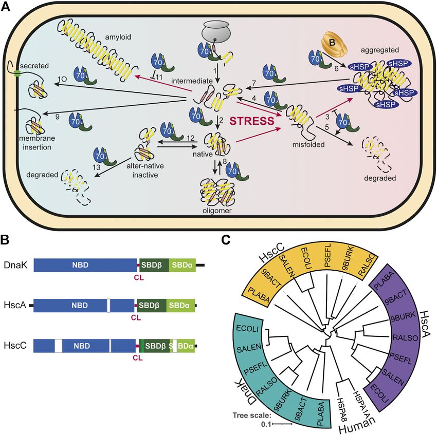
Mayer The Hsp70-Chaperone Machines in Bacteria
flexible C-terminal hinge (Sha et al., 2000; Wu et al., 2005; of organisms onto E. coli DnaJ to study their functionality [e.g.,
Barends et al., 2013) (Figure 4B). Thus, they could bind (Kelley and Georgopoulos, 1997; Nicoll et al., 2007; Maillot et al.,
simultaneously to at least two sites within misfolded 2019)]. However, there is also specificity as some of the residues of
polypeptide and aggregates, which might be an efficient way to the J-domain that interact in the crystal structure with DnaK are
distinguish native from non-native proteins. Class C JDPs are different in specific subgroups of JDPs in particular those that do
very heterogeneous with 54 to more than 1,000 amino acids in not interact with DnaK and well conserved within the respective
length and only share with DnaJ the J-domain that might be JDP subfamily as sequence alignments revealed (Figure 4E). The
found anywhere within the sequence. They may contain a functional significance of these differences has not been analyzed
number of other domains, most notably specific protein- in detail and it is currently not known, which of the differences
protein interaction domains, DNA and RNA binding domains, are the result of coevolution of functional Hsp70-JDP pairs and
and transmembrane regions. An extensive analysis of JDP which are the result of phylogenetic relationship. An extensive
associated domains in prokaryotes can be found in (Barriot phylogenetic analysis of prokaryotic JDPs was recently published
et al., 2020). In some cases, it seems that the J-domain was an (Barriot et al., 2020).
add-on late in evolution to make cellular processes more efficient A recent NMR study elucidated that class A and class B JDPs
by providing chaperone power (Sahi et al., 2010). E. coli contains bind polypeptides in a highly dynamic multivalent manner using
one class A (DnaJ), one class B (CbpA) and four class C JDPs up to four low-affinity sites, one in each of the four β-sandwich
(HscB, DjlA, DjlB, and DjlC), whereby DnaJ, CbpA, and DjlA domains of the JDP-dimer (Jiang et al., 2019). This explains the
functionally interact with DnaK; HscB with HscA (Silberg et al., earlier observation that JDPs generally bind peptides with much
1998); and DjlB and DjlC with HscC (Kluck et al., 2002) lower affinity than protein clients (Rüdiger et al., 2001). Such a
(Figure 4E). binding mode has two consequences. First, JDPs only bind
How the J-domain stimulates ATP hydrolysis was recently proteins stably when a sufficient number of binding sites for
elucidated by crystallization of the J-domain of E. coli DnaJ in the JDP are exposed, which is generally only the case in the
complex with DnaK in the ATP bound state (Kityk et al., 2018) nascent, not yet folded, and the misfolded state. The more
(Figure 4C). The J-domain binds on top of the interdomain binding sites are exposed within a polypeptide in a suitable
linker that is important for the stimulation of the ATPase activity geometry, the higher the overall affinity of JDPs to the client
and interacts with NBD and SBDβ (Vogel et al., 2006b; Swain due to the avidity effect. This also explains why the human class B
et al., 2007). It is positioned by electrostatic interaction between JDP DnaJB1 distinguishes α-synuclein amyloid fibrils from the
positively charged residues in the J-domain (R22, K26, R27, K48, intrinsically disordered monomer: at least two binding sites in
K51) and negatively charged residues in the NBD (E206, D211, neighboring α-synuclein protomers within the amyloid fibril are
E217) and SBDβ (D477) as had been proposed based on NMR necessary for high affinity interaction (Gao et al., 2015; Wentink
and computational data (Ahmad et al., 2011; Malinverni et al., et al., 2020). Second, such a binding mode allows for rapid
2017; Tomiczek et al., 2020). Genetic screens had identified the association and dissociation of individual binding sites from
highly conserved histidine-proline-aspartate (HPD) motif as the JDP favoring an efficient transfer of the client onto
essential for the functional interaction of the J-domain with Hsp70s. The NMR investigation further revealed that JDPs
Hsp70. The replacement of histidine or aspartate within this mainly interact with amino acid sidechains and not with the
motif for glutamine or asparagine, respectively, abrogated the backbone (Jiang et al., 2019), consistent with a binding mode that
ability of the J-domain to stimulate the ATPase activity of Hsp70s was proposed earlier based on peptide library scanning (Rüdiger
in every system tested so far [e.g., (Wall et al., 1994; Tsai and et al., 2001) and with hydrogen exchange mass spectrometry data
Douglas, 1996; Kelley and Georgopoulos, 1997; Liu et al., 1998; (Rodriguez et al., 2008).
Chevalier et al., 2000; Morgan et al., 2001; Mokranjac et al., Interestingly, the yeast class A JDP Ydj1 sports an intrinsically
2003)]. H33 of the DnaJ HPD motif forms a hydrogen bond with disordered C-terminal tail that binds to the substrate binding site
the backbone carbonyl of L391 of the interdomain linker of in the second β-sandwich domain and seems to compete with
DnaK, P34 forms hydrophobic contacts to P419 in the SBDβ client binding (Wu et al., 2005; Jiang et al., 2019) (Figure 4B).
of DnaK, and D35 forms hydrogen bonds to R167 and Q378 of Similar disordered tails are also found in prokaryotic class A JDPs
DnaK. L391 had previously been implicated in allosteric as multiple sequence alignments reveal. A competition of the
regulation (Kumar et al., 2011) and R167 in interaction with C-terminal tail with substrates for binding to the second
DnaJ (Suh et al., 1998). The J-domain interacts directly with the β-sandwich domain might limit the overall affinity of JDPs to
network of hydrogen bonds that converge in two branches onto very hydrophobic substrates by autoinhibition to prevent quasi
the γ-phosphate of the ATP (Figures 4C,D). Intriguing was the irreversible binding. It also might facilitate client transfer onto
finding that the J-domain contacts the SBDβ through a salt bridge Hsp70s or release of the JDP from the substrate polypeptide after
(J-domain K48→DnaK-D477) and thereby seems to stabilize the transfer of a single binding site to Hsp70. Such an autoinhibitory
signal transduction pathway that transmits the signal of the C-terminal tail is missing in class B JDPs. Intriguingly, eukaryotic
bound client to the NBD for triggering ATP hydrolysis (Kityk class B JDPs seem to be self-inhibited in a different way by a small
et al., 2018). The residues of the J-domain that interact with DnaK α-helix in the G/F-region that binds to the J-domain and
are well conserved in JDPs known to interact with a DnaK-type apparently blocks its interaction with Hsp70 (Faust et al.,
Hsp70 (Figure 4E), explaining the promiscuity of J-domains as 2020). This block is relieved by binding of the EEVD motif to
demonstrated by grafting J-domains from JDPs of a wide variety the first β-sandwich domain (Li et al., 2006; Yu et al., 2015; Faust
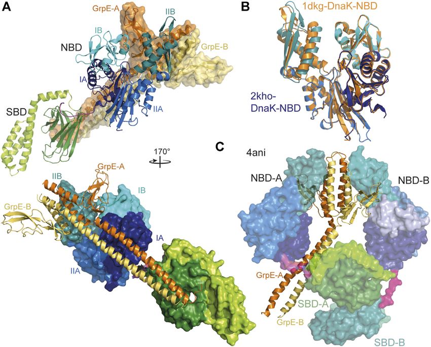
Frontiers in Molecular Biosciences | www.frontiersin.org 8 June 2021 | Volume 8 | Article 694012Mayer The Hsp70-Chaperone Machines in Bacteria FIGURE 5 | Interaction of GrpE with DnaK. (A), overlay of the crystal structure of E. coli GrpE in complex with the NBD of E. coli DnaK [1DKG, (Harrison et al., 1997)] onto the solution structure of E. coli DnaK in the ADP-bound conformation [2KHO, (Bertelsen et al., 2009)]. Top panel, DnaK as cartoon, GrpE in surface representation. Bottom panel, rotated by 170° as compared to the top panel and DnaK in surface representation and GrpE as cartoon. (B), overlay of the NBD of E. coli DnaK in the absence (colored different shades of blue according to subdomains) and presence (orange) of GrpE indicating the 14° outward tilt of subdomain IIB. (C), crystal structure of two molecules of Geobacillus kaustophilus DnaK (surface representation) in complex with the GrpE dimer (cartoon). et al., 2020). The molecular mechanism of how binding of the background of a ΔdnaJ strain phenocopies a ΔdnaJ ΔcbpA strain. EEVD motif unlocks the J-domain of class B JDPs is still a Why the inhibition of the CbpA-DnaK interaction is mystery. When the EEVD motif at the C-terminus of the advantageous is not clear. eukaryotic Hsp70 is deleted it still can refold a misfolded In eukaryotic Hsp70 systems JDPs of class A and class B model substrate in cooperation with the class A JDP but not cooperate in protein disaggregation (Nillegoda et al., 2015). anymore with a class B JDP. Whether such an inhibitory However, this does not seem to be the case for prokaryotic mechanism also exists in prokaryotic class B JDPs is currently JDPs (Nillegoda et al., 2017). unknown. The sequence of prokaryotic Hsp70s generally does not end in an EEVD motif. However, many DnaK-type prokaryotic Hsp70s contain a glutamate and aspartate rich Nucleotide Exchange Factors: Timing the sequence close to the very C-terminus and deletion of the last Hsp70-Client Interaction seven residues including an EEV sequence in E. coli DnaK Since at physiological ATP concentrations nucleotide exchange is reduces its ability to complement the temperature sensitivity rate-limiting for release of bound polypeptide clients, NEFs phenotype of a ΔdnaK strain (Smock et al., 2011). regulate the lifetime of the Hsp70-client complex. Currently, Furthermore, the crystal structure of the class B JDP of four evolutionarily unrelated families of NEFs for Hsp70s are Thermus thermophilus revealed an α-helix within the known that use different mechanisms to open the nucleotide G/F-region that was docked onto the J-domain (Figure 4B) binding cleft of Hsp70s and thereby to accelerate nucleotide (Barends et al., 2013). Furthermore, CbpA is inhibited in vitro dissociation. Three of the four families of NEFs are only found and in vivo by a small protein CbpM that is encoded in the same in eukaryotic cells and are not further discussed here [for detailed operon downstream of cbpA in E. coli and conserved in discussion see (Bracher et al., 2015; Mayer and Gierasch, 2019)]. γ-proteobacteria (Chae et al., 2004; Chenoweth et al., 2007). In prokaryotes, mitochondria and chloroplasts nucleotide CbpM is specific for CbpA and does not inhibit the exchange in Hsp70s is stimulated by GrpE, a homodimeric interaction of DnaJ with DnaK. CbpM binds to the J-domain protein that consists of an N-terminal intrinsically disordered of CbpA in a way that blocks access to DnaK (Sarraf et al., 2014) region of some 40 residues followed by an unusually long (Figure 4B lower right panel). Overexpression of CbpM in the α-helical dimerization domain and a C-terminal β-sheet Frontiers in Molecular Biosciences | www.frontiersin.org 9 June 2021 | Volume 8 | Article 694012
Mayer                                                                                                         The Hsp70-Chaperone Machines in Bacteria

domain. GrpE interacts with DnaK in an asymmetric 2-to-1                        proteins in E. coli cells deleted for the ribosome-associated
complex, inserting the β-sheet domain into the nucleotide                       chaperone trigger factor. In fact, it was shown that the DnaK-
binding cleft and opening it by tilting subdomain IIB by 14°                    DnaJ-GrpE team can keep proteins in an active state under
outward (Harrison et al., 1997) (Figures 5A,B). In contrast,                    conditions when the thermodynamic equilibrium would drive
Geobacillus kaustophilus GrpE and DnaK crystallized in a                        the protein into the denatured state (Goloubinoff et al., 2018).
GrpE2·DnaK2 complex that was nevertheless asymmetric (Wu                        Thus, DnaK uses ATP to continuously drive the protein out of
et al., 2012) (Figure 5C). So far there is no evidence that this                thermodynamic equilibrium.
structure represents a functional state that also exists in other                   At 37°C most proteins are bound transiently by DnaK in the
prokaryotic organisms, and that GrpE in this way triggers                       nascent state during synthesis at the ribosome. This observation is
nucleotide exchange and thus client release by two Hsp70                        well explained by peptide library scanning data (Rüdiger et al.,
chaperones in a coordinated fashion. GrpE was also proposed                     1997) that revealed the recognition motif for DnaK binding. This
to induce polypeptide client release in the absence of ATP. This                motif consists of a core of five residues enriched in hydrophobic
hypothesis was based on the position of GrpE in the crystal                     amino acids with a strong preference for leucine, flanked by regions
structure suggesting that the intrinsically disordered region at the            enriched in positively charged residues. Negatively charged
N-terminus of GrpE, which is well conserved in length within                    residues disfavor DnaK binding. Such motifs are found on
prokaryotic GrpE homologues, might be close to the polypeptide                  average every 30–40 residues in practically all proteins except
binding groove of Hsp70 (Figures 5A,B). However, careful                        for intrinsically disordered proteins that are generally depleted
analysis revealed that GrpE does not accelerate client                          of hydrophobic amino acids. In the structure of most proteins the
dissociation but prevents rebinding by competing with its                       DnaK binding motifs are found in the hydrophobic core and only
N-terminal tail for the client binding groove (Brehmer et al.,                  accessible in the nascent and denatured state.
2004). GrpE might thus act in a similar way as was recently shown                   The crystal structures of the SBD of DnaK in complex with
for the HspBP1 NEF in eukaryotic cells (Gowda et al., 2018).                    different substrate peptides (Zhu et al., 1996; Zahn et al., 2013)
    Some Hsp70s do not seem to need a NEF since they have a very                show the peptide well engulfed by the upward protruding loops
high intrinsic ADP dissociation rate (Brehmer et al., 2001). This               forming hydrophobic contacts with the sidechains of the peptide
raises the question why NEFs are needed at all, since ADP                       and hydrogen bonds with the peptide backbone (Figures 6A,B).
dissociation rates could be tuned to the optimal value. Such an                 Therefore, DnaK in contrast to DnaJ distinguishes well between
optimal tuning might be advantageous for Hsp70s that interact with              peptides made from L-and D-amino acids (Rüdiger et al., 2001).
one or a small number of defined clients, but not for Hsp70s that are            The SBDα lid forming a latch of a salt bridge and hydrogen bonds
generalists and interact with a wide variety of clients that need               with the outer loop contributes to the affinity of DnaK to its
different residence times on Hsp70. Maybe stochastic interaction of             substrate peptide decreasing peptide dissociation rates
GrpE with DnaK will yield at least in a fraction of the cycles the exact        substantially (Mayer et al., 2000; Moro et al., 2004). However,
optimal lifetime of the DnaK-client complex. Another advantage of               electron paramagnetic resonance spectroscopy and Förster
NEFs could be localized nucleotide exchange. Some eukaryotic NEFs               resonance energy transfer (FRET) measurements revealed that
are targeted to specific locations within the cell, for example the ER           the lid does not necessarily close entirely over bound protein
or plasma membrane, and for these NEFs it seems plausible that                  clients (Marcinowski et al., 2011; Schlecht et al., 2011). In fact,
nucleotide exchange and therefore release of client from Hsp70s                 optical tweezer experiments showed that DnaK can bind to
occurs at specific subcellular sites. In contrast, GrpE in E. coli is            folding intermediates preventing their unfolding against
homogenously distributed throughout the cell at optimal growth                  external pulling force (Mashaghi et al., 2016). For the latter
conditions, as well as, during heat shock (Kumar and Sourjik, 2012),            binding mode, the lid was more important than the peptide
refuting such a hypothesis for GrpE. Alternatively, NEFs could link             binding groove as an amino acid replacement that lowered the
nucleotide exchange and thereby polypeptide release to                          affinity for peptide binding to 1/40th of the wild type affinity (KD
environmental conditions. At heat shock temperatures above                      40-fold increased), had little effect on the force induced unfolding
42°C for E. coli or 85°C for Thermus thermophilus GrpE starts to                of the client protein, whereas a truncation of the lid in the middle
unfold reversibly and becomes inactive (Grimshaw et al., 2001;                  of helix B abrogated the ability of DnaK to counteract force
Groemping and Reinstein, 2001; Grimshaw et al., 2003). Such an                  induced client unfolding. Therefore, the picture that the crystal
unfolding would slow down nucleotide exchange and client release                structures convey may be representative for Hsp70 binding to
from Hsp70 under condition when reaching the native state is                    nascent polypeptide chains but not so much for interaction of
unlikely. Upon return to normal growth temperatures GrpE refolds                Hsp70 with folding intermediates or misfolded proteins.
and becomes active again.                                                           Moreover, the C-terminal intrinsically disordered region also
                                                                                seems to contribute to the interaction with client proteins as
                                                                                C-terminally truncated DnaKs are less efficient in
HSP70 INTERACTION WITH SUBSTRATES                                               complementing the temperature sensitivity phenotype of a
                                                                                ΔdnaK strain than wild type DnaK and also less efficiently
A proteomic study showed that DnaK in E. coli interacts with at                 assists refolding of denatured model clients in vitro (Smock
least 700 proteins among which are some 180 aggregation-prone                   et al., 2011). In addition, electron paramagnetic resonance
proteins that remained bound to DnaK for an extended period of                  spectroscopy using a nitroxide label in the C-terminal tail
time (Calloni et al., 2012). This number increases to some 1,000                indicates high mobility of the tail in the absence of a client

Frontiers in Molecular Biosciences | www.frontiersin.org                   10                                      June 2021 | Volume 8 | Article 694012
Mayer The Hsp70-Chaperone Machines in Bacteria
FIGURE 6 | Interaction of Hsp70s with peptide substrates. (A,B), crystal structure of E. coli DnaK SBD in complex with a peptide substrate (NRLLLTG) in space-
filling representation (A) and as cartoon (B) [1DKX, (Zhu et al., 1996)]. Polar contacts between SBDβ and SBDα, as well as, between SBDβ and substrate peptide are
shown as black dashed lines. Arch forming residues M404 and A429 are labeled. Lower panel, SBDβ rotated as indicated with substrate peptide in space-filling
representation and N-and C-terminus of the bound peptide labeled with N and C. (C,D), crystal structure of the SBD of E. coli HscA in complex with a peptide
(ELPPVKIHC) comprising the interaction sequence in IscU [1U00, (Cupp-Vickery et al., 2004)] in space-filling (C) and cartoon (D) representation. Arch forming residues
M401 and F426 are labeled. Whether the single hydrogen bond between SBDα and outer loops of SBDβ functions as a latch is unclear. Lower panel, SBDβ rotated as
indicated with substrate peptide in space-filling representation and N-and C-terminus of the bound peptide labeled with N and C.
protein or in the presence of short peptide substrate but restricted within the 53-residue client protein and in some cases two DnaK
mobility in the presence of a misfolded protein client, suggesting molecules could bind to the same client molecule (Rosenzweig
direct interaction of the tail with the misfolded client protein. et al., 2017). Single molecule FRET measurements monitored a
How are Hsp70s able to refold denatured inactive proteins to large expansion of a protein in the presence of DnaJ and DnaK
the native active state? Several studies suggest that the major and ATP (Kellner et al., 2014). Albeit, it should be noted that
action of Hsp70s is local unfolding. Sharma and colleagues found rhodanese the model substrate used in this study cannot be
that thioflavin T fluorescence and protease resistance of a refolded by the DnaK-DnaJ-GrpE chaperone team but requires
misfolded protein client decrease upon addition of DnaK, the GroEL-GroES machinery (Mendoza et al., 1991; Mayhew
DnaJ, GrpE and ATP (Sharma et al., 2010), indicating et al., 1996). A more recent hydrogen exchange mass
unfolding of the client. NMR experiments with a small single spectrometry and single molecule FRET study using luciferase
domain protein indicated that DnaK binds to a transiently as DnaK model client revealed also extensive unfolding by DnaK
unfolding state of the protein (conformational selection) and (Imamoglu et al., 2020). The unfolding was achieved by binding
keeps the protein in a state devoid of tertiary structure but that of several DnaK molecules to a single misfolded client protein. It
still contained secondary structure distal of the DnaK binding site could be imagined that at an initial stage DnaK binds to an
(Sekhar et al., 2015; Sekhar et al., 2016; Sekhar et al., 2018). exposed site and prevents this hydrophobic region to associate
Interestingly, in this case the conformation of the client protein with similar regions to form aggregates. As the bound client
was independent of the nucleotide state of DnaK, suggesting that protein undergoes thermal movements additional sites are
DnaK did not alter the conformation of the client in the binding exposed that then can be bound by DnaK. Alternatively or in
process. However, DnaK could bind to four different binding sites addition, DnaJ may play a more active role in the unfolding
Frontiers in Molecular Biosciences | www.frontiersin.org 11 June 2021 | Volume 8 | Article 694012Mayer The Hsp70-Chaperone Machines in Bacteria process. Since DnaJ interacts with sidechains of hydrophobic molecules preventing folding to proceed and causing a deadlock. amino acids and does not need the peptide backbone for Such a deadlock can be resolved by the Hsp90 chaperone HtpG of interaction, it could scan the surface of misfolded proteins for E. coli (Morán Luengo et al., 2018). This cooperation between regions prone to aggregation. DnaJ was also shown to induce Hsp70 and Hsp90 chaperones is also found in eukaryotic cells partial unfolding (Rodriguez et al., 2008; Kellner et al., 2014) that and does not require the Hsp70-Hsp90 organizing protein Hop could favor exposure of DnaK binding sites. Furthermore, (Bhattacharya et al., 2020). entropic pulling was introduced as mode of action for Hsp70- mediated force exertion. Originally this concept was introduced to explain import of polypeptides into the mitochondrial matrix HSP70 COMPLEXES–A NEW MODE OF and for solubilization of protein aggregates (De Los Rios et al., HSP70 ACTION? 2006). Briefly, translocating polypeptide chains that reach the mitochondrial matrix through the Tim23 import pore are bound Hsp70 oligomerization/polymerization in the nucleotide-free or close to the membrane by the matrix resident Hsp70. Since Hsp70 ADP bound state has been known for a number of years (Schmid constitutes a bulky entity that restricts the conformational et al., 1985; Freiden et al., 1992; Blond-Elguindi et al., 1993; freedom of the incoming polypeptide this state has a low Benaroudj et al., 1995; King et al., 1995; Schönfeld et al., 1995; entropy and entropy increases as the Hsp70 moves away from Angelidis et al., 1999; Thompson et al., 2012; Preissler et al., the membrane taking the bound polypeptide with it. As chemical 2015). More recently, based on the dimer assembly in crystal reaction can be driven by increasing entropy, this mechanism structures Hsp70-dimerization was proposed to also occur in the leads to import of the polypeptides and exerts a considerable force ATP bound state (Sarbeng et al., 2015). on the polypeptide, driving unfolding of the transport protein on Hsp70 oligomerization in the ADP bound state or upon ATP the other side of the membrane. The entropic pulling force hydrolysis has been suggested to be substrate-like binding of decreases with increasing polypeptide length translocated into Hsp70 to itself based on the fact that 1) ATP converts the the matrix and reaches zero at a translocated length of about 30 oligomer into monomers, which is analog to substrate release residues, whereupon a second Hsp70 has to bind the incoming (Schmid et al., 1985); 2) substrates could compete with chain close to the membrane. Experimental proof for such a mode oligomerization (Freiden et al., 1992; Angelidis et al., 1999); 3) of action was recently achieved for the disassembly of trimeric JDPs catalyze this type of interaction similar to substrate trapping human heat shock transcription factor (Kmiecik et al., 2020). (King et al., 1995). 4) Mutations that abrogate ATPase activity or Hsp70s bind close to the trimerization domain and monomerize decrease the affinity for substrates reduce oligomerization Hsf1 trimers. If the Hsp70 binding site is moved away from the tendency (Thompson et al., 2012; Preissler et al., 2015). More trimerization domain along an intrinsically disordered region, precisely, crystallographic and biochemical data suggest that the Hsf1 monomerization occurs at lower rates and cease when the SBD of one Hsp70 binds to the highly conserved hydrophobic binding site is 20 or more residues away from the trimerization NBD-SBD linker (KDVLLLD) of a second Hsp70 molecule domain. Binding of several Hsp70 to a single Hsf1 trimer (Chang et al., 2008a; Preissler et al., 2015; Preissler et al., accelerates monomerization, providing additional evidence for 2020) (Figure 7A). entropic pulling as physical principal for the reaction, as local Such a mode of interaction would have the consequence that crowding would be expected to increase the entropic pulling the Hsp70 engaged with the linker of another Hsp70 molecule force. Local crowding also seems to drive Hsp70 action in clathrin would not be able to bind clients. Thus, oligomerization could be uncoating (Sousa et al., 2016) and in the fragmentation of a mean for inactivation of Hsp70s when they are in unwanted α-synuclein fibrils (Wentink et al., 2020). It was also suggested excess. This function was proposed to neutralize excess of the that Hsp70s facilitate the sliding of nascent chains through the endoplasmic reticulum Hsp70 BiP in the wake of the unfolded ribosomal exit tunnel by entropic pulling. Translation elongation protein response. A dynamic monomer-oligomer equilibrium pauses under conditions in which Hsp70 activity is limiting, as could rapidly adapt the amount of active BiP to fluctuations in during heat shock, sever proteotoxic stress, or upon expression of unfolded protein load (Preissler et al., 2015). a dominant negative Hsp70. Such a pausing is not observed when A different type of Hsp70 dimer was recently proposed based intracellular Hsp70 concentrations are increased prior to stress on cross-linking and native mass-spectrometry data. This dimer, exposure (Liu et al., 2013; Shalgi et al., 2013). Moreover, which is also believed to be promoted by JDPs, is envisioned to ribosomal profiling revealed that translation speed increases contain the two Hsp70 molecules in an anti-parallel arrangement, when the yeast Hsp70 Ssb1 binds to the nascent chain which with the SBD of one Hsp70 being close to the NBD of the other, would be consistent with Ssb1 speeding-up translation by without engaging the interdomain linker like a substrate facilitating the sliding of the nascent chain through the (Morgner et al., 2015) (Figure 7B). Most of the data provided ribosomal exit tunnel by entropic pulling (Döring et al., 2017). in this publication are also consistent with the substrate-type Similarly, entropic pulling could lead to stepwise unfolding of a oligomerization model described above. An exception is that an misfolded protein when several Hsp70s and a JDP bind to the Hsp70 variant predicted to have a lower affinity for clients forms protein creating local crowding and a state of low entropy. dimers to a similar extend as the wild-type protein, arguing for a At physiologically high concentrations of DnaK (15–20 µM), different type of interaction. It was proposed that such an the association rates of new DnaK molecules binding to the client arrangement aids the loading of a native client onto Hsp90, might be higher than the dissociation rate of already bound DnaK albeit without supporting evidence. Frontiers in Molecular Biosciences | www.frontiersin.org 12 June 2021 | Volume 8 | Article 694012
You can also read