Global discovery of bacterial RNA-binding proteins by RNase-sensitive gradient profiles reports a new FinO domain protein - magtivio
←
→
Page content transcription
If your browser does not render page correctly, please read the page content below
Downloaded from rnajournal.cshlp.org on December 7, 2020 - Published by Cold Spring Harbor Laboratory Press

Global discovery of bacterial RNA-binding proteins
by RNase-sensitive gradient profiles reports a new
FinO domain protein

MILAN GEROVAC,1 YOUSSEF EL MOUALI,2 JOCHEN KUPER,3 CAROLINE KISKER,3 LARS BARQUIST,2
and JÖRG VOGEL1,2
1
    Institute for Molecular Infection Biology (IMIB), University of Würzburg, 97080 Würzburg, Germany
2
    Helmholtz Institute for RNA-based Infection Research (HIRI), 97080 Würzburg, Germany
3
 Rudolf Virchow Center for Integrative and Translational Bioimaging, Institute for Structural Biology, University of Würzburg, 97080 Würzburg,
Germany

ABSTRACT
RNA-binding proteins (RBPs) play important roles in bacterial gene expression and physiology but their true number and
functional scope remain little understood even in model microbes. To advance global RBP discovery in bacteria, we here
establish glycerol gradient sedimentation with RNase treatment and mass spectrometry (GradR). Applied to Salmonella
enterica, GradR confirms many known RBPs such as CsrA, Hfq, and ProQ by their RNase-sensitive sedimentation profiles,
and discovers the FopA protein as a new member of the emerging family of FinO/ProQ-like RBPs. FopA, encoded on
resistance plasmid pCol1B9, primarily targets a small RNA associated with plasmid replication. The target suite of FopA
dramatically differs from the related global RBP ProQ, revealing context-dependent selective RNA recognition by FinO-
domain RBPs. Numerous other unexpected RNase-induced changes in gradient profiles suggest that cellular RNA helps
to organize macromolecular complexes in bacteria. By enabling poly(A)-independent generic RBP discovery, GradR pro-
vides an important element in the quest to build a comprehensive catalog of microbial RBPs.
Keywords: RNA-binding protein; RNase; FinO/ProQ protein; Hfq; CsrA; FopA

INTRODUCTION                                                               standing of RNA-centric regulation and the importance
                                                                           of RNA–protein interactions in these many understudied
As they do in the other kingdoms of life, RNA-binding pro-
                                                                           species demands new global approaches to identify bac-
teins (RBPs) play vital roles in many cellular processes in
                                                                           terial RBPs in a generic manner.
bacteria. Their activities range from enabling basic protein
                                                                              Experimental screens for bacterial RBPs have been ham-
synthesis to facilitating regulatory RNA networks required
                                                                           pered by the lack of two important features that have ac-
for bacterial adaptation and genome defence (Holmqvist
                                                                           celerated global RBP discovery in eukaryotes: functional
and Vogel 2018; Babitzke et al. 2019). However, while
                                                                           poly(A) tails on transcripts, and efficient incorporation of
the collective results of recent global screens have pro-
                                                                           crosslink-enhancing artificial nucleotides (Bao et al. 2018;
duced nearly saturated RPB catalogs in several model eu-
                                                                           Hör et al. 2018). However, there has recently been a surge
karyotes (Hentze et al. 2018), our knowledge about
                                                                           in poly(A)-independent RBP discovery methods (Smirnov
bacterial RBPs has remained patchy, having accumulated
                                                                           et al. 2016; Asencio et al. 2018; Queiroz et al. 2019;
largely through studies of individual proteins and seren-
                                                                           Shchepachev et al. 2019; Trendel et al. 2019; Urdaneta
dipitous discoveries. As a result, Escherichia coli has
                                                                           et al. 2019), an example of which is Grad-seq, which pre-
∼180 annotated RBPs (Holmqvist and Vogel 2018), and
                                                                           dicts new RNA–protein complexes by RNA-seq and mass
far fewer are known for other bacterial species. At the
                                                                           spectrometry (MS) of cellular lysates after fractionation on
same time, the surging interest in microbiomes keeps in-
                                                                           glycerol gradients (Smirnov et al. 2016; Hör et al. 2020a,
creasing the number of bacteria with relevance to human
                                                                           b). Applied to Salmonella enterica serovar Typhimurium
health (Browne et al. 2016; Lagier et al. 2016). An under-
                                                                           (henceforth Salmonella), Grad-seq identified the protein

  Corresponding author: joerg.vogel@uni-wuerzburg.de                       © 2020 Gerovac et al. This article, published in RNA, is available
  Article is online at http://www.rnajournal.org/cgi/doi/10.1261/rna.      under a Creative Commons License (Attribution-NonCommercial 4.0
076992.120. Freely available online through the RNA Open Access            International), as described at http://creativecommons.org/licenses/
option.                                                                    by-nc/4.0/.

1448                                          RNA (2020) 26:1448–1463; Published by Cold Spring Harbor Laboratory Press for the RNA Society
Downloaded from rnajournal.cshlp.org on December 7, 2020 - Published by Cold Spring Harbor Laboratory Press
Bacterial RBP discovery
ProQ as the third major RBP to be associated with small tracts were pretreated with ribonuclease (RNase), this
regulatory RNAs (sRNAs) (Smirnov et al. 2016), after the would free proteins of RNA ligands and, in a glycerol gra-
Sm-like protein Hfq and the translational repressor CsrA. dient, shift RBPs to lighter fractions (Fig. 1A). RBPs will then
ProQ belongs to an emerging class of RBPs whose hall- be identified by comparative mass spectrometry of the
mark is a conserved FinO domain (Chaulk et al. 2011; treated and untreated gradients, so the theory. As do the
Attaiech et al. 2016, 2017; Olejniczak and Storz 2017; conceptually related R-DeeP (Caudron-Herger et al.
Holmqvist and Vogel 2018; Babitzke et al. 2019). This 2019) and DIF-FRAC (Mallam et al. 2019) approaches for
RBP is exciting as it binds hundreds of different E. coli RBP discovery in eukaryotes, GradR works without a UV-
and Salmonella mRNAs and sRNAs with no obvious con- crosslinking step.
sensus sequence (Smirnov et al. 2016; Holmqvist et al. We first optimized the treatment of bacterial lysates for
2018; Melamed et al. 2020), which raises the possibility rapid degradation of RNA while keeping proteolysis to a
of FinO domain-mediated global posttranscriptional con- minimum, settling at nuclease concentrations of 0.3 µg/
trol via the recognition of a complex structural RNA code µL RNase A and 0.8 U/µL RNase T1, and a 20-min reaction
(Gonzalez et al. 2017; Holmqvist et al. 2018). at 20°C for a Salmonella lysate of an A260 of ∼150.
ProQ ranked high in several recent proof-of-concept ex-
periments applying eukaryotic poly(A)-independent RBP
enrichment protocols to enteric bacteria. These protocols A C
generally used organic extraction steps or silica-based sol-
id-phase purification to specifically recover RBPs after in
vivo UV-crosslinking (Asencio et al. 2018; Queiroz et al.
2019; Shchepachev et al. 2019; Trendel et al. 2019;
Urdaneta et al. 2019). Notwithstanding the great potential
of these methods, a quick cross-comparison of the pub-
lished results suggests that each of them has their own
bias and dropout rate (Smith et al. 2020). For example, in-
stead of getting enriched, CsrA was depleted upon UV
cross-linking in some of these studies (Queiroz et al.
2019; Urdaneta et al. 2019). Thus, additional approaches
are needed to unravel the full scope of RBPs in bacteria. B D
Here, we present such an alternative approach termed
GradR, which predicts bacterial RBPs through their
changed sedimentation profile in a glycerol gradient
when associated RNA partners are removed. We demon-
strate proof-of-principle by unveiling the YafB protein of
previously unknown function as the third FinO-domain
RBP in Salmonella. Like FinO, the founding member of
this RBP family (Biesen and Frost 1994), YafB is encoded
on a plasmid, and we have renamed it here FopA (FinO
domain protein on plasmid/phage A). Intriguingly, the tar-
get transcripts of FopA are distinct from both, those of FinO
and ProQ, suggesting that the highly conserved FinO
domain recognizes very different RNAs despite the fact FIGURE 1. RNase-dependent gradient fraction reveals RNA-binding
character of proteins. (A) The Salmonella lysate was digested with
that all these proteins act in the same cytosolic environ- RNase A/T1, and sedimented in a glycerol gradient that yielded
ment. Furthermore, RNase-treatment causes many other RNA-dependent shifts for RNA-binding proteins (RBPs). The profile
intriguing changes in in-gradient distributions, indicating shows changes in protein abundance, color-coded from black (shifting
a much more prominent role of RNA in organizing the cel- from) to red (shifting toward). (B) Sedimentation profile of the global
lular proteome of bacteria than currently appreciated. RNA-binding proteins CsrA, Hfq, and ProQ were detected by
FLAG-tagged variants and western blotting. After RNase treatment,
RBPs shifted to top fractions. The Hfq monomer accumulated after
RNase digestion in top fractions. GroEL sedimentation was not affect-
RESULTS
ed by RNase treatment. n = 2. (C) One-third of the Salmonella prote-
ome was recovered by MS. RNA-binding proteins (red) were enriched
Differential sedimentation profiles of RBPs upon in the group of proteins with a high relative shift to the top (Downloaded from rnajournal.cshlp.org on December 7, 2020 - Published by Cold Spring Harbor Laboratory Press
Gerovac et al.
Gradients were prepared with 10 mM magnesium concen- CspC, CspE, and CspB (a.k.a. CspJ), which shifted only
tration in order to ensure 70S ribosome integrity and mRNA marginally (Supplemental Fig. S2E) despite the fact that
comigration with 70S ribosomes or polysomes (Gros et al. each of them binds hundreds of different cellular tran-
1961; Zitomer and Flaks 1972) in the reference sample. scripts (Michaux et al. 2017). These RBPs are difficult to as-
Probing for the model RBPs CsrA, Hfq, and ProQ on west- sess; with the present glycerol concentration they are LMW
ern blots, we observed the expected RNase-induced shifts to begin with (Supplemental Fig. S2E). Regarding false
toward low-molecular weight (LMW) fractions (Fig. 1B). In positives, proteins might shift in the gradient not because
the case of Hfq, the RNase treatment converted most of they themselves loose an RNA ligand but because they are
the hexameric form to monomers that accumulated in early in a complex with a bona fide RBP.
LMW fractions, supporting previous suggestions (Argaman
et al. 2012; Panja and Woodson 2012). As expected, the
Classification and visualization of RNase-induced
protein chaperone GroEL serving as a negative control
changes
did not show an RNase-induced shift.
To classify RNase-dependent sedimentation profiles of
proteins, we used two different descriptors: the number
Global analysis of RNase-treated gradient fractions
of fractions shifted, and the change in general presence
For a global RBP prediction, we determined protein sedi- and distribution in the gradient. We consider the latter as
mentation profiles by mass spectrometry (MS) across the a rough indicator of the extent of a protein’s interactions
20 gradient fractions plus the pellet and compared these with cellular transcripts which themselves vary greatly in
profiles between the RNase-treated and untreated refer- length (thus, molecular weight) and shape. The result is a
ence samples. For normalization, we spiked the gradient map with four quadrants (Fig. 2).
fractions with a protein marker prior to digestion and Quadrant I at the top left was enriched with well-estab-
directly proceeded to MS analysis, that is, without precip- lished RBPs; for example, CsrA, Hfq, and ProQ. In general,
itation, in order to minimize the risk of losing low-abun- the proteins in this quadrant each spread over several neigh-
dance proteins. boring fractions but their peak both sharpens and shifts to-
Analyzing bacteria in the early stationary phase of growth ward lighter fractions upon removal of their RNA partners.
(OD600 of 2), that is, our standard condition in several pre- We will describe further below how this quadrant unveiled
vious RBP studies (Holmqvist et al. 2016, 2018; Smirnov a new FinO-domain RBP, but before this discuss several in-
et al. 2016; Michaux et al. 2017), we detected 2555 Salmo- triguing patterns in the other three quadrants, which repre-
nella proteins in the original sample. Sedimentation pro- sent opposing and more complex scenarios (Fig. 2).
files (gradient samples) were obtained for 2225 proteins
(Fig. 1C; Supplemental Table 1), which is similar to the num-
Sedimentation profiles of many Salmonella proteins
ber of detected proteins in a recent RBP discovery study in
are sensitive to RNase treatment
Salmonella (Urdaneta et al. 2019). We observed a good
correlation of overall protein abundance between the two Quadrant II is dominated by r-proteins of the ribosome
gradients (Supplemental Figs. S1 and S2A,B), arguing (Fig. 2), which is the largest known ribonucleoprotein par-
that there would be few false positives resulting from po- ticle (RNP) in Salmonella (Smirnov et al. 2016; Burley et al.
tential RNase-induced protein insolubility. Permitting a 2018). 70S ribosomes typically sediment in high molecular
less than or equal to fivefold difference in protein intensi- weight (HMW) fractions 19–20 as well as the pellet
ty-based absolute quantitation (iBAQ [Schwanhäusser (Supplemental Figs. S1A,B, S2C), but as RNase fragments
et al. 2011]) between the treated and untreated gradient, ribosomal RNA, the 70S RNP decomposes and subcom-
we proceeded with 1914 proteins (Supplemental Fig. plexes appear (Supplemental Fig. S1B). Concomitantly,
S2B), which covered ∼75% of the detectable Salmonella r-proteins of the small (30S) and large (50S) subunits ap-
proteome and showed an expected enrichment of cytosol- pear in LMW fractions and tend to spread over more frac-
ic localization (Fig. 1C). Importantly, the 98 proteins with tions, that is, increase in distribution (Fig. 3A).
clear RNase-induced shifts toward lighter fractions were In quadrant III, we note several proteins that normally oc-
strongly enriched in annotated RBPs including ribosomal cur in LMW fractions (in line with their molecular weight),
proteins (Fig. 1C). Moreover, the MS data matched well but after RNase treatment show shift to larger complexes
the western blot profiles of individual proteins, for exam- in HMW fractions and concomitantly broader distribution
ple, CsrA, Hfq, ProQ, and GroEL (Fig. 1D), suggesting (Figs. 2, 3A). These include several ribosome-associated
that the global proteomics-based GradR approach could factors such as the 16S rRNA methyltransferase KsgA
be used to discover new RBPs. and the 50S GTPase ObgE, which upon RNase-treatment
Not all known RPBs shifted fractions upon RNase treat- occupied the 30S or 50S fractions, respectively. KsgA and
ment (Fig 1C; Supplemental Fig. S2C–E). Obvious false ObgE have been observed in cryo-EM reconstruction to in-
negatives include the cold shock-like proteins CspA, teract with 30S and 50S ribosomes, respectively
1450 RNA (2020) Vol. 26, No. 10Downloaded from rnajournal.cshlp.org on December 7, 2020 - Published by Cold Spring Harbor Laboratory Press
Bacterial RBP discovery
that control promoter binding. In
addition, RNAP is an RNP when in
complex with 6S RNA (Wassarman
and Storz 2000). While RNAP itself
did not shift upon RNase treatment,
several transcription factors (TFs)
changed distribution (Fig. 3B). One
striking example is the TF RtcR, which
together with σ54 controls transcrip-
tion of the RNA repair system RtcAB
(Genschik et al. 1998). RtcR sediment-
ed in the same fractions (4-6) as did
its own mRNA (Smirnov et al. 2016);
these fractions partly overlap with
RNAP. Yet, RNase treatment shifted
RtcR to HMW fractions 7–9 (Fig. 3B).
It is tempting to speculate that bind-
ing to its own mRNA modulates the
interaction of RtcR with RNAP, and
more generally, that RNA-dependent
FIGURE 2. RBPs shift and reduce distribution in the gradient. In addition to a shift to LMW frac- associations with RNAP could provide
tions, the presence and distribution of Hfq, ProQ, CspD, and CsrA were reduced in the gradi- specific feedback mechanisms in tran-
ent upon RNase treatment (quadrant I). Ribosomal proteins increased distribution in the scriptional regulons (see discussion in
gradient while still shifting to LMW fractions (II). Some proteins shifted to HMW fractions Holmqvist and Vogel 2018).
upon RNase treatment or pelleted (III, IV). Another prominent RNase-induced
shift toward RNAP fractions is seen
(Boehringer et al. 2012; Feng et al. 2014), and both were with SmpB, the protein in the tmRNA-containing
predicted to interact with rRNA in these structures. Many RNP that rescues stalled ribosomes (Rae et al. 2019).
other proteins show similar behavior (Supplemental Fig. Interestingly, tmRNA was previously observed to partially
S3), and these may be candidates for presently unrecog- sediment in the RNAP fractions as well (Smirnov et al.
nized functions in protein synthesis. 2016), indicating a potential link between translation and
Quadrant IV represents proteins that shift to HMW frac- transcription of tmRNA (Fig. 3C).
tions and become less spread upon RNase treatment;
most of them end up in the pellet, which could mean either
association with very large complexes (e.g., 70S) or simply, A candidate FinO-domain RBP
insolubility (Fig. 3A). The signal recognition particle (SRP), Quadrant I (Fig. 2) represents profiles most expected for a
composed of protein Ffh and 4.5S RNA, stalls translating ri- typical RBP, that is, RNase treatment would induce a shift
bosomes until their recruitment by receptor FtsY to the toward LMW fractions and cause a more compact distribu-
membrane for cotranslational translocation of the protein tion in the gradient. After hierarchical clustering of sedi-
to be synthesized (Walter et al. 1981; Halic et al. 2004). mentation profile changes, we obtained two main
The Ffh protein shifted almost completely to the pellet as clusters of proteins with RNase-driven enrichment in either
its 4.5S RNA partner was digested. In contrast, several pro- fraction 1 or fraction 3 (red, blue, respectively, constrains:
teins did not pellet but appeared to associate with RNA po- shiftDownloaded from rnajournal.cshlp.org on December 7, 2020 - Published by Cold Spring Harbor Laboratory Press
Gerovac et al.
Available RNA-seq data (Canals
A B et al. 2019) predict differential expres-
sion of fopA under specific condi-
tions. In western blot analysis, FopA
accumulated in stationary phase
(OD600 of 2 + 6 h), but also in media
that induce the Salmonella pathoge-
nicity island 2 (SPI-2) (Fig. 4H). By
semi-quantitative comparison with
ProQ for which a copy number of
600–1200 monomers per E. coli cell
has been determined (Supplemental
Fig. S4A; Wisn ́ iewski and Rakus
C 2014; Soufi et al. 2015), we infer that
FopA is present in 200–400 copies
per Salmonella cell (stationary phase).
In other words, FopA is an abundant
candidate RBP with an intracellular
concentration in the micromolar
range.
FIGURE 3. Complex scenarios are possible for ribosome or polymerase associated proteins.
(A) Map from Figure 2 indicating locations of protein candidates in further panels. In quadrant
FopA is a plasmid-encoded RBP
II, 70S ribosomes disintegrated and proteins shifted toward the LMW fractions corresponding with a unique target suite
to 30S and 50S fractions, increasing the distribution in the gradient. In quadrant III, translation
factors occupied subunit fractions, shifting from LMW fractions to ribosomal fractions. In quad-
To test RBP activity of FopA and iden-
rant IV, a number of factors pelleted upon RNase treatment that may require RNA for integrity. tify potential RNA ligands in vivo, we
An interesting example was the SRP protein, but also some transcription and translation factors performed a RIP-seq analysis (Chao
were shifted to HMW fractions. (B) The RNAP complex did not shift. RNAP proteins correlated et al. 2012), sequencing transcripts af-
with 6S RNA (RNA sedimentation profiles are represented as blue bars) (Smirnov et al. 2016). ter coimmunoprecipitation (coIP) with
The σ S factor shifted to LMW fractions. Interestingly, transcription factors shifted between po-
lymerase-associated fractions; for example, RtcR that was allocated before RNase treatment
the tagged protein in the fopA::3×-
closely to rtcR RNA (Smirnov et al. 2016). (C) The trans-translation factor SmpB shifted to po- FLAG strain grown to early stationary
lymerase associated fractions after RNase treatment, and partly overlapped with its associated phase. Comparison with previous
tmRNA (Smirnov et al. 2016). RIP-seq results for ProQ obtained in
the same growth phase (Smirnov
et al. 2016) yielded two key observa-
S. enterica strain SL1344 used here (Fig. 4D; Asano et al. tions. First, despite their sharing a FinO domain, FopA
1999). pCol1B9 expresses colicin Ib, a narrow-spectrum and ProQ have very different target suites (Fig. 5A). Reads
bacteriocin against Enterobacteriaceae. The fopA gene obtained with FopA are dominated by Inc (Fig. 5A–C),
lies between yafA and yagA; all of these genes encode pro- which is a ∼70-nt regulatory RNA expressed from the
teins of unknown function. Intriguingly, according to a re- same plasmid that encodes FopA. In contrast, Inc is hardly
cent update of the Pfam database (El-Gebali et al. 2018), recovered in RIP-seq of ProQ (Smirnov et al. 2016) (reana-
FopA carries a FinO domain (entry: PF04352, Fig. 4E), mak- lyzed in Fig. 5A).
ing it the third such protein in Salmonella, in addition to Second, although their targets are distinct in primary se-
FinO from the conjugative transfer locus on plasmid quence, FopA and ProQ both associate with highly struc-
pSLT, and ProQ from the chromosome. Moreover, FopA tured transcripts (Fig. 5B). Interestingly, Inc and STnc700
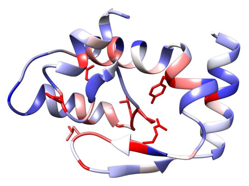
possesses the conserved FinO domain residues Arg157 both end with a 4U stretch, the latter of which is a recently
and Tyr146, which are essential for RNA binding by ProQ proposed 3′ end-located recognition element for binding
(Pandey et al. 2020), and key residues in a homology model by the FinO-domain (Stein et al. 2020). As previously ob-
of FopA (Fig. 4F). Apart from that, FopA represents a dis- served with ProQ (Holmqvist et al. 2018), the targets of
tinct branch of FinO-domain proteins (Fig. 4G), with plas- FopA share no obvious primary sequence or structural mo-
mid or chromosome encoded homologs in Escherichia, tif as none could be predicated that fits all the main targets
Shigella, and Klebsiella. Specifically, FopA lacks the ami- (Supplemental Fig. S4B), hence we conclude that FopA
no-terminal domain of FinO and the carboxy-terminal binds structured RNAs by a hidden code.
domain of ProQ but exhibits a distinct amino-terminal Inc is a cis-antisense RNA in the 5′ region of repZ encod-
domain with two stretches of positively charged residues. ing the replication initiator protein of pCol1B9 (Asano et al.
1452 RNA (2020) Vol. 26, No. 10Downloaded from rnajournal.cshlp.org on December 7, 2020 - Published by Cold Spring Harbor Laboratory Press
Bacterial RBP discovery
A B latter targets include STnc1590,
which is a cis-antisense RNA to
STnc1580 (found in several different
enterobacteria); the noncoding RNA
STnc700 from the leader region of
the histidine operon mRNA; and the
C
transcribed mgtM region that is im-
portant for ATP-sensing and regula-
tion of the mgtCBR operon (Lee and
Groisman 2012). Interestingly, the
D F mgtCBR operon is also regulated by
ProQ (Westermann et al. 2019). In
summary, FopA is the third FinO-
domain protein of Salmonella, with
targets in both a plasmid and the
core genome of this bacterium.
FopA accelerates RNA duplex
E G formation
Seeking to confirm the predicted RBP
activity of FopA, we selected the Inc–
repZ RNA pair for in vitro binding
experiments. These two RNAs form a
kissing complex that subsequently
progresses to a four-way junction (for
review, see Kolb et al. 2001). For our
binding experiments, we purified re-
H
combinant Salmonella FopA protein
after expression in E. coli and ren-
dered it RNA-free by ion-exchange
chromatography, thus reaching a pu-
rity of more than 95% (Fig. 6A;
Supplemental Fig. S5).
Using electrophoretic mobility shift
FIGURE 4. The FinO-domain protein FopA is abundant with a unique amino-terminal domain. assays (EMSA), we determined an ap-
(A) Hierarchical clustering of candidates that shifted to LMW and reduced distribution (Fig. 2, parent affinity constant of ∼1 µM for
blue box, filtered to >40% shifting protein fraction) resulted in clustering of Hfq, ProQ, CspD, FopA and Inc RNA (Fig. 6), which is a
and the RBP candidate FopA in the red cluster. In the blue cluster, proteins shifted toward frac- good fit with both the intracellular
tion 3. (B) The MS quantification of FopA showed a similar sedimentation shift as for ProQ,
which was confirmed by western blot analysis (C ). (D) Salmonella harbors three FinO-domain
concentration of FopA and reported
proteins. ProQ is encoded on the chromosome whereas FinO and FopA are encoded on plas- affinities of other FinO-domain pro-
mids. (E) FopA homologs show a unique amino-terminal domain that is different to the FinO teins (Attaiech et al. 2016; Smirnov
amino-terminal domain. The central FinO domain in FopA (PF04352, gray background) shows et al. 2016; Holmqvist et al. 2018;
conserved key residues. (F) Homology model of the FinO domain of FopA with color-coded Bauriedl et al. 2020). FopA displayed
conservation (red indicates high conservation). (G) FopA proteins represent a distinct branch
(red) in a phylogenetic analysis based on protein sequences of 2569 FinO-domain containing
similar affinities for the repZ 5′ UTR
proteins (PF04352, Pfam 32.0, Clustal Omega analysis). (H) FopA levels are strongly elevated at fragment fully antisense to Inc or for
late stationary phase and increased in SPI-2-inducing conditions. (∗ ) Anti-body cross-reaction. a preformed Inc–repZ RNA complex
(Fig. 6B). Since FopA bound Inc and
repZ both individually and as a com-
1998). The low-abundance repZ mRNA is also enriched by plex, we also assessed whether it affects the kinetics of
coIP with FopA (Fig. 5C). Interestingly, Inc/repZ and FopA Inc–repZ complex formation. Once formed, the Inc–repZ
show equal phylogenetic distribution, indicating function- complex has no apparent off-rate, hence the initial associ-
al linkage (Supplemental Fig. S4C). While Inc and repZ are ation rate is equivalent to its formation rate (k2, as previous-
plasmid-encoded transcripts, other top targets of FopA ly established for Inc and orthogonal systems [Persson
are transcribed from the Salmonella chromosome. These et al. 1988; Tomizawa 1990; Asano et al. 1998]).
www.rnajournal.org 1453Downloaded from rnajournal.cshlp.org on December 7, 2020 - Published by Cold Spring Harbor Laboratory Press
Gerovac et al.
A RNA chaperone, similar to the molecular function of its
plasmid-encoded sibling FinO in the FinP-traJ conjugation
control system (Arthur et al. 2003).
To map FopA binding sites within Inc, we performed
RNA structure probing with the single-strand specific
endoribonucleases, RNase A (cuts between unpaired py-
rimidines) and RNase T1 (cuts at unpaired guanosines).
As shown in Figure 6D, FopA protected Inc from cleavage
in the loop region at G46, and in general, protected Inc
from complete degradation by RNase A. In the Inc–repZ
complex, the interbridged region was protected between
U34–U37 and at A57. Concurrently, Inc was deprotected at
positions U53, C54, and U56, indicating an effect on the
RNA structure at the cross-junction point. Taken together,
by showing binding of FopA to its major RNA ligands in vi-
B tro, we established proof-of-principle that GradR can pre-
dict a previously unrecognized bacterial RBP.
DISCUSSION
GradR is conceptually related to two recently published ap-
proaches for eukaryotes, R-DeeP and DIF-FRAC, which
used sucrose instead of glycerol gradients (Caudron-
Herger et al. 2019) or size-exclusion chromatography
(Mallam et al. 2019). Together, these two latter studies pre-
dicted >1400 so-called RNA-dependent proteins or com-
plexes in human cells; in other words, >7% of the human
proteome would be sensitive to RNase treatment, which
is in the same range as the >5.5% predicted here for a bac-
C terial proteome. In order to visualize these RNase sensitiv-
ities and evaluate individual proteins in the context of their
physiological role and published literature, we provide
an interactive browser (Fig. 7, www.helmholtz-hiri.de/en/
datasets/GradRSeT) that also allows one to quickly com-
pare proteins from related organisms via Pfam protein
domain annotations (El-Gebali et al. 2018).
There has been a flurry of new approaches using organic
extraction or silica-based purification to globally predict
RBPs, several of which also established proof-of-principle
for usability beyond eukaryotes with E. coli or Salmonella
FIGURE 5. FopA is a global RBP that targets structured RNAs. (A) The (Queiroz et al. 2019; Shchepachev et al. 2019; Urdaneta
major targeted RNA was the antisense RNA lnc, followed by et al. 2019). We cross-compared our Salmonella GradR
STnc1590, mgtM, and STnc700 (log2-fold change >4, n = 17). The data set with the list of Salmonella RBP candidates predict-
RNA targetome of FopA was very different to ProQ recovered RNAs ed by Phenol Toluol extraction (PTex [Urdaneta et al.
that were reanalyzed differently from Smirnov et al. (2016). (B) FopA
2019]). PTex predicted 172 RBP candidates, of which 29
targeted RNAs were analyzed for conservation motifs by the
CMfinder algorithm. Homologs were searched by GLASSgo (1.5.0, had an annotated GO-term “RNA-binding” representing
Freiburg RNA tools [Lott et al. 2018]). The CMfinder algorithm deter- ∼21% of all Salmonella proteins within that GO term. For
mined RNA motifs (0.4.1.18 [Yao et al. 2005]), and R2R was used for comparison, GradR detects 210 RNase-sensitive proteins,
visualization (1.0.6 [Weinberg and Breaker 2011]). (C ) FopA-coIP 34 of which (∼25%) have the GO term “RNA binding.”
read-coverage of the repZ locus. Inc is heavily enriched, together
Interestingly, the GradR and PTex predictions share only
with the sparsely expressed repZ mRNA.
24 RBP candidates, 62% of which are established RBPs
(Supplemental Fig. S6A). As far as cross-comparison of
Importantly, FopA accelerated complex formation of the Salmonella with E. coli data permits, we correlated
Inc and repZ RNAs by approximately sevenfold (Fig. 6C). GradR with silica-based recovery of UV-crosslinked RBP
As such, the primary function of FopA may be that of an candidates to RNA by TRAPP (Shchepachev et al. 2019).
1454 RNA (2020) Vol. 26, No. 10Downloaded from rnajournal.cshlp.org on December 7, 2020 - Published by Cold Spring Harbor Laboratory Press
Bacterial RBP discovery
A B C
D
FIGURE 6. FopA protects Inc and anneals it to the 5′ UTR of repZ. (A) Purified FopA with cleaved-off tag. (B) FopA bound Inc, repZ, and the Inc–
repZ complex with an apparent KD about 1 µM. n = 2. (C) FopA accelerated Inc∗ –repZ complex formation. k2 represents the rate constant. n = 2.
(D) FopA protected full-length Inc alone and in the complex with repZ from RNase A degradation at loop regions. n = 2.
Of the 377 candidates from TRAPP, GradR found 42; interest. While previous RNA-binding domain based
again, most (67%) of these shared proteins are established searches failed to predict RBPs among Salmonella’s secret-
RBPs (Supplemental Fig. S6B). It is possible that the cross- ed virulence factors (Sharan et al. 2017; Tawk et al. 2017),
linking step used in these other methods (Asencio et al. we note that the virulence-associated protein YopD of
2018; Queiroz et al. 2019; Shchepachev et al. 2019; the related pathogen Yersinia pestis is well-known to
Trendel et al. 2019; Urdaneta et al. 2019) helps to enrich moonlight as an RBP (Chen and Anderson 2011). In addi-
weakly interacting candidates. In contrast, GradR requires tion, Listeria monocytogenes has just been shown to secret
RNPs to remain stable after lysate preparation. Therefore, an RBP to manipulate infected host cells (Gerovac and
we consider GradR as an orthogonal RBP discovery meth- Vogel 2019; Pagliuso et al. 2019). Interrogating RNase sen-
od to UV-crosslinking based approaches. Most important- sitivity of Salmonella virulence-associated proteins
ly, its generic nature provides an experimental route to (Supplemental Fig. S7A), we observe pronounced shifts to-
glance at the major RBPs of any microbe of interest, which ward central fractions for the invasion protein InvF,
becomes important in light of the thousands of understud- the secretion system apparatus protein PrgJ, and the pro-
ied bacteria now known to populate the human body. tein encoded by orf319. The invasion protein OrgB, the
Salmonella being a well-characterized pathogen, poten- bacteriophage-encoded virulence factor GtgE, effector
tial RBPs among its virulence factors are of special protein SseK1, and an uncharacterized prophage-encoded
www.rnajournal.org 1455Downloaded from rnajournal.cshlp.org on December 7, 2020 - Published by Cold Spring Harbor Laboratory Press
Gerovac et al.
AcnB protein is present in fraction 3
in a single peak that is unaffected by
RNase treatment (Supplemental Fig.
S7B,C). Potential reasons for failure
to detect this established RBP by
GradR include the use of iron-rich me-
dia in our study, and a possible insta-
bility of the AcnB-mRNA complex in
lysate. However, we do observe shifts
upon RNase treatment such that sev-
eral metabolic proteins move to
HMW fractions (AraA, AmyA, CbiK,
RfaG) or the pellet (RfaK, NarG,
FrdB, CyoB, FadJ, AroL), which may
indicate that certain metabolic en-
zymes require intracellular association
with RNA for integrity.
The power of GradR is well illustrated
by its ability to validate not only all
three sRNA-related RBPs (CsrA, Hfq,
and ProQ) of Salmonella but also to
predict a new FinO-domain RBP from
a plasmid, that is, a common type of
extrachromosomal element for which
there has been almost no systematic
annotation effort (Pilla and Tang
2018). FinO-domain proteins are a
growing class of RBPs that target struc-
tured RNA molecules in the cell, dis-
tinct from tRNA and rRNA (Attaiech
et al. 2016; Smirnov et al. 2016; Hol-
mqvist et al. 2018; Bauriedl et al.
FIGURE 7. Visualizing sedimentation patterns and RNase sensitivity data. Complex GradR 2020; Melamed et al. 2020). While
data can be visualized and explored in a browser, available at www.helmholtz-hiri.de/en/
most of the recent attention to this
datasets/GradRSeT.
RBP class has been on global posttran-
scriptional regulation by the chromo-
NTPase (SL1344_2214) pelleted upon RNase treatment. somally encoded ProQ and RocC proteins, the founding
Hypothetical virulence protein MsgA, sedimenting in frac- member of this class (the FinO protein) is encoded on a plas-
tions 1–3, shifted to fraction 1 upon RNase treatment, mak- mid (Timmis et al. 1978), just as is FopA. FinO’s assumed
ing it a good RBP candidate. Importantly, GradR might find main cellular RNA targets (the FinP sRNA-traJ mRNA pair)
even more potential RBPs when Salmonella is grown under are also plasmid-encoded (Mark Glover et al. 2015), as is
conditions that fully induce its virulence genes, so more FopA’s main target, the Inc RNA. In contrast, ProQ stably as-
proteins accumulate to levels that permit detection by MS. sociates with transcripts from the chromosome. Nonethe-
Other classes of proteins lend themselves for inspection less, all these RBPs select their very different targets from
in GradR data. For example, studies in eukaryotes have ac- the same pool of cytoplasmic transcripts, with the same
cumulated evidence that many enzymes from intermediary type of FinO domain. Importantly, since the Salmonella vir-
metabolism may moonlight as RBPs, often by binding their ulence plasmid pSLT encodes a homolog of E. coli FinO (Fig.
own mRNAs (Hentze et al. 2018). Of studies in bacteria, 4D), our work establishes this bacterium to express all three
one in E. coli proposed ∼1000 RBP candidates that com- such RBPs (FinO, ProQ, FopA). Salmonella could thus be a
prised mainly metabolic proteins (Shchepachev et al. test bed for domain swapping experiments to address
2019). A primary example of a metabolic RBP in E. coli is what intramolecular or cellular factors, including intracellular
the iron-sensing protein Aconitase B. This enzyme acts in localization, determine target selection. In addition, the
the tricarboxylic acid (TCA) cycle and directly stabilizes RNA interaction by ligation (RIL)-seq technique (Melamed
its own mRNA (acnB) when intracellular iron becomes et al. 2016), which in E. coli revealed diverse overlapping,
scarce (Benjamin and Massé 2014). Here, the 93.5 kDa complementary, or competing roles for ProQ and Hfq
1456 RNA (2020) Vol. 26, No. 10Downloaded from rnajournal.cshlp.org on December 7, 2020 - Published by Cold Spring Harbor Laboratory Press
Bacterial RBP discovery
(Melamed et al. 2020), may offer a more sensitive tool than room temperature for 20 min. An amount of 10 mM iodoaceta-
coIP to study currently unknown RNA interactions among mide (IAA) was added and samples were incubated at room tem-
the target suites of these three FinO domain RBPs. perature for 15 min in the dark. An amount of 0.25 µg Lys-C
Progress on defining its RNA interface notwithstanding protease (Promega) was added and incubated for 4 h at 37°C.
Samples were diluted with three volumes digestion buffer (100
(Ghetu et al. 2000, 2002; Arthur et al. 2011; Attaiech
mM Tris/HCl pH 8.5, 1 mM CaCl2) and 0.25 µg Trypsin
et al. 2016; Immer et al. 2018; Pandey et al. 2020), the
(Sequencing Grade, Promega) were added and incubated o/n
structure of a FinO domain with a bound RNA remains to at 37°C. Five percentage of formic acid (FA) was added for acid-
be solved. The 22 kDa FopA protein can be purified to ho- ification and samples were cleared by centrifugation at full-speed
mogeneity (Fig. 6A) and its RNA ligands offer tight folds, for 10 min. Acidified sample supernatant was loaded onto meth-
both of which is conducive to structural studies. Given anol activated stage-tips (C18) by centrifugation for 5–10 min at
FopA’s distinct sequence (Fig. 4E,G), we expect a structur- 2000g. Tips were washed three times with washing solution (2%
ally resolved FopA–RNA complex to provide important acetonitrile [ACN], 0.3% trifluoroacetic acid [TFA]) and eluted in
complementary information in the quest for the rules that 0.5 mL protein-low bind tubes with two times 20 µL elution solu-
govern the intriguing selectivity of the FinO domain for tion (60% ACN, 0.3% FA). Samples were snap-frozen in liquid ni-
RNA structure. trogen and lyophilized. For liquid chromatography analysis,
samples were solubilized in application solution (2% ACN, 0.1%
FA), sonicated, and 12 µL transferred to HPLC tubes.
MATERIALS AND METHODS
Cell lysis, RNase digestion, and gradient NanoLC-MS/MS analysis and MS analysis
fractionation Samples were MS analyzed in the laboratory of Andreas Schlosser
Salmonella SL1344 cells were grown in Lysogeny broth (LB) media by Stephanie Lamer. NanoLC-MS/MS analyses were performed
to transition phase at OD600 of 2.0, pelleted at full speed, and fro- by an Orbitrap Fusion (Thermo Scientific) equipped with a
zen at −20°C. In total, 60 OD of cells were lysed in 500 µL lysis buff- PicoView Ion Source (New Objective) and coupled to an EASY-
er (20 mM Tris/HCl pH 7.5, 150 mM KCl, 10 mM MgCl2, 1 mM DTT, nLC 1000 (Thermo Scientific). Peptides were loaded on capillary
2 mM PMSF) by glass beads (0.1 mm, BioSpec Products) in the columns (PicoFrit, 30 cm × 150 µm ID, New Objective) self-
Retsch MM200 at 30 Hz for 10 min at 4°C. The cell lysate was packed with ReproSil-Pur 120 C18-AQ, 1.9 µm (r119.aq., Dr.
cleared at full speed for 10 min and the supernatant was trans- Maisch). Samples were analyzed by a 120 min linear gradient
ferred. 20 µL RNase A/T1 mix (2 µg µL−1/5 U µL−1, Thermo from 3%–40% acetonitrile and 0.1% formic acid at a flow rate of
Scientific) was added to 100 µL lysate (OD260 ∼ 150) for RNase 500 nL/min. MS and MS/MS scans were acquired in the
digestion for 20 min at 20°C. The reaction was stopped on ice, di- Orbitrap analyzer with a resolution of 60,000 and 15,000, respec-
luted with 1 volume lysis buffer, and loaded completely onto a tively. Higher-energy collisional dissociation (HCD) fragmentation
10%–40% glycerol (w/v) gradient in lysis buffer. The proteins was applied with 35% normalized collision energy. We used top
were sedimented in the SW40Ti rotor (Beckman Coulter) at speed data-dependent MS/MS method with a fixed cycle time
100,000g for 17 h at 4°C. The gradient was fractionated in 20 equal of 3 sec. Dynamic exclusion was applied with a repeat count of
samples (∼600 µL) from the top and the pellet was resuspended in 1 and an exclusion duration of 60 sec (singly charged precursors
an additional fraction. RNA was extracted by PCI for RNA (X985.3, were excluded from selection). Minimum signal threshold for pre-
Carl Roth) for RNA integrity control gels. Protein samples were pre- cursor selection was set to 50,000. Predictive AGC was used with
pared for SDS-PAGE analysis and western blotting. AGC a target value of 2 × 1005 for MS scans and 5 × 1004 for MS/
We analyzed gradients with and without 0.2% Triton X100 in MS scans. EASY-IC was used for internal calibration.
the lysis and gradient buffers (Supplemental Fig. S2G–I). Raw MS data files were analyzed with MaxQuant version 1.6.2.2
Detergents solubilize membrane-associated proteins and pre- (Cox and Mann 2008). Database search was performed with
vent unspecific interactions, but also interfere with peptide recov- Andromeda, which is integrated in the utilized version of
ery and mass spectrometric analysis. Both GradR experimental MaxQuant. The search was performed against the UniProt
condition replicates were well correlated (Supplemental Fig. Salmonella Typhimurium UP000008962 (strain SL1344) and a da-
S2G–I, for details see section about data analysis) and we present tabase containing the proteins of the UPS2 proteomic standard.
here data of the GradR experiment without Triton X100 and com- Additionally, a database containing common contaminants was
plete in-solution MS sample preparation. used. The search was performed with tryptic cleavage specificity
with three allowed missed cleavages. Protein identification was
under control of the false-discovery rate (FDR, 1% on protein
In-solution MS sample preparation and peptide level). In addition to MaxQuant default settings,
the search was additionally performed for the following variable
An amount of 0.2 µg proteomics dynamic range standard set modifications: Protein amino-terminal acetylation, glutamine to
(UPS2, Sigma-Aldrich) was added as spike-in to 50 µL protein pyro-glutamic acid formation (N-term. glutamine) and oxidation
samples of each fraction. Samples were diluted with two volumes (methionine). Carbamidomethyl (cysteine) was set as fixed modi-
denaturation buffer (100 mM Tris/HCl pH 8.5, 12 M urea). 5 mM fication. For protein quantitation, the iBAQ intensities were used
tris(2-carboxyethyl)phosphine (TCEP, Bond-Breaker, neutral pH, (Schwanhäusser et al. 2011). Proteins that could not be distin-
Thermo Scientific) was added and samples were incubated at guished by peptides were listed individually.
www.rnajournal.org 1457Downloaded from rnajournal.cshlp.org on December 7, 2020 - Published by Cold Spring Harbor Laboratory Press
Gerovac et al.
Data analysis DNA-binding; “nucleo,” “metal,” “zinc,” “magnesium,” “iron,”
“ATP,” and “GTP” for the class metal- and nucleotide-binding;
Relative protein abundance in gradient fractions were calculated metabolism class proteins were derived from the Kyoto
by iBAQ. Absolute abundances were estimated by correcting for Encyclopaedia of Genes and Genomes (KEGG) database
differences in digestion and C18 purification efficiency in each (sey00001, A09100 Metabolism, and subgroups); proteins anno-
gradient fraction, through normalization to human albumin tated as both metal- and nucleotide-binding and metabolic pro-
(Supplemental Table S1, norm. to spike-in, P02768ups|ALBU_- teins were extracted and are indicated as an isolated shared class;
HUMAN_UPS Serum albumin, chain 26–609, Supplemental Fig. “membrane” for the two times class; “uncharacterized” for the
S2A) that was added in equal amounts to each fraction as part uncharacterized class; proteins that remained unclassified were
of the UPS2 standard (Sigma-Aldrich). We selected albumin for classified as others, or if only RNA- and DNA-binding proteins
normalization as the highest number of peptides (74) were recov- were shown, all others were classified as others). All other classifi-
ered for it of all spike-in proteins. The protein ratio between refer- cations are listed in Supplemental Table S4.
ence and +RNase gradients was determined by the normalized All figures were assembled in Adobe Illustrator. Pearson corre-
total absolute protein abundance per gradient. For bar-diagram lation coefficients between individual gradients or sedimentation
representation, the abundances for each individual protein were profiles of proteins were calculated in Excel with the function
transformed into distributions across the gradient by dividing “CORREL” considering norm. to gradient protein abundance.
the abundance in each gradient fraction by the total abundance We observed well correlated sedimentation profiles for the refer-
across the gradient (Supplemental Table S1, norm. to grad.). ence gradient with Grad-seq profiles (Supplemental Fig. S2F). In
For visualization purposes, the y-axis of individual protein gradi- addition, the nonshifting proteins were highly correlated between
ent profile plots were scaled to the largest value in either the ref- reference and RNase treated gradients in experimental condition
erence or RNase treated gradient. replicates with (0.0) and without (0.1) 0.2% Triton X100
Relative protein level changes (d) per fraction (Fig. 4A) were de- (Supplemental Fig. S2G–I).
termined by subtraction of the reference protein abundances
from the RNase treated normalized protein abundances in each
gradient fraction. These relative protein level changes per fraction Genomic 3×FLAG-tag labeling of fopA in Salmonella
were used for hierarchical clustering. The fraction of shifted pro- enterica Typhimurium SL1344
tein was the sum of all positive relative protein level changes.
The mean position of a protein in a gradient was calculated as Constructs for recombination were amplified by PCR from geno-
the mean of proportions of protein times each fraction number mic DNA with 35–45 nt 5′ and 3′ overhangs that were homolo-
(pellet equals 21). The shift in protein distribution was calculated gous to the recombination sites of the gene of interest. Wild-
to capture the difference in relative protein positions between the type Salmonella strain (JVO-1574) was transformed with pKD46
reference and the RNase treated gradients (Supplemental Fig. (Datsenko and Wanner 2000), selected by ampicillin resistance
S2A). Protein proportions were converted to binary vectors at on a plate at 28°C, picked and grown o/n in LB at 28°C. LB media
two thresholds: 0.5% (to capture low abundance presence), and was inoculated with 1:300 o/n Salmonella +pKD46 (JVO-3013)
2.5% (to capture more robust presence in a fraction). The culture with 0.2% L arabinose for λRed gene induction and ex-
Hamming distance was calculated for each protein between the pression. At OD 0.3 cells were placed on ice for 30 min and the
reference and RNase-treated gradient at each threshold, provid- culture was pelleted at 4000g for 20 min at 4°C and washed
ing a measure of similarity in protein presence and distribution with ice-chilled water. The cell pellet was dissolved in 1/100 vol-
profiles. The change in presence and distribution was then calcu- ume of initial culture. 50 µL of competent cells were mixed with
lated as the mean of the Hamming distance at these two thresh- 100–300 ng of the PCR product in a chilled 0.2 cm electroporation
olds (Supplemental Fig. S2A). cuvette and transformed (200 Ω, 25 µF, and 2.5 kV). Transformed
Calculations were conducted in Microsoft Excel (Supplemental cells were recovered in 500 µL prewarmed LB media at 37°C and
Table S1). All normalization operations are implemented in the incubated for 2 h. Cells were pelleted at 6000g for 3 min at 25°C
Excel spread sheet and can be used for analysis of new data and selected by kanamycin resistance on a plate at 37°C.
sets. Single-linkage hierarchical clustering on Euclidean distances Single colonies were verified by PCR amplification of the cas-
was performed in Orange (3.23, University of Ljubljana [Demsar sette and chromosomal elements aside by verification primers,
et al. 2013]). Thresholds were applied as indicated in the panels and selective growth with kanamycin and not with ampicillin;
through the select row widget. The distance widget calculated hence, the pKD46 plasmid was lost. To ensure only single inser-
the distances between rows, and hence individual proteins, and tion in the chromosomal region, P22 phage lysis and chromosom-
the hierarchical clustering widget yielded clusters in continuous al integration was performed. The recipient strain (JVS-1574) was
order that were visualized as a heat map in Orange or Excel P22 phage lysed and the phage lysate was used for lysis of the
with color-coding that represented the shifting protein fraction donor strain. The donor strain phage lysate was then again used
from −1 to +1 normalized relative protein levels per gradient. for transduction in the recipient strain. In detail, a single colony
Scatter plots were assembled in Origin (OriginLabs) as bubble of the recipient strain was inoculated in 5 mL LB media and grown
charts. Classification of proteins was achieved through consecu- to OD 1.0. An amount of 10 µL of P22 lysate was added and the
tively requesting terms from protein names and full GO term an- cells were grown o/n at 37°C. The suspension was pelleted at
notation (without case sensitivity, from UniProt, UP000008962, 14,000g for 10 min at 4°C. The supernatant was transferred to
release 12/2019 [The UniProt Consortium 2018]) in Microsoft glass tubes and extracted with 200 µL chloroform to kill all remain-
Excel (“RNA,” “riboso,” “transla,” and “nuclease” for the class ing bacteria. The recipient P22 lysate was stored at 4°C and used
RNA-binding; “DNA,” “transcript,” and “plasmid” for the class for lysis of the donor strain.
1458 RNA (2020) Vol. 26, No. 10Downloaded from rnajournal.cshlp.org on December 7, 2020 - Published by Cold Spring Harbor Laboratory Press
Bacterial RBP discovery
For integration of the fragment from the P22 lysate into the re- added for 5′ -end phosphorylation for 60 min at 37°C. RNA was
cipient strain, 100 µL of OD1 recipient strain were infected with purified by silica-based columns (Zymo RNA Clean & Con-
10–30 µL phage lysate for 20 min at room temperature. The infec- centrator kit, R1013, Zymo Research) and eluted in 15 µL nucle-
tion was quenched with 20 mM EGTA to allow only one round of ase-free water (436912C, VWR). RNA was denatured again and
infection. The cells were recovered in 1 mL LB media for 2 h pyrophosphates were removed from the 5′ -end by 10 U RppH
and plated on plates with kanamycin. The kanamycin cassette (M0356S, NEB) in 1× NEB buffer 2 (10 mM Tris–HCl pH 7.9,
was removed by FRT recombination and transformation of 50 mM NaCl, 10 mM MgCl2, 1 mM DTT) in the presence of
pCP20 at 28°C. pKD46 and pCP20 were removed by o/n incuba- 20 U RNase inhibitor for 60 min at 37°C, and again column puri-
tion at 42°C and selection of single colonies on plates with corre- fied and eluted in 6 µL. For 3′ adaptor ligation the NEBNext Small
sponding antibiotics. Strains that were only growing on LB plates RNA Kit (E7560S, NEB) was used: 200 ng RNA were mixed with
were considered as cured and used for studies. 1 µL 1:3 diluted 3′ SR adaptors for Illumina, and denatured, 1×
3′ ligation reaction buffer was added and 3 µL ligation enzyme
mix in a total volume of 20 µL for 60 min at 25°C. An amount of
Western blotting 1 µL 1:3 diluted SR RT-primer for Illumina was added in a total
volume of 25 µL. The annealing was conducted at 75°C for
Protein samples were diluted in reducing 1× protein-sample load- 5 min, 37°C at 15 min, 25°C at 15 min, cooling at 4°C. An amount
ing buffer (60 mM Tris–HCl pH 6.8, 0.2 g/mL SDS, 0.1 mg/mL brom- of 1 µl 5′ SR adaptor for Illumina (freshly solubilized, denatured,
phenol blue, 77 mg/mL DTT, 10% glycerol) and boiled at 100°C for and cooled on ice), 1 µL 10× 5′ ligation reaction buffer, and
5 min and loaded on SDS-PAGE. In the case of Hfq-FLAG, samples 2.5 µL 5′ ligation enzyme mix were added to the reaction and in-
were heated to 90°C. The gel was blotted by semi-dry transfer on cubated for 60 min at 25°C. For reverse transcription, 8 µL first
polyvinylidene fluoride (PVDF) membranes and probed with tag- strand synthesis reaction buffer, 1 µL murine RNase inhibitor,
specific antibodies. For FLAG-tag detection, a monoclonal anti- and 1 µL ProtoScript II reverse transcriptase were added and incu-
body was used (#F1804, 1:2000 dilution, Sigma-Aldrich). GroEL bated for 60 min at 50°C. PCR amplification of reverse transcripts
was detected after stripping with anti-GroEL rabbit anti-body was conducted by addition of 50 µL LongAmp Taq 2× master mix,
(#G6532, 1:10,000 dilution, Sigma-Aldrich). 5 µL nuclease free water, 2.5 µL 1:3 diluted SR primer for Illumina,
and 2.5 µL 1:3 diluted index primers. The reaction was cycled at
94°C for 30 sec, 14× 94°C for 15 sec—62°C for 30 s—70°C for
Coimmunoprecipitation (Co-IP) 90 sec, 70°C for 5 min, and cooled at 4°C.
Sixty OD of cells at OD 2 were resuspended in 800 µL lysis buffer DNA was purified by MagSi-NGSPREP Plus (MDKT00010005,
(20 mM Tris/HCl pH 8.0, 150 mM KCl, 1 mM MgCl2, 1 mM DTT), Steinbrenner Laborsysteme GmbH) beads with a ratio of 1.8× to
and 800 µL 0.1 mm glass beads (BioSpecs). Cells were lysed at reaction volume. All steps were executed in DNA-LoBind tubes
30 Hz for 10 min in the Retsch MM200 at 4°C. The lysate was (#0030108051, Eppendorf). The final supernatant was analyzed
cleared twice for 10 min at full-speed at 4°C. An amount of by Qubit 2.0 (Thermo Scientific) with the dsDNA HS Assay Kit
35 µL monoclonal anti-FLAG M2 antibody produced in mouse (Q32854, Thermo Scientific) and the Bioanalyzer 2100
(Sigma-Aldrich) was added to the supernatant and incubated for (G2939BA, Agilent) with the DNA 1000 kit (5067-1504) or HS
30 min at 4°C. An amount of 75 µL prewashed protein A sepharose DNA 7500 kit (5067-1506). The sample was sequenced on the
(Sigma-Aldrich) was added for recovery of FLAG-antibody and in- NextSeq-500 system (Illumina) with a read depth of 5 mio. reads
cubated for 30 min. Beads were pelleted at 300g and washed five per sample and a read length of 75 nt single end.
times with lysis buffer, resuspended in 500 µL lysis buffer and ex-
tracted with PCI. The aqueous layer was precipitated with ethanol
RNA-seq analysis
and 1:30 3 M sodium acetate at pH 5.2 at −20°C overnight. The
precipitated RNA was pelleted and washed with 500 µL 75% eth- Reads were mapped to the Salmonella reference sequences
anol. The pellet was dried and solubilized in 15.5 µL water. DNA (NC_016810.1, NC_017718.1, NC_017719.1, and NC_0177
was degraded by DNase I (Thermo Scientific) as described in the 20.1) by the READemption align function (READemption version
manual in the presence of RNase inhibitor for 30 min at 37°C. 0.4.3 [Förstner et al. 2014]). Coverage wig-files were generated
The sample was diluted with 100 µL water and the RNA was ex- with the coverage function and read allocation to genomic fea-
tracted by PCI. The RNA was solubilized to an OD260 of 1 and an- tures was quantified by the gene_quanti function. Annotation of
alyzed in the Bioanalyzer (Agilent) for quality and quantity 3′ /5′ UTRs was used from the Hinton laboratory and sRNAs from
estimation for RNA-seq. Protein samples were recovered from the Vogel laboratory. Overlaid annotations that caused multiple
the organic layer by 10 vol methanol precipitation. entries were manually corrected. Sequencing coverages were vi-
sualized in the Integrative Genomics Viewer (IGV, Broad Institute
[Robinson et al. 2011]) based on uniquely mapped reads and nor-
RNA-seq malized to the total number of aligned reads.
RNA library preparation was conducted by the Core Unit SysMed
(Kristina Döring, Würzburg). An amount of 200 ng eluted RNA RNA production
from the coIP was denatured at 70°C for 2 min, cooled down on
ice, and 3′ -end dephosphorylated by 10 U T4-PNK (M0201S, Template DNA for transcription from a T7 promoter was produced
NEB) in the presence of 20 U RNase inhibitor (M0314L, NEB) for by PCR and extracted from agarose gel or purified by anion-ex-
60 min at 37°C. An amount of 1.6 mM ATP and 10 U PNK was change columns, or reverse-complementary primers were
www.rnajournal.org 1459You can also read