Mating type switching and transcriptional silencing in Kluyveromyces lactis - Emad Barsoum
←
→
Page content transcription
If your browser does not render page correctly, please read the page content below
Mating type switching and transcriptional
     silencing in Kluyveromyces lactis

                  Emad Barsoum

                Wenner-Gren Institute
          Department of Developmental Biology
                 Stockholm University
                   Stockholm 2010

                          1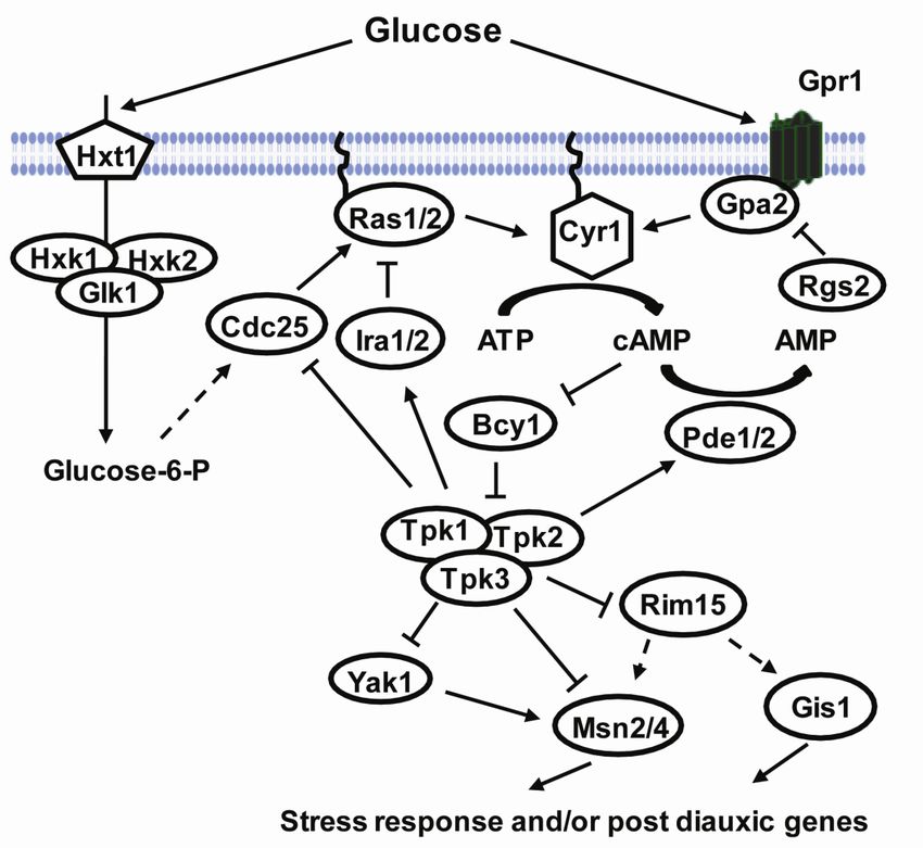
Doctoral dissertation 2010
Department of Developmental Biology
Wenner-Gren Institute
Stockholm University
Stockholm, Sweden

© Emad Barsoum, Stockholm 2010
ISBN (978-91-7447-105-2)
Distributor: Department of Developmental Biology, Wenner-Gren Institute
Printed in Sweden by US-AB, Stockholm 2010

                                      2
To my family

3
4
Abstract
To explore the similarities and differences of regulatory circuits among budding yeasts, we
characterized the role of unscheduled meiotic gene expression 6 (UME6) and a novel mating
type switching pathway in Kluyveromyces lactis. We found that Ume6 was required for
transcriptional silencing of the cryptic mating-type loci HML and HMRa. Ume6 acted
directly at these loci by binding to the cis-regulatory silencers. A mating type a (MATa) ume6
strain was mating proficient whereas a MAT ume6 strain was sterile. Consistently, absence
of Ume6 strongly derepressed HMRa, but only weakly derepressed HML. In addition, two
haploid specific genes, STE4 and MTS1, were repressed in MAT ume6 but expressed in the
MATa ume6 strain. Surprisingly, ume6 partially suppressed the mating defect of a MATa sir2
strain. Both STE12 and MATa2/HMRa2 genes were overexpressed in the MATa sir2 ume6
strain, suggesting that this deregulation was responsible for suppressing the mating defect.
Ume6 also served as a block to polyploidy and was required for repression of three meiotic
genes, independently of the Rpd3 and Sin3 corepressors.
Mating type switching from MAT to MATa required the 3 protein. The protein was
similar to transposases of the mutator like elements (MULEs). Mutational analysis showed
that the DDE-motif in 3, which is conserved in MULEs was necessary for switching. During
switching 3 mobilizes from the genome in the form of a DNA circle. The sequences
encompassing the 3 gene circle junctions in the MAT locus were essential for switching
from MAT to MATa. Switching also required a DNA binding protein, Mating type switch 1
(Mts1), whose binding sites in MAT were important. Expression of Mts1 was repressed in
MATa/MAT diploids and by nutrients, limiting switching to haploids in low nutrient
conditions.
In a genetic selection for strains with increased switching rates we found a mutation in the
RAS1 gene. By measuring the levels of the MTS1 mRNA and switching rates in ras1, pde2
and msn2 mutant strains we show that mating type switching in K. lactis was regulated by the
RAS/cAMP pathway and the transcription factor Msn2. ras1 mutants contained 20-fold
higher levels of MTS1 mRNA compared to wild type whereas pde2 and msn2 expressed less
MTS1 mRNA and had decreased switching rates. Furthermore we found that MTS1 contained
several potential Msn2 binding sites upstream of its ORF. We suggest that these observations
explain the nutrient regulation of switching.
5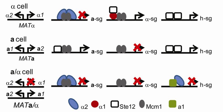
List of Publications
I. Ume6 is required for the MATa/MAT cellular identity and transcriptional
silencing in Kluyveromyces lactis.
Barsoum E, Sjöstrand JO, Åström SU. Genetics. 2010 184:999-1011.
II. 3, a transposable element that promotes host sexual reproduction.
Barsoum E, Martinez P, Åström SU. Genes Dev. 2009 24:33-44.
III. Regulation of mating type switching in Kluyveromyces lactis by the RAS/cAMP
pathway and the transcription factor Msn2.
Barsoum E and Åström SU. (Manuscript)
6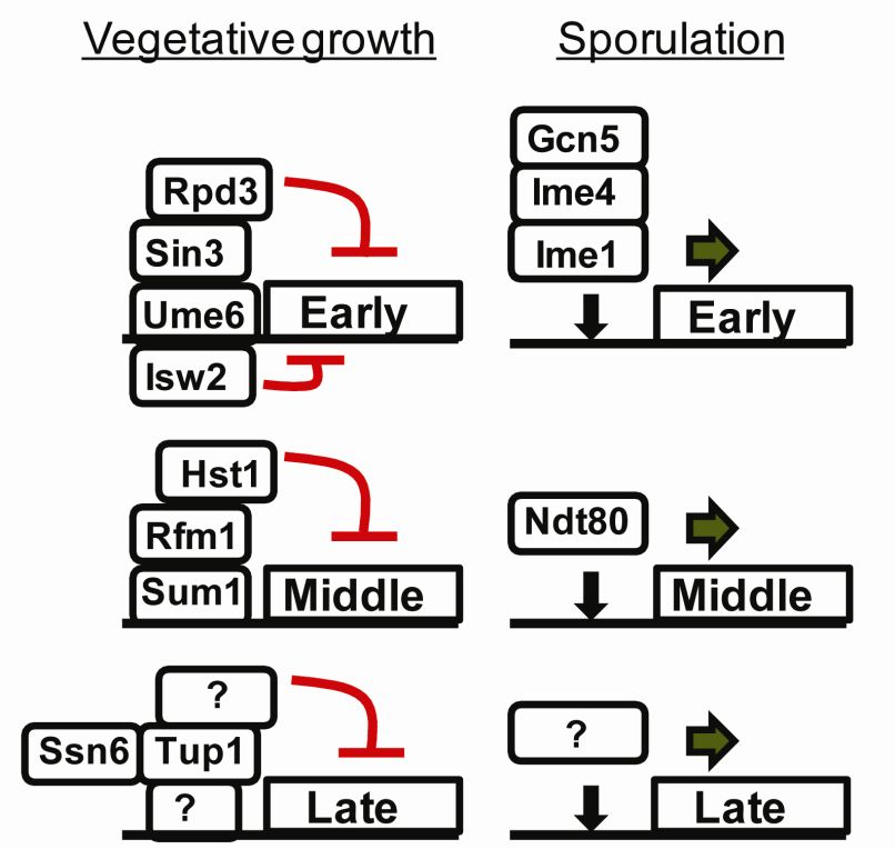
Table of contents
Introduction
1. Life cycle of Saccharomyces cerevisiae 9
2 Chromatin and silencing in Saccharomyces cerevisiae 10
3 Silent loci and components of silent chromatin 11
3.1 Transcriptional silencing at the HML and HMR loci 11
3.1.1 The silencer and silencer binding proteins 12
3.1.2 Establishment and maintenance of silent chromatin at the silencer 13
4. Mating in S. cerevisiae 16
5. Regulation of mating type specific gene expression 18
6. Mating type switching in S. cerevisiae 20
6.1 The mating type switch model 21
6.2 Regulation of mating type switching in S. cerevisiae 22
6.3 Directionality of switching 24
7. Sporulation 24
8. The cAMP-PKA pathway and stress response 27
9 Transposons 30
9.1 Definition and classification of transposons 31
9.2 Function and characteristics of the DDE-motif in transposons 32
9.3 Transposition pathways of DDE-motif containing transposons 32
9.3.1 “copy-in” transposition 33
9.3.2 “cut-out, paste in” transposition 33
9.3.3 “copy-out, paste-in” transposition 33
9.4 Molecular domestication of TEs 35
9.4.1 V(D)J recombination 35
9.4.2 FAR1/FHY3 36
Aims of this thesis 37
Results and discussion 38
Ume6 is required for the MATa/MAT cellular identity and
transcriptional silencing in Kluyveromyces lactis (Paper I) 38
7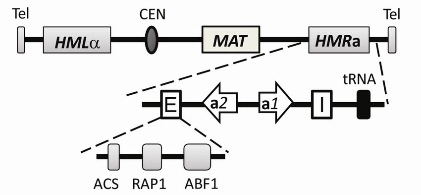
3, a transposable element that promotes host sexual
reproduction (Paper II) 41
Regulation of mating type switching in Kluyveromyces lactis
by the RAS/cAMP pathway and the transcription
factor Msn2 (Manuscript) 46
Conclusions 49
Acknowledgements 50
References 52
81. Life cycle of Saccharomyces cerevisiae
Saccharomyces cerevisiae is a unicellular eukaryotic organism that contains approximately
6000 genes distributed among 16 chromosomes. S. cerevisiae can exist as three different cell-
types. The a and cell-types are haploid, which means they contain one copy of each of the
16 chromosomes, while the a/ cell-type is diploid, which means that it contains two copies
of each chromosome. In the presence of sufficient supplies of carbon and nitrogen all three
cell-types proliferate asexually through budding. Cells undergoing asexual proliferation are
said to undergo mitotic or vegetative growth. Yeast cells abandon the proliferation mode only
in response to nutrient limitation or when another yeast cell is in its vicinity with which it can
mate. When the cells run out of nutrients they arrest in the G1 phase of the cell cycle, but can
resume growth if nutrients are re-supplied.
Sexual reproduction is a property found among almost all eukaryotes. It enables the
organism to rearrange its genome and hence increases genetic variability. In addition, diploid
cells can better cope with recessive mutations compared to haploid cells. In yeast, sexual
reproduction involves the cellular and nuclear fusion of the two haploid cell types (a and )
creating the diploid a/ cell-type. When diploid cells encounter harsh environmental
conditions such as limited amounts of both carbon and nitrogen, they undergo meiosis and
spore formation. Formation of resilient spores serves as a defense against the harsh
environment until conditions become more favorable and also completes the sexual cycle of
yeast to produce the haploid cells.
The life cycle of S. cerevisiae has an additional aspect besides proliferation, mating and
meiosis. Haploid cells can change their mating type through a process called mating type
switching. This process, also called the homothallic life cycle, allows a single haploid cell to
give rise to diploid cells capable of meiosis and spore formation. Mating type switching
requires the HO gene. Most laboratory S. cerevisiae strains lack a functional HO gene. These
strains have a heterothallic life cycle in which a single haploid cell is not able to produce
diploids. The HO gene codes for a site specific endonuclease which makes a DNA double
strand break (DSB) and this triggers a gene conversion process that allows the cells to change
mating type (Herskowitz, 1988; Madhani, 2007).
The genetic differences that define the three cell-types are in the mating type (MAT) locus.
The transcriptionally active MAT locus can harbor either a genes or genes. Strains of cell-
type contain the MAT allele and express the 1 and 2 genes, while strains of a cell-type
9contain the MATa allele and express the a1 and a2 genes. In addition, the cells contain two
transcriptionally silent loci called HML and HMRa. HML contains the genes while
HMRa contains the a genes. Both HML and HMR are silent and function as a reservoir of
mating type information that are used as donor sequences during mating type switching.
Mating of haploids of opposite mating types form the a/ diploid, which is heterozygous for
the two MAT alleles. Cells that express both MAT alleles are unable to mate, but can undergo
meiosis and sporulation unlike haploids (Laurenson and Rine, 1992).
2. Chromatin and silencing in S. cerevisiae
In eukaryotic cells the DNA interacts with proteins forming a complex called chromatin. The
nucleosome core particle is the fundamental subunit of chromatin and is composed of 147
base pairs (bp) of DNA wrapped around an octamer of the core histones: H2A, H2B, H3 and
H4. The histone octamer is composed of two H2A-H2B dimers and a stable H3-H4 tetramer.
Nucleosomes separated by linker DNA create a regularly spaced nucleosomal array structure
called “beads on a string”. Chromatin can be further organized into more complex and
condensed structures such as the 30nm chromatin fiber. Histone H1, is another component of
the chromatin. It binds to the linker DNA coming in and out from the nucleosomes. The
nucleosome is highly dynamic and can be modified through substitution of core histones with
other histone variants such as H2A.Z, found in active chromatin. In addition, the chromatin
structure can be modulated by post translational modifications of the histones such as
acetylation, methylation, phosphorylation and ubiquitination (Bradbury, 1998; Campos and
Reinberg, 2009).
Silencing involves the formation of a specialized chromatin structure that blocks the
expression of genes by preventing the transcription machinery from gaining access to the
promoters. In contrast to silencing, gene-specific repression is mediated by operators at or
near, the site of transcription initiation of a single gene whereas silencing is a form of long
range transcriptional repression occurring at certain chromosomal regions rather than at the
promoter sequences of different genes. Silencing is mediated by regulatory sites, known as
silencers, that act at some distance from their target genes (Rusche et al., 2003). The silent
region occupies distances larger than a single gene and is stably inherited during cell division,
making transcriptional silencing in S. cerevisiae functionally akin to position-effect
variegation in Drosophlia and X-chromosome inactivation in female mammals (Fox and
McConnell, 2005). Position effect variegation usually results from chromosomal
10rearrangements where euchromatin is juxtaposed to heterochromatin. During development the
heterochromatin spreads into the euchromatin on the chromosome to different degrees in
different cells, hence leading to variable silencing of the genes close to the
heterochromatin/euchromatin boundary. The heterochromatin state is then fixed and
propagated for several cell divisions (Schotta et al., 2003), which means that patches of cells
expressing or not expressing a marker gene can be observed for example in the Drosophila
eye. During mammalian X-chromosome inactivation, one X-chromosome in females is
randomly inactivated early in development and stably inherited throughout the life of the
animal (Rastan, 1994). In this case, a noncoding RNA molecule (Xist) plays a pivotal role
coating the inactivated X- chromosome, but not the active X- chromosome (Wutz and
Gribnau, 2007). Although the precise molecules involved in the formation of repressive
chromatin may vary between the organisms, silent chromatin domains contain particular post-
translational modifications of nucleosomes and specialized chromatin-binding proteins (Fox
and McConnell, 2005).
3. Silent loci and components of the silent chromatin
In S. cerevisiae, three different loci are subjected to transcriptional silencing. These are the
cryptic mating type loci, the telomeres (chromosome ends) and the rDNA (encoding
ribosomal RNA). The silencing of these loci use partly shared mechanisms, but also displays
some differences. In this thesis, I will focus solely on silencing of the cryptic mating type loci,
HML and HMRa.
3.1 Transcriptional silencing at the HML and HMR loci
Because loci that are not expressed normally are lost due to natural selection, I would like to
begin by defining the function of the cryptic mating type loci. As mentioned above, MATa
and MAT haploids can change their sex in a process called mating type switching. Mating
type switching starts with a cleavage of the MAT locus by a site specific endonuclease called
HO. The break is repaired through gene conversion where one of the cryptic mating type loci,
HML or HMR, is copied into the MAT locus. Hence, the function of the cryptic mating type
loci are to serve as a reservoir of mating type information (Haber, 1998).
113.1.1 The silencer and silencer binding proteins
The HM loci are flanked by cis-acting DNA elements that are necessary for the establishment,
stability and specificity of silent chromatin. These silencers known as E (essential) and I
(important) were defined by deletion studies on plasmids (Abraham et al., 1984; Feldman et
al., 1984). HMR-E is the best studied silencer providing most of the knowledge of what is
required for a functional silencer.
HMR-E is a small 150bp DNA element required for silent chromatin to form at HMRa.
HMR-E contains binding sites for three different sequence specific DNA-binding proteins, the
origin recognition complex (Orc), repressor activator protein 1 (Rap1) and ARS -binding
factor 1 (Abf1) (Lustig, 1998) (Figure 1). Mutation in any two of the three protein binding
sites at HMR-E leads to silencing defects (Brand et al., 1987). In addition to silencing, all
three factors bind other sites in the genome and contribute to other unrelated processes. The
Orc complex is a six-subunit ATP-dependent DNA binding complex (Orc1-6), which is
required for DNA replication and binds chromatin at replication origins throughout the cell
cycle (Bell, 2002; Bell and Dutta, 2002). Both of Abf1 and Rap1 are essential proteins that
participate in multiple nuclear events such as transcriptional activation and DNA replication.
In addition, Rap1 is involved in telomere silencing, and telomere length regulation as well as
other aspects of DNA metabolism such as heterochromatin boundary element formation,
creation of hotspots for meiotic recombination and chromatin opening (Feeser and Wolberger,
2008; Morse, 2000).
The three silencer binding proteins are not by themselves able to create silenced
heterochromatin. Silencing at the HM loci also requires the silent information regulator (Sir)
complex, which is composed of Sir1, Sir2, Sir3 and Sir4 (Ivy et al., 1986; Rine and
Herskowitz, 1987). Each of the three silencer binding proteins has some affinity for one or
more of the Sir proteins.
12Figure 1. Chromosome III with the relative positions of the mating type locus (MAT) and the cryptic
mating type loci (HML and HMR). The HMRa locus is enlarged showing the E and I silencers, the
open reading frames of a1 and a2 and the tRNA involved in the boundary element. Binding sites in the
E silencer for Orc (ACS, autonomously replicating sequence consensus sequence), Rap1 and Abf1 are
indicated. The figure is modified from (Rusche et al., 2003).
3.1.2 Establishment and maintenance of silent chromatin at the silencer
S. cerevisiae contains four SIR genes, which were identified through mutations that activated
the silent mating type loci. The Sir2, Sir3 and Sir4 gene products are essential for silencing
and acts as structural components of silent chromatin while Sir1 unlike the other Sir proteins
contributes but is not essential for silencing (Rusche et al., 2002). Excision of mini circles of
HML and HMR through site specific recombination in vivo, revealed that the assembly of the
Sir proteins into silenced chromatin creates a highly ordered structure thus contributing to
silenced loci being more negatively supercoiled in the mini circles and less accessible to DNA
modifying enzymes in wild-type compared to sir deleted cells (Bi and Broach, 1997; Cheng et
al., 1998).
The establishment of silent chromatin occurs in two discrete steps (Figure 2). First, the Sir
proteins are recruited to the silencers and then the Sir proteins spread over the target locus.
The Sir proteins are recruited to the silencers through protein-protein interactions. The
assembly of the Sir proteins follows a specific order. First Sir1 is localized to the silencer
though binding to Orc1 (Gardner et al., 1999; Triolo and Sternglanz, 1996). By being located
to the silencer, Sir1 enhances the probability of the other Sir proteins to be recruited to the
same silencer (Rusche et al., 2002). Next Sir4 is recruited to the silencer by interacting with
Rap1 (Cockell et al., 1995; Moretti et al., 1994; Moretti and Shore, 2001) and Sir1 (Bose et
13al., 2004; Triolo and Sternglanz, 1996). Sir4 in turn is thought to recruit Sir2 as a member of
the Sir4-Sir2 complex (Ghidelli et al., 2001; Hoppe et al., 2002). Sir3 is located to the silencer
by binding to both Rap1 (Moretti et al., 1994; Moretti and Shore, 2001) and Sir4 (Moazed et
al., 1997). Once the network of the Sir-complex has assembled at the silencer, they spread
from the silencer forming silent chromatin, which inhibits transcription of the target genes.
A common feature of all silent chromatin domains is that the lysine residues on the histone
tails within the nucleosomes are hypoacetylated (Grunstein, 1997). Binding of Sir3 and Sir 4
to these deacetylated histone tails of H3 and H4 enables the Sir-complex to spread along the
chromosome (Carmen et al., 2002; Hecht et al., 1995). A major breakthrough in
understanding silencing in S. cerevisiae came with the discovery of an enzymatic activity
displayed by Sir2. Sir2 is a member of a large protein family of nicotinamide adenine
dinucleotide (NAD)-dependent histone deacetylases (Imai et al., 2000; Smith et al., 2000;
Tanny et al., 1999), conserved from bacteria to humans (Brachmann et al., 1995; Frye, 2000).
In addition to Sir2, S. cerevisiae encodes four other paralogs called Hst1, Hst2, Hst3 and Hst4
(Homolog of SIR Two) (Brachmann et al., 1995; Derbyshire et al., 1996). Hence, the
spreading and silencing conducted by the Sir-complex is achieved through Sir2 deacetylating
the tails of Histones H3 and H4 thus creating high-affinity binding sites for Sir3 and Sir4.
This in turn enables the recruitment of additional Sir proteins to the neighboring nucleosomes
leading to another round of deacetylation and silencing taking place. This self-perpetuating
feature allows the Sir-complex to spread and form a stable silent chromatin structure
extending over several kb of DNA.
14Figure 2. A model of the assembly of silent chromatin at the HMR-E silencer. The Sir proteins are
recruited to the HMR-E silencer by Orc1 and Rap1. Sir2 deacetylates the lysines on histones H3 and
H4 thus promoting the recruitment of an additional Sir2-4 molecules to the histone tails. The Sir
complex spreads along the chromosome and generates silent chromatin (modified from (Fox and
McConnell, 2005)).
How do the cells prevent the spreading of the silent chromatin beyond the borders of the silent
cassette and inhibit silencing of essential genes? Cells have evolved several mechanisms to
prevent this from happening; one is the use of boundary elements, that is, a defined DNA
sequence that prevents the silent chromatin from advancing into active chromatin domains
and inappropriately repress transcription. Boundary elements are chromatin domains that have
reduced affinity for Sir proteins and have the opposite function compared to silencers. The
best understood boundary element is the one on the telomere-proximal side of HMR that
prevents silent chromatin from spreading towards the telomere (Donze et al., 1999). This
boundary element consists of the promoter of an actively transcribed tRNA. The boundary
effect lies in the ability of the promoter to recruit proteins such as chromatin remodelers and
histone acetyltransferases that make the promoter permissive for transcription (Donze and
Kamakaka, 2001; Oki et al., 2004). In addition, this boundary element also relies on
nucleosomes containing the conserved histone variant H2A.Z as an alternative of
conventional H2A. H2A.Z functions to protect genes from Sir-dependent silencing by
preventing the encroachment of Sir2-Sir4 dependent complexes onto active gene promoters
(Meneghini et al., 2003).
154. Mating in S. cerevisiae
When MATa and MAT cells are in close proximity to each other they start to undergo a
series of physiological changes in preparation for mating. These changes include cell cycle
arrest and polarized growth toward the mating partner (shmoo formation), which is followed
by cell and nuclear fusion. The trigger of these changes is the stimulation by a pheromone
secreted by a nearby cell of the opposite mating type (Dohlman and Thorner, 2001). Mating
type a produce a pheromone called a-factor while mating type produce a pheromone called
-factor. These pheromones are small peptides (a-factor is 12 residues long and factor is
13 residues long) and are distinct from each other. For the cells to respond to the pheromones
they have specific receptors on their cell surface that recognize the pheromones produced by
the opposite mating type (Figure3). MATa cells express the STE2 receptor for the -factor and
MAT cell express the STE3 receptor for the a-factor (Bardwell, 2005).
Collectively the Ste2 and Ste3 mating pheromone receptors are called G protein-coupled
receptors (GPCRs) to reflect their ability to interact with an intracellular protein complex
called a heterotrimeric G protein. This G protein is made up of three subunits called Gpa1,
Ste4 and Ste18 also referred to as G, G and G, respectively. The G and the G form a
complex that binds strongly to a GDP bound form of G (Dohlman, 2002). However, binding
of pheromone to its receptor causes the receptor to interact with G, which in turn releases its
bound GDP and exchanges it with GTP. When this occurs, the G subunit loses its ability to
bind to the G complex. The free G complex is then capable to interact with proteins that
it could not when being in complex with the G. If pheromone is no longer bound to its
receptor, G bound GTP is hydrolyzed to GDP and returned to its inactive state where it
binds and inhibits the G complex (Dohlman and Thorner, 2001). The free G complex
activates a mitogen-activated protein (MAP) kinase cascade where activation of the final
kinase triggers both the arrest of the cell cycle and transcription of genes involved in mating.
During mating, the free G complex (anchored to the cell membrane) recruits the scaffold
protein Ste5 (Feng et al., 1998; Inouye et al., 1997). Ste5 in turn is in a complex with the
protein kinases Ste11, Ste7 and Fus3 (Choi et al., 1994). G binds also the kinase Ste20.
Ste20 gets activated at the plasma membrane by binding to Cdc42, a plasma membrane
anchored small GTPase (Ash et al., 2003). The active Ste20 activates Ste11 through
phosphorylation (Drogen et al., 2000). Active Ste11 phosphorylate and activate Ste7 (Neiman
16and Herskowitz, 1994). Active Ste7 in turn phosphorylates and activates Fus3 (Bardwell et
al., 1996), which translocates from the plasma membrane to the nucleus where it targets the
proteins Dig1/Dig2 which are in complex with the transcription factor Ste12 (Olson et al.,
2000). Phosphorylation of Dig1/Dig2 releases Ste12, which results in the induction of genes
that are involved in mating (Tedford et al., 1997). Active Fus3 can also phosphorylate the
protein Far1, which causes the cell cycle to arrest in G1 prior to mating (Gartner et al., 1998).
Figure 3. A schematic presentation of the yeast mating pheromone pathway(modified from (Bardwell,
2005)). See text for details.
Although Ste12 requires pheromone signaling to be fully activated there is a basal level of
signaling taking place even in the absence of pheromone. This basal level of signaling is
enough for limited activation of Ste12 and the transcription of the a-specific genes, -specific
genes and haploid-specific genes (see below), but not to arrest the cells. To regulate the
signaling to proper levels the cells use protein phosphatases that remove the phosphorylations
from the kinases in the pathway hence rendering them inactive. Another way to regulate
signaling is through negative feedback where production of the final product above a certain
threshold triggers a response to shut down the signaling. When cells no longer are exposed to
mating pheromones, phosphatases in the cell will rapidly shut down the kinase cascade. Two
17well characterized negative feedback pathways of pheromone signaling involve the protein
phosphatase, Msg5 and Sst2, the GTPase activating protein for Gpa1.
Induction of Msg5 protein expression due to pheromone signaling leads to dephosphorylation
of active Fus3 and its translocation from the nucleus (Doi et al., 1994). Sst2 work upstream in
the pathway. It binds to G and accelerates the hydrolysis of GTP to GDP thus allowing G
to bind and inhibit the G complex (Apanovitch et al., 1998).
5. Regulation of mating type specific gene expression
The different alleles present at the MAT locus underlies the differences in phenotype and gene
expression between the different cell types. The way this regulatory circuit works provides a
simple example of combinatorial gene control, which is summarized below (Figure 4). Alpha
cells have the MAT allele, a cells have the MATa allele and the a/ diploid have both. Genes
that are only expressed in cells are called specific genes (sg), those that are only
expressed in a cells are referred as a-specific genes (asg) and those that are expressed in both
and a cells but not in the a/ diploid are referred as haploid-specific genes (hsg).
MATcells express the 1 and 2 proteins. The 1 protein is required for the activation of
sg while 2 represses the asg (Strathern et al., 1981). The activation of specific genes is
accomplished by the cooperative binding of 1 and the Mcm1 homodimer to their respective
binding sites in the promoters of the sg (Primig et al., 1991). However the binding of 1 and
Mcm1 to the promoter is not enough to trigger transcription, also -mediated recruitment of
Ste12 is required for the transcription of sg (Yuan et al., 1993). Both of the transcription
factors Mcm1 and Ste12 are present in all three cell types. Like 1, 2 binds DNA
cooperatively with the Mcm1 homodimer, however to a different DNA sequence present at
the promoters of the asg. Two 2 molecules, one on each side of the Mcm1 dimer bind to its
proximal Mcm1 protein and to each other (Johnson, 1995). In addition, 2 recruits the
evolutionary conserved Tup1-Ssn6 co-repressor complex to the promoters of asg. The Tup1-
Ssn6 co-repressor complex mediates repression by blocking the recruitment of RNA
polymerase ll and through recruitment of histone deacetylases to the promoters thus forming a
repressed and inaccessible chromatin structure (Malave and Dent, 2006).
MATa cells express the a1 and a2 genes. One might assume that these two genes have
analogous functions as 1 and 2 in cells, but this is not the case. The MATa locus does not
encode an activator for asg, rather this activation is performed by the cooperative binding of
18Ste12 and the Mcm1 dimer to the promoters of the asg (Hwang-Shum et al., 1991; Primig et
al., 1991). The lack of the 2 repressor complex in a cells allow the asg to be expressed. The
function of the a2 gene is unknown and in S. cerevisiae this gene is thought to be generating a
noncoding RNA (Herskowitz, 1988).
The diploid a/ cells contain both of the MATa and MAT alleles and are restricted to
mate but they can undergo meiosis and sporulation if the environment becomes harsh and
limited in nutrients. Since a/ cells are prohibited to mate they need to shut off the a- and -
specific genes together with the haploid genes to ensure no mating will occur. The genes
encoding the heterotrimeric G proteins (Gpa1, Ste4 and Ste18), belong to the hsgs. Although
a/ cells have both of the MATa and the MAT alleles they only express three genes from the
MAT locus: 2, a1 and a2. Because 1 is not expressed the sg are not activated. The
expression of 2 on the other hand represses the asg (Strathern et al., 1981). The
simultaneous expression of both a1 and 2 generates a diploid-specific repressor complex
that binds DNA at specific sites. This a1-2 repressor complex binds to the promoter of 1
and the hsg and represses their expression.
Figure 4. Regulation of a-, - and haploid specific genes in the different cell types in S. cerevisiae
(modified from (Madhani, 2007)).
Although mating in nature is restricted to haploids, there are some exceptions. First, not all
haploids will mate, even if they have an intact pheromone response. Haploids with mutations
19that derepress the transcriptionally silent HML and HMRa loci will form the a1-2
heterodimer and hence become sterile. In addition, it is not the ploidy that determines a
successful mating instead it is the presence of the a1-2 repressor complex. This is the reason
why MATa/MATa or MAT/MAT diploids can mate as their respective haploids
(Herskowitz, 1988). Second, in some mutant backgrounds MATa/MAT diploids can mate
and form triploids. MATa/MATcells where both DIG1 and DIG2 are deleted can mate as
pseudo a-cells (Gelli, 2002). The absence of DIG1 and DIG2 leads to the increased activity of
Ste12 resulting in increased transcription of a-specific genes (Fields and Herskowitz, 1985;
Gelli, 2002; Tedford et al., 1997). Increased Ste12 activity was critical for the pseudo-a
mating type of the dig1/dig2 diploids, because deletion of one STE12 allele made the strain
sterile (Gelli, 2002). In addition, the MAT2 function was compromised in dig1/dig2 cells
compared to the wild type (Gelli, 2002).
6. Mating type switching in S. cerevisiae
Mating type switching is a tightly regulated process where a programmed DSB at the MAT
locus results in a gene conversion event that switches the mating type of the cell. For
switching to occur there are some requirements that need to be fulfilled. The cells must be of
haploid cell type and they must contain the transcriptionally silent HML and HMR loci
(donors). In addition, presence of a functional HO gene is critical for switching. There is also
a mechanism that regulates the selective use of the two donors, HML and HMR, known as
donor preference.
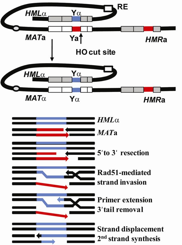
The MAT locus can be divided into five regions; W, X, Y, Z1 and Z2 (Figure 5). The two
mating type alleles MATa and MAT differ by a region designated Ya (650bp) and
Y(~750bp) containing a-specific sequence and specific genes respectively.
Figure 5. The structure of the MATa and MAT alleles( modified from (Haber, 1998)). MATa and
MAT differ by their Y region. MATa have a Ya (650bp) containing a specific sequence while MAT
have a Y (750bp) containing -specific sequence.
206.1 The mating type switch model
Mating type switching is initiated by a DSB at the Y-Z1 junction sequence at the MAT locus
by the HO endonuclease. Since, left of the cut site (as drawn), in the Y region there is no
homology (between MATa and HML or MAT and HMRa), the recombination is initiated to
the right of the cut site in the homologous region. Here MAT shares 230bp with HMR (Z1)
and 320bp with HML (regions Z1 and Z2) (Haber, 1998, 2006).
The HO endonuclease creates a 4bp 3´overhang where the 5´ of both DNA ends are further
resectioned by several DNA nucleases including the MRX complex, Sae2, Exo1, Sgs1 and
Dna2 into long 3´ ssDNA tails (Kostriken and Heffron, 1984; Mimitou and Symington, 2008;
Zhu et al., 2008) (Figure 6). The ssDNA is initially bound by replication protein A (RPA) and
then replaced by Rad51, with the assistance of accessory factors including Rad52 , forming a
nucleoprotein filament that facilitates homology searches along a donor DNA (San Filippo et
al., 2008). In a process called synapsis the nucleoprotein filament invades and anneals to the
complementary sequence in either of the donor loci, HML or HMR. The invasion and the
displacement of the non-complementary strand forms a structure called the D-loop. The
invading 3´end is used to prime new DNA synthesis using the donor duplex as a template.
The newly synthesized strand is displaced from the donor duplex and anneals to the other side
of the DSB on the original molecule. Before the second strand synthesis occurs the non
homologous Y segment is removed by the Rad1-Rad10 endonuclease-complex together with
the mismatch repair proteins Msh2 and Msh3. During mating type switching no crossover
products are formed and neither are there any alterations of the donor sequences (Haber,
2006; Krogh and Symington, 2004; San Filippo et al., 2008).
21Figure 6. Mating type switching in S. cerevisiae. An HO induced DSB at MATa is repaired by gene
conversion with the silent HML as a donor. MATa cells preferentially recombine with HML while
MAT prefer to recombine with HMRa. The donor preference is regulated by the recombination
enhancer, RE. Mating-type switch occurs through a synthesis-dependent strand annealing mechanism.
The molecular steps are shown in the lower panel. The figure is modified from (Haber, 2006).
6.2 Regulation of mating type switching in S. cerevisiae
DSBs are potentially lethal lesions that can occur spontaneously during normal cell
metabolism or through environmental factors such as ionizing radiation and mutagenic
chemicals. If left unrepaired or repaired inappropriately the DSBs can lead to chromosome
loss, deletion, duplication or translocation, events that can lead to cancerogenesis in higher
animals. There are two major ways to repair DSBs. Non homologous endjoining (NHEJ) is an
error prone repair mechanism where the ends are simply religated. After completion the site
22of repair often contains deletions or base insertions (Schiestl et al., 1993). The other major
repair mechanism is homologous recombination (HR), where an undamaged homologous
DNA sequence is used as a template. HR is a high fidelity mechanism that is usually referred
to as error-free. There are several subgroups of HR and mating type switching uses a
mechanism referred to as the synthesis dependent strand annealing (SDSA) (Haber, 1998).
The HO gene is a haploid-specific gene. It is therefore repressed by the a1-2 repressor
complex in diploids, hence limiting switching to haploids (Jensen et al., 1983). Moreover,
mating type switching occurs only in mother cells where HO is transiently expressed in late
G1 (Nasmyth, 1993). HO activation in late G1 requires the key cell cycle regulator Cdc28
(Cdk1). The expression of HO in G1 triggers the switching process to start before the DNA
replication and thus results in that both of the progeny of the mother inherit a new mating type
(Connolly et al., 1988). Since the major player of mating type switch is an endonuclease that
yields a DSB when expressed, the slightest disruption of its regulation could upset the whole
cell cycle and even be lethal for the cells. The HO gene is therefore tightly regulated and has a
large regulatory region (~2kb) containing binding sites for several transcriptional regulators.
The region can be roughly divided into a TATA-like element (-90), upstream regulatory
sequence 1 (URS1; -1000 to -1400) concerned with mother-cell specificity and a more
proximal URS2 (-900 to -150) region, which is necessary for the G1-specific transcription of
HO (Nasmyth, 1985a, b). The transcription factor Swi5 enters both mother and daughter
nuclei upon activation by the Cdc14 phosphatase during late anaphase (Nasmyth et al., 1990;
Visintin et al., 1998). Swi5 binds to sites in URS1 cooperatively with the transcription factor
Pho2 (McBride et al., 1997). The binding of Swi5 to the HO regulatory region recruits the
Swi/Snf complex to both URS1 and URS2 (Cosma et al., 1999). Swi5 also activates the
transcription of Ash1, by binding to its promoter. Unlike HO where the transcription is
delayed until late G1, Ash1 is immediately transcribed in both mother and daughter cells
(Cosma et al., 1999). The ASH1 mRNA is recognized by the nuclear RNA binding proteins
Loc1 and She2 and transported through the nuclear pores into the cytoplasm where it is
assembled into a ribonucleoprotein (RNP) complex. The RNP consists of She2, the ASH1
mRNA and the myosin motor protein Myo4 and She3. The assembled RNP is transported by
Myo4 to the distal growth tip along the actin filaments, where it is anchored by
Khd1/Bud6/She5 (Beach and Bloom, 2001; Bohl et al., 2000; Irie et al., 2002; Long et al.,
2001). The Ash1 protein is then translated and accumulates in the daughter cell nucleus where
it binds and aborts the recruitment of the Swi/Snf complex by Swi5. Both Ash1 and Swi5 are
23degraded shortly after they bind the HO promoter. Swi/Snf is therefore only recruited to the
HO regulatory region in mother cells. Swi/Snf recruits the SAGA complex which facilitates
the binding of the activator SBF (Swi4/Swi6) to the URS2 region through histone acetylation
and chromatin remodeling of the region (Cosma et al., 1999; Krebs et al., 1999). SBF is
inhibited to activate transcription by a protein called Whi5. Phosphorylation of Whi5 by
Cdc28 renders it inactive and SBF is free to recruit RNA polymerase II and associated factors,
allowing transcription to occur (Wittenberg and Reed, 2005).
6.3 Directionality of switching
Mating type switch is a biased gene conversion where cells choose to recombine their MAT
allele with the opposite allele almost all of the time, a phenomenon known as donor
preference. Donor preference is regulated by a 700bp cis-acting recombination enhancer (RE)
located 17kb from HML (Figure 6). The RE is only active in MATa cells and it is
responsible for HML being used as a donor in MATa cells. The RE activity is suppressed in
MAT cells due to the expression of 2. In the absence of RE activity HMRa is used as donor
during mating type switching. The inactivation of the RE requires the cooperative binding of
2-Mcm1 to the RE sequence and the recruitment of the Tup1-Ssn6 repressor complex,
similar to repression of asg (Haber, 1998). Exactly how the active RE promotes use of HML
as donor is unknown, but experiments have shown that the RE activates the entire left arm of
chromosome III for recombination. In MATa cells, Mcm1 binding at the RE is thought to
open up the structure thus allowing other factors to bind such as the Forkhead protein 1
(Fkh1), the Ku80 protein and the SBF (Swi4/Swi6) complex (Coic et al., 2006; Ruan et al.,
2005; Sun et al., 2002). The binding of these proteins are thought to release the left arm of
chromosome III from the nuclear periphery thus making it accessible for recombination (Ruan
et al., 2005). Despite several studies it is still unknown exactly how the RE works but its
regulation seems to be very complex.
7. Sporulation
For sporulation to occur in yeast two requirements need to be fulfilled. Firstly, the cells have
to be of the MATa/MAT cell type and secondly, they need to be starved for both nitrogen and
carbon. The diploids need to have a functional a1-2 repressor complex to undergo
sporulation. As mentioned before, a1-2 represses haploid-specific genes, and this property is
24crucial for cells which are set to undergo sporulation (Goutte and Johnson, 1988; Herskowitz,
1988). RME1 (repressor of meiosis 1) is a haploid-specific gene that is expressed in both a-
and cells (Mitchell and Herskowitz, 1986). The expression of the zinc finger protein Rme1
in haploid cells leads to the repression of a gene called IME1 (inducer of meiosis 1) (Covitz
and Mitchell, 1993) mediated by Rme1 binding sites in the IME1 regulatory region. Ime1 is a
transcriptional activator whose expression is sufficient to induce meiosis and sporulation
(Kassir et al., 1988). Therefore haploids are prohibited to undergo meiosis even under
conditions of starvation. However, in diploid MATa/MAT cells Rme1 is repressed by the a1-
2 repressor complex leading to the expression of Ime1 and the induction of meiosis. Many
genes are specifically expressed during sporulation. These genes are divided in three main
groups referred to early, middle and late genes, depending on their time of expression during
the sporulation process (Figure 7). The early sporulation genes are involved in the preparation
and execution of the meiosis I. The middle genes are expressed when the cells initiate the first
meiotic division, while the late genes are involved in post meiotic differentiation and spore
wall maturation.
During vegetative growth, where nutrients are available the sporulation genes are
repressed. This repression mechanism differs between the early, middle and late sporulation
genes (Govin and Berger, 2009).
Ume6, a DNA binding protein represses early sporulation genes by binding to an upstream
regulatory sequence, URS1 that is found in the promoters of several early genes (Anderson et
al., 1995; Strich et al., 1994). Ume6 interacts with Sin3 which in turn recruits Rpd3, a histone
deactylase to the promoters (Kadosh and Struhl, 1997). Furthermore, Ume6 also recruits an
ATP-dependent chromatin remodeling factor called Isw2 to the promoter. This complex
facilitates histone deacetylation and creates a compact chromatin structure that is inaccessible
for the transcriptional machinery (Goldmark et al., 2000).
In contrast, middle genes are repressed by a protein called Sum1. Sum1 recognizes a
sequence called the Middle Sporulation Element, MSE, which is known to regulate the middle
genes (Pierce et al., 2003; Xie et al., 1999). Sum1 binds to the promoters and repress the
genes by recruiting the NAD+ dependent histone deacetylase, Hst1 through the interaction
with Rfm1. Deacetylation of the promoter by Hst1 represses the transcription of the gene
(McCord et al., 2003).
The repression of the late sporulation genes is not that well understood besides that the
Tup1-Ssn6 repressor complex is involved (Friesen et al., 1997).
25Upon nutrient starvation the sporulation genes are activated. There are some conflicting data
on how the early genes are activated. One model suggests that phosphorylation of Ume6 by
the yeast GSK3b homologs Rim11 and Mck1 leads to the association of Ume6 with Ime1
(Malathi et al., 1997; Xiao and Mitchell, 2000). When associated with Ime1, the
phosphorylated Ume6 is converted from a repressor to an activator thus resulting in the
transcription of meiosis-specific genes (Rubin-Bejerano et al., 1996). Another model
challenges this view by suggesting that early genes are activated through the degradation of
Ume6 (Mallory et al., 2007). In the absence of Ume6, Ime1 binds to the URS1 sequences and
activates expression through the recruitment of Ime4 (the default transcript of IME4 is a
noncoding antisense, a1-2 heterodimer blocks antisense transcription and promotes sense
transcription which is translated into Ime4 protein) and the histone acetyltransferase Gcn5
(Hongay et al., 2006; Inai et al., 2007; Mallory et al., 2007). The induction of middle and late
genes is not that well understood. Ndt80 activates the middle genes by recognizing and
binding to their MSE sequence (Govin and Berger, 2009).
Figure 7. Regulation of early-, middle- and late sporulartion genes during vegetative growth and
sporulation. Different complexes repress and activate the early, middle and late genes. The figure is
modified from (Govin and Berger, 2009).
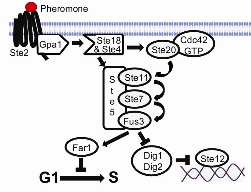
268. The cAMP-PKA pathway and the stress response
Cells of all living organisms are constantly challenged by different types of environmental
and physiological stress conditions. To cope with stress, cells have developed rapid molecular
responses to repair damage and protect against further exposure to the same or other forms of
stress. These molecular responses are particularly important in microorganisms, such as yeast
where the environment surrounding them is highly variable and conditions such as nutrient
availability, osmolarity and temperature are fluctuating. Since there is a high degree of
evolutionary conservation of these signal transduction pathways between all eukaryotes, S.
cerevisiae is often used as a model organism to study stress-induced cell signaling. There are
several pathways in yeast that are involved in stress-induced signaling. Here we will focus on
one of them, namely the cAMP-PKA pathway (Estruch, 2000; Smets et al., 2010) (Figure 8).
Yeast cells exposed to mild stress develops tolerance not only to higher doses of the same
stress but also to other kinds of stresses caused by other agents. This phenomenon called
cross-protection, suggests the existence of an integrating mechanism that senses and responds
to different forms of stress. This general stress response would act as a system that
coordinates the induction of many stress genes through a common cis-element at their
promoter. This is the case for many of the stress induced genes. They have a stress response
element (STRE) located at their promoter which gets activated by different environmental or
metabolic stresses leading to the transcriptional activation and the acquisition of a tolerant
state towards many stress conditions (Estruch, 2000; Smets et al., 2010).
In yeast, glucose signals activate the production of cellular cAMP. This pathway is called
the cAMP-PKA pathway and it plays a major role in many cellular processes, including
nutrient sensing, carbon storage and metabolism, regulation of cell proliferation and stress
response (Smets et al., 2010; Tamaki, 2007; Thevelein and de Winde, 1999). An increase of
the cAMP levels activates the cAMP dependent protein kinase (PKA). In turn PKA controls
several downstream targets through their phosphorylation. PKA is a hetero-tetramer
composed of two regulatory subunits encoded by BCY1 and catalytic subunits, redundantly
encoded by TPK1, TPK2 and TPK3 (Toda et al., 1987a; Toda et al., 1987b). The cAMP-PKA
pathway is regulated positively through two systems, intracellular detection of glucose and the
extracellular detection of glucose via the Gpr1 and Gpa2 proteins (Rolland et al., 2000).
In the intracellular cAMP-PKA activation, glucose is taken up by the cell and
phosphorylated by either one of the phosphorylating enzymes Hxk1, Hxk2 or Glk1. The
27signal is further transduced through the small G proteins, Ras1/2. The active GTP bound form
of the Ras1/2 proteins stimulates adenylate cyclase encoded by CYR1 to produce cellular
cAMP from ATP (Casperson et al., 1985; Field et al., 1988; Kataoka et al., 1985; Toda et al.,
1985). The GDP/GTP exchange and therefore the activation of the Ras proteins is catalyzed
by Cdc25 while the Ira proteins (Ira1/2) hydrolyse the GTP to GDP and inactivates Ras
(Broek et al., 1987; Jones et al., 1991; Robinson et al., 1987; Tanaka et al., 1991; Tanaka et
al., 1989; Tanaka et al., 1990a; Tanaka et al., 1990b). The increased cAMP level activates
PKA by binding to its regulatory subunit, Bcy1, and dissociating the catalytic subunits
encoded by TPK1, TPK2 and TPK3, which then phosphorylates target proteins. Cellular
cAMP is hydrolysed by the low- and high-affinity phosphodiesterases, encoded by PDE1 and
PDE2 respectively, which hence contributes to cAMP homeostasis (Nikawa et al., 1987; Sass
et al., 1986; Toda et al., 1987a; Toda et al., 1987b).
The extra cellular detection system of glucose occurs through a G protein coupled receptor
(GPCR) system composed of Gpr1 and Gpa2 (Kraakman et al., 1999). Gpr1 belongs to the
GPCR super family while Gpa2 belongs to the heterotrimeric G protein alpha subunit protein
family, the same family as Gpa1 involved in mating. Addition of glucose to glucose-starved
cells activates Gpr1 which stimulates the exchange of GDP to GTP on Gpa2, which in turn
activates the cAMP-PKA pathway through the stimulation of adenylate cyclase (Kraakman et
al., 1999; Nakafuku et al., 1988; Xue et al., 1998; Yun et al., 1998). The protein Rgs2
belonging to the family of regulators of G protein signaling (RGS) inhibits the Gpa2 signaling
through GTPase activity (Versele et al., 1999).
cAMP activated PKA has a major impact on gene expression through its targets, which
effect gene transcription either directly or indirectly. Two of the PKA targets are the partially
redundant C2H2 zinc finger proteins Msn2 and Msn4. Msn2 and Msn4 are transcription
factors which mediate the transcription of the stress response element (STRE) controlled
genes (Estruch and Carlson, 1993; Martinez-Pastor et al., 1996; Schmitt and McEntee, 1996).
The STRE (CCCCT or AGGGG) regulated genes are involved in a variety of processes
including stress such as heat, osmotic and oxidative stress, carbohydrate metabolism and
growth control (Gasch et al., 2000; Moskvina et al., 1998; Smith et al., 1998). During growth
on glucose Msn2 and Msn4 are cytosolic and inhibited to enter the nucleus by PKA mediated
phosphorylation of their nuclear localization signal. However, during glucose depravation
they get activated by protein phosphate 1 (Ppt1) and translocated to the nucleus where they
can induce the STRE regulated genes (De Wever et al., 2005; Garreau et al., 2000; Gorner et
28al., 1998; Gorner et al., 2002). There are two additional PKA targets that PKA phosphorylates
to inhibit the activation of Msn2/4. These targets are protein kinases called Yak1 and Rim15.
Yak1 activates Msn2/4 through phosphorylation of Msn2 by an unknown mechanism but does
not affect neither the DNA binding nor the cellular localization of Msn2 (Lee et al., 2008).
Rim15 was first identified as an activator for meiotic gene expression and was later shown to
be essential in the induction of several stress response genes, thermotolerance and the proper
induction of G1-arrest upon nutrient starvation (Cameroni et al., 2004; Reinders et al., 1998;
Vidan and Mitchell, 1997; Zhang et al., 2009). In addition, Rim15 controls an expression
program during the diauxic shift mediated by the transcription factors Msn2, Msn4 and Gis1.
Gis1 activates stress response genes containing the post diauxic shift (PDS) element, some of
which are required for respiratory growth (Cameroni et al., 2004; Pedruzzi et al., 2000; Zhang
et al., 2009).
Deletion of both RAS1 and RAS2 or CYR1 is lethal for the cells. This is due to that the
cAMP-PKA pathway is essential for cell viability. Mutation of BCY1 suppresses the lethal
effects of the deletion of both RAS1 and RAS2 and also the phenotype of cyr1 mutation. In
addition, overexpression of Cyr1 also suppresses the lethal phenotype of the deleted RAS
genes. (Kataoka et al., 1985; Matsumoto et al., 1982; Toda et al., 1985)
29Figure 8. The cAMP-PKA pathway and stress response in S. cerevisiae. Addition of glucose to
glucose-starved respiring cells triggers a rapid cAMP synthesis and PKA activation. Two glucose-
induced sensing systems are required for cAMP synthesis (i) extra cellular detection of glucose via the
Gpr1-Gpa2 system and (ii) intracellular detection of glucose. Activated PKA mediates the fast switch
from respiratory to fermentative growth via several downstream targets. Arrows and bars represent
positive and negative interactions and dashed lines represent putative or indirect interactions. See text
for further details. The figure is modified from (Smets et al., 2010).
9. Transposons
Transposable elements (TEs) are a heterogeneous class of genetic elements that can jump
from one location in the genome through excision or duplication and integrate into a second
location without any requirement for DNA homology (Curcio and Derbyshire, 2003).
Transposition of TEs in the genome can either activate or inactivate genes depending on the
location where the TE integrates relative to the target gene. If it integrates upstream of a gene
it might activate its expression, but if it integrates within a gene the gene may get inactivated.
30Transposons can also promote inversions, deletions and the transduction of flanking DNA. In
addition TEs may cause chromosome rearrangements by generating blocks of homology that
result in unequal sister chromatid exchanges (Moran et al., 1999). TEs are widely distributed
in bacteria, plants and animals. The proportion of TEs in these genomes varies from 1-5% in
microorganisms, to more than 25% in rice and to about 50% of the human genome (Lander et
al., 2001; Yu et al., 2002)
9.1 Definition and classification of transposons
TEs exist as either autonomous or non autonomous elements. Autonomous TEs encodes all
the enzymes necessary to mobilize and integrate whereas non autonomous TEs lack that
capacity and therefore depend on their autonomous relatives for their enzymatic machinery to
move (Sinzelle et al., 2009).
The transposons are divided into two major classes according to their transposition
mechanism. Class 1 transposons or retrotransposons are mobilized through an RNA
intermediate and use a “copy and paste” mechanism, which means that they remain at the
original site. This class of TEs is not going to be further addressed in this thesis. Class II TEs
or DNA transposons move as DNA molecules and often use a “cut and paste” mechanism,
which means that they leave the original site. Both classes contain autonomous and
nonautonomous elements. Class II elements are further subdivided in two subclasses, which
are distinguished from each other based on the generation of a single- or double-strand cuts
during the transposition process. Subclass I contains TEs that transpose through the “cut and
paste” mechanism and are flanked by terminal inverted repeats (TIRs) of variable length. So
far this subclass is composed of nine superfamilies that are distinguished by the TIR sequence
and the length of the target site duplications (TSDs). TSDs are short DNA sequences from the
host that gets duplicated and flank the transposon upon insertion (Wicker et al., 2007). The
second subclass of DNA transposons contains the recently indentified Helitron and Maverick
families of transposons. Their transposition involves a copy and paste mechanism and relies
on replication without any DNA double strand breaks (Kapitonov and Jurka, 2001; Pritham et
al., 2007)
The classical DNA transposon usually transcribe at least one protein, the transposase,
which binds to DNA at the TIR sequence, through its N-terminal domain and cuts the DNA
though the catalytic domain at the C-terminus. Except from the P, piggyback and the CACTA
superfamilies where the catalytic domain is not well established, eukaryotic transposases
31contain a conserved DDE/D-motif also found in retroviral integrases (Haren et al., 1999;
Sinzelle et al., 2009)
9.2 Function and characteristics of the DDE-motif in transposons
The best characterized transposons belong to the class II transposons including those that
were first originally described by Barabara McClintock. Most of these transposons encode a
DDE-containing transposase, which is responsible for the excision and integration of the
transposon into the target (Haren et al., 1999). In vitro and in vivo mutagenic studies of the
DDE motif established its importance and showed that it was essential for transposition
(Haren et al., 1999). Structural studies have located divalent metal ions and both of the
transposon ends to the active site of the transposase. Furthermore, these studies support a role
for the DDE motif in coordination of the metal ions to facilitate catalysis (Davies et al., 2000;
Lovell et al., 2002). The DDE-enzymes catalyzes their reactions within the transpososome, a
DNA-protein complex containing the transposon, the target and the transposase protein and
sometimes other proteins. The transpososome ensures the fidelity of the reaction (Mizuuchi et
al., 1995).
The DDE-enzymes catalyze two chemical reactions. First, the phosphodiester backbone at
each end of the transposon undergoes water-mediated hydrolysis generating free 3’ OH ends.
Second, the exposed 3’ ends function as nucleophiles and directly attack the target DNA in a
transesterification reaction. The nucleophilic attack by the two 3’ ends on each strand of the
target is staggered and separated by 2-9 nucleotides. Repair of this segments results in the
duplication of these 2-9 nucleotides, a hallmark of DDE-transposition. The length of the
duplication is characteristic for each transposon (Craig, 2002). It has been shown that for
transposases and retroviral integrases the structure of the catalytic domains and the location of
the DDE-motifs are largely super imposable despite of the lack of sequence similarity (Rice
and Mizuuchi, 1995).
9.3 Transposition pathways of DDE-motif containing transposons
How do the different DDE-motif containing transposable elements move? Although the
chemical reactions performed by the different DDE-transposases are the same, the
mechanisms they use are different. The transpositions can be categorized into “Copy in”, “Cut
out, paste in” and “Copy out, paste in” (Curcio and Derbyshire, 2003) (Figure 9).
329.3.1 “copy-in” transposition
The Mu and the Tn3 families represent transposons using this mechanism. These transposases
generates a nick at the 3’ ends of the transposon and joins them to the target, resulting in a
branched strand-transfer intermediate (Mizuuchi, 1984). Replication from the newly exposed
3’ ends in the flanking target DNA generates a co-integration molecule with two copies of the
transposon (Curcio and Derbyshire, 2003). Resolution of the co-integrate results in a new
copy of the TE at the target site, but also leaves an intact copy at the donor site.
9.3.2 “cut-out, paste in” transposition
A major trait of this mechanism is that the DNA transposons excise themselves from the
donor DNA trough a hairpin intermediate. In the bacterial IS10 and IS50 elements the 3´
nicked transposon ends attack the phosphate backbone of the 5’ ends of the complementary
strand of the transposon forming a hairpin on the transposon ends. The hairpin is resolved
through hydrolysis at the 3´ends and leads to integration into the genome. For the eukaryotic
Ac/Dc and Hermes elements it is proposed that nicks are introduced at the 5’ ends of the
transposon, generating free 3’ ends in the flanking DNA. Direct transesterification by the
3´ends on the opposing strand results in hairpin formation at the flanking DNA (Curcio and
Derbyshire, 2003). Hence, a difference between prokaryotes and eukaryotes in this respect is
the formation of hairpin ends on the transposon ends (prokaryotes) versus on the flanking
DNA ends (eukaryotes). V(D)J recombination shares this mechanism, thus forming hairpins
on the coding ends through transesterification. The coding ends are quickly opened, processed
and joined through the non-homologous end-joining DNA repair apparatus thus releasing the
excised blunt ended signal sequences which is equivalent to excised transposon (Jones and
Gellert, 2004).
9.3.3“copy-out, paste-in” transposition
The first excision step for IS911, IS2, IS3 and other related elements involves the generation
of an asymmetric nick at just one 3’ end of the transposon. The exposed 3’ end directly attack
the phosphodiester backbone of the same strand outside the transposon, circularizing one
strand of the transposon and generating a free 3’ end in the flanking DNA. DNA replication
starting from the nick of the transposon junction generates a new copy of the transposon as an
33You can also read