The end protection problem-an unexpected twist in the tail
←
→
Page content transcription
If your browser does not render page correctly, please read the page content below
Downloaded from genesdev.cshlp.org on February 11, 2021 - Published by Cold Spring Harbor Laboratory Press

PERSPECTIVE

The end protection problem—an
unexpected twist in the tail
Phil Ruis and Simon J. Boulton
The Francis Crick Institute, London NW1 1AT, United Kingdom

In this perspective, we introduce shelterin and the                         and cell death, the question of how shelterin—and, espe-
mechanisms of ATM activation and NHEJ at telomeres,                         cially, TRF2—protects chromosome ends from DNA re-
before discussing the following questions: How are t-                       pair activities has been paramount. A succession of
loops proposed to protect chromosome ends and what is                       papers followed, showing that loss of TRF2 leads to activa-
the evidence for this model? Can other models explain                       tion of the DNA double strand break (DSB) kinase ATM
how TRF2 mediates end protection? Could t-loops be                          and classical nonhomologous end joining (NHEJ) at telo-
pathological structures? How is end protection achieved                     meres. The question of how TRF2 protects chromosome
in pluripotent cells? What do the insights into telomere                    ends has thus focused on how TRF2 prevents the activa-
end protection in pluripotent cells mean for the t-loop                     tion of these dual pathways—ATM and NHEJ—at telo-
model of end protection? Why might different cell states                    meres. Both pathways rely on the binding of DSB
have evolved different mechanisms of end protection?                        sensors (MRE11/RAD50/NBS1 [MRN] and KU70/80, re-
Finally, we offer support for an updated t-loop model of                    spectively) to the same substrate (DNA ends), suggesting
end protection, suggesting that the data is supportive of                   that one possible way TRF2 could inhibit these pathways
a critical role for t-loops in protecting chromosome ends                   would be to hide these DSB ends.
from NHEJ and ATM activation, but that other                                   The field has now broadly coalesced around the t-loop
mechanisms are involved. Finally, we propose that t-                        model of end protection, which hypothesises that TRF2
loops are likely dynamic, rather than static, structures.                   mediates the invasion of the single stranded telomere ter-
                                                                            minus into the chromosome-proximal telomeric dsDNA,
                                                                            or stabilizes this structure, sequestering the chromosome
Chromosome ends present a dual danger to cells. Semi-                       end in a lariat loop structure, elegantly hiding it from the
conservative DNA replication is unable to replicate the                     DSB end sensors that trigger ATM and NHEJ activation.
extreme chromosome terminus and, because chromo-                            This model was first proposed in 1999 and, to a large ex-
some ends resemble DNA breaks, these ends can poten-                        tent, fits with published data. In particular, telomere loops
tially activate the DNA damage response (DDR) and                           have been observed via both electron microscopy (EM)
elicit misrepair. Mammalian chromosome ends mitigate                        and, more recently, super-resolution microscopy. More-
these dual dangers through telomeres, nucleoprotein                         over, TRF2 has been shown to stimulate the formation
structures consisting of double-stranded (ds) TTAGGG re-                    of these t-loops in vitro, while the depletion of TRF2 leads
peats culminating in a single-stranded (ss) G-rich 3′ over-                 to the reduction of these loops in cells. Although this
hang that are bound by the hexameric shelterin complex                      makes TRF2-mediated t-loop formation a plausible candi-
(Fig. 1A). Shelterin facilitates the addition of new telo-                  date for the mechanism through which TRF2 protects
meric repeats by regulating the reverse transcriptase                       chromosome ends, it has remained impossible to test
telomerase and also inhibits at least seven distinct mech-                  this directly. Indeed, some have suggested that these t-
anisms of DNA repair, counteracting telomere loss and                       loops are a pathological structure formed through aberrant
DDR activation at chromosome ends, respectively.                            telomere recombination. Furthermore, TRF2 interacts
   Understanding how telomeres protect the ends of chro-                    directly with multiple ATM and NHEJ components, and
mosomes from DNA damage signaling has been a focus of                       TRF2 has also been suggested to mediate telomere com-
the telomere field for >20 yr. Since the seminal discovery                  paction, providing alternative means through which it
that removal of the shelterin component TRF2 from telo-                     could mediate end protection. Therefore, the exact role
meres leads to telomere end-to-end fusions, senescence                      of t-loops and TRF2 in end protection has remained con-
                                                                            troversial. One key test, identifying whether t-loops pro-
                                                                            tect chromosome ends in the absence of TRF2, has
[Keywords: DNA damage response; NHEJ; TRF2; pluripotency; somatic           never before been possible as no factor, other than TRF2,
cells; t-loops; telomeres]
Corresponding author: simon.boulton@crick.ac.uk
Article published online ahead of print. Article and publication date are
online at http://www.genesdev.org/cgi/doi/10.1101/gad.344044.120. Free-     © 2021 Ruis and Boulton This article, published in Genes & Develop-
ly available online through the Genes & Development Open Access             ment, is available under a Creative Commons License (Attribution 4.0 In-
option.                                                                     ternational), as described at http://creativecommons.org/licenses/by/4.0/.

                  GENES & DEVELOPMENT 35:1–21 Published by Cold Spring Harbor Laboratory Press; ISSN 0890-9369/21; www.genesdev.org                 1
Downloaded from genesdev.cshlp.org on February 11, 2021 - Published by Cold Spring Harbor Laboratory Press

Ruis and Boulton

               A

               B

               C

               D

               E

Figure 1. The mammalian telomere. (A) Depiction of a mammalian chromosome highlighting key features of the telomeres that cap each
chromosome end. (B) Depiction of how the hexameric complex shelterin associates with telomeres. (C) TRF1 promotes efficient telomere
replication, avoiding replication fork stalling that leads to ATR activation, telomere loss and telomere fragility. (D) RAP1 represses homol-
ogous recombination (HDR) within mammalian telomeres, avoiding deleterious telomere length alterations. (E) TPP1 recruits POT1 to
telomeres, with POT1 repressing ATR activation and HDR, restricting excessive G overhang resection and telomere length alterations
respectively.

has been found that can mediate t-loop formation and/or                  liferate without TRF2, apparently indefinitely. However,
stabilization.                                                           end protection in ESCs is still dependent on the shelterin
   Several unexpected new discoveries have enabled pro-                  complex, as depletion of the entire shelterin complex
gress to be made in this direction. Surprisingly, together               from ESC telomeres leads to ATM activation, NHEJ-de-
with the Cesare laboratory (Ruis et al. 2020), we recently               pendent telomere end-to-end fusions and rapid cell death.
identified that in mouse embryonic stem cells (ESCs),                    While multiple pluripotent lineages, including ESCs and
chromosome end protection is achieved largely indepen-                   epiblast stem cells (EpiSCs) apparently possess this largely
dently of the shelterin component TRF2. Similar findings                 TRF2-independent end protection mechanism, the differ-
have been obtained independently from parallel studies                   entiation of TRF2-null ESCs leads to a dramatic loss of end
from the Lazzerini-Denchi laboratory (Markiewicz-                        protection, robust activation of ATM and NHEJ at telo-
Potoczny et al. 2020). While TRF2 localizes to ESC telo-                 meres and cell death at the point that these cells exit
meres as part of the shelterin complex, ESCs without                     the pluripotent state. Thus, in the context of early devel-
TRF2 show a severely attenuated telomeric DDR and do                     opment, this alternate end protection mechanism is ap-
not undergo telomeric NHEJ. Consistently, ESCs can pro-                  parently restricted to the pluripotent state and TRF2

2      GENES & DEVELOPMENT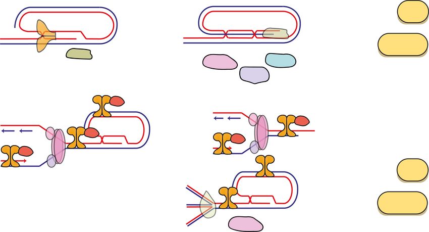
Downloaded from genesdev.cshlp.org on February 11, 2021 - Published by Cold Spring Harbor Laboratory Press

                                                                                             Telomeres protection of chromosome ends

becomes essential for chromosome end protection when               et al. 2006). Finally, TRF2 interacts with RAP1, the
cells exit pluripotency upon differentiation. Finally, to-         most evolutionarily conserved component of shelterin
gether with the Cesare laboratory (Ruis et al. 2020), we           (Li et al. 2000; Li and de Lange 2003). RAP1 has nontelo-
also observed that ESCs possess t-loops with a similar fre-        meric functions: It impacts nuclear factor κB (NF-κB) sig-
quency to somatic cells, but unlike somatic cells, the for-        naling (Teo et al. 2010), regulates transcription
mation of these loops is not dependent on TRF2.                    (Martinez et al. 2010, 2013), and is the only nonessential
   This work, and other recent studies, cast new light on          component of shelterin; knockout of any other compo-
the t-loop model of chromosome end protection. In this             nent leads to cell and organismal inviability (Karlseder
perspective, we introduce shelterin and the mechanisms             et al. 2003; Chiang et al. 2004; Celli and de Lange 2005).
of ATM activation and NHEJ at telomeres, before discuss-              Shelterin is thought to be expressed, localized to telo-
ing the following questions: How are t-loops proposed to           meres and responsible for end protection in all known
protect chromosome ends and what is the evidence for               mammalian systems (de Lange 2005, 2010, 2018; Sfeir
this model? Can other models explain how TRF2 medi-                and de Lange 2012). As the only complex that binds telo-
ates end protection? Could t-loops be pathological struc-          meres with a high affinity and sequence specificity, inter-
tures? How is end protection achieved in pluripotent               actions with shelterin components represent the primary
cells? What do the insights into telomere end protection           means through which other proteins and complexes are re-
in pluripotent cells mean for the t-loop model of end pro-         cruited to telomeres. These other proteins, which include
tection? Why might different cell states have evolved dif-         nucleases, helicases, DNA damage factors and DNA
ferent mechanisms of end protection? Finally, we offer             replication proteins, act as shelterin cofactors, assisting
support for an updated t-loop model of end protection,             shelterin in execution of end protection and telomere rep-
suggesting that the data is supportive of a critical role          lication and extension (Oganesian and Karlseder 2009; de
for t-loops in protecting chromosome ends from NHEJ                Lange 2018). They perform important functions including
and ATM activation, but that other mechanisms are in-              unwinding telomere secondary structures to enable pas-
volved. Finally, we propose that t-loops are likely dynam-         sage of the replication fork (RTEL1 and BLM), resolving
ic, rather than static, structures.                                telomere secondary structures that could not be unwound
                                                                   (SLX1/4), generating 3′ G overhangs of an appropriate
The shelterin complex                                              length for end protection (CST complex, APOLLO, and
                                                                   EXOI) and processing telomere ends (MRN complex,
Telomeric DNA is bound by specialized sets of proteins,            KU70/80) (d’Adda di Fagagna et al. 2001; Dimitrova and
whose composition, structure and function has diverged             de Lange 2009; Sfeir et al. 2009; Wu et al. 2010, 2012; Ye
across species. In mammalian cells, six bona fide proteins         et al. 2010; Vannier et al. 2012, 2013; Zimmermann et al.
specifically associate with telomeres in a complex termed          2014). The shelterin complex both possesses its own in-
shelterin (de Lange 2005): TRF1 (telomeric repeat-binding          trinsic functions and co-opts the functions of these various
factor 1, also known as TERF1), TRF2 (telomeric repeat-            cofactors to maintain end protection. Emerging data sug-
binding factor 2, also known as TERF2), RAP1 (TERF2-in-            gests these activities are tightly regulated throughout the
teracting protein, also known as TERF2IP), TIN2 (TRF1-             cell cycle (Sarek et al. 2019).
interacting nuclear factor 2, also known as TINF2),                   Since TRF1 and TRF2 are the only proteins to bind telo-
TPP1 (adrenocortical dysplasia protein homolog, also               meres with a high affinity and sequence specificity, code-
known as ACD), and POT1 (protection of telomeres 1)                pletion of both TRF1 and TRF2 leads to “shelterin-free”
(Fig. 1B). Although a complete atomic-level structure of           telomeres that lose the end protective functions of shel-
the shelterin complex is currently lacking, how the shel-          terin and its cofactors. This reveals the full scope of the
terin components interact with each other and with telo-           telomere end protection problem (Sfeir and de Lange
meric DNA, recruit other proteins to telomeres and                 2012). Telomeres lacking shelterin are subject to the re-
mediate end protection is generally well-known. The shel-          sponse of at least seven independent DDR pathways,
terin complex components TRF1 and TRF2 bind with                   any of which could cause gross genome instability if inap-
high (nanomolar) affinity to double stranded telomeric             propriately activated at telomeres. The basic mechanisms
TTAGGG repeats via their Myb domains (Chong et al.                 through which shelterin and its cofactors inhibit these
1995; Broccoli et al. 1997; Smogorzewska et al. 2000).             DDR pathways to achieve end protection are now general-
TIN2 interacts with TRF1, TRF2 and TPP1, bridging these            ly understood (Fig. 1C,E; Palm and de Lange 2008; Sfeir
three proteins, while TPP1 interacts with POT1 (O’Con-             et al. 2009; de Lange 2010, 2018; Kim et al. 2017). Howev-
nor et al. 2006; Hockemeyer et al. 2007; Wang et al.               er, one topic that has remained controversial has been the
2007; Kibe et al. 2010; Takai et al. 2011; Hu et al. 2017),        protection of chromosome ends from the ATM and NHEJ
enabling POT1 to coat the 3′ single-stranded G overhang            DSB pathways by the shelterin component TRF2.
by virtue of its oligonucleotide/oligosaccharide-binding
(OB) folds (Kim et al. 1999; Baumann and Cech 2001; Bau-
mann et al. 2002; Loayza and De Lange 2003; Lei et al.
                                                                   The activation of ATM and NHEJ at DNA double-strand
2004; O’Connor et al. 2006; Hu et al. 2017). Rodents ex-
                                                                   breaks
press two POT1 paralogs that emerged via gene duplica-
tion (POT1a and POT1b) and are structurally similar,               DSBs are highly toxic lesions that can be repaired via
yet functionally divergent (Hockemeyer et al. 2006; Wu             three independent mechanisms: homology-directed

                                                                                                   GENES & DEVELOPMENT            3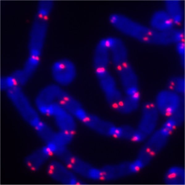
Downloaded from genesdev.cshlp.org on February 11, 2021 - Published by Cold Spring Harbor Laboratory Press
Ruis and Boulton
repair (HDR, also known as HR), nonhomologous end cell death (Shieh et al. 1997; Banin et al. 1998; Meek 2004;
joining (NHEJ), and microhomology-mediated repair Roos et al. 2016); P53-binding protein 1 (53BP1) (Adams
(MMEJ, also known as alt-NHEJ) (Jackson and Bartek and Carpenter 2006; Chapman et al. 2013); the histone
2009; Chapman et al. 2012; Panier and Boulton 2014). variant H2AX, which is phosphorylated on Ser139 to gen-
The activation of HR and alt-NHEJ at telomeres has erate γH2AX (Burma et al. 2001).
been extensively reviewed elsewhere (Doksani and de Once generated by ATM at DSBs, γH2AX binds MDC1.
Lange 2014; de Lange 2018). DSBs are recognized by two MDC1 binds MRN, which in turn recruits and activates
parallel DSB end sensor complexes—KU70/80 and MRN additional ATM at the DSB, producing a feed-forward
—that associate with DSB ends within seconds of their loop that results in focal accumulation of γH2AX (Lukas
formation (Fig. 2A; Uziel et al. 2003). Once bound to et al. 2011). This facilitates a cascade of protein recruit-
DSB ends, MRN recruits and activates the apical DDR sig- ment to, and ubiquitylation of, chromatin surrounding
naling kinase Ataxia telangiectasia mutated (ATM) (for re- DNA breaks, involving the RING finger 8 (RNF8), RING
view, see Paull 2015). NBS1 and RAD50 both bind ATM finger 168 (RNF168) E3 Ubiquitin ligases and their E2 li-
directly, facilitating its recruitment to DSBs, while gase UBC13 (Huen et al. 2007; Kolas et al. 2007; Mailand
RAD50 promotes short distance (15 bp) unwinding of et al. 2007; Doil et al. 2009; Peuscher and Jacobs 2011;
the DSB end, producing an optimal end for ATM activa- Mattiroli et al. 2012). This ubiquitylation enables the sta-
tion (Cannon et al. 2013) and MRN acts as a cofactor for ble recruitment of additional repair factors to DSBs, in-
ATM activation (Paull 2015). ATM is autoinhibited in cluding 53BP1, which binds to H2A ubiquitylated by
its typical dimeric form (Lee and Paull 2005; 2007), but RNF168 on Lys15 (Anderson et al. 2001; Bekker-Jensen
when recruited to DSB ends by MRN, ATM monomer- et al. 2005). 53BP1 then acts as a scaffold with a range of
izes, becomes active and undergoes autophosphorylation protein-protein interaction sites that enable the recruit-
on Ser1981. ATM activation thus requires both MRN ment of other factors involved in DSB repair to the lesion
and accessible DSB ends. Once activated, ATM coordi- (Mirman and de Lange 2020). 53BP1 is itself phosphorylat-
nates the local and cellular response to DSBs, by phos- ed by ATM on multiple N-terminal SQ/TQ sites; this
phorylating in excess of 200 protein targets that promote phosphorylation enables the binding of 53BP1 to cofactors
lesion repair and cell cycle arrest to provide time for repair including the NHEJ mediator RIF1, with RIF1 then re-
to ensue (Matsuoka et al. 2007; Jackson and Bartek 2009). cruiting the Shieldin complex to DSBs (Chapman et al.
Important ATM targets include: checkpoint kinase 2 2013; Boersma et al. 2015; Xu et al. 2015; Dev et al.
(CHK2), which inhibits CDKs to stall cell cycling at the 2018; Ghezraoui et al. 2018; Gupta et al. 2018; Mirman
G1/S and G2/M transitions (Chehab et al. 2000; Matsuoka et al. 2018; Noordermeer et al. 2018).
et al. 2000); P53, which coordinates both transient cell cy- Unlike MRN, which is involved in both NHEJ and HR,
cle arrest and (if P53 activation is prolonged) senescence or the KU70/80 DNA end sensor is a specific component of
A B Figure 2. TRF2 prevents ATM and NHEJ at
mammalian telomeres. (A) TRF2 inhibits the
localization of the MRE11, RAD50, and
NBS1 (MRN) and KU70/80 DNA double-
strand break (DSB) sensors to chromosome
ends. In somatic cells, TRF2 depletion depro-
tects telomeres leading to MRN and KU70/
80 binding to telomeres. KU70/80 facilitates
ligase IV-mediated NHEJ, leading to telo-
mere fusions. Concurrently, MRN recruits
ATM to chromosome ends, triggering a chain
C of protein modification and positive feedback
loops that culminate in stable recruitment of
MRN, ATM, MDC1, 53BP1, and the 53BP1
effectors RIF1, REV7, and Shieldin to telo-
meres, where they promote NHEJ. ATM
also phosphorylates and activates CHK2
and P53, amongst other targets, leading to
checkpoint arrest, apoptosis and senescence.
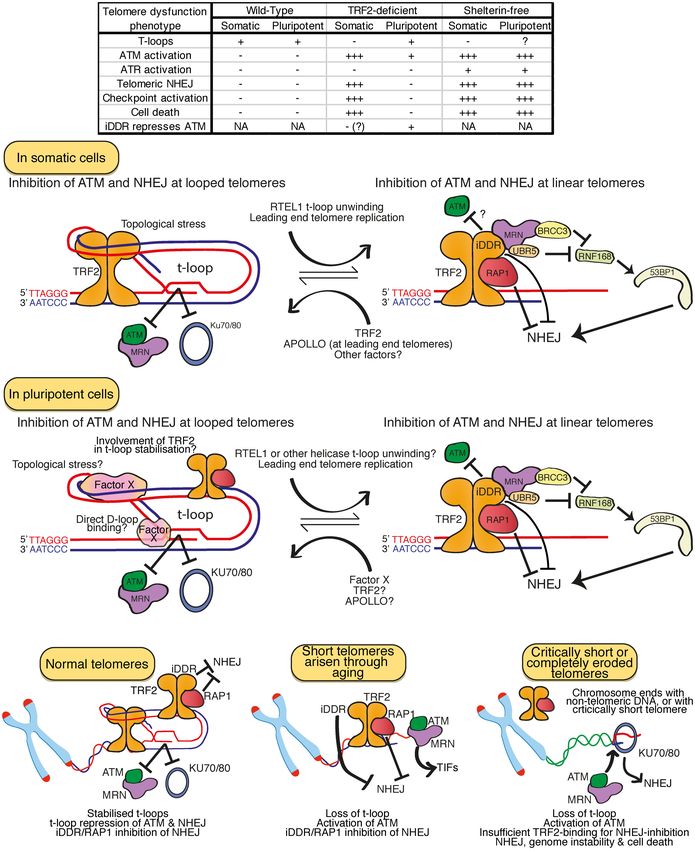
(B,C) The removal of TRF2 from somatic
D cells leads to abundant telomere end-to-end
fusions, visible in metaphase spreads, and
telomeric dysfunction-induced foci (TIFs),
which represent telomeres bound by the
DNA repair proteins mentioned in A. (D) Lin-
ear telomeres have accessible ends that re-
semble “DSBs,” but telomere ends within looped (t-loop) conformations would remain inaccessible to, and hence protected from, DSB
sensors.
4 GENES & DEVELOPMENT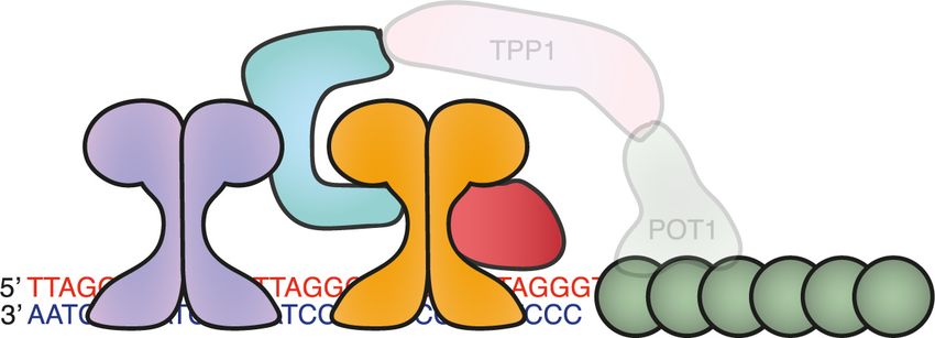
Downloaded from genesdev.cshlp.org on February 11, 2021 - Published by Cold Spring Harbor Laboratory Press

                                                                                             Telomeres protection of chromosome ends

classical NHEJ-mediated DSB repair (Weterings and van              clearly solve this end protection problem; chromosome
Gent 2004; Weterings and Chen 2008). KU70/80 forms a               ends in mammalian cells do not constitutively activate
ring that binds DNA ends with a remarkable affinity                ATM or undergo NHEJ. A series of seminal papers re-
(binding constant of 2 × 10⁹ M−1), is highly abundant with-        vealed that the removal of TRF2 from telomeres leads to
in most cells (∼ 500,000 molecules per cell) and binds DSB         chromosome end-to-end fusions, CHK2-mediated G2/M
ends in a sequence independent manner (Blier et al. 1993;          cell cycle arrest and p53-mediated senescence and cell
Downs and Jackson 2004; Davis and Chen 2013). Once                 death (van Steensel et al. 1998; Karlseder et al. 1999).
bound to DSB ends, KU70/80 serves as a scaffold to recruit         These telomere fusions are dependent on the core NHEJ
other factors involved in NHEJ, including DNA-PKcs,                factors KU70/80, ligase IV, and DNA-PKcs; the ATM ki-
DNA ligase IV, XRCC4-like factor (XLF), X-ray cross-com-           nase and its DNA sensor MRN; and the NHEJ accessory
plementing protein 4 (XRCC4), and aprataxin and PNK-               factors 53BP1, RIF1, and Shieldin (Fig. 2A,B; Smogorzew-
like factor (APLF) (Davis and Chen 2013; Davis et al.              ska et al. 2002; Celli and de Lange 2005; Celli et al. 2006;
2014). These factors process DSB ends, producing a blunt           Denchi and de Lange 2007; Dimitrova et al. 2008; Deng
ended substrate suitable for DNA ligase IV-mediated liga-          et al. 2009; Dimitrova and de Lange 2009; Chapman
tion (Wilson et al. 1997). This contrasts with HR, which           et al. 2013; Zimmermann et al. 2013; Ghezraoui et al.
requires extensive resection of DSB to produce ssDNA               2018). Like at bona fide DSBs, ATM is recruited to telo-
suitable for RAD51 loading and the homology search                 meres by MRN upon TRF2-loss, generating γH2AX and
that is essential for HR. Recent evidence suggests the cen-        triggering the γH2AX/MDC1/MRN/ATM feed-forward
tral axis of the HR versus NHEJ pathway choice is the reg-         mechanism that leads to stable recruitment of RNF8,
ulation of resection: Resection of DSBs favors HR, while           RNF168, and 53BP1 and its cofactors RIF1 and Shieldin
the retention of relatively blunt ends favors NHEJ (Panier         to telomeres (de Lange 2018). Once recruited to TRF2-
and Boulton 2014).                                                 null telomeres, 53BP1 cooperates with the LINC complex
   While the core NHEJ components are sufficient for               to promote their microtubule-dependent motility and, to-
NHEJ in vitro and at simple blunt-ended DSBs in G1, addi-          gether with RIF1/Shieldin, counteracts excessive telo-
tional factors including ATM, 53BP1 and its cofactors RIF1         mere resection to maintain overhangs of an appropriate
and Shieldin are required in many in vivo contexts, includ-        length for NHEJ (Dimitrova et al. 2008; Lottersberger
ing at physiological DSBs generated during class switch re-        et al. 2015; Mirman et al. 2018). 53BP1, γH2AX and
combination (CSR), DSBs within heterochromatin and at              many of these other DSB repair factors can be readily de-
dysfunctional telomeres (Escribano-Díaz et al. 2013; Zim-          tected at telomeres in TRF2-null cells via indirect immu-
mermann et al. 2013; Xu et al. 2015; Ceccaldi et al. 2016;         nofluorescence (IF) staining, where they form discrete foci
Mirman et al. 2018; Noordermeer et al. 2018; Mirman                termed telomere dysfunction induced foci (TIFs) (Fig. 2C;
and de Lange 2020). 53BP1 promotes the three-dimension-            Takai et al. 2003). The accumulation of TIFs, CHK2-medi-
al motility of DNA breaks, facilitating synapsis of distal         ated cell cycle arrest, telomere fusions and p53 activation
DNA ends, while Shieldin counteracts DNA end resection             in TRF2-null cells is entirely dependent on ATM, suggest-
to promote NHEJ over HR at DSBs (Lottersberger et al.              ing this is the sole kinase responsible for coordinating the
2015; Dev et al. 2018; Ghezraoui et al. 2018; Mirman               DDR at TRF2-null telomeres (Celli and de Lange 2005;
et al. 2018). Shieldin may achieve this by directly inhibit-       Denchi and de Lange 2007). Likewise, the TIFs in TRF2-
ing DNA end resection, promoting CST/Polα/Primase-                 null cells are dependent on MRN, while these telomere
mediated fill-in of already resected DNA ends or via both          fusions are dependent on both MRN and KU70/80 (Celli
pathways. Highly heterochromatic regions are also refrac-          et al. 2006; Deng et al. 2009; Dimitrova and de Lange
tory to NHEJ (Goodarzi et al. 2008). At DSBs in such re-           2009).
gions, 53BP1 amplifies MRE11-NBS1 accumulation,                       Somewhat counterintuitively, multiple components of
concentrating active ATM to facilitate repair through ro-          ATM and NHEJ signaling that contribute to NHEJ at
bust local phosphorylation of KAP1, which then promotes            TRF2-null telomeres are actually required to maintain
repair through chromatin relaxation (Noon et al. 2010).            complete chromosome end protection in TRF2-proficient
Thus, while not essential components of NHEJ per se,               cells. MRE11, NBS1, and ATM are specifically recruited
53BP1 and its cofactors are essential for NHEJ in particular       to functional telomeres in G2 (Verdun et al. 2005; Verdun
contexts. Since ATM is required for stable 53BP1, RIF1 and         and Karlseder 2006), following telomere replication, while
Shieldin recruitment to DSBs, NHEJ that relies on 53BP1 is         the loss of KU70, KU80, DNA-PKcs, or MRN is sufficient
also largely dependent on ATM.                                     to induce a small number of telomere fusions in cells with
                                                                   functional TRF2, presumably via alt-NHEJ (d’Adda di
                                                                   Fagagna et al. 2001; Gilley et al. 2001; Gao et al. 2009;
The importance of TRF2 for mammalian chromosome                    Rybanska-Spaeder et al. 2014). It seems likely that these
end protection                                                     proteins, which all possess DSB end processing functions,
                                                                   promote proper processing of chromosome ends to medi-
The efficient mechanisms that recognize and repair DSBs            ate end protection, perhaps assisting in the production
discussed above pose an obvious threat to eukaryotes with          of 3′ G overhangs suitable for t-loop formation. How these
linear chromosomes; the ends of these chromosomes are              functions are coordinated remains unknown.
ideal substrates for the binding of MRN and KU70/80                   Thus, TRF2 is a crucial component of the mammalian
and hence the activation of ATM and NHEJ. Telomeres                solution to the end protection problem and prevents the

                                                                                                   GENES & DEVELOPMENT            5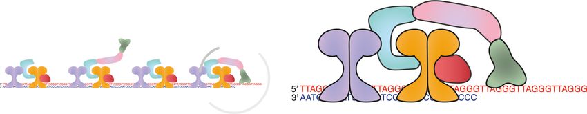
Downloaded from genesdev.cshlp.org on February 11, 2021 - Published by Cold Spring Harbor Laboratory Press

Ruis and Boulton

activation of ATM and NHEJ at chromosome ends. Un-                      should possess some specific domain/mechanism that
like at bona fide DSBs, NHEJ at TRF2-null telomeres                     can promote the formation or stabilization of t-loops;
does not require extensive end processing, but rather de-               the loss of this domain/mechanism should remove t-loops
pends on endonucleolytic cleavage of the 3′ G overhang,                 and induce ATM and NHEJ at telomeres. Finally, if t-loop
possibly by ERCC1/XPF, as part of the ligation reaction                 formation is the central component of protection from
(Zhu et al. 2003; Celli and de Lange 2005). The protection              ATM and NHEJ, t-loops alone should be sufficient to pro-
of chromosome ends from ATM and NHEJ by TRF2 has,                       tect chromosome ends from ATM and NHEJ.
until recently, been regarded as a universal feature of
mammalian chromosome end protection. The depletion
of TRF2 from somatic mouse cells and various human                      T-loops exist and are lost concurrently
cell lines leads to telomeric ATM activation, NHEJ and                  with end protection
is incompatible with viability (van Steensel et al. 1998;
Karlseder et al. 1999; Kim et al. 2017). Likewise, Cre-lox              Obtaining an accurate estimate of the frequency of t-loops
mediated systems used to knockout TRF2 specifically in                  within a population of cells is a significant challenge. The
mouse epidermal, liver, and neural tissues in each case in-             two available techniques, EM and superresolution mi-
duces ATM activation and NHEJ at telomeres, which in                    croscopy, use the same basic method to prepare telomeres
tissues with cycling cells leads to cell death and loss of              for visualization. DNA is harvested and cross-linked via
that tissue (Zhu et al. 2003; Lazzerini Denchi et al.                   UV and Psoralen, preserving DNA secondary structures
2006; Martinez et al. 2014; Lobanova et al. 2017). The                  that arise in vivo. For EM visualization, DNA is incubated
question that remains is how TRF2 achieves this inhibi-                 with frequent-cutter restriction endonucleases that exclu-
tion of NHEJ and ATM.                                                   sively digest nontelomeric DNA, freeing individual telo-
                                                                        meres from the intervening DNA (Griffith et al. 1999).
                                                                        Telomeric DNA can then be purified from genomic
The t-loop model of end protection                                      DNA via a gel-filtration column and spread onto EM grids.
                                                                        For superresolution microscopy visualization, DNA ex-
Microscopic interrogation of telomeres has revealed that                tracts are spread onto slides and telomeric DNA is labeled
mammalian telomeres often end in a looped structure,                    with a sequence-specific telomere probe (Doksani et al.
termed a telomere (t)-loop (Griffith et al. 1999; Doksani               2013). In each case, the macroscopic structure of cross-
et al. 2013). It is proposed that this structure forms                  linked telomeric DNA is visualized, allowing an estima-
through the invasion of the 3′ G overhang into the up-                  tion of the frequency of telomeres with terminal loops
stream telomeric dsDNA, where it base-pairs with the C                  to be made. EM detects loops at roughly 15%–40% of telo-
strand, displacing the G strand in this region. The ob-                 meres, while superresolution microscopy has produced
served loops vary in size from 1 to 20 kb, suggesting this              estimates of 25%–35% looped telomeres in a diverse
invasion occurs in a positionally blind manner, with any                range of contexts, from human somatic and cancer cells
telomeric dsDNA available for the invasion of the 3′ G                  and pluripotent and somatic mouse cells, amongst others,
overhang. Given telomeres share a single hexameric re-                  suggesting t-loops exist with similar frequencies across
petitive sequence, this is certainly plausible. The t-loop              different mammalian organisms and cell types (Griffith
is an attractive structure to mediate end protection as it              et al. 1999; Doksani et al. 2013; Van Ly et al. 2018).
would sequester the extreme chromosome terminus, hid-                      Could these loops simply be an artefact of the cross-
ing the linear DNA end that would otherwise bind KU70/                  linking and/or visualization process? Several pieces of ev-
80 and MRN. Thus, t-loops would be expected to simulta-                 idence strongly dispute this notion. T-loops are never ob-
neously block the initiation steps of both ATM activation               served at both ends of a purified single telomere via EM,
and NHEJ. Since TRF2 blocks both these pathways at telo-                indicating that the “t-loop” objects being observed pos-
meres, a hypothesis quickly developed that TRF2 might                   sess end specificity and are not simply the result of
mediate end protection primarily through promoting the                  “sticky” DNA ends (Griffith et al. 1999). Moreover, t-
formation and/or stabilization of these loops. In its sim-              loops can be observed without cross-linking, albeit at sig-
plest formulation, this is the t-loop model of end protec-              nificantly lower frequencies (Griffith et al. 1999). Most
tion: The TRF2-dependent formation of t-loops at                        compellingly, multiple studies have now shown that the
chromosome ends prevents their misidentification as                     depletion of TRF2 leads to a dramatic reduction in the fre-
DSBs and the activation of ATM and NHEJ (Fig. 2D).                      quency of t-loops when measured by superresolution mi-
   Testing this model directly is challenging, but it makes             croscopy, from 25%–35% to 5%–10% (Fig. 3A; Doksani
many predictions that can be experimentally tested. If                  et al. 2013; Benarroch-Popivker et al. 2016; Van Ly et al.
they are a key mediator of end protection, t-loops should               2018; Tomáška et al. 2020). This indicates that the struc-
be present at the majority of chromosome ends; t-loops                  tures observed as “t-loops” are specific products of TRF2
should be present when protection from ATM and NHEJ                     function and not an artefact of the telomere preparation
is achieved, but t-loops should disappear when this end                 and visualization process. Therefore, t-loops really do
protection is lost; the unwinding of t-loops should induce              form at chromosome ends in vivo and they arise in a large-
ATM and/or NHEJ activation. Likewise, if TRF2 mediates                  ly TRF2-dependent manner.
the formation of these loops, t-loops should be present in                 However, if cross-linking is incomplete, telomeres
cells with TRF2 but disappear when TRF2 is lost; TRF2                   break during spreading or loops reside along the z-axis

6      GENES & DEVELOPMENT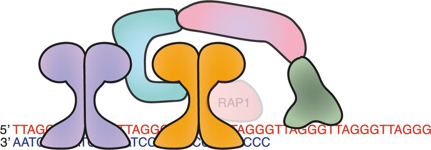
Downloaded from genesdev.cshlp.org on February 11, 2021 - Published by Cold Spring Harbor Laboratory Press

                                                                                             Telomeres protection of chromosome ends

A                                                                                      Figure 3. TRF2 promotes t-loop stabiliza-
                                                                                       tion to protect chromosome ends. (A) T-
                                                                                       loops require telomeric DNA possessing a
                                                                                       3′ overhang. TRF2 associates with the telo-
                                                                                       meric dsDNA sequence, wrapping 90 bp of
                                                                                       DNA with various lysine/alanine residues
                                                                                       in its TRFH domain, applying a topological
B                                                                                      stress that is proposed to promote the inva-
                                                                                       sion of the 3′ overhang into the ds telomeric
                                                                                       DNA. (B) Depiction of the multiple discrete
                                                                                       domains of TRF2, their interactions and
                                                                                       functions. (C) Complete depletion of TRF2
                                                                                       leads to loss of t-loops and t-loop indepen-
                                                                                       dent functions of TRF2, leading to strong ac-
                                                                                       tivation of ATM and ATM and NHEJ-
C                                                                                      dependent telomere fusions. (D) Mutants of
                                                                                       TRF2 that are unable to form t-loops, or teth-
                                                                                       ering of RTEL1 to TRF2 to promote promis-
                                                                                       cuous t-loop unwinding, leads to a
                                                                                       reduction in t-loops and activation of ATM
                                                                                       but not NHEJ. These telomeres that are com-
                                                                                       promised for t-loop stabilization therefore re-
D                                                                                      tain some protection from NHEJ that is lost
                                                                                       when TRF2 is removed entirely. This t-loop
                                                                                       independent protection from NHEJ is enact-
                                                                                       ed, at least in part, by RAP1 and the TRF2-
                                                                                       iDDR domain.

during imaging, looped telomeres will be misidentified as          ing various protein–protein interactions (Fig. 3B; de Lange
linear. Moreover, superresolution microscopy techniques            2010; Okamoto et al. 2013). TRF2, but not TRF1, can pro-
have maximal resolutions of 20 nm (STORM) and 140 nm               mote the formation of t-loops when added to model telo-
(Airyscan), corresponding to ∼ 70 bp and ∼ 500 bp of               meric DNA in vitro. This is dependent on the presence
dsDNA respectively. These maximal technical resolu-                of ds TTAGGG repeats and a 3′ G overhang; termini
tions are rarely reached in practice. Small t-loop structures      with 5′ overhangs, blunt ends, or 3′ termini with nontelo-
would thus be indistinguishable from linear telomeres in           meric sequences at the ds/ss junction cannot form loops
these analyses. Similarly, linear telomeres that crossed           in vitro, and involve the binding of TRF2 near the ds/ss
themselves during spreading might be misidentified as              junction point (Griffith et al. 1999; Stansel et al. 2001).
looped. Collectively therefore, the false-negative rate            Thus, TRF2 likely promotes invasion of the 3′ G overhang
and, to a lesser extent false-positive rate, for t-loop identi-    into the ds telomeric DNA, as predicted by the t-loop
fication is likely very high, meaning these techniques can         model. This is analogous to the formation of D-loops dur-
only be used to estimate relative t-loop frequency and not         ing RAD51-mediated HR, in which RAD51 coats the
the absolute frequency of looped telomeres. These tech-            ssDNA molecule and catalyzes its invasion into a homol-
niques demonstrate that t-loops exist, but leave open the          ogous template. However, unlike RAD51, TRF2 lacks en-
possibility that anything from 30% to 100% of telomeres            zymatic domains or ATP-hydrolyzing activity so it cannot
reside in t-loops within a population of cells.                    actively catalyze the invasion event that leads to t-loop
                                                                   formation. It therefore seems reasonable to propose that
                                                                   TRF2 stabilizes t-loops rather than directly mediates their
The mechanism of TRF2-mediated t-loop stabilization                formation.
                                                                     Insights into how TRF2 might stabilize t-loops have
As mentioned above, the presence of t-loops within cells           come from genetic studies with various mutants of
is largely dependent on TRF2 (Fig. 3A). TRF2 is expressed          TRF2. The TRFH domain contains a series of exposed Ly-
as two functionally indistinct proteins, distinguished by          sines and Arginines that interact with DNA in a sequence-
the addition of a 42-amino-acid N-terminal extension,              independent manner. This allows dimeric TRF2 to wrap
and has a series of highly conserved domains, including:           90 bp of telomeric DNA around itself, promoting DNA
the TRFH domain, which enables the homodimerization                condensation and exerting a topological stress onto the
of TRF2; a C-terminal Myb domain, which allows TRF2                dsDNA (Amiard et al. 2007; Poulet et al. 2012; Benar-
to bind ds telomere (TTAGGG) repeats directly; the N-ter-          roch-Popivker et al. 2016). A TRF2 mutant in which these
minal basic domain, which binds to DNA junctions; and              lysines and arginines are substituted for alanines, named
the Hinge domain, which has been implicated in mediat-             Top-less TRF2, can no longer condense telomeric DNA.

                                                                                                    GENES & DEVELOPMENT             7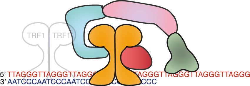
Downloaded from genesdev.cshlp.org on February 11, 2021 - Published by Cold Spring Harbor Laboratory Press

Ruis and Boulton

Cells expressing Top-less TRF2, and cells expressing a                  cedes telomeric NHEJ by 12–24 h (Van Ly et al. 2018). An-
TRFcT mutant lacking the entire TRFH domain of                          other recent study investigated the impact of reduced
TRF2, possess significantly fewer t-loops than cells ex-                levels of t-loops in the presence of functional TRF2. The
pressing wild type TRF2 (Benarroch-Popivker et al. 2016;                TRF2-S365A mutant constitutively binds the t-loop un-
Van Ly et al. 2018). Indeed, the level of t-loops in these              winding helicase regulator of telomere length 1 (RTEL1),
cells is indistinguishable from cells completely lacking                tethering RTEL1 to telomeres. Expression of TRF2-
TRF2. Thus, the TRFH domain of TRF2 is necessary for                    S365A induces promiscuous t-loop unwinding, producing
t-loop formation, suggesting TRF2 exerts a topological                  somatic cells with fewer t-loops but otherwise fully func-
stress onto ds telomeric DNA, entropically favoring the                 tional TRF2 (Sarek et al. 2019). The linear telomeres re-
product of this invasion over individual linear telomeres               sulting from excessive RTEL1 t-loop unwinding induce
(Okamoto et al. 2013; Benarroch-Popivker et al. 2016;                   an ATM-dependent DDR but do not activate NHEJ.
Van Ly et al. 2018).                                                    This confirms that t-loops are required to fully repress
                                                                        ATM activation at telomeres and that TRF2 is able to pro-
                                                                        tect telomeres from NHEJ when ATM is activated at lin-
T-loops protect chromosome ends from ATM activation                     ear telomeres. This ATM-activated but NHEJ-repressed
and NHEJ but are not solely responsible for this                        state has been proposed to represent an “intermediate
                                                                        state” of telomere end protection and has now been ob-
T-loops exist at some chromosome ends and arise in a                    served in multiple scenarios. Telomeres in cells blocked
manner that likely depends on TRF2 exerting topological                 in mitosis activate ATM but not NHEJ (Cesare et al.
stress on dsDNA to facilitate 3′ G overhang invasion or                 2009, 2013; Van Ly et al. 2018); telomeres in cells with
stabilize the t-loop three-way junction. What is the impact             partially reduced TRF2 expression activate ATM without
of the t-loop on protection from ATM and NHEJ? The for-                 strongly activating NHEJ (Cesare et al. 2009, 2013; Van Ly
mation of t-loops depends on both TRF2 and the 3′ G over-               et al. 2018); critically short telomeres produced through
hang. Complete removal of TRF2, as discussed, leads to                  aging activate ATM but very rarely undergo NHEJ (Hewitt
dramatically fewer t-loops and robust activation of                     et al. 2012; Kaul et al. 2012; Hayashi et al. 2015); cells ex-
NHEJ and ATM at telomeres, resulting in inviability                     pressing Top-less or TRFcT mutants (which lack t-loops)
(Fig. 2; Doksani et al. 2013). However, it is impossible to             robustly activate telomeric ATM but not NHEJ (Okamoto
say from these data alone whether the loss of t-loops is re-            et al. 2013; Benarroch-Popivker et al. 2016; Van Ly et al.
sponsible for the activation of ATM and NHEJ—TRF2                       2018); telomeres engineered to possess TRF2 but fewer
could have other functions that are important for end pro-              t-loops via the TRF2-S365A mutant activate ATM but
tection that are also lost coincidentally with t-loops in               not NHEJ (Fig. 3C; Sarek et al. 2019). Thus, telomeres
these experiments (Arnoult and Karlseder 2015).                         can activate ATM without NHEJ and do so when the fre-
   Due to the nature of semi-conservative DNA replica-                  quency of t-loops is reduced but some TRF2 functionality
tion, the 3′ G overhang is retained at lagging telomeres                remains present. This suggests that t-loops protect telo-
but must be generated de novo at each leading end telo-                 meres from ATM activation, but that t-loops are not es-
mere after telomere replication. This is achieved by the                sential for protection from NHEJ and hence ATM and
TRF2-mediated recruitment of the exonuclease APOLLO                     NHEJ activation can be uncoupled at telomeres.
to leading end telomeres. The knockout of APOLLO, or
abrogation of the APOLLO-TRF2 interaction through a
TRF2-F162 mutant, prevents timely formation of leading                  Alternative mechanisms of TRF2-mediated end
end G overhangs and, since t-loops require an overhang,                 protection
this necessarily restricts t-loop formation at leading end
telomeres (van Overbeek and de Lange 2006; Lam et al.                   While t-loops are observed in organisms as diverse as ver-
2010; Wu et al. 2010). Consistent with the proposed pro-                tebrates (Griffith et al. 1999), plants (Cesare et al. 2003,
tective role of t-loops, cells lacking APOLLO or expressing             2008) and trypanosomes (Munoz-Jordan et al. 2001),
an APOLLO-binding deficient TRF2-F162A mutant show                      they are not universal (Tomáška et al. 2020). Chromo-
robust activation of ATM and NHEJ specifically at leading               some end protection is maintained without t-loops in
end telomeres (Wu et al. 2010). Likewise, cells expressing              the small linear DNA fragments found in the macronuclei
the Top-less or TRFcT mutants show a similar reduction                  of hypotrichous ciliates, where end protection relies on a
in t-loop frequencies to cells lacking TRF2 and show ro-                proteinaceous cap (Gottschling and Zakian 1986), and in
bust telomeric activation of ATM. However, the linear                   dipteran insects, which lack G-rich telomeric sequences
telomeres formed in cells expressing Top-less or TRFcT                  entirely and instead cap their chromosome ends with
mutants are still protected from NHEJ (Benarroch-                       long retrotransposons (Young et al. 1983; Biessmann and
Popivker et al. 2016; Van Ly et al. 2018). Thus, linear telo-           Mason 1997). Thus, end protection can be achieved with-
meres are still protected from NHEJ by TRF2 mutants that                out t-loops in certain species. Indeed, mammalian cells
cannot form t-loops, suggesting TRF2 protects telomeres                 expressing TRF2-S365A, TRFcT or Top-less (which have
from NHEJ independently of t-loop formation.                            reduced t-loops) robustly activate telomeric ATM but do
   Consistent with this notion, time course studies of t-               not robustly activate NHEJ. Therefore, in mammals t-
loops and the DDR upon removal of TRF2 reveal that                      loop stabilization cannot be the sole means through
ATM activation coincides with the loss of t-loops and pre-              which TRF2 mediates end protection.

8      GENES & DEVELOPMENT
Downloaded from genesdev.cshlp.org on February 11, 2021 - Published by Cold Spring Harbor Laboratory Press

                                                                                            Telomeres protection of chromosome ends

   One proposal is that TRF2 contributes to the compac-           not activate either ATM or NHEJ (Sfeir et al. 2010). How-
tion of telomeric chromatin through a complex network             ever, the tethering of RAP1 to TRF2-deficient telomeres
of interactions between shelterin subunits and telomeric          reduces telomeric NHEJ (Sarthy et al. 2009), TRF2/
DNA. In this model, the tightly compacted telomeric               RAP1, but not TRF2 alone, can inhibit NHEJ at telomeric
chromatin would prevent KU70/80 and MRN from ac-                  substrates in vitro (Bae and Baumann 2007) and the deple-
cessing the end of the chromosome, removing the require-          tion of RAP1 from cells expressing the Top-less TRF2 mu-
ment for a t-loop. Consistent with this possibility, the          tant (which lack t-loops and activate ATM without
removal of individual shelterin subunits, including               concomitant NHEJ) induces telomeric NHEJ (Benarroch-
TRF2, or mutations that abrogate shelterin assembly,              Popivker et al. 2016). Thus, while RAP1 is not essential
were suggested to induce a 10-fold increase in telomere           for end protection per se, TRF2 appears to recruit RAP1
volume, coincident with the activation of telomeric               to inhibit NHEJ at linear telomeres independently of
ATM signaling (Bandaria et al. 2016). However, multiple           ATM activation.
independent studies, in mouse and human cells, have
failed to recapitulate the essential phenotype predicted
by this model, namely that the removal of TRF2, or the en-        T-loops as pathological structures
tire shelterin complex, from telomeres should induce
three-dimensional decompaction, concomitant with                  While proposed to be protective structures, t-loops can ac-
DDR activation (Timashev et al. 2017; Vancevska et al.            tually pose a threat to end protection, leading to the alter-
2017). One possible explanation for this disparity is that        nate notion that t-loops might be pathological, not
dysfunctional telomeres can become clustered, giving              protective, structures. If a t-loop undergoes branch migra-
the misleading impression of telomeric decompaction               tion to form a double Holliday junction (dHJ) (Fig. 4A), it
(Dimitrova et al. 2008). Indeed, if chromatin compaction          becomes an optimal substrate for cleavage by HJ resol-
is responsible for the inhibition of ATM at telomeres,            vases, including MUS81, SLX1/SLX4, EMI1 and/or
this should prevent ATM activation at breaks within the           GEN1 (Wyatt and West 2014). This cleavage results in
telomere. However, telomere-internal DSBs generated               large telomere deletions and Telomere Circle (TC) forma-
by TRF1-FOK1 induce the robust activation of ATM                  tion and is promoted by poly(ADP-ribose) polymerase
(Doksani and de Lange 2016). Shelterin-mediated telo-             (PARP1), which can bind to 5′ ss-dsDNA junctions, and re-
mere compaction is therefore insufficient to explain the          pressed by the basic domain of TRF2 (TRF2B) (Wang et al.
repression of ATM and NHEJ at telomeres.                          2004; Saint-Léger et al. 2014; Schmutz et al. 2017). TRF2B
   Another alternate proposal is that TRF2 might inhibit          binds to branched DNA junctions in vitro and in vivo,
DDR factors directly. TRF2 interacts with ATM, MRN                while deletion of TRF2B induces telomeric accumulation
and KU70/80, providing multiple avenues through which             of PARP1 and TC formation via t-loop cleavage (Fouché
this could occur (Song et al. 2000; Karlseder et al. 2004;        et al. 2006; Poulet et al. 2012; Schmutz et al. 2017).
Okamoto et al. 2013). Indeed, TRF2 has been proposed              TRF2B can be functionally replaced with bacterial
to bind to chromosomal DSBs, where it might influence             branched DNA-binding domains, suggesting the key func-
both ATM activation and repair, and TRF2 contains mul-            tion of TRF2B is to bind the DNA junction at the base of
tiple canonical S/T-Q ATM phosphosites, suggesting                the t-loop, blocking branch migration at the base of the
ATM could regulate TRF2 function (Bradshaw et al.                 t-loop to prevent dHJ formation (Schmutz et al. 2017).
2005; Matsuoka et al. 2007). A short 30-amino-acid inhib-         The TRF2 basic domain also directly blocks the telomeric
itor of the DDR (iDDR) domain has been found within the           recruitment of PARP1 (Rai et al. 2016). The BLM helicase,
TRF2 Hinge domain (Okamoto et al. 2013). TRF2-iDDR                which dissolves dHJs, represses the cleavage of t-loops in
interacts with the MRN complex and, via MRN, recruits             the absence of the TRF2 basic domain, presumably by me-
the deubiquitination enzyme BRCA1–BRCA2-containing                diating the reversion of telomeric dHJs into three-way
complex 3 (BRCC3), which prevents H2A polyubiquitina-             junctions (Fig. 4A).
tion–dependent recruitment of RNF168, and ubiquitin                  Like other DNA secondary structures, t-loops can stall
protein ligase 5 (UBR5), an enzyme that mediates degrada-         the replication fork and therefore require unwinding dur-
tion of RNF168, to telomeres (Okamoto et al. 2013). The           ing S phase (Mirkin and Mirkin 2007; Vannier et al. 2012,
expression of TRF1 fused to the TRF2-iDDR domain is               2013). This is achieved by the RTEL1 helicase, which is re-
sufficient to reduce, but not abolish, NHEJ at TRF2-null          cruited to telomeres in S phase when TRF2-S365 is de-
telomeres, without impacting ATM activation. Thus,                phosphorylated by PP6R3, enabling TRF2 to interact
TRF2-iDDR is proposed to repress NHEJ downstream                  with and recruit RTEL1 to telomeres (Vannier et al.
from ATM activation, by limiting the accumulation of              2012, 2013; Sarek et al. 2015a,b; Sarek et al. 2019). This
RNF168, and thereby 53BP1, at telomeres. This provides            provides a brief window for RTEL1 to unwind t-loops, fa-
at least one t-loop independent mechanism through                 cilitating the passage of the DNA replication machinery.
which TRF2 can repress NHEJ.                                      RTEL1 unwinds D-loops in vitro, while tethering of
   TRF2 has also been proposed to limit telomeric NHEJ            RTEL1 to telomeres via a TRF2-S365A mutant reduces
through an undefined function of its interacting partner          t-loop levels in vivo, suggesting RTEL1 unwinds t-loops
RAP1. Unlike other shelterin components, RAP1 is not              directly (Barber et al. 2008; Vannier et al. 2012). However,
essential for viability or end protection; cells lacking          RTEL1 also unwinds telomeric G-quadruplex structures
RAP1 show no telomere dysfunction phenotypes and do               and possibly RNA-DNA hybrids (R-loops), although

                                                                                                  GENES & DEVELOPMENT            9
Downloaded from genesdev.cshlp.org on February 11, 2021 - Published by Cold Spring Harbor Laboratory Press
Ruis and Boulton
A Removal of TRF2B
TRF2B branched DNA binding
Branch
Migration dHJ T-circles
Resolution
TRF2B dHJ PARP1
5’ TTAGGG BLM 5’ TTAGGG
5’ TTAGGG Telomere
3’ AATCCC 3’ AATCCC Shortening &
3’ AATCCC
PARP1 loss
SLX1/4 MUS81
Branch
Migration
GEN1
B
TRF2
RTEL1
DNA Replication 5’ TTAGGG Lagging end telomere
Fork 3’ AATCCC 5’ TTAGGG
RTEL1
5’ TTAGGG 3’ AATCCC
3’ AATCCC Leading end telomere
RTEL1 3’ AATCCC
TRF2 RTEL1 5’ TTAGGG
5’ TTAGGG
3’ AATCCC
5’ TTAGGG
3’ AATCCC Reversed
Failure of Rtel1- T-circles
mediated t-loop DNA Replication
S-phase t-loop Fork
unwinding unwinding
Telomerase 5’ TTAGGG Telomere
3’ AATCCC Shortening &
loss
SLX1/4
Figure 4. TRF2 coordinates t-loop transactions to ensure they remain protective, not pathological, structures. (A) The TRF2 basic
domain (TRF2B) binds to branched DNA junctions, including those found at the base of the t-loop, and prevents their migration from
three-way into four-way junctions, which are idealized substrates for double Holliday junction (dHJ) resolvases, including SLX1/4,
GEN1 and MUS81. TRF2B also inhibits PARP1 activity at telomeres. Collectively, these TRF2B functions prevent dHJ resolution and
hence telomere shorterning and T-circle formation. (B) TRF2 recruits RTEL1 to telomeres in S phase through a phosphorylation-regulated
interaction between TRF2-S365 and RTEL1. RTEL1 then unwinds t-loops, enabling the passage of the replication fork through this DNA
Secondary structure and complete telomere replication. Failure of RTEL1-mediated t-loop unwinding leads to replication fork reversal
within the telomere and cleavage of either the reversed fork or residual t-loops themselves by SLX1/4, leading to potentially catastrophic
telomere shorterning and T-circle formation.
RTEL1 is recruited to these secondary structures through at t-loops, through the TRF2B domain. Thus, TRF2 en-
an interaction with PCNA, not with TRF2 (Vannier et al. sures t-loops are protective, not pathological, structures
2012; Björkman et al. 2020; Wu et al. 2020). Therefore, in normal conditions.
RTEL1 might also impact t-loops indirectly, by unwind-
ing secondary structures that alter topological stress with-
in the telomere. Loss of RTEL1, or abrogation of TRF2- End protection in ESCs
mediated RTEL1 recruitment to telomeres, leads to repli-
cation fork stalling and reversal, in a manner dependent The absolute dependency of end protection on TRF2 has
on telomerase and the fork reversal machinery (Margalef been confirmed in numerous scenarios, including somatic
et al. 2018). These reversed forks and/or t-loops them- mouse and human cells and in mouse liver, skin and neu-
selves are then cleaved by SLX1/SLX4, inducing TC for- ronal compartments in vivo (van Steensel et al. 1998; Karl-
mation and telomere shortening (Fig. 4B; Vannier et al. seder et al. 1999; Lazzerini Denchi et al. 2006; Martinez
2012). et al. 2014; Kim et al. 2017; Lobanova et al. 2017). In all
Progressive telomere shortening in cells that fail to un- of these contexts, the loss of TRF2 leads to robust ATM
wind t-loops, or in cells with unrestrained dHJ formation, activation and rapid accumulation of telomere fusions as
eventually generates critically short telomeres that are a result of NHEJ. On this basis, TRF2 was assumed to
unable to mediate end protection. This is exemplified by have a universal role in end protection throughout mam-
mutations in RTEL1, which cause Hoyeraal–Hreidarsson malian development. Surprisingly, two recent studies
syndrome (HHS), a syndrome typified by very short, heter- have revealed that chromosome end protection in the plu-
ogenous telomeres that drive senescence, premature aging ripotent stages of early development occurs largely inde-
and cancer (Sarek et al. 2015). Thus, when t-loops persist pendently of TRF2 (Markiewicz-Potoczny et al. 2020;
throughout telomeric DNA replication or are converted Ruis et al. 2020). While these studies confirmed previous
into dHJs, they become pathological structures. Mam- findings (that TRF2-depletion in somatic cells leads to
mals have evolved an elegant solution to this potential ATM activation, NHEJ, cell cycle arrest and cell death),
problem: The same protein that mediates t-loop forma- they establish that the loss of TRF2 from mouse ESCs
tion (TRF2) ensures the t-loop does not become toxic by leads to a mild activation of ATM but with no evidence
coordinating timely t-loop unwinding, through RTEL1, of significant telomeric NHEJ, CHK2 activation, cell cy-
and by blocking PARP1 recruitment and branch migration cle arrest, or cell death. However, the depletion of the
10 GENES & DEVELOPMENTDownloaded from genesdev.cshlp.org on February 11, 2021 - Published by Cold Spring Harbor Laboratory Press
Telomeres protection of chromosome ends
entire shelterin complex from ESC telomeres leads to ro- nicely complements recent findings showing that the per-
bust telomeric activation of ATM and NHEJ, CHK2 acti- sistent unwinding of t-loops activates ATM, despite the
vation, G2/M-phase cell cycle arrest and rapid loss of presence of TRF2 (Sarek et al. 2019). Together, these re-
viability (Ruis et al. 2020). Thus, in ESCs the inhibition sults demonstrate that t-loops are necessary but not suffi-
of ATM and NHEJ at telomeres is achieved in a largely cient for the complete inhibition of ATM at telomeres.
TRF2-independent, but shelterin-dependent, manner. Intriguingly, in somatic cells TRF2-iDDR has only previ-
While this largely TRF2-independent end protection is ap- ously been shown to repress NHEJ, downstream from
parently present in multiple distinct pluripotent states, ATM activation, while in ESCs TRF2-iDDR is necessary
including ESCs, EpiSCs, and pluripotent cells in murine to suppress the activation of ATM itself (Okamoto et al.
E3.5 embryos, differentiation of TRF2-null ESCs rapidly 2013; Van Ly et al. 2018). TRF2-iDDR interacts with
leads to robust telomeric ATM activation, telomere fu- MRN directly, providing one possible means through
sions by NHEJ and cell death at the point that TRF2- which TRF2-iDDR could impact upon ATM activation in-
null cells exit from pluripotency (Markiewicz-Potoczny dependently of t-loop formation.
et al. 2020; Ruis et al. 2020). Thus, the TRF2-independent Together, these new studies complement existing data
protection of telomeres from ATM and NHEJ is, in the to create a more complete picture of t-loop mediated end
context of early development, uniquely restricted to the protection. The protection of chromosome ends requires
pluripotent stage. t-loops, which both efficiently suppress NHEJ and signifi-
Given the inextricable link between TRF2, end protec- cantly suppress ATM activation. The removal of t-loops
tion and t-loop formation in somatic cells, it was impor- leads to ATM activation, but if the other activities of
tant to consider the status of t-loops in ESCs. The TRF2 are retained, low levels of NHEJ. Likewise, the
Boulton and Cesare laboratories (Ruis et al. 2020) visual- loss of these other functions of TRF2, most notably the
ized telomeric secondary structures in ESCs via superre- TRF2 iDDR domain, can induce ATM activation but
solution microscopy. This revealed that ESCs possess t- not NHEJ in the presence of t-loops. Thus, the presence
loops with similar size and frequency (∼30% of telomeres) of physiologically normal levels of t-loops is insufficient
to somatic cells. However, unlike somatic cells where to completely suppress ATM in the absence of TRF2. Col-
these loops are lost upon TRF2 depletion, in ESCs the fre- lectively therefore, protection from ATM and NHEJ is
quency and size of t-loops is unaffected by the loss of achieved via a two-step mechanism that involves t-loops
TRF2, suggesting that in ESCs the formation and stabili- and t-loop independent functions of TRF2. Loss of either
zation of t-loops occurs in a TRF2-independent manner t-loops or TRF2-mediated ATM inhibition alone leads to
(Fig. 5A; Ruis et al. 2020). Collectively, these unexpected attenuated telomeric DDR signaling. However, the dual
discoveries prompt a series of questions and also cast new loss of t-loops and these t-loop independent functions of
light on the existing paradigms of telomere end protec- TRF2 unleashes complete telomeric DDR signaling in-
tion, both discussed below. volving robust ATM activation, NHEJ, cell cycle arrest
and cell death (Fig. 5B,C).
In ESCs lacking TRF2, the mild telomeric ATM re-
Insights regarding chromosome end protection from ESCs sponse is not accompanied by NHEJ, cell cycle arrest or
dramatic loss of viability. This confirms previous reports
In these new studies, we discovered that ESCs possess t- that telomere fusions, cell death, and genomic instability
loops that are unaffected by the loss of TRF2. These do not accompany telomeric ATM activation per se, but
TRF2-null, but looped, telomeres remain shielded from rather the specific activation of NHEJ at telomeres. This
NHEJ but activate a mild ATM-dependent telomeric is consistent with previous reports of an “intermediate
DDR. This DDR is attenuated both qualitatively and state” of telomere end protection based on observations
quantitatively relative to in TRF2-null somatic cells, as that in normal somatic cells the telomere DDR can be ac-
TRF2-null ESCs have dramatically fewer TIFs and do tivated without accompanying telomere fusions (Cesare
not undergo checkpoint arrest or cell death (Ruis et al. et al. 2009, 2013). In this context, the telomere DDR is
2020). Indeed, unlike somatic cells in which the TRFH thought to primarily serve as a signaling mechanism to
domain is required to inhibit ATM activation at telo- identify shortened telomeres in aged somatic cells, arrest
meres, in ESCs apparently only the TRF2-iDDR domain proliferation and trigger senescence (d’Adda di Fagagna
is necessary to inhibit ATM activation (Ruis et al. 2020). et al. 2003; Kaul et al. 2012; Cesare et al. 2013). This has
Thus, as predicted by the t-loop model of end protection, important implications as short telomeres in aged cells ac-
t-loops that persist in the absence of TRF2 are sufficient tivate ATM but, apparently, very rarely activate NHEJ;
to protect chromosome ends from NHEJ. This is the es- telomere fusions in somatic tissues only arise upon com-
sential, long awaited evidence that conclusively demon- plete erosion of the telomeric DNA substrate (d’Adda di
strates t-loops are a key mediator of end protection (de Fagagna et al. 2003; Kaul et al. 2012; Cesare et al. 2013;
Lange 2018). Hayashi et al. 2015). Given t-loops require the exertion
However, consistent with other reports that the repres- of topological stress onto ds telomeric DNA, presumably
sion of ATM and NHEJ are uncoupled, telomeres in TRF2- there is a minimum telomere length below which t-loops
null ESCs still activate ATM, albeit weakly. The presence can no longer form. TRF2 would still be expected to sup-
of t-loops is unable to completely protect chromosome press NHEJ, but not fully repress ATM, at short but linear
ends from ATM activation in the absence of TRF2. This telomeres, but critically short telomeres or telomere-free
GENES & DEVELOPMENT 11You can also read