Promising Therapeutic Targets for Treatment of Rheumatoid Arthritis
←
→
Page content transcription
If your browser does not render page correctly, please read the page content below
REVIEW
                                                                                                                                              published: 09 July 2021
                                                                                                                                    doi: 10.3389/fimmu.2021.686155

                                          Promising Therapeutic Targets for
                                          Treatment of Rheumatoid Arthritis
                                          Jie Huang 1†, Xuekun Fu 1†, Xinxin Chen 1, Zheng Li 1, Yuhong Huang 1 and Chao Liang 1,2*
                                          1 Department of Biology, Southern University of Science and Technology, Shenzhen, China, 2 Institute of Integrated

                                          Bioinfomedicine and Translational Science (IBTS), School of Chinese Medicine, Hong Kong Baptist University,
                                          Hong Kong, China

                                          Rheumatoid arthritis (RA) is a systemic poly-articular chronic autoimmune joint disease
                                          that mainly damages the hands and feet, which affects 0.5% to 1.0% of the population
                                          worldwide. With the sustained development of disease-modifying antirheumatic drugs
                                          (DMARDs), significant success has been achieved for preventing and relieving disease
                                          activity in RA patients. Unfortunately, some patients still show limited response to
                                          DMARDs, which puts forward new requirements for special targets and novel
                                          therapies. Understanding the pathogenetic roles of the various molecules in RA could
                                          facilitate discovery of potential therapeutic targets and approaches. In this review, both
                         Edited by:
                                          existing and emerging targets, including the proteins, small molecular metabolites, and
                Trine N. Jorgensen,       epigenetic regulators related to RA, are discussed, with a focus on the mechanisms that
    Case Western Reserve University,      result in inflammation and the development of new drugs for blocking the various
                      United States
                                          modulators in RA.
                      Reviewed by:
                     Åsa Andersson,       Keywords: rheumatoid arthritis, targets, proteins, small molecular metabolites, epigenetic regulators
         Halmstad University, Sweden
                Abdurrahman Tufan,
               Gazi University, Turkey

                   *Correspondence:       INTRODUCTION
                          Chao Liang
               liangc@sustech.edu.cn      Rheumatoid arthritis (RA) is classified as a systemic poly-articular chronic autoimmune joint
     †
      These authors have contributed
                                          disease that primarily affects hands and feet. RA is pathologically manifested as immune cell
                 equally to this work     infiltration, hyperplasia of the synovial lining, pannus formation, and destruction of articular
                                          cartilage and bone (1, 2). Although the exact etiology of RA is unclear, it is certain that genetic and
                   Specialty section:     environmental factors have influences on RA occurrence. At present, RA affects approximately 0.5%
         This article was submitted to    to 1.0% of the population worldwide (3), and in particular, females are at higher risk of the disease
                      Autoimmune and      (two to three times than males) (4). RA patients typically experience morning stiffness. If left
         Autoinflammatory Disorders,       untreated, they could appear small focal necrosis, adhesion of granulation, and fibrous tissue on the
               a section of the journal   articular surface, which lead to progressive joint ankylosis, destruction, deformities, and
              Frontiers in Immunology
                                          disability (5).
           Received: 26 March 2021            To date, a large number of clinical trials have been performed by scientists and clinicians for
            Accepted: 23 June 2021        testing different types of agents in RA treatment. Some of these agents have been approved for daily
            Published: 09 July 2021
                                          clinical practice. In the first place, nonsteroidal anti-inflammatory drugs (NSAIDs), including
                            Citation:     acetylsalicylate, naproxen, ibuprofen, and etodolac, are used to alleviate pain, swelling, and decrease
Huang J, Fu X, Chen X, Li Z, Huang Y
                                          inflammation. NSAIDs exert their actions by inhibiting the enzymatic activity of the cyclooxygenase
       and Liang C (2021) Promising
   Therapeutic Targets for Treatment
                                          (COX) involved in the synthesis of prostaglandins (PG). Inhibition of COX-2 by NSAIDs blocks PG
             of Rheumatoid Arthritis.     production at sites of inflammation, whereas inhibition of COX-1 in other tissues (platelets and the
         Front. Immunol. 12:686155.       gastroduodenal mucosa) leads to common adverse effects of NSAIDs, such as bleeding and
   doi: 10.3389/fimmu.2021.686155         gastrointestinal ulceration (6). In addition, corticosteroids, like glucocorticoids, are another kind

Frontiers in Immunology | www.frontiersin.org                                    1                                           July 2021 | Volume 12 | Article 686155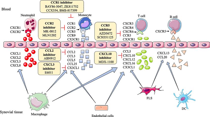
Huang et al.                                                                                               Therapeutic Targets Rheumatoid Arthritis

of potent anti-inflammatory drug, which modulates gene                     searched literature published between 2005 and 2021 using
expression by binding to glucocorticoid receptors to display              keywords cytokines,” “chemokines,” “protein targets,” “small
anti-inflammatory and immunosuppressive effects. However,                  molecular metabolites,” and “epigenetics,” and summarizes
their side effects include nausea, abdominal pain, ulcers,                recent advances in these novel targets. The review provides
osteoporosis, and diabetes (7).                                           insights that contribute to future directions and drug
    Owing to the adverse effects of NSAIDs and corticosteroids,           discoveries for the treatment of RA.
disease-modifying antirheumatic drugs (DMARDs), a class of
immunosuppressive and immunomodulatory agents, are
developed to prevent and relieve RA aggression. As the first               PROTEIN TARGETS FOR TREATMENT OF
treatment strategy, conventional synthetic (cs) DMARDs like               RHEUMATOID ARTHRITIS
methotrexate, hydroxychloroquine, sulfasalazine, leflunomide,
chloroquine, and gold salts should be used as soon as RA is               Currently, many agents aiming at various protein targets have
diagnosed. Notably, methotrexate is preferred for use in patients.        been explored and tested to ease the progression of RA, and some
csDMARDs are popular because of their low price and good                  agents have been used in the clinic to treat RA patients (Table 1).
efficacies. However, their mechanisms of action are not fully              In addition to cytokine targets and chemokine targets, several
understood and multiple signal pathways could be involved. If a           important proteins participate in inflammatory cellular pathways
patient shows nonresponse for csDMARDs, biological (b)                    as well, such as JAK and IRAK-4.
DMARDs or targeted synthetic (ts) DMARDs should be
added. bDMARDs (i.e., adalimumab, infliximab, certolizumab,                Cytokine Targets
canakinumab, tocilizumab, sarilumab, and secukinumab) are                 Cytokines have long been explored and studied as potential
monoclonal antibodies and have special targets like tumor                 targets of RA because cytokines are directly involved in the RA
necrosis factor (TNF)-a, interleukin (IL)-6, IL-1b, and IL-17             process, which can be classified as pro- and anti-inflammatory
(8–10). tsDMARDs also have special targets, for example, janus            cytokines based on their different functions against
kinases (JAK) is the special target of tofacitinib, baricitinib,          antigen response.
filgotinib, upadacitinib, and decernotinib (11).                               Pro-inflammatory cytokines, including TNF-a, IL-1b, IL-6,
    Although the above mentioned DMARDs have been quite                   IL-7, IL-15, IL-17, IL-18, IL-23, interferon (IFN)-g, granulocyte-
successful in mitigating RA, it is still an undeniable fact that a        macrophage colony-stimulating factor (GM-CSF) have been
significant proportion of patients could experience treatment              found to govern inflammation in RA occurrence. The level of
failure, including nonresponse and limited efficacy (12). To               these cytokines elevated in the synovium, synovial fluid, serum,
achieve the maximum therapeutic effectiveness, rheumatologists            or peripheral blood of RA patients (61–67). In addition, IL-15,
recommend using combination therapy for RA patients (13, 14).             IL-17, IL-23, and GM-CSF have a strong relation to rheumatoid
For instance, the combination of methotrexate and glucocorticoid          factor (RF), anti-cyclic citrullinated peptide (CCP) seropositivity,
can relieve RA in about 25% of patients within 6 months. If               and RA activity, which could become diagnostic biomarkers for
methotrexate plus glucocorticoid is insufficient, any bDMARDs or           RA (64, 67–69). IL-7 would also be taken as a diagnostic
tsDMARDs can be recommended to add to csDMARDs, such as                   biomarker for early RA because of the different levels in the
methotrexate plus tocilizumab, methotrexate plus rituximab,               stages of RA occurrence (63).
methotrexate plus tofacitinib, and so on (15). Apart from                     Macrophages can secrete various cytokines, such as TNF-a,
nonresponse, some DMARDs do cause adverse clinical effects, such          IL-1b, IL-6, IL-7, IL-15, IL-18, IL-23. TNF-a can induce the
as stomatitis, exanthema, diarrhea, anemia, pneumonia, and                proliferation of fibroblast-like synoviocytes (FLS) and synovial
nephritis, further aggravating the disease condition (16–18).             cells by activating nuclear factor kappa-B (NF-kB) and
    With the deepening of research and exploration, many                  extracellular regulated protein kinases (Erk)-1/2-E26
molecules are identified to exert important roles and bring                transformation-specific (ETS)-1 signaling pathway, respectively
novel insights to prevent RA. For example, emerging protein               (70, 71), resulting in the secretion of a variety of inflammatory
targets like IL-4, IL-10, IL-15, IL-17, IL-18, IL-23, interleukin-1       mediators like IL-6, matrix metalloproteinases (MMP)-1, and
receptor-associated kinase (IRAK)-4 have been revealed to have            MMP-3 to increase inflammation (72). IL-1b enhances MMPs
a strong implication with innate and adaptive immune response             production and the adhesion of leukocytes to RA FLS by
in RA (19). Small molecular metabolites, including                        activating ERK, c-Jun N-terminal kinase (JNK), apetala (AP)-1,
prostaglandins (PGs), lipoxins (LXs), platelet-activating factor          and NF-kB (73, 74). IL-6 causes bone resorption and cartilage
(PAF) and leukotrienes (LTs), nitric oxide (NO), and reactive             degeneration by inducing the production of MMPs and NF-kB
oxygen species (ROS), are also vital participants and mediators in        ligand (RANKL) receptors (75, 76). Blockade of IL-7 ameliorates
the physiopathology of RA (20). Besides, an increasing number             joint inflammation by reducing T cells trafficking and
of studies show that epigenetic regulators play important roles in        proinflammatory factors like TNF-a, IL-1b, IL-6, and MMPs
RA, like non-coding RNAs, DNA methylation, RNA                            (77). IL-15 increases the level of major histocompatibility
methylation, and histone modifications (21). Up to now,                    complex (MHC)-II on macrophages to result in enhancing
researchers have explored and developed some new agents for               proliferation of antigen-specific cluster of differentiation (CD)
RA according to these classical or emerging targets. This review          4+ T cells (78). IL-18 acts in synergy with IL-12 to stimulate T

Frontiers in Immunology | www.frontiersin.org                         2                                    July 2021 | Volume 12 | Article 686155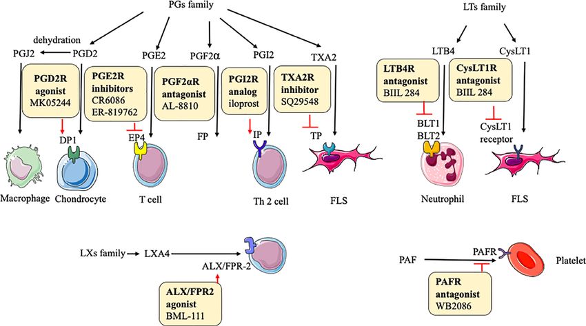
Huang et al. Therapeutic Targets Rheumatoid Arthritis
TABLE 1 | Protein targets and their agents in rheumatoid arthritis.
Targets Agents Phases References
Cytokines
TNF Adalimumab Marketed (7)
Infliximab Marketed (7)
Etanercept Marketed (7)
Certolizumab Marketed (7)
Golimumab Marketed (7)
IL-1R Anakinra Marketed (7)
IL-1 Canakinumab Marketed (7)
Gevokizumab Marketed (7)
Rilonacept Terminated Clinicaltrials.gov
IL-6R Tocilizumab Marketed (7)
IL-6a Sarilumab Marketed (7)
Clazakizumab Marketed (7)
Olokizumab Marketed (7)
Sirukumab Marketed (7)
Il-2 MEDI5117 Terminated Clinicaltrials.gov
IL-10 Dekavil Phase 1 (22)
IL-15 AMG-714 Phase 2 (23)
IL-18 rhIL-18BP Phase 1 (24)
IL-17 Secukinumab Phase 3 (25)
Ixekizumab Phase 2 (26)
IL-17R Brodalumab Terminated Clinicaltrials.gov
IFN-g Fontolizumab Terminated Clinicaltrials.gov
Chemokines
CCL2 p8A MCP-1 Animal study (27)
ABN912 Phase 1 (28)
CCR9 CCX8037 Animal study (29)
CX3CL1 E6011 Phase 1 (30)
CCR1 J−113863 Animal study (31)
BX147 Animal study (32)
BAY86-5047 Phase 2 Clinicaltrials.gov
ZK811752 Phase 2 Clinicaltrials.gov
CCX354 Phase 2 (33)
BMS-817399 Phase 2 Clinicaltrials.gov
CCR2 MK−0812 Phase 2 Clinicaltrials.gov
MC−21 Animal study (34)
MLN1202 Phase 2a (35)
CCR5 SCH−X82 Phase 2 (32)
Met-RANTES Phase 2 (36)
AZD5672 Phase 2 (37)
Maraviroc Terminated (38)
SCH351125 Phase 1b (39)
Other proteins
TLR4 NI-0101 Phase 2 Clinicaltrials.gov
GRK2 Paroxetine Phase 2 Clinicaltrials.gov
MEK ARRY-162 Phase 2 Clinicaltrials.gov
MMP-9 Andecaliximab Phase 2 (40)
CD3 Otelixizumab Phase 1 (41)
CD80 Abatacept Marketed (42)
BTK ICP-022 Phase 1 Clinicaltrials.gov
CC-292 Phase 2 (43)
HM71224 Phase 1 Clinicaltrials.gov
Il-23 STA 5326 mesylate Phase 2 (44)
Guselkumab Terminated Clinicaltrials.gov
GM-CSF Otilimab Phase 3 (45)
Gimsilumab Phase 1 (45)
Namilumab Phase 2 (45)
Mavrilimumab Phase 2 (45)
Lenzilumab Terminated Clinicaltrials.gov
Chemokines
CXCL10 MDX−1100 Phase 2 (25)
CXCL12 30D8 Animal study (46)
CXCL13 mAb470 Animal study (47)
(Continued)
Frontiers in Immunology | www.frontiersin.org 3 July 2021 | Volume 12 | Article 686155Huang et al. Therapeutic Targets Rheumatoid Arthritis
TABLE 1 | Continued
Targets Agents Phases References
CXCL16 IgG1 12-81 Animal study (48)
CXCR1/2 Repertaxin Animal study (49)
DF2162 Animal study (50)
CXCR3 SCH546738 Animal study (51)
AMG487 Animal study (52)
JN-2 Animal study (53)
CXCR4 Plerixafor Animal study (54)
T140 Animal study (55)
AMD3100 Animal study (56)
CXCR7 CCX733 Animal study (56)
CCR7 8H3-16A12 Animal study (57)
Other proteins
JAK Tofacitinib Approved (58)
Baricitinib Approved (58)
Filgotinib Phase 3 Clinicaltrials.gov
Upadacitinib Approved (58)
Peficitinib Phase 3 (58)
Ruxolitinib Phase 2 Clinicaltrials.gov
Itacitinib Phase 2 Clinicaltrials.gov
Tasocitinib Phase 2 Clinicaltrials.gov
INCB018424 Phase 2 Clinicaltrials.gov
VX-509 Phase 3 Clinicaltrials.gov
p38 MAPK RO4402257 Phase 2 Clinicaltrials.gov
PH-797804 Phase 2 Clinicaltrials.gov
VX-702 Phase 2 Clinicaltrials.gov
BMS-582949 Phase 2 Clinicaltrials.gov
ARRY-371797 Phase 1 Clinicaltrials.gov
SCIO-469 Phase 2 Clinicaltrials.gov
SB-681323 Phase 2 Clinicaltrials.gov
IRAK-4 PF-06650833 Phase 2 (59)
BAY1834845 Phase 1 (59)
BAY1830839 Phase 1 (59)
CA-4948 Phase 2 (59)
CD20 Rituximab Phase 3 (60)
Ocrelizumab Terminated Clinicaltrials.gov
Ofatumumab Phase 3 Clinicaltrials.gov
CD11a Efalizumab Phase 2 Clinicaltrials.gov
BTK M2951 Phase 2 Clinicaltrials.gov
GS-4059 Phase 1 Clinicaltrials.gov
CD19 MDX-1342 Phase 1 Clinicaltrials.gov
TNF, tumor necrosis factor; IL-1R, IL-1b, IL-6R, IL-6a, IL-2, IL-10, IL-15, IL-17, IL-17R, IL-18, IL-23, interleukin (IL)-1 receptor, -1 beta, -6 receptor, -6 antibody, -2, -10, -15, -17, -17
receptor, -18, -23, respectively; TGF-b, transforming growth factor-beta; IFN-g, interferon-gamma; GM-CSF, granulocyte-macrophage colony stimulating factor; GM-CSFR, granulocyte-
macrophage colony stimulating factor receptor; Ab, antibody; JAK, Janus kinase; IRAK-4, interleukin (IL)-1 receptor associated kinase 4; p38 MAPK, mitogen-activated protein kinases;
MMP-9, matrix metalloproteinase 9; CD20, CD80, CD3, CD11a, CD19, cluster of differentiation (CD)-20, -80, -3, -11a, -19, respectively; GRK2, G protein-coupled receptor kinase 2;
BMP9, bone morphogenetic protein 9; TLR4, toll like receptor 4; MEK, mitogen-activated protein kinase; BTK, Bruton’s tyrosine kinase; CXCL10, CXCL12, CXCL13, CXCL16, CXC motif
ligand-10, -12, -13, -16; CXCR1/2, CXCR3, CXCR4, CXCR7, CXC motif receptor-1/2, -3, -4, -7; CCL2, CC motif ligand 2; CCR1, CCR2, CCR5, CCR7, CCR9, CC motif receptor-1, -2, -5,
-7, -9; CX3CL1, CX3C ligand 1.
cells production of IFN-g, which in turn stimulates synovial upregulate macrophage/monocyte-derived dendritic cells (Mo-
macrophages to produce TNF-a and IL-1b, leading to joint DCs) numbers via GM-CSFR signaling (67). Understanding
inflammation and cartilage destruction (79). IL-23-induced these mechanisms is beneficial for the development of agents
synovial inflammation is primarily linked to the activation of in RA. As Table 1 shows, many agents aiming at different
JAK-STAT, tyrosine kinase 2, NF-kB, and retinoic acid receptor- cytokine targets have been developed and applied in practice,
related orphan receptors (RORs) (80). IL-17 produced by T such as TNF inhibitors, IL-6 inhibitors, IL-1 inhibitors, IL-15
helper (Th) 17 cells upregulates RANKL expression, which is inhibitors, IL-17 inhibitors, and so on. Most of these agents act as
dependent on the IL-17/IL-17 receptor A (IL-17RA)/STAT-3 inhibitors to affect downstream pro-inflammatory cytokines by
signaling cascade in FLS (81). IFN-g is produced mainly by blocking the corresponding targets, reducing symptoms and pain
nature killing (NK) cells and increases CD31 and vascular cell (Figure 1). For example, adalimumab as a TNF inhibitor blocks
adhesion molecule (VCAM)-1, resulting in the expansion of the bind of TNF and its receptors to reduce cytokines (like
innate immune cell infiltration (82). Th1 cells are the MMP-1 and MMP-3)-driven inflammatory processes, which
predominant Th cell subset to produce GM-CSF, which can suppresses the destruction of cartilage and bone (72).
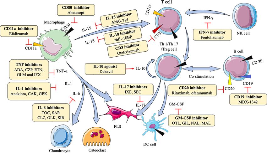
Frontiers in Immunology | www.frontiersin.org 4 July 2021 | Volume 12 | Article 686155Huang et al. Therapeutic Targets Rheumatoid Arthritis FIGURE 1 | Action of drugs targeting cytokines in rheumatoid arthritis. ADA, adalimumab; CZP, certolizumab; ETN, etanercept; GLM, golimumab; IFX, infliximab; CAK, canakinumab; GEK, gevokizumab; TOC, tocilizumab; SAR, sarilumab; CLZ, clazakizumab; OLK, olokizumab; SIR, sirukumab; OTL, otelixizumab; IXE, ixekizumab; SEC, secukinumab; OTL, otilimab; GIL, gimsilumab; NAL, namilumab; MAL, mavrilimumab; FLS, fibroblast-like synoviocytes; DC, dendritic cell; NK, natural killer cell; TNF-a, tumor necrosis factor-a; IL-1, IL-6, IL-10, IL-15, IL-17, IL-18, interleukin (IL)-1, -6, -10, -15, -17, -17, respectively; IFN-g, interferon-gamma; GM-CSF, granulocyte-macrophage colony stimulating factor. On the other hand, several cytokines, including IL-4, IL-10, activating Smad-2/3 in RA (92). Clinical trials observed that IL-13, and TGF-b, exert anti-inflammatory effects in RA. dekavil (an agonist of IL-10) shows a significant efficacy in RA Unsurprisingly, serum IL-10 level is remarkably lower in RA patients (93) (Figure 1). These types of agents (agonist) can bind patients (83). A higher expression of IL-4 and IL-13 is uncovered and initiate receptors to induce corresponding target reactions. in the synovial fluid of early RA rather than established RA, which means IL-4 and IL-13 would be the diagnostic biomarkers Chemokine Targets for early RA patients (84, 85). However, the level of TGF-b is It is reported that chemokines are involved in the underlying high in the FLS and synovial fluids from RA patients (86, 87). pathogenesis of RA by recruiting leukocyte and affecting As anti-inflammatory factors, the injection of L-4, IL-10, angiogenesis. Chemokines are divided into four categories IL-13, TGF-b, or their agonist can play therapeutic roles. IL-4 based on different structures, which are as follows: CXC secreted by activated T cells has anti-angiogenic effects by chemokines, CC chemokines, XC −chemokines, and inhibiting VEGF production in RA FLS, which helps relieve CX3C chemokines. RA (88). IL-10, produced by regulatory T (Treg) cells, suppresses CXC chemokines, including CXCL1, CXCL2, CXCL5, Th17 cells and promotes Treg cells in the CD4+ T cells CXCL6, CXCL8, CXCL9, CXCL10, CXCL12, CXCL13, and population (89). IL-13 is a cytokine of Th2 cell-mediated CXCL16, have been identified with abnormal expression levels immune response. IL-13 exerts its anti-angiogenic function via in synovial fluids, synovial tissues, fibroblasts, and endothelial activation of protein kinase C (PKC) a/b II and ERK-1/2, with cells of RA patients (2, 94, 95). In addition, CXC chemokine concomitant down-regulation of the NF-kB/p65 pathway (90), receptors also implicate in RA, such as CXCR1, CXCR2, CXCR3, and it also can reduce the death of chondrocytes to protect the CXCR4, CXCR5, CXCR6, and CXCR8. The level of these cartilage from destruction probably because of the reduction of receptors is higher in RA patients than in healthy controls Fc gamma receptor I (FcgRI) (91). Transforming growth factor (96–98). (TGF)-b is principally expressed by macrophages and T CXC chemokines,like CXCL1, CXCL2, CXCL5, CXCL8, lymphocytes. TGF-b1 promotes FLS migration and invasion by CXCR1, and CXCR2, generally, are involved in neutrophil inducing epithelial-mesenchymal transition (EMT) via chemotaxis (99), but CXCL10 and CXCL13 promote effector T Frontiers in Immunology | www.frontiersin.org 5 July 2021 | Volume 12 | Article 686155
Huang et al.                                                                                                             Therapeutic Targets Rheumatoid Arthritis

cells and B cells recruitment into the joint, respectively (100,                   and CCR6 recruit T cells into the joint. CCL20 induces B cells,
101). CXCL12, CXCL16, and CXCR6 increase the endothelial                           and CCL14, CCL16, CCR3 recruit endothelial cells to enter into
progenitor cell recruitment and blood vessel formation in the RA                   the inflamed joint (115–118). CCL13 has chemoattractant
joint (102). CXCR3, CXCR4, and CXCR5 enhance Th1 cells,                            activity for both human myeloid leukemia mononuclear cells
lymphocytes, B cells, and T follicular helper (Tfh) cells into joint,              and human umbilical vein endothelial cells (119). CCR4 can
respectively (100, 103). However, CXCL9 can diminish                               attract skin-specific memory T cells to enter the joints (120).
neutrophil recruitment of joints (104). As shown in Table 1,                       CCR9 can also increase the number of dendritic cells in the joint
inhibitors or antagonists of these targets have shown good results                 (116). Researchers find blocking or reducing these CC
in animals, such as CXCR3, CXCR4, CXCL10, CXCL12, and                              chemokines and their receptors, such as CCL2, CCL3, CCL5,
CXCL13, especially the antibody of CXCL10 (MDX-1100) has                           CCL7, CCR1-5, CCR9, and CCR10, can ameliorate tissue
entered clinical trials (25) (Figure 2).                                           swelling and bone erosion (27, 29, 121–124). Among them,
    CC chemokines including CCL2, CCL3, CCL4, CCL5, CCL7,                          CCL2, CCR1, CCR2, and CCR5 have achieved good clinical
CCL13, CCL14, CCL16, CCL18, CCL19, CCL20, CCL21, and                               results (28, 33, 35, 37, 39, 125, 126) (Figure 2).
CCL25 are abnormally expressed in plasma and synovia in RA.                            XC- chemokines and their receptors (like XCL1, XCR1),
Levels of some CC chemokines are significantly correlated with                      CX3C chemokines and their receptors (like CX3CL1 and
the swollen joints, erythrocyte sedimentation rate (ESR), c-                       CX3CR1) have an up-regulation in mononuclear cells (MNCs)
reaction protein (CRP), like CCL2, CCL5, CCL17, CCL18,                             and FLS, respectively, in RA patients (98, 127). Many
CCL19 (105–107). Besides, the synovium also is rich in CCR1,                       inflammatory chemokines are mainly produced by synovial
CCR2, CCR3, CCR4, CCR5, CCR6, CCR7, CCR9, and CCR10 in                             macrophages and FLS in the joints of RA patients, whereas
RA (96, 108–112). CCR2, CCR4, and CCR6 are proven to                               CX3CL1 is produced by synovial endothelial cells. XC and CX3C
positively implicate disease activity in RA (113, 114).                            chemokines are involved in the recruitment of T cells and
    In RA, CCL2, CCL3, CCL4, CCL5, CCL7, CCR1, CCR2,                               synovial fibroblasts. Moreover, CX3CL1 and XCL1 also
CCR5-7, CCR9, and CCR10 induce monocytes to enter the                              promote the migration of monocytes and subchondral
joint synovial. CCL18, CCL19, CCL20, CCL21, CCL25, CCR5,                           mesenchymal progenitor cells, respectively, into RA synovium

 FIGURE 2 | Action of drugs targeting chemokines in rheumatoid arthritis. FLS, fibroblast-like synoviocytes; DC, dendritic cell; CXCL1, CXCL2, CXCL5, CXCL8,
 CXCL10, CXCL12, CXCL13, CXCL16, CXC motif ligand-1, -2, -5, -8, -10, -12, -13, -16; CXCR1/2, CXCR3, CXCR4, CXCR6, CXC motif receptor-1/2, -3, -4, -6;
 CCL2, CCL3, CCL5, CCL20, CCL25, CC motif ligand -2, -3, -5, -20, -25; CCR1, CCR2, CCR5, CCR9, CC motif receptor-1, -2, -5, -9; CX3CL1, CX3C ligand 1.

Frontiers in Immunology | www.frontiersin.org                                  6                                         July 2021 | Volume 12 | Article 686155
Huang et al. Therapeutic Targets Rheumatoid Arthritis
(128, 129). Currently, a clinical trial of E6011 (an anti-CX3CL1 SMALL MOLECULAR METABOLITE
mAb) has been demonstrated to have a promising role in active TARGETS FOR TREATMENT OF
RA patients (30) (Figure 2).
RHEUMATOID ARTHRITIS
Other Protein Targets Previous research has shown that small molecular metabolites,
Similar to the cytokine targets described above, many other like PGs, LTs, LXs, PAF, ROS, and NO, support to induce,
important proteins also play remarkable roles in the maintain, or relieve inflammation in RA (20). Therefore, such
pathogenesis of RA, and corresponding agents have been used compounds could be potential therapeutic targets (Table 2).
in clinical settings as part of continuous research into treating
RA (Table 1). PGs Targets
In a large number of experiments, JAK, p38 mitogen- In some reports, the key function of PGs is shown in physiological
activated protein kinase (MAPK), ERK, JNK, IRAK-4, MMPs, immune responses and pathological conditions related to
toll-like receptor (TLR)-4, G protein-coupled receptor kinase inflammation and tissue damage. The expressions of PGs,
(GRK)-2, Bruton’s tyrosine kinase (BTK), CD3, CD11a, CD19, including PGD2, PGE2, PGF2a, PGI2, PGJ2, and TXA2, are
CD20, and CD80 are demonstrated as examples of such abnormal in RA (20). It is worth noting that PGD2 and PGJ2 are
important proteins in RA. JAK are a part of the JAK/STAT anti-inflammatory small molecules. The binding of PGD2 to DP1(a
pathway, and this signaling is continuously activated, resulting in PGD receptor) inhibits IL-1–induced production of MMP-1 and
the elevated level of MMPs and apoptotic chondrocytes in RA MMP-13 by chondrocytes (160). It is likely that PGJ2 decreases the
synovial joints (130). p38 MAPK, ERK, and JNK activations are production of IL-1b and reactive oxygen species (ROS) through the
almost exclusively found in synovial. As a member of the MAPK NF-kB pathway (146). 15d-PGJ2, as the metabolite of PGD2,
family, p38MAPK, ERK, JNK are activated by MAPKK to ameliorates disease through the suppression of Th17 cells and the
influence pro-inflammatory cytokines, such as TNF, IL-6, and induction of CD4+CD25-FOXP3 +cells (146). PGE2 enhances cyclic
IL-1 (131). Specifically, p38MAPK may phosphorylate AMP production by an EP4 (a PGE2 receptor)-dependent
MAPKAP2, which in turn affects downstream cells (132). JNK mechanism to increase immune inflammation (161). Although
involves in effector T cells function by stimulating Th1 the mechanism of PGF2a in RA is unclear, it can prevent cell
differentiation in RA synovial tissue (131). IRAK-4 is an proliferation, inflammation, tissue remodeling via MMP-3, and
essential protein kinase in mediating pathogen recognition and angiogenesis via VEGF (147). PGI2 probably increases Th2 cell
local cytokine release (like IL-1, IL-6, TNF) through TLR and IL- function by IP (PGI2 receptor) to reduce the production of IL-1b,
1R signaling. Furthermore, the activity of IRAK-4 kinase IL-6, and monocyte chemoattractant protein (MCP)-1 (162). The
regulates Th17-mediated autoimmune diseases like RA TP receptor antagonist (SQ29548) inhibits both cyclooxygenase
through the involvement of Th17 differentiation (133). MMPs (COX)-2 expression and FLS proliferation, and TP agonist U46619
break cartilage and bone by degrading all components of the enhances them. Therefore, TXA2 exerts its function probably
extracellular matrix (134). TLR4 can enhance the production of through the IP-COX-2 pathway (148) (Figure 3).
pro-inflammatory cytokines and chemokines, such as IL-6 and
IL-17, by binding with exogenous ligands, like peptidoglycan, in LTs Targets
FLS and peripheral blood mononuclear (PBMC) from RA In the LTs family, LTB-4 and cysteinyl (Cys)LT-1 are involved in
patients, and trigger cartilage inflammation and degeneration the inflammatory response of RA. Synovial fluid LTB4 levels are
(135). GRK2 prevents the shift of M1 into M2 macrophages by upregulated in RA patients. Although the underlying mechanism
mediating PGE2-EP4-cAMP-CREB signaling in synovial of LTs is not rather clear, some trials show their function in RA.
macrophages (136). BTK activation induces B cells survival, The major effect of LTB4 and its receptor is to enhance the
proliferation, and differentiation by the SYK-BTK axis (137), movement of leucocytes from the circulation toward the site of
which is an attractive therapeutic target for RA. CD3 expressed tissue damage (163). An antagonist of LTB4 receptor, BIIL 284
by mature T cells and thymocytes can activate T cells signaling inhibits the LTB4-stimulated expression of Mac-1 on neutrophils
and regulate TCR expression by the formation of T cell receptor in RA patients (164). A potent CysLT1 receptor antagonist,
(TCR)/CD3 complex (138). As an adhesion molecule, CD11a montelukast inhibits the activation of the NF-kB pathway and
can facilitate the recruitment and entry of T cells into the secretion of IL-6 and IL-8 in FLS (165), next to reducing the
synovial tissue via LFA-1(CD11a/CD18)/intercellular cell disease incidence and its activity (166). These results mean that
adhesion molecule (ICAM)-1 pathway (139). CD19 amplifies the inhibition of CysLT1 and LTB4 receptors would be a
the activation of Lyn and Src-family protein tyrosine kinases, potential and promising new therapeutic method to prevent
thereby enhancing the signals generated by the B-cell antigen inflammation and disease progression in RA patients (Figure 3).
receptor to regulate B-cell development, activation, and
differentiation (140). Although the biological activity of CD20 LXs Targets
and CD80 are not fully elucidated, CD20 allows specific and The family of LXs, like LXA4 and LXB4, generated from
effective B-cell depletion and CD80 involves in T cell arachidonic acid display anti-inflammatory activities. LXA4 can
co−stimulation (141). Inhibitors for these abovementioned decrease memory B-cell response via engagement of lipoxin A4
protein targets have entered clinical trials (Figure 1). receptor (ALX)/formyl peptide receptor-2 (FPR-2) in synovial
Frontiers in Immunology | www.frontiersin.org 7 July 2021 | Volume 12 | Article 686155Huang et al. Therapeutic Targets Rheumatoid Arthritis
TABLE 2 | Small molecular metabolite targets and their agents in rheumatoid arthritis.
Targets Agents Phases References
PGs targets
PGD2 MK0524 Animal study (142)
PGE2 ER-819762 Animal study (143)
CR6086 Animal study (144)
PGI2 Iloprost Phase 2 (145)
PGJ2 15d-PGJ2 Animal study (146)
PGF2a AL‐8810 Animal study (147)
TXA2 SQ29548 Animal study (148)
LTs targets
LTB4R BIIL 284 Phase 1 (149)
CysLT1R Montelukast Animal study (150)
LXs targets
ALX BML-111 Animal study (151)
PAF targets
PAFR WB2086 Animal study (152)
ROS targets
ROS Cinnamaldehyde Cell culture (153)
Eugenol Cell culture (153)
NO targets
iNOS GW274150 Phase 2 (154)
L-NAME Animal study (155)
Other small molecular targets
CB2 HU-308 Animal study (156)
JWH-015 Animal study (157)
FFAH URB597 Animal study (158)
NAGly Animal study (159)
PGD2, prostaglandin D2; PGE2, prostaglandin E2; PGI2, prostaglandin I2; PGJ2, prostaglandin J2; TXA2, thromboxane A2; LTB4R, leukotriene B4 receptor; CysLT1R, cysteine
leukotrienes 1 receptor; ALX, lipoxin A4 receptor; PAF, platelet-activating factor; PAFR, platelet-activating factor receptor; iNOS, inducible nitric oxide synthase; CB2, cannabinoid receptor
2; FFAH, specific fatty acid amide hydrolase; NAGly, N-arachidonic glycine.
tissues of patients with RA, further enhancing the reduction of pathways, further affecting cell proliferation, angiogenesis, and
inflammation (167, 168). BML-111 (an ALX/FPR2 agonist) partly apoptosis in joints (173). NO and inducible nitric oxide synthase
downregulates the immune response in CIA (151). LXB4 has anti- (iNOS) expressions are changed in patients with RA (174). NO
inflammatory effects by regulating the adhesion and motility of regulates T and B cells infiltration by inhibiting their chemotaxis
monocytes and neutrophils and enhancing antibody production by and adhesion into joints (175). Treatments with the NOS
human memory B cells (47, 169) (Figure 3). inhibitor L-NAME and iNOS inhibitor GW274150 are proven
to improve inflammation response, and a trend toward a
PAF Targets reduction in synovial thickness is observed (154, 155).
Studies suggest that PAF plays a prominent role in RA. The
activation of circulating platelets further influences leukocyte Other Small Molecular Targets
activity and participates in inflammation formation in RA The receptor activation of cannabinoid (CB)-1/2 involves the
patients (170). It is reported that pathways based on TNF-a production of endocannabinoids, subsequently, endocannabinoids
regulate PAF, and TNF-a antagonists inhibit platelet activation in are quickly metabolized by specific fatty acid amide hydrolase
active patients with RA (171). WB2086, a human PAF receptor (FAAH) (176). The levels of CB1 and CB2 increase in the synovial
antagonist, inhibits PAF-induced platelet aggregation in animal membrane with RA. CB2 inhibits IL-1b–induced proliferation of RA
models (152). Based on current studies, targeted agents inhibiting FLS and the activation of MAPK pathway (157). In addition, the
PAF and its receptor needed more research because of the limitation reduction of arthritis severity and activity in FAAH knock-down mice
of experience with the therapeutic effects of this target. is observed (158). Similarly, the treatment with URB597 and N-
In these mechanisms of PGs, LTs, LXs, PAF, receptors play arachidonic glycine (FAAH inhibitors) prevents the occurrence of
significant roles, therefore inhibitors or agonists of these CIA in mice as well (158, 159). These results mean that the new
receptors can exert therapeutic functions, and the therapeutic agents targeting the endocannabinoid system are potential and
effects are proven in animal experiments (Figure 3). promising therapeutics for further research.
ROS and NO Targets
ROS and NO belong to oxidant molecules, which involve in the EPIGENETIC TARGETS FOR TREATMENT
pathogenesis of many chronic autoimmune diseases, including OF RHEUMATOID ARTHRITIS
RA. There is a strong positive correlation between serum ROS
level and disease severity in both RA patients and arthritic rodent Epigenetic modifications can regulate gene expression without
models (172). ROS regulates MAPK and NF-kB signaling altering the DNA sequence. Non-coding RNAs (ncRNAs)
Frontiers in Immunology | www.frontiersin.org 8 July 2021 | Volume 12 | Article 686155Huang et al. Therapeutic Targets Rheumatoid Arthritis
A B
C D
FIGURE 3 | Action of drugs targeting small molecular metabolites in rheumatoid arthritis. (A) action of drugs targeting PGs family in rheumatoid arthritis; (B) action of
drugs targeting LTs family in rheumatoid arthritis; (C) action of drugs targeting LXs family in rheumatoid arthritis; (D) action of drugs targeting PAF in rheumatoid
arthritis. PGD2, prostaglandin D2; PGE2, prostaglandin E2; PGI2, prostaglandin I2; PGJ2, prostaglandin J2; TXA2, thromboxane A2; PGF2a, prostaglandin F2a;
DP1, prostaglandin D2 receptor 1; EP4, prostaglandin E receptor 4; TP, prostaglandin TXA2 receptor; FP, prostaglandin PGF2a receptor; IP, prostaglandin PGI2
receptor; LTB4, leukotriene B4; CysLT1, cysteine leukotrienes 1; ALX, lipoxin A4 receptor; FPR-2, formyl peptide receptor-2; PAF, platelet-activating factor; PAFR,
platelet-activating factor receptor; iNOS, inducible nitric oxide synthase.
regulation, DNA methylation, RNA methylation, and histone correlate with the ESR, rheumatoid factor (RF), CRP, disease
modifications are seen as the main mechanisms of epigenetic activity score (DAS), and anti-citrullinated protein antibodies
regulations. Numerous research has established that several (ACPA), which will become promising targets for RA detection
abnormalities in these mechanisms eventuate in the development (31, 204). miR-16 and miR-223 are also identified as targets to
of RA. Corresponding target agents have been applied for distinguish patients with early RA from healthy individuals (34).
patients (Table 3). Most of the aberrant miRNA levels can alter the secretion of
inflammatory cytokines or MMPs, further affecting the
ncRNAs procession of RA. In FLS, miR-34a-3p (193), miR-129-5p
With the increasing advancement of bioinformatics analysis (202), miR-410-3p (207), miR-506 (208), miR-22 (213), miR-
and microarray sequencing techniques, tremendous ncRNAs 101-3p (216), miR-495 (217), miRNA-17-5p (218), miRNA-140-
are identified in different tissues. Compared with healthy 5p (219), miR-21 (223), miRNA-15a/16 (224), miR-9 (230),
individuals, aberrant levels of abundant ncRNAs are observed, miR-20a (234), miR-145-5p (235), miR-365 (236), and miR-
including microRNAs (miRNAs), long non-coding RNAs 124a (238) overexpression significantly inhibit the proliferation
(lncRNAs), and circular RNAs (circRNAs). Because a large and promote apoptosis by aiming different proteins or other
number of ncRNAs especially miRNAs are found, ncRNAs targets. On the contrary, miR-138 (192), miR-142-3p (198), miR-
involved in the development and progression of RA are 98 (201), miR-26a-5p (203), miR-191 (205), miR-15 (222), and
summarized in this review since 2019 sourcing from miR-483-3p (282) enhance the inflammatory milieu and
PubMed (Table 4). subsequently tissues could be damaged. Moreover, miRNA-
miRNAs are small, mature, non-coding RNA molecules 486-5p upregulation in exosomes can repress FLS proliferation
(about 22 nucleotides long) that can affect the processing of and migration, which proves exosomes to be a suitable vector for
target mRNAs at the post-transcriptional level by translational the delivery of therapeutic miRNA-486-5p (283).
inhibition or promoting mRNA degradation (281). As shown in Table 4, miRNA levels change various
Accumulating studies have revealed that altered expression and intracellular pathways, and the most prominent implicated
dysregulation of miRNAs have something to do with RA pathways are those of NF-kB (192, 198, 207, 212, 213, 222,
occurrence. miRNA-23b, miR-221/222 levels positively 229, 230, 238), PI3K/Akt (194, 199, 203, 236), JAK/STAT (218,
Frontiers in Immunology | www.frontiersin.org 9 July 2021 | Volume 12 | Article 686155Huang et al. Therapeutic Targets Rheumatoid Arthritis
TABLE 3 | Epigenetic targets and their agents in rheumatoid arthritis.
Targets Agents Phases References
DNA methylation
DNMT Azacitidine Animal study (177)
Decitabine Animal study (177)
Procainamide Animal study (177)
Hydralazine Animal study (177)
EGCG Animal study (177)
RNA methylation
METTL3 – Cell culture (178)
Histone modification
HAT Delphinidin Cell culture (179)
Anacardic acid Animal study (180)
HMT GSK-J4 Animal study (181)
EZH2 Cell culture
HDAC MS-275 Animal study (182)
Entinostat Cell culture (183)
Histone modification
HDAC MI192 Cell culture (183)
Trichostatin A Cell culture (183)
Valproic acid Animal study (183)
Vorinostat Cell culture (183)
Nicotinamide Cell culture (184)
MPT0G009 Animal study (183)
CKD-506 Animal study (185)
CKD-L Animal study (186)
NK-HDAC-1 Animal study (187)
SAHA Animal study (182)
Largazole Cell culture (188)
Givinostat Cell culture (189)
BET I-BET151 Animal study (190)
JQ1 Animal study (191)
DNMT, DNA methyltransferase; EGCG, epigallocatechin-3-gallate; METTL3, methyltransferase-like 3; HAT, histone acetyltransferase; HDAC, histone deacetylases; HMT, histone
methyltransferase; BET, bromodomain and extra-terminal; SAHA, suberoylanilide hydroxamic acid; EZH2, zeste homolog 2.
219), TLR (208, 220), b-catenin (215, 217, 235), and Wnt in RA FLS (263, 288). Overexpression of lncRNA MEG3 plays
(223, 235). In parallel, the efficacy of several miRNAs against an anti-inflammatory effect by regulating the AKT/mTOR
RA has been verified in animal experiments. Injections of signaling pathway (258). lncRNA PICSAR alters cell
miR-141-3p agomir, miR-411 mimics, miR-9, miR-21 proliferation, migration, invasion, IL-6, IL-8, and MMP-3
lentivirus, miR-26a, miRNA-147 mimics have been proven to production through sponging miR-4701-5p, in other words,
ameliorate cartilage injury and bone erosion, further inhibiting lncRNA PICSAR can competitively combine miR-4701-5p to
inflammatory arthritic development in CIA animal models affect downstream target genes (257). As a competitive
(223, 226, 229, 230, 284). On the other hand, miRNA-17-5p endogenous RNAs (ceRNA), lncRNA HIX003209 exaggerates
lipoplex and miR−145−5p agomir given to mice are found to inflammation through sponging miR-6089 via TLR-4/NF-kB
increase inflammatory cytokine levels (MMP−3, MMP−9, MMP pathway in RA macrophages (262). miR-222-3p/Sirt1 axis is
−13), aggravating arthritis in the future (212, 236). The also found to be critical for the function of lncRNA GAS5 in
abovementioned studies about the mechanism of miRNAs mitigating the proliferation, inflammation, and apoptosis of RA
have shown promising results in experimental models of FLS (289). In particular, lncRNA NEAT1 and lncRNA HAND2-
arthritis, and their efficacy needs more clinical trials to prove. AS1 are found in PBMC and mesenchymal stem cells (MSC),
Consisting of more than 200 nucleotides in length, lncRNAs are respectively, and are involved in the regulation of RA (241, 255).
identified as long non-coding RNAs and are widely expressed in In an arthritic model experiment, silencing lncRNA ZFAS1
various tissues of the human body. Many studies have suggested can mitigate inflammation and hyperplasia by competitively
lncRNA could become a diagnostic tool for RA. For example, lnc- binding to miR-296-5p and regulating MMP-15 expression
AL928768.3 and lnc-AC091493.1 are positively associated with (249). Additionally, injecting lentivirus expressing shRNA for
CRP, DAS, and RF (260). Apart from those two, lncRNA lncRNA-H19 intra-articularly at the ankle of CIA mice
ENST00000483588, ENST00000456270, RNA143598, ameliorates the progression of CIA by competitively binding
RNA143596, HIX0032090, IGHCg1, and XLOC_002730 could with miR-124a, which directly targets CDK2 and MCP-1 (267).
also become targets to diagnose RA (285–287). The injection of lentivirus carrying sh-lncRNA XIST plasmids
lncRNA FER1L4 and MEG3 regulate RA via targeting reduces levels of TNF-a, IL-2, and IL-6 to suppress inflammatory
nucleotide oligomerization domain-like receptors 5 (NLRC5) and damage in cartilage tissues (270). Taken together, these
Frontiers in Immunology | www.frontiersin.org 10 July 2021 | Volume 12 | Article 686155Huang et al. Therapeutic Targets Rheumatoid Arthritis
TABLE 4 | NcRNAs targets in rheumatoid arthritis.
NcRNAs Expression Tissue Signalings Phases References
miRNAs
miR-138 Up FLS NF-kB signaling Cell culture (192)
miR-34a-3p Down FLS – Animal study (193)
miR-23b Up FLS, STs – – (31)
miR-125 Down ST PI3K/Akt/mTOR pathway Cell culture (194)
miR-27b-3p Down ST HIPK2 signaling Cell culture (195)
MiR-19a-3p Up ST IGFBP5 signaling Cell culture (196)
Down Plasma SOCS3 Cell culture (197)
miR-142-3p Up ST, FLS NF-kB signaling Cell culture (198)
miRNA‐135a Up ST PI3K/AKT pathway Cell culture (199)
miR-192-5p Down BM-MSC-exos – – (200)
miR-98 Up FLS IL-10 signaling Cell culture (201)
miR-129-5p Down FLS IGF-1R/SRC/ERK/EGR-1 pathway Cell culture (202)
miR-26a-5p Up FLS PTEN/PI3K/AKT pathway Cell culture (203)
miR-221/222 Up PBMC – – (204)
miR-191 Up FLS miR-191-C/EBPb pathway Cell culture (205)
miR-449a Down ST HMGB1 signaling Cell culture (206)
miR-410-3p Down SF, FLS NF-kB signaling Cell culture (207)
miR-506 Down ST, FLS TLR4 signaling Cell culture (208)
miR-320a Down ST MAPK-ERK1/2 pathway Cell culture (209)
miR-29b Up PBM HBP1 signaling Cell culture (210)
miR-155 Up ST FOXO3a signaling Cell culture (211)
miR−145−5p Up FLS NF−kB pathway Animal study (212)
miR-22 Down FLS IL6R signaling/NF-kB pathway Cell culture (213)
Down ST SIRT1 signaling Cell culture (214)
miRNA-141-3p Down FLS FoxC1/b-catenin axis Animal culture (215)
miR-101-3p Down FLS PTGS2 signaling Cell culture (216)
miR-495 Down FLS b-catenin pathway Cell culture (217)
miRNA-17-5p Down FLS JAK/STAT pathway Animal study (218)
miRNA-140-5p Down FLS STAT3 signaling Cell culture (219)
miR-3926 Down FLS TLR 5 signaling Cell culture (220)
miR-613 Down FLS, ST DKK1 signaling Cell culture (221)
miR-15 Up FLS NF-kB pathway Cell culture (222)
miR-21 Down FLS Wnt pathway Animal study (223)
miRNA-15a/16 Down FLS SOX5 axis Cell culture (224)
miRNA-155 Up FLS – – (225)
miR-26a Down CT, AC CTGF signaling Animal study (226)
miR-106b Down SFDE PDK4 signaling Cell culture (227)
miR-223 Up FLS – – (228)
miR-411 Down ST, FLS NF-kB pathway Animal study (229)
miR-9 Down FLS NF-kB1-RANKL pathway Animal study (230)
miRNA-486-5p Down FLS-exos Tob1/BMP/Smad pathway Animal study (231)
miR-49 Up PBMC – – (232)
miR-326 Down PBMC – – (232)
miR-34a-5p Down ST XBP1 signaling Cell culture (233)
miR-20a Down FLS ADAM10 signaling Cell culture (234)
miR-145-5p Down FLS Wnt1/b-catenin pathway Cell culture (235)
miR-365 Down FLS IGF1 signaling or PI3K/AKT/mTOR pathway Animal study (236)
miR-34a Down BM-MSC-Evs cyclin I/ATM/ATR/p53 axis Cell culture (237)
miR-124a Down FLS PIK3/NF-kB pathway Cell culture (238)
miR-9-5p Down Serum REST/miR-132 pathway Cell culture (239)
miR-34a-5p Down ST XBP1 signaling Cell culture (233)
lncRNAs
linc01197 Down ST miRNA-150/THBS2 axis Cell culture (240)
lncRNA NEAT1 Up PBMC- exos miRNA-23a/MDM2/SIRT6 Axis Cell culture (241)
Up ST, FLS miR-204-5p signaling Cell culture (242)
Up ST, FLS MAPK/ERK pathway Cell culture (243)
Up FLS miR-410-3p/YY1 axis Cell culture (244)
lncRNA PVT1 Up FLS miRNA-145-5p Cell culture (245)
Up ST miR-543-dependent SCUBE2 Cell culture (246)
Up FLS SIRT6 Cell culture (247)
lncRNA OIP5-AS1 Down FLS miR-448-PON1/TLR3/NF-kB axis Cell culture (248)
lncRNA ZFAS1 Up FLS miR-296-5p/MMP-15 Animal study (249)
(Continued)
Frontiers in Immunology | www.frontiersin.org 11 July 2021 | Volume 12 | Article 686155Huang et al. Therapeutic Targets Rheumatoid Arthritis
TABLE 4 | Continued
NcRNAs Expression Tissue Signalings Phases References
Up FLS miR-2682-5p/ADAMTS9 axis Cell culture (250)
linc00152 Up FLS Wnt/b-catenin pathway Cell culture (251)
lncRNA MALAT1 Down PBMC Notch pathway Cell culture (252)
lncRNA GAS5 Down FLS miR-128-3p/HDAC4 axis Cell culture (253)
Down ST, FS HIPK2 signaling Cell culture (254)
lncRNA HAND2-AS1 Down MSC-exos miR-143-3p/TNFAIP3/NF-kB pathway Cell culture (255)
lncRNAS56464.1 Up FLS miR−152−3p/Wnt pathway Cell culture (256)
lncRNA PICSAR Up FLS miRNA-4701-5p signaling Cell culture (257)
lncRNA MEG3 Down FLS miR-141/AKT/mTOR pathway Animal study (258)
lncRNA ITSN1-2 Up FLS NOD2/RIP2 pathway Cell culture (259)
lncAL928768.3 Up FLS – – (260)
lncAC091493.1 Up FLS – – (260)
lncRNA HOTTIP Up FLS SFRP1 demethylation Cell culture (261)
lncRNA HIX003209 Up PBMC TLR4/NF-kB pathway Cell culture (262)
lncRNA FER1L4 Down ST, FLS NLRC5 signaling Cell culture (263)
lncRNA CASC2 Down Plasma IL−17 signaling Cell culture (264)
lncRNA PlncRNA-1 Down Serum, SF TGF-b1 signaling Cell culture (265)
lncRNA H19 Up FLS Notch pathway Cell culture (266)
Up FLS miR-124a Animal study (267)
lncRNA RP11-83J16.1 Down FLS b-catenin pathway Cell culture (268)
lncRNA H19 Down FLS NF-kB and JNK/p38 MAPK pathways Cell culture (269)
lncRNA XIST Up CT STAT3 signaling Animal study (270)
lncRNA SNHG1 Up FLS PTBP1 signaling Cell culture (271)
lncRNA THRIL Up Serum PI3K/AKT pathway Cell culture (272)
circRNAs
circ_0088036 Up FLS miR-140-3p/SIRT 1 axis Cell culture (273)
circ_0000396 Down FLS miR-203/HBP1 axis Cell culture (274)
circ_AFF2 Up FLS miR-375/TAB2 axis Cell culture (275)
circ_0130438 Down PBMC – – (276)
circ_0002715 Up PB – – (277)
circ_0035197 Up PB – – (277)
circRNA_09505 Up PBMC miR-6089/AKT1/NF-kB axis Animal study (278)
circFADS2 Down AC miR-498/mTOR pathway Cell culture (279)
circ_0000175 Down PBMC – – (280)
Circ_0008410 Up PBMC – – (280)
FLS, fibroblast-like synoviocytes; CT, cartilage tissues; AC, articular chondrocytes; ST, synovial tissues; BM-MSC-Evs, bone marrow mesenchymal stem cell -derived extracellular vesicles;
MSC-exos, mesenchymal stem cell-derived exosomes; PB, peripheral blood; FoxC1, forkhead box C1; PTGS2, prostaglandin-endoperoxide synthase 2; SOX5, sex determining region Y-
box protein 5; CTGF, connective tissue growth factor; SFDE, synovial fibroblast-derived exosomes; PDK4, pyruvate dehydrogenase kinase 4; NF-ΚB, nuclear factor kappa-B; RANKL,
receptor activator of nuclear factor-kb ligand; MDM2, murine double minute-2; SIRT6, sirtuin 6; TLR3, toll-like receptor 3; BMP, bone morphogenetic protein; Smad, mouse signal
transduction molecule; MMP-15, matrix metalloproteinase 15; XBP1, X-box binding protein 1; Wnt, wingless and integration-1; ADAM, a disintegrin and metalloprotease 10; SCUBE2,
signal peptide-CUB-EGF-like containing protein 2; MAPK, mitogen-activated protein kinase; ERK, extracellular regulated protein kinase; HBP1, HMG-box transcription factor 1; HDAC4,
histone deacetylase 4; TNFAIP3, tumor necrosis factor alpha-inducible protein 3; TAB2, TAK1-binding 2; ATM, ataxia‐telangiectasia mutated; ATR, ATM-Rad3-related; JAK, janus kinase;
STAT, signal transducers and activators of transcription; TLR 5, toll-like receptor 5; DDK1, dickkopf 1; PI3K, phosphatidylinositol 3-kinase; REST, presentational state transfer; PBM,
peripheral blood monocytes; FOXO3a, forkhead box O3 alpha; SIRT6, sirtuin 6; HMGB1, high-mobility group box protein 1; C/EBPb, CCAAT enhancer-binding proteinb; IGF-1R, insulin-
like growth factor 1 receptor; HIPK2, homeodomain-interacting protein kinase 2; SOCS 3, suppressors of cytokine signaling 3; NOD2, nucleotide-binding oligomerization domain 2; RIPK2,
receptor interacting serine threonine kinase 2; NLRC5, nucleotide oligomerisation domain-like receptors 5; TGF-b1, transforming growth factor beta 1; PTBP1, polypyridine tract-binding
protein 1; HIPK2, homeodomain-interacting protein kinase 2; THBS2, thrombospondin 2; PBMC, peripheral blood monouclear cell; exos: exosomes; ADAMTS9, ADAM metallopeptidase
with thrombospondin type 1 motif 9; mTOR, mammalian target of rapamycinMammalian target of rapamycin.
mechanisms described above reveal that identification of circRNAs might be important targets in clinical blood samples
lncRNA-miRNA interaction provides new insights into the for RA diagnosis, which show a significant association with
pathogenesis of RA. Thus, lncRNAs are potential and valuable DAS28, RF, CRP, such as circ0003972 (291), circ0002715 (277).
targets for treating RA. Several studies reveal the partial and hidden molecular
circRNAs are a category of newly endogenous non-coding mechanisms of circRNAs in the pathogenesis of RA.
RNA, and they are becoming significant members of the gene circ0088036 is found to be aberrantly upregulated in RA FLS,
regulation environment, the most representative characteristic of and it facilitates RA progression by acting as a miR-140-3p
which is the covalently closed RNA circle without 5′ end caps sponge to upregulate SIRT 1 expression (273). circFADS2
and 3′ poly tails. circRNAs have been reported to involve in the protects chondrocytes from apoptosis by acting as an
pathogenesis of some autoimmune diseases and have a wide interceptor in miR-498/mTOR singling pathway (292).
range of functions, such as RNA polymerase (RNAP) II circ_0000396 regulates miR-203/HBP1 axis to inhibit the
elongation, miRNA and RBP sponge, RNA maturation growth of RA FLS (274). Besides, circRNA_09505 can promote
regulation, protein localization, and so on (290). Also, several AKT1 expression via regulating the IkBa/NF-kB signaling
Frontiers in Immunology | www.frontiersin.org 12 July 2021 | Volume 12 | Article 686155Huang et al. Therapeutic Targets Rheumatoid Arthritis
pathway in macrophages, the most interesting is circRNA_09505 There are four types of HDAC, including class I (HDAC1-3,
knockdown significantly alleviates arthritis and inflammation in HDAC8), class II (HDAC4-7, HDAC9-10), class III (SIRT1-7),
CIA mice (278). These emerging studies elucidate that circRNAs and class IV (HDAC11) (183). It has been reported that HDAC
are potential and promising targets for RA therapy. In the future, activity is involved in RA synovial are significantly increased
more mechanisms of circRNAs in RA need to be uncovered. compared with normal controls and are in direct proportion to
TNF-a mRNA levels (301). In contrast, inflammatory stimuli
DNA Methylation diminish the expression of HDAC5 to regulate the generation
Methylation of DNA is carried out by the activity of DNA of cytokines and chemokines through modulation of IRF1 in
methyltransferases (DNMT) and leads to the formation of 5- RA FLS (302). In addition, the mRNA and protein levels of
methylcytosine (5-mC), which further affect various life SIRT1 in RA LFS are lower than normal FLS, and increased
activities. Altered DNA methylation patterns have been SIRT1 expression strikingly suppresses the invasiveness of
identified in clinical RA. In RA patients, an alteration of DNA RA FLS by inhibiting MMP1 and MMP13 expression (303).
methylome signature in PBMC and a reduction of 5-mC An animal study also shows that knockout specifically of
amounts in synovial tissues were observed, the alteration of SIRT1 in myeloid cells alleviates synovial inflammation
FLS gene expression, including chitinase-3-like protein 1 and bone destruction of RA by decreasing Th1 and Th17
(CHI3L1), caspase 1 (CASP1), STAT3, mitogen-activated differentiation (304).
protein kinase kinase kinase 5 (MAP3K5), familial Two inhibitors of histone acetyltransferase (delphinidin and
Mediterranean fever (MEFV), and wnt1-inducible signaling anacardic acid) suppress FLS proliferation and invasion, which
protein 3 (WISP3), is caused by differentially methylated genes further ameliorates inflammatory (179, 180). In addition, many
leading to the pathogenesis of RA (293). In addition, the change HDAC inhibitors aiming at different HDAC types are also shown
of DNA methylation is rather different between early and late to suppress inflammation in cell or animal experiments,
stage (294, 295). Studies have shown that several DNMT entinostat, MI192, trichostatin A, and valproic acid (Table 2).
inhibitors including azacitidine, decitabine, procainamide, These experiment results display potent therapeutic efficacies of
hydralazine, and epigallocatechin-3-gallate are applied in HDAC inhibitors in RA remission (186, 305). Moreover,
animals, which show excellent efficacies via decreasing pro- bromodomain and extra-terminal (BET) family proteins can
inflammatory cytokines (i.e., IL-6, TNF-a, and TGF) (177). identify acetylated histones. I-BET151 (a selective inhibitor of
BET) can reduce joint inflammation and bone loss by blocking
RNA Methylation MMP-1, MMP-3, IL-6, and IL-8 production in RA FLS (190,
The most common methylated modification of RNA is N6- 306). Similar results are seen with another BET inhibitor, JQ1,
methyladenosine (m6A). m6A methyltransferase, m6A suggesting that this family of proteins may be promising
demethylase, and m6A RNA-binding protein are essential for therapeutic targets (191).
m6A RNA modification (296, 297). It is reported that WTAP,
RIPK2, JAK3, and TNFRSF10A genes identified by Other Histone Modifications
transcriptome-wide high-throughput m6A sequencing are in The involvement of histone methylation is important in the
accordance with m6A, which are increased in inflammation- pathogenesis of RA. H3K4me3 in SF is associated with the onset
related pathways, cell proliferation, and apoptosis in FLS (298). of arthritis-activated chromatin. GSK-J4 can inhibit the
In the RA patient’s peripheral blood, m6A demethylases H3K27me3 methylation at the TLR2 promoter, which
(ALKBH5 and FTO) and m6A RNA-binding protein significantly relieves the articular cartilage destruction and
(YTHDF2) are proven to have an association with DAS28, inflammation (307). The histone methyltransferase enhancer of
complement 3 (C3), and immunoglobulin G (IgG) (297). As a zeste homolog 2 (EZH2) is discovered to overexpress in RA FLS
key methyltransferase of m6A, METTL3 can inhibit the and can be induced by TNF-a through the JAK and NF-kB
activation of pTHP-1 macrophages by preventing the pathways. EZH2-mediated epigenetic alteration of secreted
generation of IL-6 and TNF-a, and attenuate inflammatory frizzled-related protein 1 (SFRP1) significantly correlates with
response induced by LPS via the NF-kB signaling pathway the activation of RA FLS (308).
(178). These studies provide novel prospects for us in
recognizing the pathogenesis of RA and finding promising
targets for RA. CONCLUSION AND PERSPECTIVE
Histone Modifications RA is an autoimmune disease with complex etiology. To date,
The histones can be modified in many ways posttranslationally, under unremitting efforts, RA has been altered from a highly
including acetylation, methylation, citrullination, ubiquitination, disabling disease without effective treatment to a disease that can
phosphorylation, and sumoylation (299, 300). These be well controlled. Many patients achieve remission or low
modifications implicate RA occurrence. disease state, which can be attributed to the development of
specific DMARDs. However, the main problems of marketed
Histone Acetylation biologics/drugs are nonresponses and partial responses as well as
For histone acetylation, most studies mainly focus on histone the occurrence of adverse effects like stomatitis, exanthema, and
acetyltransferase (HAT) and histone deacetylases (HDAC). diarrhea. This review summarizes new targets, including
Frontiers in Immunology | www.frontiersin.org 13 July 2021 | Volume 12 | Article 686155You can also read