Heart Failure Association of the ESC, Heart Failure Society of America and Japanese Heart Failure Society Position statement on endomyocardial ...

Page created by Joan Becker
 
CONTINUE READING
Heart Failure Association of the ESC, Heart Failure Society of America and Japanese Heart Failure Society Position statement on endomyocardial ...
European Journal of Heart Failure (2021) 23, 854–871                                                 POSITION PAPER
                                     doi:10.1002/ejhf.2190

Heart Failure Association of the ESC, Heart
Failure Society of America and Japanese Heart
Failure Society Position statement
on endomyocardial biopsy
Petar M. Seferović1*, Hiroyuki Tsutsui2, Dennis M. McNamara3, Arsen D. Ristić4,5,
Cristina Basso6, Biykem Bozkurt7, Leslie T. Cooper Jr8, Gerasimos Filippatos9,
Tomomi Ide2, Takayuki Inomata10, Karin Klingel11, Aleš Linhart12,
Alexander R. Lyon13, Mandeep R. Mehra14, Marija Polovina4,5, Ivan Milinković4,5,
Kazufumi Nakamura15, Stefan D. Anker16, Ivana Veljić4, Tomohito Ohtani17,
Takahiro Okumura18, Thomas Thum19,20, Carsten Tschöpe21, Giuseppe Rosano22,23,
Andrew J.S. Coats24,25, and Randall C. Starling26
1 Serbian Academy of Sciences and Arts, Belgrade, Serbia; 2 Department of Cardiovascular Medicine, Faculty of Medical Sciences, Kyushu University, Fukuoka, Japan; 3 Heart and
Vascur Institute, University of Pittsburgh Medical Center, Pittsburgh, PA, USA; 4 Department of Cardiology, Clinical Center of Serbia, Belgrade, Serbia; 5 Faculty of Medicine,
University of Belgrade, Belgrade, Serbia; 6 Cardiovascular Pathology Unit, Department of Cardiac, Thoracic, Vascular Sciences and Public Health, University of Padua, Padua, Italy;
7 Winters Center for Heart Failure, Cardiovascular Research Institute, Baylor College of Medicine, Michael E. DeBakey VA Medical Center, Houston, TX, USA; 8 Department of

Cardiovascular Medicine, Mayo Clinic, Jacksonville, FL, USA; 9 Attikon University Hospital, Department of Cardiology, National and Kapodistrian University of Athens, School of
Medicine, Athens, Greece; 10 Department of Cardiovascular Medicine, Kitasato University Kitasato Institute Hospital, Tokyo, Japan; 11 Cardiopathology, Institute for Pathology,
University Hospital, Tuebingen, Germany; 12 Department of Cardiovascular Medicine, Charles University, Prague, Czech Republic; 13 National Heart and Lung Institute, Imperial
College and Royal Brompton Hospital, London, UK; 14 Heart and Vascular Center, Brigham and Women’s Hospital and Harvard Medical School, Boston, MA, USA; 15 Department
of Cardiovascular Medicine, Okayama University Graduate School of Medicine, Dentistry and Pharmaceutical Sciences, Okayama, Japan; 16 Department of Cardiology (CVK); and
Berlin Institute of Health Center for Regenerative Therapies (BCRT), German Centre for Cardiovascular Research (DZHK) partner site Berlin; Charité Universitätsmedizin,
Berlin, Germany; 17 Department of Cardiovascular Medicine, Osaka University Graduate School of Medicine, Osaka, Japan; 18 Department of Cardiology, Nagoya University
Graduate School of Medicine, Nagoya, Japan; 19 Institute of Molecular and Translational Therapeutic Strategies, Hannover Medical School, Hannover, Germany; 20 Fraunhofer
Institute for Toxicology and Experimental Medicine, Hannover, Germany; 21 Berlin Institute of Health (BIH) and Berlin-Brandenburg Center for Regenerative Therapies (BCRT),
Department of Cardiology, Campus Virchow Klinikum, Charite University, Berlin, Germany; 22 Department of Medical Sciences, IRCCS San Raffaele, Rome, Italy; 23 Cardiology
Clinical Academic Group, St George’s Hospitals NHS Trust, London, UK; 24 Monash University, Melbourne, Australia; 25 University of Warwick, Coventry, UK; and 26 Cleveland
Clinic, Cleveland, OH, USA

Received 26 November 2020; revised 23 March 2021; accepted 8 April 2021 ; online publish-ahead-of-print 19 May 2021

Endomyocardial biopsy (EMB) is an invasive procedure, globally most often used for the monitoring of heart transplant (HTx) rejection.
In addition, EMB can have an important complementary role to the clinical assessment in establishing the diagnosis of diverse cardiac
disorders, including myocarditis, cardiomyopathies, drug-related cardiotoxicity, amyloidosis, other infiltrative and storage disorders, and
cardiac tumours. Improvements in EMB equipment and the development of new techniques for the analysis of EMB samples have significantly
improved diagnostic precision of EMB. The present document is the result of the Trilateral Cooperation Project between the Heart Failure
Association of the European Society of Cardiology, the Heart Failure Society of America, and the Japanese Heart Failure Society. It represents
an expert consensus aiming to provide a comprehensive, up-to-date perspective on EMB, with a focus on the following main issues: (i) an
overview of the practical approach to EMB, (ii) an update on indications for EMB, (iii) a revised plan for HTx rejection surveillance, (iv) the
impact of multimodality imaging on EMB, and (v) the current clinical practice in the worldwide use of EMB.
..........................................................................................................

*Corresponding author. University of Belgrade, Faculty of Medicine and Heart Failure Center, Belgrade University Medical Center, Koste Todorovica 8, 11000 Belgrade, Serbia.
Tel/Fax: +381 11 361 47 38, Email: seferovic.petar@gmail.com

                                                           © 2021 Elsevier Inc. and Journal of Cardiac Failure. [Published by Elsevier Inc.] All rights reserved.
Heart Failure Association of the ESC, Heart Failure Society of America and Japanese Heart Failure Society Position statement on endomyocardial ...
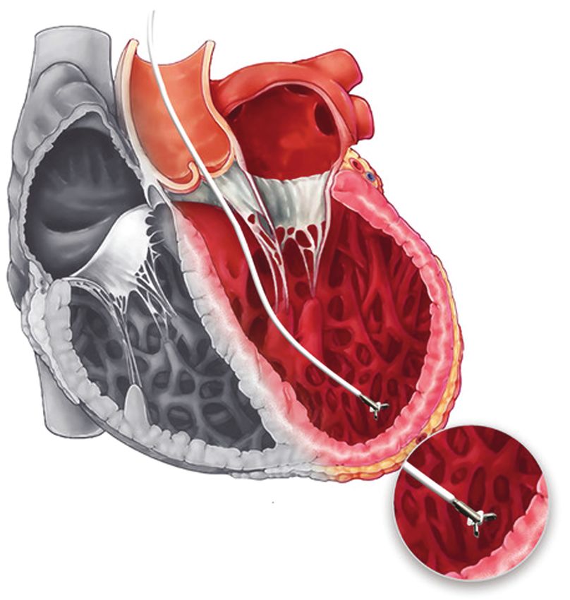
HFA/HFSA/JHFS Position statement on endomyocardial biopsy                                                                                                                                                                    855

Graphical Abstract

The contemporary perspective of endomyocardial biopsy.

..........................................................................................................
Keywords                 Endomyocardial biopsy • Heart failure • Myocarditis •                                                                                           Cardiomyopathy •       Amyloidosis •
                         Sarcoidosis • Cardiotoxicity • Heart transplantation •                                                                                          Cardiac tumours

Introduction                                                                                                                                                Historical milestones
                                                                          ...............................................................................

Endomyocardial biopsy (EMB) is an established invasive procedure                                                                                            Konno and Sakakibara first reported percutaneous EMB proce-
most frequently used for the monitoring of heart transplant (HTx)                                                                                           dure (Figure 1), using a flexible bioptome with sharpened cusps
rejection. EMB also has a complementary role to the clinical assess-                                                                                        that allowed EMB by pinching, as opposed to the surgical cutting
ment in establishing the diagnosis of several cardiac disorders,                                                                                            technique used since 1950.3,4 Subsequently, Sekiguchi described
including myocarditis, cardiomyopathies, drug-induced cardiotox-
                                                                                                                                                            the use of EMB in diagnostic assessment of myocardial diseases
icity, amyloidosis, other infiltrative and storage disorders and car-
                                                                                                                                                            such as glycogen storage disorders, sarcoidosis and myocarditis.5
diac tumours. Improvements in EMB equipment and a significant
                                                                                                                                                            He proposed a systematic histopathological classification, including
progress in the analysis of EMB samples have led to an improvement
                                                                                                                                                            analysis of cardiomyocyte hypertrophy, degeneration, disarrange-
in diagnostic precision of EMB. This document is the result of the
                                                                                                                                                            ment and/or fragmentation of muscle bundles, as well as the extent
Trilateral Cooperation Project between the Heart Failure Asso-
                                                                                                                                                            of interstitial fibrosis, and endocardial thickening.5,6
ciation (HFA) of the European Society of Cardiology (ESC), the
                                                                                                                                                               Caves and Schultz modified the Konno-Sakakibara forceps to
Heart Failure Society of America (HFSA), and the Japanese Heart
Failure Society (JHFS). It was developed during the first Trilateral                                                                                        allow percutaneous biopsies through the right internal jugular vein
Cooperation Workshop held in Munich, in March 2019.                                                                                                         under local anaesthesia with rapid tissue extraction.7 The reusable
   The role of EMB in the management of cardiovascular disorders                                                                                            Stanford Caves-Schultz bioptome and its subsequent modifications
has been previously discussed.1,2 The present document, based on                                                                                            became the standard device for EMB for approximately two
the Trilateral Cooperative Project between ESC-HFA/HFSA/JHFS,                                                                                               decades, predominantly used for monitoring of HTx rejection.8,9
represents an expert consensus aiming to provide a comprehen-                                                                                               Since then, the use of EMB had extended to diverse cardiac dis-
sive, up-to-date perspective on EMB, with a focus on the following                                                                                          eases, including myocarditis, cardiomyopathies, drug-induced car-
main issues: (i) an overview of the practical approach to EMB, (ii)                                                                                         diotoxicity, amyloidosis, other infiltrative and storage disorders and
an update on the indications for EMB, (iii) a revised plan for HTx                                                                                          cardiac tumours.
rejection surveillance, (iv) the impact of multimodality imaging on                                                                                            Simultaneously, the long-sheath technique was developed, which
EMB, and (v) the current clinical practice in the worldwide use                                                                                             improved feasibility and safety of the procedure. In 1974, a flexi-
of EMB. All the relevant points are summarised in the Graphical                                                                                             ble King’s College bioptome was introduced by Richardson.9 This
Abstract.                                                                                                                                                   bioptome, and its subsequent modifications, could be inserted

© 2021 Elsevier Inc. and Journal of Cardiac Failure. [Published by Elsevier Inc.] All rights reserved.
Heart Failure Association of the ESC, Heart Failure Society of America and Japanese Heart Failure Society Position statement on endomyocardial ...
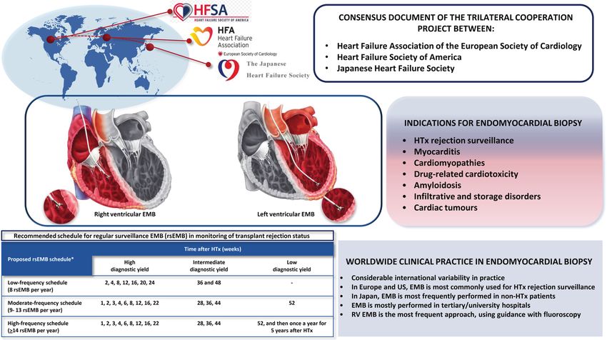
856                                                                                                                                                                                                                      P.M. Seferović et al.

  Figure 1 (A) Original illustration by Konno and Sakakibara of the percutaneous technique of endomyocardial biopsy. (B) Opening and closing
  of the cutting claw at the tip of the catheter (3). LA, left atrium; LV, left ventricle; RA, right atrium; RV, right ventricle. Reproduced with
  permission from Konno and Sakakibara,3 copyright Elsevier.

through the long sheath using either jugular or subclavian veins,                                                                                                      fewer vascular complications, earlier ambulation and lower costs;
                                                                         ...........................................................................................

femoral veins, and right and left femoral arteries. The first study                                                                                                    however, radial thrombosis may occur if the inner vessel diameter
on radial approach using sheetless guiding catheters for left ven-                                                                                                     is small (≤2.5 mm) and peak systolic velocity is low.
tricular (LV) EMB was reported by Bagur and co-workers.10                                                                                                                 Endomyocardial biopsy is most commonly performed as a sin-
The safety of EMB was established both for the right and left                                                                                                          gle procedure in HTx patients, while in non-HTx patients it can
ventricle.11 With the improvement of the technique and tissue pro-                                                                                                     be combined with right heart catheterisation, coronary angiog-
cessing, EMB has gradually gained worldwide acceptance. Besides                                                                                                        raphy, and/or electrophysiological study for the purpose of elec-
the significant progress in the technique, various imaging modal-                                                                                                      troanatomic voltage mapping-guided procedure.14
ities were introduced for EMB guidance, and several new tech-
niques were developed for tissue processing and viral genome
detection (Figure 2).
                                                                                                                                                                       The number of endomyocardial biopsy
Practical approach to                                                                                                                                                  procedures per operator for the
                                                                                                                                                                       maintenance of procedural skill
endomyocardial biopsy
                                                                                                                                                                       The number of EMBs per operator required to maintain the
Selection of the access site                                                                                                                                           procedural skill may vary between institutions and is not accurately
Endomyocardial biopsy is usually performed in a cardiac catheter-                                                                                                      defined. Training and yearly volumes for operators should be
isation laboratory, under fluoroscopic guidance, using jugular,                                                                                                        consistent with the recommendations of the appropriate medical
femoral, or brachial veins, or femoral or radial arteries for vascular                                                                                                 societies. The opinion of the Trilateral Cooperative Project experts
access.12 Patient monitoring (heart rhythm, non-invasive blood                                                                                                         is that a range between 20 and 50 procedures per operator per
pressure and blood oxygen saturation monitoring) is mandatory                                                                                                          year may be reasonable. The report of the American College of
during the procedure. To minimise the risk of bleeding, an                                                                                                             Cardiology Competency Management Committee recommends 50
international normalised ratio should be ≤1.5–1.8 and platelet                                                                                                         EMBs per operator per year.15 In addition to the procedural skill, it
count ≥50 × 109 /L.13                                                                                                                                                  is essential that an experienced cardiac pathologist is available for
   The internal jugular vein is the most common access site for right                                                                                                  the timely analysis and communication of EMB findings.
ventricular (RV) EMB in HTx patients, whereas the right femoral                                                                                                           Details of EMB technique are described in the online supple-
vein is most frequently used in non-HTx patients. Other access                                                                                                         mentary Appendix 1 and a video tutorial on EMB procedure as it is
sites include brachial venous access for RV EMB,12 and right femoral                                                                                                   performed in expert centres in Europe, the US and Japan is available
and radial arteries for LV EMB. Radial access is associated with                                                                                                       online (online supplementary video links).

                                                © 2021 Elsevier Inc. and Journal of Cardiac Failure. [Published by Elsevier Inc.] All rights reserved.
Heart Failure Association of the ESC, Heart Failure Society of America and Japanese Heart Failure Society Position statement on endomyocardial ...
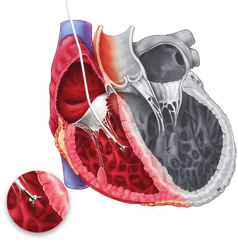
HFA/HFSA/JHFS Position statement on endomyocardial biopsy                                                                                                                     857

  Figure 2 Historical cornerstones in the development of endomyocardial biopsy (EMB): procedural technique (upper panel), imaging guidance
  (middle panel), myocardial tissue processing (lower panel). 3D, three-dimensional; PCR, polymerase chain reaction; RF, radiofrequency; TEE,
  transoesophageal echocardiography; TTE, transthoracic echocardiography.

Selection of endomyocardial biopsy site,                                                                      disorders) indicated that biventricular EMB can increase diagnos-
                                                                          .................................

                                                                                                              tic accuracy compared with selective LV or RV EMB.11 Sampling
sampling error and biopsy of non-cardiac
                                                                                                              error is the major limitation of the diagnostic utility of EMB. It is
tissues                                                                                                       suggested that at least five samples should be taken from differ-
The most common site of EMB is RV EMB (Figure 3), but                                                         ent sites in the right and left ventricle in order to reduce the risk
occasionally LV (Figure 4) or biventricular EMB may be needed.                                                of sampling error in the setting of diseases with focal pattern or
                                                                                                              intracardiac tumours.1,17
The decision on EMB site should be based on the clinical indi-
                                                                                                                 In patients with infiltrative and storage disorders affecting
cation, findings of preprocedural imaging, and on the operator                                                multiple organs, biopsies taken from the most affected organ
expertise.16 A study of 755 patients with suspected myocarditis and                                           are most likely to provide the diagnosis, but occasionally their
non-ischaemic cardiomyopathy (including infiltrative and storage                                              utility may be hampered by low sensitivity. In patients with

© 2021 Elsevier Inc. and Journal of Cardiac Failure. [Published by Elsevier Inc.] All rights reserved.
858                                                                                                                                                                                                                                                                                                   P.M. Seferović et al.

                                                                        ........................................................................................................................................................................
  Figure 3 An artistic presentation of right ventricular endomy-
  ocardial biopsy. Endomyocardial biopsy samples are typically taken                                                                                                                                                                                 Figure 4 An artistic presentation of left ventricular endomy-
  from the interventricular septum.                                                                                                                                                                                                                  ocardial biopsy. Endomyocardial biopsy samples are typically taken
                                                                                                                                                                                                                                                     from the ventricular apex.

amyloidosis, abdominal fat pad biopsies have a sensitivity of 75%                                                                                                                                                                                  Since T2 mapping has a greater sensitivity for detecting inflamma-
for immunoglobulin light-chain (AL) amyloidosis, but the sensitivity                                                                                                                                                                               tion, this technique may be further explored for directing EMB
is significantly lower in both hereditary and wild-type transthyretin                                                                                                                                                                              to the most affected regions of the heart in myocarditis and
amyloidosis (ATTR) (∼45% and ∼15%, respectively) and thus, a                                                                                                                                                                                       other inflammatory disorders.24 However, small cohort studies
negative result does not rule out cardiac involvement.18                                                                                                                                                                                           of patients with cardiomyopathies indicate that the concordance
                                                                                                                                                                                                                                                   between CMR and EMB findings is only partial and that these pro-
                                                                                                                                                                                                                                                   cedures have a complementary role in diagnostic assessment.25,26
Imaging guidance                                                                                                                                                                                                                                      Electroanatomic voltage mapping has been used for the guid-
In most centres, EMB is performed using fluoroscopic guid-                                                                                                                                                                                         ance of EMB in diseases with focal pattern associated with ven-
ance; however, novel guidance modalities have emerged aim-                                                                                                                                                                                         tricular arrhythmias (myocarditis, sarcoidosis and arrhythmogenic
ing to improve the feasibility and enable targeted EMB. The                                                                                                                                                                                        right ventricular cardiomyopathy, ARVC).27,28 Areas of low-voltage
role of imaging in EMB guidance is twofold. Firstly, preprocedu-                                                                                                                                                                                   or abnormal electrogram on electroanatomic voltage mapping
ral imaging with echocardiography, cardiac magnetic resonance                                                                                                                                                                                      have a high sensitivity and specificity to identify the pathological
(CMR) imaging, computed tomography and/or positron emission                                                                                                                                                                                        substrate.27 EMB procedure may be further facilitated by using
tomography (PET) can be used to direct EMB to the specific                                                                                                                                                                                         bioptomes with an integrated electrode at the tip, as well as with
sites of myocardial disease. Secondly, procedural imaging (e.g.                                                                                                                                                                                    the use of three-dimensional electroanatomic voltage mapping sys-
real-time three-dimensional echocardiography) can be performed                                                                                                                                                                                     tems and intracardiac echocardiography.29,30
simultaneously with fluoroscopy to improve the accuracy of the
EMB procedure.19 Intracardiac echocardiography has also been
successfully employed to guide EMB of cardiac tumours.20                                                                                                                                                                                           Complications
   Preprocedural diagnostics with CMR has been demonstrated                                                                                                                                                                                        Endomyocardial biopsy is associated with a low rate of major com-
to improve diagnostic performance of EMB in several cardiac dis-                                                                                                                                                                                   plications (∼1%),11,16 which can be classified as major and minor
orders. CMR-directed EMB can improve procedural accuracy in                                                                                                                                                                                        (Table 1). Patient characteristics, EMB site, procedural volume and
diseases with focal pattern (e.g. sarcoidosis),21 and in the set-                                                                                                                                                                                  operator expertise are the most important determinants of EMB
ting of soft tissue masses, which may be difficult to visualize                                                                                                                                                                                    risk (details in online supplementary Table S1). The risk of major
by fluoroscopy.22 Likewise, a small study suggested that direct-                                                                                                                                                                                   complications is lower in HTx recipients compared with non-HTx
ing EMB to the regions of late gadolinium enhancement on CMR                                                                                                                                                                                       patients (0.19% vs. 0.70%).31 Haemodynamically unstable patients
can increase diagnostic utility in myocarditis.23 However, a larger                                                                                                                                                                                with acute or advanced heart failure (HF) and those with dilated
study failed to confirm this finding, perhaps because late gadolinium                                                                                                                                                                              ventricles may be at a higher risk of cardiac perforation, tamponade
enhancement is a non-specific sign, which may correspond to both                                                                                                                                                                                   and malignant arrhythmias.32 Cardiac perforation and tamponade
acute necrosis/inflammation, as well as fibrosis in myocarditis.11                                                                                                                                                                                 are more frequently observed with RV than with LV EMB16 but

                                                © 2021 Elsevier Inc. and Journal of Cardiac Failure. [Published by Elsevier Inc.] All rights reserved.
HFA/HFSA/JHFS Position statement on endomyocardial biopsy                                                                                                                                                                                                                                                           859

LV EMB is more frequently complicated by stroke or systemic                                                                                                                                                                                          disorders, when EMB findings may provide information pertinent

                                                                          ........................................................................................................................................................................
embolism. High-volume centres have a lower complication rate                                                                                                                                                                                         to further management.17
compared with low-volume centres, and high procedural volume                                                                                                                                                                                            Details on EMB sample processing and analyses are presented
has been identified as an independent predictor of a lower risk of                                                                                                                                                                                   in Table 2 and considered in the online supplementary Appendix
major complications.33                                                                                                                                                                                                                               S2. In addition, typical histopathological findings of the normal
    There is a risk of tricuspid valve damage during EMB, both at                                                                                                                                                                                    myocardium, lymphocytic myocarditis, HTx rejection and cardiac
the valvular and sub-valvular level.34 The risk of complication can                                                                                                                                                                                  amyloidosis are presented in Figure 5.
be minimised by using a correctly located long sheath across the
tricuspid valve with the tip in the right ventricle, to avoid repeated
exposure of the valve leaflets to the bioptome. Infection/sepsis is
a very rare risk of EMB if the procedure follows recommendations
                                                                                                                                                                                                                                                     Indications for endomyocardial
for the aseptic technique.                                                                                                                                                                                                                           biopsy
    The risk of periprocedural mortality is low (0–0.07%),16,35                                                                                                                                                                                      Endomyocardial biopsy can provide important histological,
and most frequently caused by stroke, malignant arrhythmias,                                                                                                                                                                                         immunohistochemical, and molecular information about the
high-degree atrioventricular block, and cardiac tamponade.36 The                                                                                                                                                                                     heart. Since EMB is an invasive procedure with limited availability,
risk of stroke and systemic embolism can be decreased by iden-                                                                                                                                                                                       risk and benefits of the procedure should be taken into account.
tification of a thrombus (an absolute contraindication for EMB)                                                                                                                                                                                      In establishing an indication for EMB, it is important to identify
and administration of low-dose heparin during the procedure in
                                                                                                                                                                                                                                                     clinical situations in which EMB can complement the diagnostic
patients with high thromboembolic risk.1
                                                                                                                                                                                                                                                     process in order to confirm clinically suspected diagnosis and
    Management of cardiac perforation during EMB includes imme-
                                                                                                                                                                                                                                                     provide information relevant for the management. Diagnostic
diate pericardiocentesis and autotransfusion from the pericardium
                                                                                                                                                                                                                                                     value of EMB also depends on the myocardial disease (i.e. lower
to a large central vein (femoral or jugular) until the bleeding
                                                                                                                                                                                                                                                     sensitivity in diseases with focal involvement), and on the centre’s
has stopped.37 If cardiac perforation has occurred, these patients
                                                                                                                                                                                                                                                     proficiency in sample processing and analysis. The most frequent
require close monitoring and consultation with a cardiac surgi-
                                                                                                                                                                                                                                                     indications for EMB are summarised in Table 3.
cal service. Urgent surgical repair of the perforation site may be
required in patients with ongoing bleeding or instability related to
the perforation.
                                                                                                                                                                                                                                                     Clinically suspected myocarditis
                                                                                                                                                                                                                                                     Endomyocardial biopsy is indicated in patients with fulminant/acute
Evaluation of endomyocardial biopsy                                                                                                                                                                                                                  myocarditis presenting with cardiogenic shock or acute HF and
samples                                                                                                                                                                                                                                              LV dysfunction, with or without malignant ventricular arrhythmias
The choice of the technique for the analysis of EMB speci-                                                                                                                                                                                           and/or conduction abnormalities. It may also be considered in
mens depends on the clinical presentation and suspected under-                                                                                                                                                                                       haemodynamically stable patients with clinical symptoms and diag-
lying cardiac disorder. First, the pathologist performing the anal-                                                                                                                                                                                  nostic criteria (electrocardiographic abnormalities, elevated tro-
ysis should be well trained in specimen processing and profi-                                                                                                                                                                                        ponin levels, imaging findings) suggestive of myocarditis, in the
cient in analysis techniques. Standardized diagnostic criteria for                                                                                                                                                                                   absence of significant coronary artery disease.17
histopathological analyses (e.g. Dallas criteria for myocarditis)                                                                                                                                                                                       A retrospective registry-based analysis of 220 patients (mean age
should be used to minimise EMB reporting variability. Second,                                                                                                                                                                                        42 years) from the US, Europe and Japan with acute myocarditis and
the use of vital stains is indicated to demonstrate myocyte                                                                                                                                                                                          LV dysfunction has shown that patients with fulminant myocardi-
hypertrophy, patterns of myocyte disarray or vacuolization. Infil-                                                                                                                                                                                   tis have significantly worse short-term (60-day mortality/HTx rate:
trative disorders such as amyloidosis can be characterized by                                                                                                                                                                                        27.8% vs. 1.8%) and long-term prognosis (7-year mortality/HTx
Congo red stain, immunohistochemistry, immunogold electron                                                                                                                                                                                           rate: 43.0% vs. 9.0%) compared with non-fulminant course and
microscopy and mass spectroscopy. Immunostaining can be used                                                                                                                                                                                         that EMB-proven diagnosis of giant cell myocarditis carries the
to quantify resident and infiltrating macrophages, myofibroblasts,                                                                                                                                                                                   worst prognosis.39 A recent analysis of 443 individuals with sus-
and lymphocytes. Quantitative polymerase chain reaction (PCR),                                                                                                                                                                                       pected myocarditis has shown that among high-risk patients with
reverse transcription (RT)-PCR and direct sequencing should be                                                                                                                                                                                       LV dysfunction, sustained ventricular arrhythmias and/or haemo-
used to identify infectious agents.38 Simultaneously, blood sam-                                                                                                                                                                                     dynamic instability (n = 118, EMB performed in 56 patients)
ples should be assessed with PCR to identify systemic infection,                                                                                                                                                                                     EMB-established diagnosis (89.3%) offered information relevant for
and to exclude potential contamination of heart tissue by persis-                                                                                                                                                                                    the management and prognosis (e.g. institution of immunosuppres-
tently/latently infected blood cells.17 Electron microscopy is useful                                                                                                                                                                                sive therapy in giant cell myocarditis, sarcoidosis or eosinophilic
to detect and quantify changes in cardiomyopathies and storage                                                                                                                                                                                       myocarditis).40 In addition, EMB can provide differential diagnosis
disease.                                                                                                                                                                                                                                             in patients with severe clinical course, when non-invasive assess-
   The most frequent indication for a repeat EMB procedure is                                                                                                                                                                                        ment is inconclusive or unfeasible.40 Accordingly, in unexplained
the follow-up of graft rejection status after HTx. Rarely, a repeat                                                                                                                                                                                  acute HF with haemodynamic compromise, a cohort study of
EMB may be considered if sampling error is suspected in a patient                                                                                                                                                                                    851 patients demonstrated that EMB provided a diagnosis in 39%,
with unexplained deterioration of HF and/or malignant rhythm                                                                                                                                                                                         and that the most common finding was acute myocarditis.41 In

© 2021 Elsevier Inc. and Journal of Cardiac Failure. [Published by Elsevier Inc.] All rights reserved.
860                                                                                                                                                                                                                                                                        P.M. Seferović et al.

  Table 1 Major and minor complications of endomyocardial biopsy

  Major complications                                                                      Minor complications
  ...........................................................................................................................................
  Death (0–0.07%)                                                                                                                                                                                                                       Chest pain (transient) (0–1.8%)
  Cardiac perforation/haemopericardium/tamponade (0–6.9%)                                                                                                                                                                               Deep vein thrombosis (0.23–3.8%)
  Pneumothorax/air embolism (0–0.8%)                                                                                                                                                                                                    Puncture site haematoma/nerve palsy (0–0.64%)
  Thromboembolism (0–0.32%)                                                                                                                                                                                                             Hypotension/vaso-vagal syncope (0–4.3%)
  Valvular trauma (0.02–1.1%)                                                                                                                                                                                                           Arterial trauma/vascular damage/fistulae (0.32–2.8%)
  Severe arrhythmias/atrioventricular block (0–11%)

  Detailed description of complications according to the centre volume, access site, type of endomyocardial biopsy procedure and patient characteristics as well as references
  are provided in online supplementary Table S1.

this study, EMB-based diagnosis resulted in a change of ther-                                                                                                                                                           Dilated cardiomyopathy

                                                                                      ...............................................................................................................................
apy in almost a third of patients, and most clinical decisions
                                                                                                                                                                                                                        In patients with dilated cardiomyopathy (DCM), EMB may be indi-
concerned the institution or withholding of immunosuppressive
                                                                                                                                                                                                                        cated in the setting of decompensated HF with moderate-to-severe
medications.41
                                                                                                                                                                                                                        LV dysfunction, refractory to standard HF treatment, with a recent
   The common histological types of myocarditis include lym-
                                                                                                                                                                                                                        onset of the clinical syndrome, exclusion of other specific aeti-
phocytic, eosinophilic, giant cell and granulomatous myocarditis
                                                                                                                                                                                                                        ologies, absence of severe LV remodelling and negative familial
(cardiac sarcoidosis). The most prevalent is lymphocytic myocardi-
                                                                                                                                                                                                                        history and/or genetic testing for cardiomyopathy. In this set-
tis caused by viral infection, autoimmunity or drug-toxicity, which
                                                                                                                                                                                                                        ting, EMB can be used to confirm inflammatory cardiomyopathy
is frequently associated with HF of various severity. Eosinophilic
                                                                                                                                                                                                                        with a higher sensitivity compared with CMR.49 EMB may also
myocarditis is characterised by eosinophilic infiltrate in the heart
                                                                                                                                                                                                                        have a role in the assessment of Borrelia burgdorferi involvement
and is often accompanied by peripheral blood eosinophilia. Giant
                                                                                                                                                                                                                        in unexplained DCM in endemic regions for Lyme disease.50 A
cell myocarditis is rare (∼1% of acute myocarditis cases) but it may
                                                                                                                                                                                                                        study of 110 individuals with recent-onset DCM has demonstrated
take the fulminant course and carries a poor prognosis.39 EMB has
                                                                                                                                                                                                                        that Borrelia burgdorferi genome was present in 20% of EMB
a high sensitivity (80%) and positive predictive value (71%) for giant
                                                                                                                                                                                                                        samples.51
cell myocarditis, especially if performed within 2–4 weeks of symp-
tom onset.42 Non-caseating granulomatous myocarditis is the usual
histopathological finding in patients with cardiac sarcoidosis.43 EMB
may be indicated in suspected cardiac sarcoidosis (electrocardio-                                                                                                                                                       Cardiotoxicity of cancer therapy
graphic abnormalities, unexplained syncope, or palpitations), if                                                                                                                                                        Immune checkpoint inhibitors (ICI) represent a novel, highly
imaging studies (echocardiography, CMR, 18 fluorodeoxyglucose                                                                                                                                                           effective class of anti-neoplastic drugs but their use can result
PET) and lymph node or lung biopsy render inconclusive                                                                                                                                                                  in cardiac toxicity in up to 5% of cases, including myocardi-
results, as well as in cases of isolated cardiac involvement.44                                                                                                                                                         tis, non-inflammatory LV dysfunction, myocardial infarction and
The major drawback is a low sensitivity of EMB due to the                                                                                                                                                               arrhythmias.52 ICI-mediated myocarditis and pericarditis occur
focal nature of myocardial involvement, revealing non-caseating                                                                                                                                                         early (>75% cases in first four cycles), more frequently in patients
granulomatous infiltrates in ∼25% of patients.44 Small case                                                                                                                                                             on combined ICIs and can be severe or fatal in up to 50%.53,54
series have suggested that sensitivity can be improved with an                                                                                                                                                          EMB is indicated in suspected ICI-mediated cardiotoxicity, if CMR
electrogram-guided approach targeting areas with low amplitude                                                                                                                                                          or 18 F-fluorodeoxyglucose PET-computed tomography yield uncer-
and/or abnormal electrogram appearance,45 or with preprocedural                                                                                                                                                         tain findings and/or the patients cannot undergo non-invasive
CMR-guided EMB.21                                                                                                                                                                                                       assessment due to haemodynamically instability.55 In patients with
   Endomyocardial biopsy is rarely indicated in individuals with                                                                                                                                                        confirmed ICI-mediated myocarditis, ICI treatment should be dis-
suspected COVID-19 myocarditis. EMB and autopsy findings                                                                                                                                                                continued and high-dose immunosuppression should be instituted,
support the presence of SARS-CoV-2 in the myocardium,46,47                                                                                                                                                              in addition to standard HF care.52 If active inflammation has been
and histopathological studies suggest that increased interstitial                                                                                                                                                       ruled out by EMB, then ICI treatment re-challenge may be consid-
macrophage infiltration and lymphocytic myocarditis are the most                                                                                                                                                        ered once LV function has stabilized or recovered with standard
common findings.46,48                                                                                                                                                                                                   HF drugs.52
   The diagnostic value of EMB in clinically suspected myocarditis                                                                                                                                                         Endomyocardial biopsy has been used to document and
increases if the procedure is performed 2–4 weeks after symptom                                                                                                                                                         assess the degree of anthracycline-related cardiotoxicity.56
onset1,17 and the sample is analysed with the use of immunohisto-                                                                                                                                                       However, EMB is not routinely recommended in patients with
chemistry. A recent meta-analysis (61 publications with a total of                                                                                                                                                      anthracycline-related cardiotoxicity and HF when there is a clear
10 491 patients) indicated that the use of immunohistochemistry                                                                                                                                                         causal relationship. EMB may be considered in rare cases when
can increase the detection rate of inflammation in EMB specimens                                                                                                                                                        there is clinical uncertainty as to the cause of HF (e.g. suspected
to ∼51%.49                                                                                                                                                                                                              myocarditis). The role of EMB in cyclophosphamide-induced

                                                          © 2021 Elsevier Inc. and Journal of Cardiac Failure. [Published by Elsevier Inc.] All rights reserved.
HFA/HFSA/JHFS Position statement on endomyocardial biopsy                                                                                                         861

  Table 2 Sample processing, analysis and characteristic findings according to clinical presentation

  Disease           EMB processing/staining                                    Possible findings
  ...........................................................................................................................................
  Myocarditis,         Histopathology Haematoxylin and eosin, Mason or                      Dallas criteria for myocarditis: inflammatory infiltrates
    DCM                  Mallory trichrome, Elastic van Gieson, PAS, Heidenhein’s             associated with myocyte degeneration and necrosis of
                         AZAN, and Methylene blue stain (Trypanosoma cruzii)                  non-ischaemic origin (active or borderline).
                                                                                            Lymphocytic myocarditis: patchy or diffuse inflammatory
                                                                                              infiltrate mostly of lymphocytes and macrophages [viral
                                                                                              infections, immune-mediated myocarditis (systemic lupus
                                                                                              erythematosus, polymyositis/dermatomyositis, rheumatoid
                                                                                              arthritis, organ-specific autoimmune disorders, etc.)].
                                                                                            Giant cell myocarditis: myocyte necrosis and diffuse or
                                                                                              multifocal inflammatory infiltrates, with T lymphocytes,
                                                                                              macrophage-derived multinucleated giant cells and eosinophilic
                                                                                              granulocytes.
                                                                                            Granulomatous myocarditis: non-necrotizing granulomas with
                                                                                              macrophages and multinucleated giant cells, surrounded by
                                                                                              fibrosis and a lymphocytic infiltrate (sarcoidosis).
                                                                                            Eosinophilic myocarditis: interstitial inflammatory infiltrate
                                                                                              dominated by eosinophils, often without myocyte damage,
                                                                                              frequently accompanied by peripheral eosinophilia
                                                                                              (hypersensitivity, parasitic infection, Churg–Strauss syndrome,
                                                                                              endomyocardial fibrosis).
                       Quantitative real-time PCR for enteroviruses,                        Infection confirmed or not by (RT-) PCR
                         adenoviruses, herpesviruses (cytomegalovirus, herpes
                         simplex, Epstein–Barr, human herpesvirus 6), parvovirus
                         B19, influenza A and B, and SARS-CoV-2 virus + Borrelia
                       Immunohistochemistry                                                 Myocarditis confirmed by immunohistochemistry: ≥14
                       CD3 (T cells), CD68 (macrophages), MHC II, alpha                       leucocytes/mm2 including up to 4 monocytes/mm2 with the
                         SM-myofibroblasts                                                    presence of CD3+ T-lymphocytes ≥7 cells/mm2
  DCM, ARVC            Histology and PCR as above, additional                               DCM: non-specific histopathology including hypertrophy and
                         immunohistochemical stains for lamin A/C, dystrophin,                vacuolar changes of myocytes, interstitial fibrosis, foci of
                         and plakoglobin (ARVC)                                               micro-scarring.
                                                                                            ARVC: progressive myocyte atrophy/loss with fibrous or
                                                                                              fibro-fatty myocardial replacement.
  Storage              PAS, Congo Red, sulfate alcian blue, or S/T thioflavin, Sudan        PAS+ sarcoplasmic vacuoles and lysosomal glycogen accumulation
    diseases             black or Oil Red O (lipid deposits), Prussian Blue (iron),           (Pompe disease); PAS+ and LAMP2 absence, autophagic
                         TEM (Anderson–Fabry, Danon)                                          granules in TEM (Danon disease), PAS+ and lamellar bodies
                                                                                              (Anderson–Fabry), Congo Red+ and interstitial deposits
                                                                                              (amyloidosis); brownish perinuclear granules in myocytes
                                                                                              highlighted in blue by Prussian Blue stain (iron storage disease).
  Tumours              Standard histopathology + immunohistochemistry for                   Differential diagnosis between benign and malignant tumours, and
                         specific tumours                                                     in malignant tumour subtyping.
  Heart trans-         Haematoxylin and eosin, Giemsa, Movat, Masson                        Cellular rejection: Grade 0R (no rejection); Grade 1R (mild)
    plantation           trichrome, Weigert-Van Gieson, Ziehl Nielsen, PAS,                   interstitial and/or perivascular infiltrate with up to 1 focus of
                         Gram, Gomori, CD31, CD34, CD45, CD68, C4d                            myocyte damage; Grade 2R (moderate), ≥2 foci of infiltrate
                                                                                              with associated myocyte damage; Grade 3R (severe) diffuse
                                                                                              infiltrate with multifocal myocyte damage, oedema,
                                                                                              haemorrhage, or vasculitis.
                                                                                            Humoral rejection: capillary injury, endothelial cell swelling and
                                                                                              aggregation of intravascular macrophages (positive staining for
                                                                                              C4d or C3d fragments of complement by endothelial cells).

  ARVC, arrhythmogenic right ventricular cardiomyopathy; CD, cluster of differentiation; DCM, dilated cardiomyopathy, EMB, endomyocardial biopsy; LAMP2,
  lysosome-associated membrane protein 2; MHC II, major histocompatibility complex type II; PAS, periodic acid Schiff; PCR, polymerase chain reaction; RT-PCR, reverse
  transcriptase polymerase chain reaction; TEM, transmission electron microscopy.

© 2021 Elsevier Inc. and Journal of Cardiac Failure. [Published by Elsevier Inc.] All rights reserved.
862                                                                                                                                                         P.M. Seferović et al.

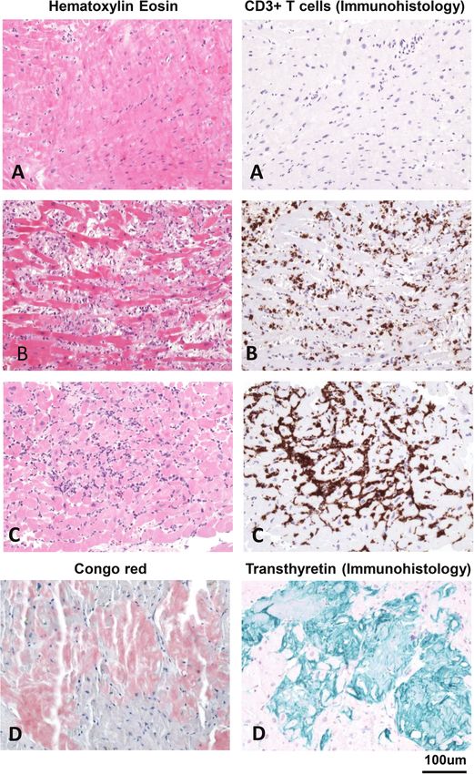
  Figure 5 Typical histopathological findings of the normal myocardium (A), lymphocytic myocarditis (B), cellular heart transplant rejection
  (C) and cardiac amyloidosis (D). (A) Normal myocardium: no myocyte necrosis, inflammation or fibrosis. (B) Acute lymphocytic myocarditis:
  many necrotic myocytes (light pink) and numerous CD3+ T cells and other immune cells (e.g. CD68+ macrophages). (C) Acute cellular heart
  transplant rejection: significant amounts of inflammatory cells including CD3+ T cells. (D) Cardiac amyloidosis: Congo red staining and subtyping
  by immunohistochemistry defines cardiac amyloidosis (presented in the figure: transthyretin amyloidosis).

cardiotoxicity, and other cancer therapy-induced HF is less                                              to treatment, without obvious cardiac disease or with mini-
                                                                        ..............................

well-established57 and EMB is not indicated.                                                             mal structural changes in order to identify potentially treatable
                                                                                                         aetiologies, such as myocarditis, ARVC, or sarcoidosis.44,58,59 Ven-
                                                                                                         tricular arrhythmias may be the only symptom of myocarditis and
Unexplained ventricular arrhythmias,                                                                     sarcoidosis,44,60 as well as the first presentation of ARVC in patients
conduction disorders and syncope                                                                         with subtle structural abnormalities, that may challenge diagnostic
Endomyocardial biopsy may be indicated in patients with                                                  evaluation. Given the focal nature of cardiac sarcoidosis and
unexplained ventricular arrhythmias/syncope (ventricular fib-                                            ARVC, undirected EMB can be false negative and electroanatomic
rillation or tachycardia, frequent multifocal ventricular premature                                      voltage mapping guidance may be considered to increase
complexes/non-sustained ventricular tachycardia), refractory                                             diagnostic yield.45,61

                                                © 2021 Elsevier Inc. and Journal of Cardiac Failure. [Published by Elsevier Inc.] All rights reserved.
HFA/HFSA/JHFS Position statement on endomyocardial biopsy                                                                                                                                                                              863

  Table 3 Indications for endomyocardial biopsy

  Clinical presentation                                                        Endomyocardial biopsy finding
  ...........................................................................................................................................
                                                                                                                                                                   Myocarditis type:
    • Suspected fulminant myocarditis or acute myocarditis with acute HF, LV
      dysfunction and/or rhythm disorders.                                                                                                                             • Lymphocytic myocarditis
    • Suspected myocarditis in haemodynamically stable patients.                                                                                                       • Eosinophilic myocarditis
                                                                                                                                                                       • Giant cell myocarditis
                                                                                                                                                                       • Granulomatous myocarditis
  Dilated cardiomyopathy with recent onset HF, moderate-to-severe LV                                                                                               Myocyte abnormalities, focal or diffuse fibrosis and inflammatory
    dysfunction, refractory to standard treatment (following exclusion of specific                                                                                   infiltrates (inflammatory cardiomyopathy).
    aetiologies).
  Suspected ICI-mediated cardiotoxicity: acute HF with/without haemodynamic             ICI-mediated myocarditis
    instability early after drug initiation (∼ first 4 cycles)
  High-degree atrioventricular block, syncope and/or unexplained ventricular
                                                                                          • Myocarditis
    arrhythmias (ventricular fibrillation, ventricular tachycardia, frequent multifocal
                                                                                          • Arrhythmogenic right ventricular cardiomyopathy
    premature ventricular complexes), refractory to treatment, without obvious
                                                                                          • Cardiac sarcoidosis
    cardiac disease or with minimal structural abnormalities.
  Autoimmune disorders with progressive HF unresponsive to treatment
                                                                                          • Autoimmune myocarditis
    with/without sustained ventricular arrhythmias and/or conduction
                                                                                          • Viral myocarditis
    abnormalities.
                                                                                          • Vasculitis/vasculopathy
  MINOCA/takotsubo syndrome with progressive LV dysfunction and HF                      Differential diagnosis of myocarditis
    with/without ventricular arrhythmias or conduction abnormalities.
  Unexplained restrictive or hypertrophic cardiomyopathy.
                                                                                          • Amyloidosis
                                                                                          • Infiltrative/storage disorders (Anderson–Fabry disease,
                                                                                            glycogen storage diseases, sarcoidosis, haemochromatosis)
  Cardiac tumours.                                                                      Histopathological diagnosis
                                                                                        HTx rejection status
   • Routine surveillance EMB
   • Symptom-triggered EMB

  EMB, endomyocardial biopsy; HF, heart failure; HTx, heart transplant; ICI, immune checkpoint inhibitor; LV, left ventricular; MINOCA, myocardial infarction without obstructive
  coronary artery disease.

   Endomyocardial biopsy may be useful in patients with                                                                                                         Myocardial infarction without
                                                                                       ......................................................................

new-onset bradycardia and conduction abnormalities, when
                                                                                                                                                                obstructive coronary artery disease
clinical presentation is suggestive of a treatable aetiology (e.g.
myocarditis, amyloidosis, sarcoidosis).62,63 Electroanatomic voltage
                                                                                                                                                                and takotsubo syndrome
mapping guidance may be useful, as suggested by a cohort of                                                                                                     Endomyocardial biopsy is rarely indicated in myocardial infarction
patients with unexplained atrioventricular block, where a com-                                                                                                  without obstructive coronary artery disease (MINOCA) and
prehensive evaluation, including electroanatomic voltage mapping                                                                                                in takotsubo syndrome. In may be considered for the purpose
guided-EMB, demonstrated cardiac sarcoidosis in 34%.64                                                                                                          of differential diagnosis of myocarditis in the setting of pro-
                                                                                                                                                                gressive LV dysfunction and HF despite standard therapy, with
                                                                                                                                                                or without life-threatening ventricular arrhythmias/conduction
Autoimmune disorders                                                                                                                                            abnormalities.68
Endomyocardial biopsy is rarely indicated in autoimmune disorders
(systemic lupus erythematosus, rheumatoid arthritis, systemic scle-
rosis, polymyositis/dermatomyositis, etc.), but it may be considered
                                                                                                                                                                Restrictive and hypertrophic
in patients with progressive HF unresponsive to usual treatment,                                                                                                cardiomyopathy
as well as in patients with sustained ventricular arrhythmias and/or                                                                                            Endomyocardial biopsy may be considered in patients with
conduction abnormalities, when there is a high clinical suspicion of                                                                                            restrictive and hypertrophic cardiomyopathy if the aetiology
myocarditis or vasculitis. In a small study of patients with systemic                                                                                           of cardiomyopathy remains inconclusive following non-invasive
sclerosis and HF, greater extent of EMB-detected inflammation                                                                                                   assessment, and there is clinical suspicion of infiltrative or storage
and fibrosis correlated with serious adverse events.65 Likewise,                                                                                                disorder (amyloidosis, sarcoidosis, Anderson–Fabry disease, and
EMB in patients with systemic lupus erythematosus can provide                                                                                                   glycogen storage diseases) with available treatment options.69–71
confirmation of lupus myocarditis, hydroxychloroquine-induced                                                                                                   In patients with cardiac amyloidosis, differentiating between AL
cardiotoxicity and/or coronary vasculitis/vasculopathy.66,67                                                                                                    amyloidosis and wild-type or hereditary ATTR amyloidosis has

© 2021 Elsevier Inc. and Journal of Cardiac Failure. [Published by Elsevier Inc.] All rights reserved.
864                                                                                                                                                                                                                                                                                                   P.M. Seferović et al.

important therapeutic implications.72 EMB is highly sensitive                                                                                                                                                                                       data in the International Society for Heart and Lung Transplanta-

                                                                         ........................................................................................................................................................................
and specific for cardiac amyloidosis,73 and may be considered                                                                                                                                                                                       tion Registry.81 In this study, rsEMB was performed monthly for the
if non-invasive assessment provides inconclusive or discordant                                                                                                                                                                                      first 6 months (with the first rsEMB being scheduled 1 month after
results (e.g. abnormal serum free light-chain assay and a positive                                                                                                                                                                                  HTx), and subsequently at months 9 and 12. Despite this relatively
99m
    Tc 3,3-diphosphono-1,2-propanodicarboxylic acid scintigra-                                                                                                                                                                                      low frequency of rsEMB procedures, only six unscheduled stEMB
phy), or in patients with plasma cell dyscrasia and ambiguous                                                                                                                                                                                       procedures were required, resulting in a change of treatment in
imaging results.72,74 Congo red staining and immunohistochemistry                                                                                                                                                                                   only two patients.
are the standard techniques used to characterize the type of
amyloid fibrils in EMB specimens, but newer technologies, such
                                                                                                                                                                                                                                                    Revised schedule for heart transplant
as immunoelectron microscopy and laser dissection mass spec-
trometry appear superior to immunohistochemistry in identifying                                                                                                                                                                                     rejection surveillance
amyloid protein type.75,76 In individuals with LV hypertrophy                                                                                                                                                                                       Currently, most HTx protocols suggest performing rsEMB every
and suspected Anderson–Fabry disease, who do not meet                                                                                                                                                                                               week during the first month, every second week for the next
all diagnostic criteria, EMB can be performed to confirm the                                                                                                                                                                                        several months, and then once monthly for the first 12 months.
diagnosis.71 Rarely, EMB may be indicated in the presence of                                                                                                                                                                                        Thereafter, rsEMB are often continued at variable frequency
iron overload and unequivocal imaging results to confirm cardiac                                                                                                                                                                                    for years, despite a low risk of late rejection and with a low
haemochromatosis.77                                                                                                                                                                                                                                 cost-effectiveness.82 Recently, non-invasive surveillance of HTx
                                                                                                                                                                                                                                                    rejection with the combined use of novel techniques, such as gene
                                                                                                                                                                                                                                                    expression profiling and donor-derived cell-free DNA has shown
Tumours of the heart                                                                                                                                                                                                                                high negative predictive validity for acute graft rejection, which may
                                                                                                                                                                                                                                                    decrease the need for rsEMB.83 In the future, multicentre prospec-
In patients with cardiac tumours, multimodality imaging plays
                                                                                                                                                                                                                                                    tive clinical trials should be planned to test the optimal approach
the pivotal role in the identification and characterisation of car-
                                                                                                                                                                                                                                                    to rsEMB after HTx. Based on the available data on diagnostic yield
diac masses. EMB may be indicated in patients with primary or
                                                                                                                                                                                                                                                    of EMB according to the time after HTx, the following schedule for
metastatic cardiac tumours when non-invasive assessment and/or
                                                                                                                                                                                                                                                    rsEMB is suggested (Figure 6).
biopsy of non-cardiac tissues have been inconclusive, and his-
tological diagnosis is relevant for the prognosis and treatment.1
EMB is not indicated for intracardiac masses with high embolic                                                                                                                                                                                      Contraindications
potential, such as left-sided tumours or typical cardiac myxomas.
EMB guidance with transthoracic, transoesophageal and intracar-                                                                                                                                                                                     In most instances, contraindications for EMB are consistent with
diac echocardiography can improve the efficacy and safety of the                                                                                                                                                                                    contraindications for cardiac catheterisation (Table 4). Additional
procedure.20,78                                                                                                                                                                                                                                     caution is required in patients with recent pacemaker implantation
                                                                                                                                                                                                                                                    (increased risk of lead dislodgement for RV EMB), marked ventric-
                                                                                                                                                                                                                                                    ular wall thinning and hypercontractility (high risk of ventricular
                                                                                                                                                                                                                                                    perforation).84
Monitoring of heart transplant
rejection status
                                                                                                                                                                                                                                                    Multimodality imaging
Despite advances in cardiac imaging and availability of novel
biomarkers, EMB remains the ‘gold-standard’ for the detection of                                                                                                                                                                                    and endomyocardial biopsy
HTx rejection. EMB after HTx can be scheduled according to a                                                                                                                                                                                        Multimodality imaging including standard two-dimensional,
protocol for routine surveillance EMB (rsEMB) in asymptomatic                                                                                                                                                                                       three-dimensional, speckle-tracking and intracardiac echocar-
patients, and it is also performed in patients with worsening clinical                                                                                                                                                                              diography, CMR, computed tomography and nuclear imaging
status, as a symptom triggered EMB (stEMB).                                                                                                                                                                                                         techniques (e.g. 18 F-fluorodeoxyglucose PET), represent key
   At present, there is a lack of consensus on the optimal timing                                                                                                                                                                                   non-invasive diagnostic tools in the evaluation of patients with sus-
and frequency of rsEMB. In the era of potent immunosuppres-                                                                                                                                                                                         pected myocarditis, cardiomyopathies, cardiotoxicity, infiltrative or
sive regimens, a decline in diagnostic utility was observed with                                                                                                                                                                                    storage disorders and cardiac tumours. These imaging techniques
surveillance protocols that utilise frequent rsEMB procedures. A                                                                                                                                                                                    allow identification of cardiac structural and functional alterations,
diagnostic yield of 1.39% for detecting clinically silent acute rejec-                                                                                                                                                                              tissue characterisation, exclusion of significant coronary artery dis-
tion was described with a protocol of 14 rsEMB procedures per                                                                                                                                                                                       ease or pericardial involvement, and the assessment of myocardial
patient in the first year after HTx.79 Another study reported a                                                                                                                                                                                     perfusion and metabolism (Table 5).85–91 In most instances, mod-
diagnostic yield of ∼3% in the first 6 months after HTx and of                                                                                                                                                                                      ern imaging techniques in combination with laboratory analyses,
0% in the next 6 months, with a protocol involving an average of                                                                                                                                                                                    biomarkers, genetic testing and/or biopsy of non-cardiac tissues can
8.7 ± 3.7 rsEMB procedures in months 0–6, and 2.0 ± 2.1 rsEMB                                                                                                                                                                                       provide the diagnosis without a requirement for EMB, thus narrow-
procedures in months 6–12.80 Recently, a low-frequency protocol                                                                                                                                                                                     ing the scope of clinical situations in which EMB may be necessary.
for rsEMB was tested in 282 HTx patients and demonstrated mor-                                                                                                                                                                                         Nevertheless, EMB cannot be fully substituted by cardiac imag-
bidity and mortality comparable with the high-frequency protocol                                                                                                                                                                                    ing. CMR and nuclear imaging are often limited by access issues and

                                                © 2021 Elsevier Inc. and Journal of Cardiac Failure. [Published by Elsevier Inc.] All rights reserved.
HFA/HFSA/JHFS Position statement on endomyocardial biopsy                                                                                                                                                                                                                865

  Figure 6 Recommended schedule for the routine surveillance endomyocardial biopsies (rsEMB) in the monitoring of heart transplant (HTx)
  rejection status. High pre-test diagnostic probability is highlighted in green, intermediate in yellow and low in blue. *If rsEMB reveals more than
  grade 1 rejection or if there is ongoing clinical concern for the patient, a follow-up EMB should be considered.

                                                                                                                                                                                                         disorders. EMB-confirmed lymphocytic myocarditis is associated

                                                                                    ..................................................................................................................
  Table 4 Contraindications for endomyocardial biopsy
                                                                                                                                                                                                         with a more favourable outcome in comparison with giant cell
                                                                                                                                                                                                         myocarditis, which confers a poor prognosis.39 Viral persistence in
     Absolute contraindications
                                                                                                                                                                                                         the myocardium in patients with LV dysfunction is associated with
        •   Intracardiac thrombus                                                                                                                                                                        a deterioration in LV function, while spontaneous viral elimination
        •   Ventricular aneurysm                                                                                                                                                                         usually leads to a significant recovery.92
        •   Severe tricuspid, pulmonary or aortic stenosis
                                                                                                                                                                                                            Endomyocardial biopsy-detected morphological changes in the
        •   Aortic and tricuspid mechanical prosthesis
                                                                                                                                                                                                         myocardium may also inform on the prognosis in DCM. Focal
     Relative contraindications                                                                                                                                                                          derangement and diffuse myofilament lysis in EMB samples are
                                                                                                                                                                                                         predictors of readmissions for worsening HF in patients with DCM,
        •   Active bleeding
        •   Infection and fever                                                                                                                                                                          while diffuse myofilament lysis is as an independent predictor
        •   Infective endocarditis                                                                                                                                                                       of mortality.93 Furthermore, findings of ultrastructural changes,
        •   Pregnancy                                                                                                                                                                                    fibrosis, apoptosis, hypertrophy, vascular density, inflammation,
        •   Recent cerebrovascular accident/TIA (
866                                                                                                                                                                                                       P.M. Seferović et al.

  Table 5 Sensitivity and specificity of magnetic resonance and nuclear imaging techniques in myocarditis, amyloidosis
  and sarcoidosis

  Disease                          Method                          Finding                                       Sensitivity     Specificity
  ...........................................................................................................................................
  Myocarditis85–87
  Early phase (14 days after             CMR                                    T2 weighted imaging: imaging modality                                                                          71%             72%
    symptom onset)                                                                with the greatest diagnostic accuracy
  Amyloidosis88,89
                                         CMR                                    Increased T1 weighted imaging, ECV                                                                             85%             92%
                                                                                Diffuse global subendocardial LGE
                                         Nuclear imaging (99m Tc                Typical finding: positive uptake in ATTR                                                                       >90%            >90%
                                          pyrophosphate, or                       cardiac amyloidosis.
                                          99m Tc-hydroxymethylene-

                                          diphosphonate full body
                                          scan)
  Sarcoidosis90,91
                                         18 F-fluorodeoxyglucose                Active inflammation and scar                                                                                   89%             78%
                                          positron emission
                                          tomography
                                         CMR                                    T2 weighted imaging:                                                                                           93%             85%
                                                                                inflammation, focal wall thickening,
                                                                                   myocardial fibrosis. Typical finding:
                                                                                   subepicardial and mid wall LGE on basal
                                                                                   septum and/or inferolateral wall

  ATTR, transthyretin amyloidosis; CMR, cardiac magnetic resonance; ECV, extracellular volume; LGE, late gadolinium enhancement.

   In patients with DCM, therapeutic implications of EMB-proven                                                                                           with EMB-proven enterovirus, adenovirus, and/or parvovirus B19
                                                                                   ....................................................................

virus-negative myocardial inflammation (i.e. inflammatory car-                                                                                            presence in the myocardium has demonstrated that 24 weeks
diomyopathy) have been addressed in two randomised trials. In                                                                                             of interferon beta-1b vs. placebo resulted in effective viral
the TIMIC study (n = 85), 6 months of prednisone and azathio-                                                                                             clearance or reduction in viral load.113 Likewise, rituximab has
prine treatment resulted in a significant improvement in LV function                                                                                      shown promising results in a small series of patients with car-
compared with placebo without major adverse effects.106 Another                                                                                           diomyopathy and CD20+ B lymphocytes in EMB samples.114
trial (n = 84) reported that 3 months of immunosuppressive ther-                                                                                          Presently, recommendations for the routine clinical use can-
apy vs. placebo provided a significant improvement in LV ejection                                                                                         not be given for these medications, pending further clinical
fraction that was maintained at 2-year follow-up, although there                                                                                          evaluation.
was no difference in survival.107 A propensity score-matched retro-                                                                                          Endomyocardial biopsy findings also have therapeutic implica-
spective analysis of patients receiving immunosuppressive therapy                                                                                         tions for individuals with storage disorders for which specific
(n = 90) vs. standard care (n = 90) also demonstrated beneficial                                                                                          enzyme replacement therapies are available (Anderson–Fabry dis-
effects of immunosuppression on HTx-free survival and improve-                                                                                            ease, glycogen storage disorders), as well as in the management of
ment in LV function after a median follow-up of 12 months.108 In                                                                                          amyloidosis and in HTx rejection.
an observational study or 110 patients with Lyme disease asso-
ciated cardiomyopathy, an improvement in cardiac function was
described with antibiotic treatment in addition to standard HF                                                                                            Worldwide use of endomyocardial
medications.51
   In patients with active viral infection, several treatment options
                                                                                                                                                          biopsy: current clinical practice
have been investigated, including intravenous immunoglob-                                                                                                 There is a considerable international variability in the clinical
ulins, interferon-alfa and beta, ganciclovir, acyclovir and                                                                                               practice of EMB. In most countries the procedure is more fre-
valacyclovir.109–112 A phase II randomised trial of 143 patients                                                                                          quently used for the surveillance of HTx rejection than for other

                                                        © 2021 Elsevier Inc. and Journal of Cardiac Failure. [Published by Elsevier Inc.] All rights reserved.
HFA/HFSA/JHFS Position statement on endomyocardial biopsy                                                                                                                                                                                                                                                                              867

indications.97,115 However, in Japan, EMB is more frequently per-                                                                                                                                                                                    Heart Failure Quality of Care Centres, providing multidisciplinary

                                                                          ........................................................................................................................................................................
formed in non-HTx patients because of the low rate of HTx                                                                                                                                                                                            care of HF patients, including the availability of EMB in tertiary
procedures.116,117 According to a nationwide study in Japan report-                                                                                                                                                                                  level centres.123 The high level of expertise provided by these cen-
ing on 9508 adult patients (EMB performed in 2010–2013), the                                                                                                                                                                                         tres will increase diagnostic value of EMB, open new clinical per-
most common indication was DCM (35%), followed by sarcoidosis                                                                                                                                                                                        spectives and decrease the risk of complications. These centres
(7.3%), amyloidosis (4.2%), and myocarditis (3.4%), whereas HTx                                                                                                                                                                                      should build multidisciplinary teams with complementary compe-
patients accounted for only 3.6% of EMB indications.33 By con-                                                                                                                                                                                       tences in EMB procedure, evaluation of samples, interpretation of
trast, in a large US survey (2002–2014), the most frequent indi-                                                                                                                                                                                     the results and clinical expertise in patient management. The teams
cation for EMB was HTx rejection surveillance (71%), followed by                                                                                                                                                                                     should include HF specialists, electrophysiologists, experts in imag-
the assessment of cardiomyopathies, amyloidosis, myocarditis and                                                                                                                                                                                     ing, cardio-pathology, molecular biology, and clinical genetics.
sarcoidosis.84 Similarly, in a large single-centre study from Brazil                                                                                                                                                                                    Endomyocardial biopsy has only partially fulfilled its earlier
reporting on 5347 EMB procedures (1978–2011), HTx rejection                                                                                                                                                                                          expectations. Its future role will be determined by advances made
surveillance was the most common indication in 67% of patients,                                                                                                                                                                                      in non-invasive assessment of cardiac disorders, progress in trans-
while the assessment of cardiomyopathies and cardiac tumours                                                                                                                                                                                         lational sciences and the development of new, targeted therapeutic
accounted for 33% and 1% of EMB procedures, respectively.35                                                                                                                                                                                          options.
   The overwhelming majority of EMB procedures are performed
in tertiary or university hospitals (99% of HTx and 94% of non-HTx
EMBs).31 RV EMB is the most frequently used approach, while LV                                                                                                                                                                                       Supplementary Information
EMB is less frequent, especially in the USA. A large single-centre                                                                                                                                                                                   Additional supporting information may be found online in the
European non-HTx study (n = 4221, over 28 years) indicates that                                                                                                                                                                                      Supporting Information section at the end of the article.
LV EMB can be safely performed (84% of patients) and provide
incremental diagnostic information to RV EMB.16 Guidance with                                                                                                                                                                                        Conflict of interest: none declared.
fluoroscopy was used in 98% of the procedures in the Brazil-
ian study, whereas two-dimensional echocardiography and guid-
ance with both fluoroscopy and two-dimensional echocardiography                                                                                                                                                                                      References
were used significantly less often (1.6% and 1.0%, respectively),                                                                                                                                                                                     1. Cooper LT, Baughman KL, Feldman AM, Frustaci A, Jessup M, Kuhl U, Levine
                                                                                                                                                                                                                                                         GN, Narula J, Starling RC, Towbin J, Virmani R. The role of endomyocardial
mostly for cardiac tumours.35 In this study, the right internal jugular                                                                                                                                                                                  biopsy in the management of cardiovascular disease: a scientific statement from
vein was used as an access site in 97% of the procedures, followed                                                                                                                                                                                       the American Heart Association, the American College of Cardiology, and
by the left internal jugular vein (0.6%), femoral (0.5%), or subcla-                                                                                                                                                                                     the European Society of Cardiology. Endorsed by the Heart Failure Society
                                                                                                                                                                                                                                                         of America and the Heart Failure Association of the European Society of
vian approach (0.3%).35 Similar practice is applied in HTx centres                                                                                                                                                                                       Cardiology. Eur Heart J 2007;28:3076–3093.
in Germany, where internal jugular vein is the prevailing vascular                                                                                                                                                                                    2. Leone O, Veinot JP, Angelini A, Baandrup UT, Basso C, Berry G, Bruneval P,
access site in 95% of EMB procedures, while femoral access is used                                                                                                                                                                                       Burke M, Butany J, Calabrese F, d’Amati G, Edwards WD, Fallon JT, Fishbein
                                                                                                                                                                                                                                                         MC, Gallagher PJ, Halushka MK, McManus B, Pucci A, Rodriguez ER, Saffitz
in 4.6%.118 By contrast, most of the specialized centres in other                                                                                                                                                                                        JE, Sheppard MN, Steenbergen C, Stone JR, Tan C, Thiene G, van der Wal
countries report performing EMB in the non-HTx population using                                                                                                                                                                                          AC, Winters GL. 2011 Consensus statement on endomyocardial biopsy from
femoral veins and/or arteries.119,120                                                                                                                                                                                                                    the Association for European Cardiovascular Pathology and the Society for
                                                                                                                                                                                                                                                         Cardiovascular Pathology. Cardiovasc Pathol 2012;21:245–274.
                                                                                                                                                                                                                                                      3. Konno S, Sakakibara S. Endo-myocardial biopsy. Dis Chest 1963;44:345–350.
                                                                                                                                                                                                                                                      4. Sakakibara S, Konno S. Endomyocardial biopsy. Jpn Heart J 1962;3:537–543.

Future perspectives                                                                                                                                                                                                                                   5. Sekiguchi M, Konno S. Histopathological differentiation employing endomyocar-
                                                                                                                                                                                                                                                         dial biopsy in the clinical assessment of primary myocardial disease. Jpn Heart J
                                                                                                                                                                                                                                                         1969;10:30–46.
Endomyocardial biopsy has gained global acceptance in the surveil-                                                                                                                                                                                    6. Sekiguchi M, Konno S. Diagnosis and classification of primary myocardial disease
lance of HTx rejection and in diagnostic assessment of select                                                                                                                                                                                            with the aid of endomyocardial biopsy. Jpn Circ J 1971;35:737–754.
patients with myocarditis, cardiomyopathies, cardiotoxicity of can-                                                                                                                                                                                   7. Caves PK, Stinson EB, Graham AF, Billingham ME, Grehl TM, Shumway NE.
                                                                                                                                                                                                                                                         Percutaneous transvenous endomyocardial biopsy. JAMA 1973;225:288–291.
cer drugs, infiltrative and storage disorders and cardiac tumours.                                                                                                                                                                                    8. Kawai C, Kitaura Y. New endomyocardial biopsy catheter for the left ventricle.
In addition, EMB was instrumental in describing the pathophysiol-                                                                                                                                                                                        Am J Cardiol 1977;40:63–65.
ogy of ICI-mediated cardiotoxicity52 and myocardial involvement in                                                                                                                                                                                    9. Richardson PJ. King’s endomyocardial bioptome. Lancet 1974;1:660–661.
                                                                                                                                                                                                                                                     10. Bagur R, Bertrand OF, Béliveau P, Gaudreault V, Potvin JM, Fillion N, Levesque P,
SARS-CoV-2 infection.48                                                                                                                                                                                                                                  Tremblay B, Yadav P, Gilchrist IC. Feasibility of using a sheathless guiding catheter
   Future improvement in technologies is expected to provide                                                                                                                                                                                             for left ventricular endomyocardial biopsy performed by transradial approach.
more flexible and steerable guidance catheters, as well as the possi-                                                                                                                                                                                    J Invasive Cardiol 2014;26:E161–E163.
                                                                                                                                                                                                                                                     11. Yilmaz A, Kindermann I, Kindermann M, Mahfoud F, Ukena C, Athanasiadis A,
bility of integrating EMB with high-resolution imaging modalities.121                                                                                                                                                                                    Hill S, Mahrholdt H, Voehringer M, Schieber M, Klingel K, Kandolf R, Böhm M,
Innovations in cardio-pathology, including new-generation PCR                                                                                                                                                                                            Sechtem U. Comparative evaluation of left and right ventricular endomyocardial
tools, confocal laser scanning microscopy and super-resolution                                                                                                                                                                                           biopsy: differences in complication rate and diagnostic performance. Circulation
                                                                                                                                                                                                                                                         2010;122:900–909.
microscopy with high-contrast and high-resolution fluorescent                                                                                                                                                                                        12. Harwani N, Chukwu E, Alvarez M, Thohan V. Comparison of brachial vein versus
imaging, will likely improve the diagnostic yield of EMB.122                                                                                                                                                                                             internal jugular vein approach for access to the right side of the heart with or
   Presently, there is an unmet need to develop a network of                                                                                                                                                                                             without myocardial biopsy. Am J Cardiol 2015;116:740–743.
                                                                                                                                                                                                                                                     13. Patel IJ, Rahim S, Davidson JC, Hanks SE, Tam AL, Walker TG, Wilkins LR,
regional and national centres with a standardized expertise in EMB                                                                                                                                                                                       Sarode R, Weinberg I. Society of Interventional Radiology consensus guidelines
practice. This issue can be addressed though the implementation of                                                                                                                                                                                       for the periprocedural management of thrombotic and bleeding risk in patients

© 2021 Elsevier Inc. and Journal of Cardiac Failure. [Published by Elsevier Inc.] All rights reserved.
You can also read