Uveal Melanoma Effective Date: June, 2021 - Guideline Resource Unit - Alberta Health Services
←
→
Page content transcription
If your browser does not render page correctly, please read the page content below

Guideline Resource Unit
guru@ahs.ca

                          Uveal Melanoma
                           Effective Date: June, 2021

                                    Clinical Practice Guideline CU-015 – Version 2
                                                                   www.ahs.ca/guru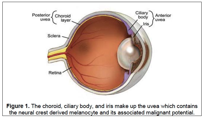
Background
Melanoma of the uveal tract (i.e., iris, ciliary body, and choroid), sometimes referred to as ‘ocular
melanoma’ (Figure 1) accounts for 5% of all melanomas and occurs at an incidence rate of about 6
cases per million person years.1, 2 Uveal melanoma is the most common primary intraocular
malignancy and the uveal tract is the second most common location for melanoma, after the skin.2
Risk factors include Caucasian race, light eye color, fair skin, cutaneous and iris nevi and freckles,
and an inability to tan, and exposure to arc welding and suntan beds.3-8 Despite these specific
similarities to cutaneous melanomas, the association between ultraviolet (UV) light and uveal
melanoma has not been clarified.4-6, 9
Uveal melanoma is distinct in molecular pathogenesis, with classically described UVB-induced
signature mutations in the cancer genomes restricted to iris uveal melanoma,10 and a median count of
nine somantic mutations per tumour compared with a median of 171 somatic mutations in cutaneous
melanoma.11 Driver mutations are also, distinct, commonly affecting GNAQ or GNA11 and BAP1,
while mutations in BRAF and NRAD, commonly seen in cutaneous melanoma, are uncommon.12
Uveal melanoma is a distinct clinical entity from other ‘ocular’ melanomas that can arise in the
conjunctiva, the eyelid, and the orbit.7 Therefore, these guidelines do not apply to melanomas that
arise in the conjunctiva, eyelid, and orbit. The choroid is the most common location for uveal
melanoma, comprising 80% of cases, the ciliary body 12%, and the iris 8%. Of these topographical
locations, the ciliary body carries the worst prognosis, while the iris carries the best.13, 14 The
Callender histological classification for uveal melanoma identified four distinct cellular types in order
of best to worst prognosis: spindle-A cells (slender nuclei and lacking visible nucleoli); spindle-B cells
(larger nuclei and distinct nucleoli); intermediate cells (similar to but smaller than epithelioid cells);
and epithelioid cells (larger polygonal cells with one or more prominent nucleoli).15, 16 Mixed-cell type
uveal melanoma (i.e., epithelioid and spindle) is the most common histological subtype of uveal
melanoma and carries an intermediate prognosis.16
Roughly 60% of patients with uveal melanoma will succumb to metastasis within 10 years, but varies
based on tumour size and other prognostic factors, including cell type, location of the anterior margin
of the tumour, degree of ciliary body involvement, extraocular extension, mitotic rate, and lymphocytic
infiltration.13, 14, 17, 18 Survival is 47% at 10 years and 25% at 20 years (Table 1). Using modern genetic
prognostic testing (gene expression profiling) further prognostic information can be obtained (Table
2). The overall local recurrence rate following plaque brachytherapy is approximately 10% at five
years (median 25.5 [12-71] months).19 The most common site of metastasis is the liver and the
second most common is the lungs. Few clinical trials focused solely on metastatic uveal melanoma,
therefore treatment decisions in the palliative setting are often based upon data from studies
conducted in the non-uveal melanoma patient population. Low quality evidence exists to support the
use of immune checkpoint inhibitors for the treatment of patients with metastatic uveal melanoma,
and recently an improvement in overall survival (OS) for patients with metastatic uveal melanoma
treated with the novel immunotherapy tebentafusp was reported.20

Last revision: June 2021
                                                                                Guideline Resource Unit   2
Table 1. Long-term survival estimates associated with uveal melanoma.18, 21
Survival Measure 5-year 10-year 15-year 20-year 25-year
All-Cause Survival 62% 47% 35% 25% 21%
Melanoma Metastasis-Free Survival 69% 60% 55% 52% 51%
Second Cancer-Free Survival 95% 89% 85% 79% 76%
Table 2. Metastasis-free survival by signature class (i.e., gene expression profile).18, 21, 22
Metastasis-Free Survival
Signature Class 3-year 5-year
1A 98% 98%
1B 93% 79%
2 50% 28%
The common differential diagnosis for uveal melanoma includes lesions such as nevus, neovascular
(‘wet’) age-related macular degeneration, congenital hypertrophy of the retinal pigment epithelium,
circumscribed choroidal hemangioma, hemorrhagic detachment of the choroid or retina,
melanocytoma, metastasis to the eye from another site and choroidal osteoma.23
Guideline Questions
1. How should patients with uveal melanoma be staged at baseline?
2. How should uveal melanoma, including patients who experience metastatic or recurrent disease,
be managed?
3. What is the recommended surveillance strategy for patients diagnosed with uveal melanoma?
Last revision: June 2021
Guideline Resource Unit 3Search Strategy
For the 2021 guideline update, PubMed was searched (2014 through 2021 Mar) for phase II and III
clinical trials, prospective studies, systematic reviews, meta-analyses, and clinical practice guidelines.
The Medical Subject Heading (MeSH) term “uveal melanoma” was used, and results were limited to
studies in humans 19+ years of age published in English. Studies that did not report outcomes related
to the efficacy of treatments or imaging modalities for uveal melanoma and studies involving less than
ten patients were excluded. Reference lists of key publications were also searched for relevant
citations. For the detailed literature search strategy, results and a summary of key evidence please
refer to the accompanying evidence table.
The ECRI Guidelines Trust, well-known cancer guideline developers and Google (search term:
“uveal melanoma guideline”) were searched for practice guidelines relevant to this topic. A total of
eight clinical practice guidelines published after 2014 were identified from the following organizations:
UpToDate, National Comprehensive Cancer Network (NCCN), National Cancer Institute (NCI),
Cancer Council Australia, European Dermatology Forum (EDF) / European Association of Dermato-
Oncology (EADO) / European Organization for Research and Treatment of Cancer (EORTC),
National Services Division (NSD) Scotland, United Kingdom Uveal melanoma Guideline Working
Group, Princess Margaret Cancer Centre.
Target Population
The recommendations outlined in this guideline apply to adults over the age of 18 with malignant
uveal melanoma (intra-ocular melanoma). Other ‘ocular’ melanomas arising in the conjunctiva, the
eyelid, and the orbit are not included in this guideline. Different principles may apply to pediatric
principles.
Recommendations
Diagnosis and Work-Up
1. All intraocular malignancies and indeterminate lesions should be evaluated by a provider trained
in all aspects of care (i.e., medical, oncologic, surgical, radiotherapy [RT], laser therapy [e.g.,
transpupillary thermotherapy]) to determine appropriate follow-up and/or treatment. (Level of
Evidence: V24-26, Strength of Recommendation: B)
2. Complete history including ophthalmic and medical history.
3. Complete ophthalmic examination and fundoscopy.
• A baseline fundus photograph of adequate quality and an objective assessment of lesion
height is required for all melanocytic lesions.
Last revision: June 2021
Guideline Resource Unit 44. Ocular ultrasonography by a certified ophthalmic ultrasonographer or ophthalmologist with training
in ultrasound (U/S).
• A-scan U/S can demonstrate initial prominent spike followed by low-to-medium internal
reflectivity or a decrescendo pattern and can be used to measure tumour height. (Level of
Evidence: IV27, Strength of Recommendation: B)
• B-scan U/S can allow for tumour measurement (height), and tumour characteristics
including solidity/hollowness, vascularity, shape, and extra-scleral (extraocular) extension.
(Level of Evidence: IV27, Strength of Recommendation: B)
• U/S biomicroscopy (UBM) is a high frequency U/S providing high resolution imaging of the
anterior segment of the eye. It is used to visualize ciliary body and iris tumours. (Level of
Evidence: IV28, 29, Strength of Recommendation: B)
5. Ancillary ocular studies, if ophthalmic examination is inconclusive, sometimes due to media
opacity. (Level of Evidence V30-32, Strength of Recommendation: B)
• Fluorescine and/or Indocyanin green angiography of the retina and choroidal vasculature is
helpful in select cases (requires clear media for visualization).
• Computed tomography (CT) of the eye is rarely needed.
• Magnetic resonance imaging (MRI) of the eye is rarely needed.
6. Staging work-up to rule out metastases for patients diagnosed with uveal melanoma.
• Serum testing
o Complete blood count (CBC)
o Liver function tests (LFTs)
• Diagnostic imaging should aim to reduce unnecessary radiation.
o All patients should receive a baseline Primovist-enhanced abdominal MRI and
ultrasound (U/S) of the liver and non-contrast enhanced CT scan of the chest.
o Or whole-body positron emission tomography (PET)/CT scan and ultrasound of the
liver. (Level of Evidence: III33 IV34, Strength of Recommendation: B)
o If there is a suspicion of metastases, refer to a tertiary cancer centre.
Primary Management
Melanocytic Choroid Tumours
1. Small (• Small lesions are observed for growth or treated based on risk factors for growth and the
associated risk of visual loss with treatment.
o Most lesions with no risk factors are observed until growth is documented. Once
growth is documented the lesion is labeled a melanoma and is treated. (Level of
Evidence: IV35-39, Strength of Recommendation: B)
o All lesions are evaluated based on their risk factors for future growth.
Risk factors for future growth include tumour thickness >2 mm, subretinal
fluid, symptoms of visual acuity loss to 20/50 or worse, orange pigment,
hollow acoustic density and tumour largest basal diameter >5 mm. (Level of
Evidence: IV40, Strength of Recommendation: B)
High-risk lesions (≥ 3 risk factors) are often offered treatment, biopsy, or close
observation based on discussions with the patient regarding visual loss, since
the risk of future growth is greater than 50%. (Level of Evidence: IV41 V31,
Strength of Recommendation: B)
When indicated, treatment is most commonly ocular brachytherapy. (Level of
Evidence: III42 IV43, Strength of Recommendation: B)
2. Medium/intermediate (3-12 mm in thickness) tumours are typically treated with ocular
brachytherapy. (Level of Evidence: I44-48, Strength of Recommendation: A)
• Enucleation is sometimes chosen by patients who cannot make the follow-up visits required
post brachytherapy.
3. Large (>12 mm in thickness) tumours
• Due to the risk of severe vision loss and neovascular glaucoma secondary to radiation
complications with large lesions, large lesions are offered enucleation or brachytherapy (if
standard dosing can be achieved with brachytherapy).
o Many centres offer enucleation for very large tumors greater than 12 mm in
thickness and 18 mm in maximal width. (Level of Evidence: IV49-52, Strength of
Recommendation: B)
o Brachytherapy for very large lesions (>12 mm thick or >18 mm in maximal basal
dimension) is sometimes performed in select cases such as contralateral vision
loss or in patients who insist on avoiding enucleation. (Level of Evidence: V24,
Strength of Recommendation: C)
• Neo-adjuvant pre-enucleation radiation does not provide a clinically or statistically
meaningful difference in mortality rates. (Level of evidence: I53, Strength of
recommendation: E)
Last revision: June 2021
Guideline Resource Unit 6Ciliary Body Lesions 1. Ciliary body lesions
•    If two or more risk factors are present or any change or growth is noted, referral to a
              subspecialist ocular oncologist is recommended.
Principles of Enucleation
1. Enucleation involves surgical removal of the eye.
2. Typically, lesions >12 mm in thickness and/or >18 mm in diameter are offered enucleation.
3. For patients undergoing enucleation, in accordance with the College of American Pathologists’
   Protocol for the Examination of Specimens from Patients with Uveal Melanoma,60 review of
   specimens should include reporting of the following elements:
         •    Specimen laterality
         •    Tumour site: iris, ciliary body, choroid
         •    Largest basal diameter and thickness
         •    Scleral and optic nerve invasion
         •    Extraocular extension
         •    Histologic type: spindle, mixed, epithelioid
         •    Mitotic count
         •    Vascular invasion
         •    Extravascular matrix pattern
         •    Inflammatory cells/tumour infiltrating lymphocytes and macrophages
         •    Invasion of Bruch’s membrane
         •    Margins
         •    Regional lymph nodes
         •    Pathologic stage classification (pTNM, AJCC 8th Edition)
         •    Molecular results (if known):
                    o Chromosome 3 and 8 loss/gain
                    o BAP1 status
                    o Gene expression profile (GEP)
                    o Multiplex ligation dependent probe amplification (MLPA) analysis
         •    Additional pathologic findings

Last revision: June 2021
                                                                                   Guideline Resource Unit   8
Principles of Primary Radiotherapy (RT)
1. Episcleral brachytherapy is the most commonly utilized treatment for uveal melanoma worldwide
and is the treatment of choice in Alberta.
2. Other RT modalities include charged-particle external beam RT (EBRT) (i.e., protons, carbon ions,
or helium ions), and photon-based radiosurgery (i.e., linear accelerator, gammaknife, or
cyberknife).
Adjuvant Local Therapy
1. Positive margins post excision:
• If margins are positive or indeterminate after resection, adjunctive plaque brachytherapy RT
of the surgical margins is often utilized.
2. Transpupillary thermotherapy (TTT):
• TTT uses an infrared laser administered through a dilated pupil for choroidal lesions.
• TTT as a primary treatment has been associated with a relatively high rate of local
recurrence, especially when the tumour abuts the optic nerve and overhangs the optic disc.
Therefore, TTT is not recommended as monotherapy for uveal melanoma in the standard
case. (Level of Evidence: II61 IV62, Strength of Recommendation: D)
• TTT can be offered as an adjunctive treatment to reduce the risk of local recurrence
following RT or as a primary treatment for medium risk nevi in select cases. (Level of
Evidence: IV63, 64, Strength of Recommendation: C)
• TTT is used in some centers to treat marginal recurrence post brachytherapy. (Level of
evidence: IV64, Strength of Recommendation: C)
• TTT can cause retinal vascular damage and retinal traction and subsequent secondary
visual loss.
3. Radiation retinopathy:
• Intravitreal anti-vascular endothelial growth factor (VEGF) agents are often utilized to
prevent and/or reduce the severity of radiation retinopathy and its associated visual loss.
(Level of Evidence: ranibizumab II65, 66 bevacizumab IV67-70, Strength of Recommendation:
B)
Genetic Prognostic Testing
1. All patients should be offered GEP or monosomy 3 and 8 testing to provide information on survival
prognosis. This will also guide systemic follow-up and consideration for inclusion in clinical trials
Last revision: June 2021
Guideline Resource Unit 9for patients at high risk of metastases (Figure 2, Table 2). (Level of Evidence: III71, 72 IV22, 73-75,
Strength of Recommendation: B)
Management of Patients with Metastatic Disease and High-Risk Patients
1. Currently there is no strong evidence to treat high-risk patients (monosomy 3 and 8q gain, GEP 2,
or tumours >9 mm thick) without identified metastasis with adjuvant treatments to reduce the risk
of disease recurrence. However, the use of systemic therapy as adjuvant treatment to enucleation
or definitive radiation is an active focus of research, and consideration for enrollment in clinical
trials is warranted where possible. (Level of Evidence: IV76, Strength of Recommendation: B)
2. Systemic therapy for the management of metastases:
• When possible, enrollment in a clinical trial is recommended.
• A phase III clinical trial comparing treatment with tebentafusp against investigator’s choice
chemo-/immunotherapy in advanced uveal melanoma patients with positive HLA-A 02:01
haplotype achieved its primary end point of OS in the intent-to-treat population with a
hazard ratio (HR) of 0.51 (95% CI, 0.36-0.71; pSurveillance Following Definitive Local Therapy
In patients who would qualify for treatment of metastatic treatment, surveillance should be offered.
This may consist of history and physical exam, chemistry, and imaging based on patient risk
categories:
1. Patients with GEP class 1a or 1b, or disomy 3 (monosomy 3 negative or undetected) OR patients
with no genetic assessment and tumour ≤9 mm thick: (Level of Evidence: V, Strength of
Recommendation B)
• Liver U/S: annually for up to 10 years.
• Physical exam: annually, for up to 10 years.
• Follow-up may be transitioned to the family physician at 5 years.
2. Patients with GEP class 2, monosomy 3 (monosomy 3 positive or detected), OR tumours >9 mm
thick with no genetic assessment: (Level of Evidence: V, Strength of Recommendation B)
• Physical exam: annually, indefinitely
• Imaging every six months consisting of an annual liver U/S alternating with annual MRI liver
for ten years. If body habitus limits U/S, consideration for other modalities should be given.
• Follow-up may be transitioned to the family physician at 5-10 years.
Discussion
Diagnosis
The timely management (including observation) of uveal melanocytic lesions, including small flat
lesions, is vitally important, as this is a major/complex eye condition that threatens both complete
visual loss and life. Any delays in referral and treatment may result in both complete loss of the eye
(enucleation) and/or life (metastasis).
Research has demonstrated that earlier treatment, allowing for treatment of a smaller lesion, portends
improved survival.106 Furthermore, waiting for observation of growth, in small lesions identified as
high-risk by an ophthalmologist, can increases the risk of metastasis by eight times.107 Therefore,
even melanocytic lesions ≤2 mm in thickness without any documentation of growth can be offered
treatment.24, 107 The diagnosis of uveal melanoma can be very difficult for the non-specialist.45, 108
Treatment options for uveal melanocytic lesions involve both medical, laser, complicated extraocular
and intraocular surgery, RT, radiosurgery, and other eye sparing treatment modalities.109 Often
observation versus treatment discussions, especially for small melanocytic lesions, require balancing
treatments and their complications with the risk of observation and its threat to life; therefore,
published international guidelines, with Canadian representation, recommend that patients be
provided an evaluation/discussion with an eye cancer specialist (eye cancer specialist is defined as
an ophthalmic oncologist, medical physicist, or radiation oncologist)107 or ophthalmologist.32 Similarly,
Last revision: June 2021
Guideline Resource Unit 11to reduce the risks of local recurrence and to reduce the extent of visual loss following eye sparing
treatments, adjuvant medical, laser, and complicated surgical treatments often need to be
administered.63, 67, 110, 111 For these reasons it is recommended that the provider be fully trained in all
treatment areas (i.e., medical, complicated surgical, RT, laser treatments of the eye, and cancer care)
to safely follow, discuss, and treat all indeterminate (uveal melanocytic lesions that have not
demonstrated growth) and malignant intraocular lesions.
Ocular U/S is used to determine tumour size, shape, and the lesion’s U/S characteristics.
Orbital/ocular CT and MRI are not commonly required in the diagnostic work-up, unless other
examinations are inconclusive.32, 108 The common lesions on the differential diagnosis for uveal
melanoma includes freckles, nevus, Lisch nodules, neovascular age-related macular degeneration,
congenital hypertrophy of the retinal pigment epithelium, choroidal hemangioma, hemorrhagic
detachment of the choroid or retina, melanocytoma, metastasis to the eye from another location, and
choroidal osteoma. Experienced ocular oncologists (ophthalmologists with a practice focus in
oncology) are able to diagnose uveal melanoma accurately (based predominantly on fundoscopy and
ultrasound) with >98% accuracy without biopsy.45
Staging
Staging is guided by the American Joint Committee on Cancer (AJCC) system for uveal
melanoma.112 Staging requires intraocular examination, serum tests, and imaging. Blood work
typically consists of complete blood count (CBC) and liver function tests (LFT).32 Historically, the most
basic baseline imaging for ruling out systemic metastases consisted of chest x-ray (CXR) with
abdominal U/S. However, these tests have since been shown to have low sensitivities113 and have
largely been replaced by positron emission tomography (PET)-CT scanning, MRI of the abdomen and
CT of the chest, or CT of the chest and abdomen. Whole body PET-CT scan has demonstrated good
sensitivity (35-100%) and positive predictive value (88-100%),114-116 while MRI has shown the highest
sensitivity (67-92%)115-117 Table 3 summarizes the accuracy of these various imaging modalities in
the detection of uveal melanoma.
Controversy exists around whether baseline imaging should be performed in this population, based
on the premise that metastases cannot be treated and the yield of positive findings at presentation is
low (2%). It should be noted, however, that more than half of patients (55%) have CT abdomen
findings that require further investigation,118 the majority of which are false positives; only 2% have
definitive metastasis at staging.118 An international, registry-based retrospective data analysis of
patients presenting with stage IV uveal melanoma found that 6% of the uveal melanoma with
metastasis at initial presentation belonged to subcategory T1a, most often detected by whole-body
PET/CT.34 Therefore, it may be best to clarify these baseline imaging findings early on to reduce the
challenges of ruling out metastasis at a later date. The treating surgeon should decide on the
appropriateness of staging investigations that balance excessive testing with patient stress, additional
testing that can arise from false positives, and potentially unnecessary surgery. Patients who
demonstrate metastasis at presentation may be spared aggressive treatment of their primary lesion.
Last revision: June 2021
Guideline Resource Unit 12Genetic Prognostic Testing
Two modern prognostic tests that require tumour sampling are currently available, including
assessment of monosomy 3 and 8 gain status and GEP. Both tests are typically performed with a fine
needle aspiration biopsy at the time of definitive treatment. Monosomy 3 associated with a gain in 8q,
and GEP class 2 carry a poor prognosis with a 3-year metastasis free survival of 53% and 50%
respectively.18, 22, 71 Due to the better biopsy yields and stronger evidence on prognostication, in
Alberta, the commercially available GEP is now utilized instead of monosomy 3 testing (Figure 2,
page 16, Table 2).71 A controlled prospective clinical observational trial that examined the
psychosocial impact of prognostic genetic testing in uveal melanoma patients found that rather than
being a burden, it may help to ease stress and support a more realistic risk perception.119
Primary Management
Observation. Observation is typically reserved for indeterminate lesions, but may be acceptable for
select patients with small melanoma (5 mm.40 If these risk factors are present, treatment should be
considered. Waiting for documented growth of lesions can increase the risk of metastasis up to eight-
fold107 and improved survival has been demonstrated with earlier management.106 Similarly, the eight
edition of the AJCC classification system has demonstrated that tumour size predicts survival.112
Furthermore, even after controlling for GEP, tumour size has been found to be an independent
predictor of metastasis at 5 years.120, 121 In contrast, several small non-comparative case-series have
suggested that patients with small indeterminate lesions who are carefully selected by an
ophthalmologist, may be observed for tumour growth before initiating treatment without adversely
affecting survival.35-37, 42, 43 The American Brachytherapy Society guidelines suggest that patients
being observed should be counseled about the small (yet still unquantified) increased risk of
metastasis with observation.24
Lesions are often labeled as high-risk nevi or indeterminate melanocytic lesions if they have 3 or
more risk factors for growth. Several small, underpowered, retrospective studies suggest there may
be a roll in certain situations to observe small lesions. A retrospective analysis of data from patients
with primary posterior uveal melanoma with documented tumour growth of ≥3 mm in basal diameter,
1.5 mm in thickness, or both (n=30), during a pretreatment interval of ≥6 months was compared with
data from a control group of promptly treated patients (n=30).35 The resulting mean ± standard error
cumulative 5-year probability of melanoma-specific mortality relative to the date of initial examination
was 17.1% ± 7% in the delayed group vs. 18.4% ± 8% in the promptly treated group (p>0.5, log rank
test). Although this study is underpowered, it and several other similar studies suggest that delaying
Last revision: June 2021
Guideline Resource Unit 13treatment in carefully selected patients may not worsen survival. In contrast, retrospective data on a large dataset of 1287 patients suggested that waiting for documentation of growth for lesions 12 mm in thickness and/or >18 mm in diameter are offered enucleation. Concern over whether enucleation was promoting the release of tumour cells throughout the body, leading to observed increases in mortality post-enucleation,123 contributed to the development of new management strategies, such as RT and TTT. Since then this Zimmerman hypothesis on seeding of tumour during enucleation has been disproved.124 Radiotherapy (RT). RT has largely replaced enucleation for tumours with suitable location and dimension (i.e.,
The use of a calculation method that corrects for plaque heterogeneities differs from the
homogeneous medium calculation method used in the original COMS study, resulting in a difference
between the treatment goal of 70 Gy recommended in these guidelines and the 85 Gy used in the
COMS study and commonly cited in the literature. This can be explained, however, as a dose of 70
Gy calculated using the more accurate method which includes heterogeneities is roughly equivalent
to 85 Gy using the homogeneous method. A review of 53 cases treated in Alberta found the 70 Gy
accounting for plaque heterogeneities was equivalent to 81.8 ± 2.2 Gy utilizing the COMS
methodology.131
The dose is delivered over 3-7 days, as per the recommendations of the American Brachytherapy
Society.132 Whenever possible, plaque size and shape, plaque loading, and plaque position are
chosen such that doses to critical structures such as the fovea, papillomacular bundle, and optic
nerve head are minimized. The fovea and papillomacular bundle are retinal tissue with an assumed
tolerance of 50 Gy and the optic nerve head has an assumed tolerance dose of 60 Gy.128, 129, 133, 134
While reports on the exact radiation tolerance of these tissues vary, it is clear that tumour proximity
and radiation dose to these structures are associated with poorer visual outcomes.134
The COMS randomized trial of 125I brachytherapy vs. enucleation as primary therapy for medium
sized melanomas found no difference in survival outcomes and little difference in quality-of-life
outcomes between groups. Five-year survival was substantially better than expected based on
published rates.45, 46, 48, 125 A retrospective case-series among patients diagnosed with uveal
melanoma without metastases (N=400) and treated with 103Pd brachytherapy (mean apical dose of
73.3 Gy over 5 to 7 continuous days) revealed a local control rate of 96.5%. Fourteen patients
required secondary enucleation (5 for tumour growth and 9 for glaucoma pain control). The expected
5- and 10-year metastases-free survival rates were 92.7% and 86.6%, respectively.135 Low
recurrence rates were reported for 125I brachytherapy as well, in a retrospective analysis of data from
87 patients with uveal melanoma ≤16 mm by largest basal diameter and large by height by the COMS
criteria.47 The COMS trial found that the risk of treatment failure (i.e., tumour growth, recurrence, or
extrascleral extension) with 125I brachytherapy was 10.3% (95% CI, 8.0%-13.2%). The Kaplan-Meier
estimate of proportion of patients undergoing enucleation by 5 years was 12.5% (95% confidence
interval [CI], 10.0%-15.6%). Risk factors for treatment failure were older age, greater tumour
thickness, and proximity of the tumour to the foveal avascular zone. Tumour control by RT is typically
95% (95% CI 93-96%) at 15 years.136 Except for select centers, the majority of radiation treatment for
uveal melanoma is administered through brachytherapy. This technique provides extremely accurate
administration of radiation to a mobile organ and provides theoretical advantages due to its
continuous dose administration. Local failure post-radiation for posterior uveal melanoma should be
retreated either by enucleation or re-treatment by brachytherapy.137 Most cases of failed local control
primarily treated with radiation are enucleated.
Charged-particle EBRT (i.e., protons, carbon ions, or helium ions), and photon-based radiosurgery
(i.e., linear accelerator, gammaknife, or cyberknife) have also been used in the setting of uveal
melanoma. Proton beam RT carries a local control rate of 93.9% at 5 years and 92.1% at 10 years.

Last revision: June 2021
                                                                               Guideline Resource Unit   15
The ocular conservation rates were 91.1% at 5 years and 87.3% at 10 years.138 Similar results have
been reported elsewhere.139-144
Adjuvant Therapies
Transpupillary Thermotherapy (TTT). TTT uses an infrared laser through a dilated pupil and is not
typically used as a primary treatment for uveal melanoma anymore due to high recurrence rates. It is
now most commonly used as an adjuvant treatment to RT or as treatment of medium risk
nevi/indeterminate lesions. Due to its penetrance limitations, this therapy is best suited for small (1.0-
3.0 mm in apical height and 5.0-16.0 mm largest basal diameter) melanomas.23 A randomized
controlled trial among patients with small choroidal melanomas (N=95) compared TTT primary
therapy with brachytherapy and found, after a mean follow-up of 56.2 months (range, 24-118 months;
standard deviation [SD], 22.6), that tumour regression occurred in 45 patients (92%) in the TTT group
versus 45 patients (98%) in the brachytherapy group (p=0.397). Recurrences developed in four TTT
patients and one brachytherapy patient.61
A retrospective case-matched comparative study (N=36) and retrospective observational study
(N=21) were conducted in parallel to compare TTT alone vs. TTT plus plaque RT. Local failure
occurred in six patients (29%) and was associated with an increased number of TTT spots per
session (p=0.023) and decreased tumour pigmentation (p=0.001). The RT plus TTT group regressed
rapidly, with no local failures. No patient developed metastasis. TTT performed as a supplemental
therapy in RT-resistant tumours (6 patients) or tumours at high risk for local failure with RT alone (3
patients) successfully induced tumour shrinkage and resolution of exudative retinal detachment in all
6 tumours RT-resistant tumours and after a mean follow-up of 32 months (range, 10–52 months), all
9 tumours regressed satisfactorily, with no local failures or enucleations.62
Intra-vitreal Anti-Vascular Endothelial Growth Factor (VEGF) Medications. One of the major
causes of visual loss in patients who have received RT is radiation retinopathy and optic neuropathy.
The main underlying cause of this visual loss is vascular damage leading to both ischemia from
capillary drop out and exudation from vascular injury and ischemia.145 Similarly, one of the primary
causes of enucleation post RT is neovascularization that occurs secondary to ischemia and VEGF
production leading to uncontrolled neovascular glaucoma. Anti-VEGF agents have been developed
and utilized to suppress the vascular permeability and neovascularization process that can result in
significant ocular morbidity. Multiple case series have shown substantial reductions of subretinal fluid,
intra-retinal hemorrhages, visible retinal infarcts, regression of neovascularization, and improved
visual function.67, 125, 146-152
Adjuvant prophylactic intravitreal bevacizumab injections every 4 months for 2 years post plaque
treatment significantly improved visual outcomes by reducing optical coherence tomography (OCT)-
evident macular edema, clinically notable radiation maculopathy, moderate vision loss, and poor final
visual acuity.147 Similarly the use of intravitreal anti-VEGF for symptomatic radiation retinopathy has
also shown improvement in macular thickness and visual function.65-67, 70, 146, 148, 153, 154
Last revision: June 2021
Guideline Resource Unit 16Systemic Adjuvant Treatments No studies to date have shown any benefit from adjuvant therapy in reducing metastasis rates in patients at high risk for future metastasis (GEP class 2 and monosomy 3). Management of Metastatic or Recurrent Disease. Local treatment failure (ocular recurrence) Local (ocular) recurrence is typically treated with enucleation or repeat brachytherapy. Some centers will utilize TTT for small recurrence at the margin of the tumour.62, 155, 156 Local therapy in the setting of limited metastatic disease There are some data to suggest that resection of uveal melanoma liver metastases may prolong survival,157, 158 including a single-arm prospective study among twelve patients who were able to achieve a median recurrence free survival (RFS) of 19 months (6-78; 5-year RFS 15.6%) and an OS of 27 months (11-86; 5-year OS 53.3%) following complete resection.159 Retrospective data also suggest that resection of liver metastases is associated with a 3.7-fold increase in median survival, as compared to no surgery.100 Similar data have been reported elsewhere.101, 160, 161 However, the results of these non-comparative cohorts may be influenced by lead-time bias and/or favorable tumour biology in patients who are candidates for resection.23 In general, surgery is a preferred option in younger patients with large tumours and in patients with a metastasis. The option of surgery needs to be carefully considered due to risk of metastatic spread to remaining liver and systemically. Surgical resection in combination with chemotherapy may offer some benefit to patients with metastatic disease. A prospective study of aggressive surgery (i.e., removal of as much liver disease as possible) and implantation of an intra-arterial catheter for delivery of chemotherapy (e.g., fotemustine and/or DTIC-platinum for 4–9 cycles) among patients with uveal melanoma metastatic to the liver (N=75) demonstrated complete resection in 27.5% and significant tumour reduction in 49.3%. Median OS was 10 months in patients who received complete treatment surgery plus chemotherapy; curative resection improved the median OS to 22 months (p
that four patients had all metastatic liver lesions addressed: one patient underwent left lateral
segmentectomy and three patients had combinations of left lateral segmentectomies, wedge
resections and radiofrequency ablation of two to four lesions. The median survival of patients who
underwent surgery alone or in conjunction with radiofrequency ablation to address all liver lesions
was 46 months.103 In a phase II trial of radioembolization for the treatment of uveal melanoma hepatic
metastasis, treatment-naïve patients (n=24) achieved a median OS of 18.5 months with a 1-year
survival of approximately 61%.102 Participants treated with radioembolization in whom prior
immunoembolization treatment failed (n=23) achieved a median OS of 19.2 months with a 1-year
survival of approximately 70%. Grade 3 treatment-related toxicities were reported in three of the
treatment-naïve patients and in one of the patients who received prior immunoembolization. These
results suggest that radioembolization is safe and effective first- or second-line treatment in this
setting.
Advanced metastatic recurrence
Systemic therapies for metastatic uveal melanoma have been largely modeled after therapies for
cutaneous melanoma. In the pre-immunotherapy era, phase II chemotherapy data did not
demonstrate clinical efficacy in metastatic uveal melanoma with any of the tested single agents or
combinations of agents.82-97, 165 However, the use of immune checkpoint inhibitors, either singly or in
combination, has demonstrated more promising results, and a recently reported phase III randomized
clinical trial supports the use of immunotherapy for the treatment of patients with unresectable
metastatic disease. Clinical trials for the uveal melanoma patient population are emerging, and where
possible enrolment in a clinical trial is recommended.23
Chemotherapy. Systemic chemotherapy alone for the management of metastases or recurrent
disease is largely ineffective, and therefore not recommended. Clinical trials have tested the efficacy
of carboplatin, paclitaxel, docosahexaenoic acid-paclitaxel, and cisplatin by transarterial
chemoembolization (TACE). Of these, the most efficacious therapy was cisplatin TACE, with a partial
response of 57%.85 None of the regimens were able to achieve a complete response in any patients.
A complete summary of phase II data is in Appendix B.
Immunotherapy. Immunotherapies have demonstrated prolonged survival in patients with metastatic
cutaneous melanoma. Ipilimumab, an anti-CTLA4 antibody, have demonstrated activity in patients
with advance uveal melanoma in retrospective and expanded access studies.166-171 These reports
varied in inclusion, exclusion, treatment protocols, and reported highly variable outcomes making the
summary data problematic. Yet if we summarize, these six reports in, 188 patients with advanced
uveal melanoma have been treated with ipilimumab with one complete response, 7 partial responses,
and 52 patients with stable disease. This translates to a response rate of 4.3% and disease control
rate of 31.9%. The tumour kinetics and response patterns in these patients with uveal melanoma
were similar to those with cutaneous melanoma treated with ipilimumab. This response rate seen in
patients with ocular melanoma would appear to be lower than that reported in the phase III cutaneous
melanoma trials of ipilimumab alone or combined with dacarbazine of 10.9% and 15.2%, respectively.

Last revision: June 2021
                                                                              Guideline Resource Unit   18
It is hypothesized that metastatic uveal melanoma is less responsive to immune checkpoint inhibitor
due to the presence of fewer somatic mutations, which results in fewer potential neoantigens that can
be targeted by antitumour immunity. Additionally, the liver as an immune-modulatory organ may
protect uveal melanoma metastases from immune surveillance.172 However, uveal melanoma
expresses several immunogenic antigens, such as glycoprotein 100 (gp100), melanoma antigen
recognized by T cells 1 (MART-1), and tyrosinase, and a subset of uveal melanoma are able to elicit
a vigorous immune response.173 Rare, marked response to immunotherapy has been reported in
uveal melanoma, and molecular investigation of these tumours revealed high tumour mutation burden
(TMB) secondary to germline, loss-of function MBD4 mutations.174, 175 Johansson et al. recently
demonstrated that iris uveal melanoma is unique among uveal melanoma subtypes in that it
demonstrates ultraviolet radiation-associated DNA damage, and like MBD4-definicient tumours, has a
high TMB.10 This suggests that metastatic iris uveal melanoma may be more likely to respond to
immunotherapy than other variants of uveal melanoma, the vast majority of which have low TMB.
Wessely et al.176 recently reviewed the evidence for immune checkpoint inhibitors (ICI) in treating
metastatic uveal melanoma. Among nine prospective and retrospective studies using anti‐CTLA‐4
(ipilimumab or tremelimumab) for the treatment of metastatic uveal melanoma, the largest
prospective observational study with 53 patients reported ORR of 0% and a median OS of
6.8 months. The largest retrospective study with 82 patients reported ORR of 4.8% and a median OS
of 6.0 months. Among 11 retrospective and prospective studies utilizing anti‐PD1 agents
(pembrolizumab or nivolumab), the largest prospective study had 34 evaluable patients and reported
ORR of 5.8% and median OS of 11 months for patients on nivolumab. The largest retrospective study
which followed 43 patients on pembrolizumab reported ORR of 7.0% and a median OS of
10.3 months. Improved responses are reported with combination CTLA-4/PD-1 ICI. One retrospective
study consisting of 64 patients reported ORR of 15.6% (3.1% CR) and a median OS of
16.1 months,177 while another with 89 patients reported ORR of 11.0% (1% CR) and a median OS of
15.0 months.178 Bol et al. reported the longest median OS in the literature at 18.9 months for
combination ipilimumab and nivolumab, with an ORR of 21.0%, albeit with a small sample size of 19
patients.179 A recent study by Klemen et al. followed 30 metastatic uveal melanoma patients treated
with ICI. The study had four patients survive >5 years, all of whom received anti‐CTLA‐4 and anti‐
PD1, either sequentially or in combination. The author suggested that exposure to ipilimumab in
addition to anti‐PD1 may be integral in achieving long‐term survival in metastatic uveal
melanoma.180 Collectively, data suggest that combined ICI may be superior to anti‐PD1 or anti‐CTLA‐
4 monotherapy, although limitations in the current data (including small sample sizes, potential
selection bias, and a lack of clinical trials with comparative study design) must be noted.
More robust data supporting the use of ICI for metastatic uveal melanoma is emerging. Nivolumab
and pembrolizumab both demonstrate activity in prospective, non-comparative phase II clinical
trials,78, 79 and a single-arm phase II clinical trial utilizing the combination of nivolumab with ipilimumab
resulted in an objective response rate of 18%, and median OS of nearly two years.77
Last revision: June 2021
Guideline Resource Unit 19Most recently, data from a randomized phase III clinical trial confirms a survival advantage for HLA-
A*02:01 positive adult patients treated with the novel immunotherapy tebentafusp, a novel bispecific
protein comprised of a soluble T cell receptor fused to an anti-CD3 immune-effector function
(clinicaltrials.gov identifier: NCT03070392).20 When compared against investigator’s choice of therapy
(including ipilimumab, pembrolizumab or dacarbazine chemotherapy), treatment with tebentafusp
improved OS (HR 0.51) with a one-year OS rate of 73% and median OS of 22 months.
Molecularly targeted agents. Greater than 80% of primary uveal melanomas carry active mutations in
the GNAQ or GNA11 genes, which encode for G protein alpha subunits, leading to activation of the
mitogen-activated protein kinase (MEK) pathway. Advancements in our understanding of the
molecular and genetic mechanisms of pathophysiology has generated interest in the use of receptor
tyrosine kinase inhibitors, including the MEK inhibitors selumetinib and trametinib, and the c-KIT
(CD117) inhibitor sunitinib.83, 181, 182 Invariably resistance to these agents develops in a matter of
months. While modest clinical activity with the use of these agents has been reported, none have yet
been shown to improve OS.98 Clinicians and patients who decide to use targeted therapies in the
metastatic setting should understand that treatment-related toxicities may be significant and a
detriment to the quality of life.
Follow-Up
There are no high-level data to inform the most appropriate way to monitor patients who have
undergone treatment for uveal melanoma. As such, there is no consensus within the ophthalmic or
oncologic community regarding the role of surveillance for detection of metastases in these
patients.183 Some lower-level data are available on the usefulness of specific imaging tests and
biochemical tests in the detection of metastasis. Since surgical resection, ablation therapy and/or
systemic immunotherapy evidence has suggested improved survival,103, 117, 184 most ocular oncology
centers perform rigorous follow-up on high-risk patients.
Liver function tests are not sufficiently sensitive to be used as a sole method of surveillance.
Regarding liver function tests (LFTs), an Israeli study among 30 uveal melanoma patients with
metastases and 80 non-metastatic controls looked retrospectively at the use of LFTs and liver
imaging in detecting metastases. At the time of diagnosis of liver metastases by imaging, only 50% of
patients had at least one abnormal LFT (vs. 5% of controls). Alkaline-phosphatase and lactate
dehydrogenase were the most predictive tests. Lactate dehydrogenase and aspartate-
aminotransferase were predictive at 80% of the upper normal limit, whereas alkaline-phosphatase
and gamma-glutamyltransferase were most predictive at the upper normal limit.185
Clinical characteristics and tumour genetics predict survival. Therefore, a customized follow-up
routine, based on the risk category of the patient is recommended. Ultrasound (U/S) has
demonstrated high specificity (i.e., 100%) but low sensitivity (i.e., 14%) for the detection of uveal
melanoma liver metastases.113 The use of U/S in the follow-up of high-risk patients should therefore
complement other more sensitive tests. Several studies have looked at the use of various imaging
modalities in detecting metastases, particularly in the liver, at follow-up.99, 114, 185-188 MRI offers
Last revision: June 2021
Guideline Resource Unit 20consistently good sensitivity (92-96%), while that of PET-CT is variable (35-100%). In a head-to-head
comparison of MRI and PET-CT, sensitivity was higher with MRI (67% vs. 41%; p=0.01), while
positive predictive value was slightly higher with PET-CT (95% vs. 100%; p=0.01); the authors
concluded that MRI was superior to PET-CT for detecting liver metastases from uveal melanoma.115
In a cohort of 188 high-risk patients, 6-monthly MRI of the abdomen detected metastases before
symptoms in 92% of patients, resulting in 14% of patients who qualified for liver resection.99
Consensus-based guidelines recommend that follow-up consist of annual history and physical exam,
LFTs, PET-CT or MRI of the abdomen, CXR, and liver U/S.32, 189, 190 High-risk patients require more
frequent imaging. To date there is no data on the impact of follow-up on survival.
Low-risk patients (i.e., GEP class 1a/1b; no monosomy 3 detected; or tumour ≤9 mm thick and no
genetic assessment) should undergo liver U/S and physical exam annually, for up to 10 years; follow-
up may be transitioned to the family physician at 5 years. High-risk patients (i.e., GEP class 2;
monosomy 3 detected; or tumour ≥9 mm thick and no genetic assessment) should undergo physical
exam annually, indefinitely, plus imaging every six months consisting of a liver U/S alternating with
abdominal/liver MRI for 10 years. If body habitus limits U/S, consideration for other modalities should
be given. Follow-up may be transitioned to the family physician at 5-10 years.
There have been several reports of an increased risk of cutaneous melanoma following a diagnosis of
uveal melanoma. The risk varies significantly between studies and may be partially related to
increased surveillance.99, 191, 192 At present there is insufficient evidence to recommend universal
screening for cutaneous melanoma of these individuals. In select individuals (those with significant
risk factors for cutaneous melanoma) consider referral to a dermatologist for baseline total skin
examination. Anecdotally, follow-up assessments have typically consisted of history and physical
exam, liver function studies, PET-CT or MRI of the chest and abdomen and/or CXR and liver U/S.189,
190 These assessments are typically performed annually, except in the case of patients with
monosomy 3 or GEP class 2, who should undergo liver U/S every 3 months due to the increased risk
of metastases.
Last revision: June 2021
Guideline Resource Unit 21Last revision: June 2021
Guideline Resource Unit 22Table 3. Accuracy of various imaging modalities in the detection of uveal melanoma metastases
Author, Year Modality Metastases Site Sensitivity Specificity Positive
Predictive Value
Marshall, 201399 MRI liver 92% n/a n/a
Orcurto, 2012116 MRI liver 96% n/a n/a
PET-CT 35%
Freton, 2012192 PET-CT Any n/a 94% 100%
(whole body)
Klingenstein, PET-CT liver or lung 100% n/a n/a
2010185 (whole body)
Servois, 2010115, MRI liver 67% n/a 95%
116 PET 45% 100%
Francken, PET liver 100% 67% 88%
2006114
Finger, 200533 PET Any 100% 94% 100%
(whole body)
Semelka, MRI liver n/a n/a 98%
2001187 CT 75%
Eskelin, 1998188 AST liver 43% 93% 62%
ALT 38% 90% 68%
AP 27% 95% 77%
LDH 67% 96% 35%
Hicks, 1998113 CXR any 1.8% 100% 100%
U/S 14% 100% 100%
AST 15% 89% 28%
AP 25% 86% 33%
Last revision: June 2021
Guideline Resource Unit 23References
1. Singh AD, Topham A. Survival rates with uveal melanoma in the United States: 1973-1997. Ophthalmology. May
2003;110(5):962-5.
2. Egan KM, Seddon JM, Glynn RJ, Gragoudas ES, Albert DM. Epidemiologic aspects of uveal melanoma. Surv
Ophthalmol. Jan-Feb 1988;32(4):239-51.
3. Gallagher RP, Elwood JM, Rootman J, Spinelli JJ, Hill GB, Threlfall WJ, et al. Risk factors for ocular melanoma:
Western Canada Melanoma Study. J Natl Cancer Inst. Apr 1985;74(4):775-8.
4. Weis E, Shah CP, Lajous M, Shields JA, Shields CL. The association of cutaneous and iris nevi with uveal melanoma:
a meta-analysis. Ophthalmology. Mar 2009;116(3):536-543.e2.
5. Weis E, Shah CP, Lajous M, Shields JA, Shields CL. The association between host susceptibility factors and uveal
melanoma: a meta-analysis. Arch Ophthalmol. Jan 2006;124(1):54-60.
6. Shah CP, Weis E, Lajous M, Shields JA, Shields CL. Intermittent and chronic ultraviolet light exposure and uveal
melanoma: a meta-analysis. Ophthalmology. Sep 2005;112(9):1599-607.
7. Canadian Cancer Society. Eye Cancer. n.d.
8. Weis E, Aghazadeh H, Roelofs K, Agi J. Sunlamp use is a risk factor for uveal melanoma: a meta-analysis. Can J
Ophthalmol. Apr 8 2021;
9. Weis E, Vrouwe SQ, LeBaron DB, Parliament MB, Shields J, Shields CL. Changes in Ultraviolet Radiation Exposure to
the Ocular Region: A Population-Based Study. Cancers (Basel). May 24 2019;11(5)
10. Johansson PA, Brooks K, Newell F, Palmer JM, Wilmott JS, Pritchard AL, et al. Whole genome landscapes of uveal
melanoma show an ultraviolet radiation signature in iris tumours. Nat Commun. May 15 2020;11(1):2408.
11. Krauthammer M, Kong Y, Ha BH, Evans P, Bacchiocchi A, McCusker JP, et al. Exome sequencing identifies recurrent
somatic RAC1 mutations in melanoma. Nat Genet. Sep 2012;44(9):1006-14.
12. Chattopadhyay C, Kim DW, Gombos DS, Oba J, Qin Y, Williams MD, et al. Uveal melanoma: From diagnosis to
treatment and the science in between. Cancer. Aug 1 2016;122(15):2299-312.
13. Edge S, Byrd D, Compton C. Malignant Melanoma of the Uvea. AJCC Cancer Staging Manual. 7th ed. New York, NY:
Springer; 2010. p. 547-559.
14. Cancer.Net. Eye Cancer. 2015.
15. Klintworth G, Scroggs M. The eye and ocular adnexa. In: Sternberg S, ed. Diagnostic surgical pathology. Lippincott
Williams & Wilkins; 1999:994-996.
16. Grossniklaus H, Green W. Uveal Tumors. In: Garner A, Klintworth G, eds. Pathobiology of Occular Disease: A
Dynamic Approach Second Edition. Marcel Dekker Inc; 1994:1423-77.
17. Singh AD, Turell ME, Topham AK. Uveal melanoma: trends in incidence, treatment, and survival. Ophthalmology. Sep
2011;118(9):1881-5.
18. Kujala E, Mäkitie T, Kivelä T. Very long-term prognosis of patients with malignant uveal melanoma. Invest Ophthalmol
Vis Sci. Nov 2003;44(11):4651-9.
19. Papageorgiou KI, Cohen VM, Bunce C, Kinsella M, Hungerford JL. Predicting local control of choroidal melanomas
following ¹⁰⁶Ru plaque brachytherapy. Br J Ophthalmol. Feb 2011;95(2):166-70.
20. Immunocore's tebentafusp granted Breakthrough Therapy Designation for unresectable or metastatic uveal melanoma
from FDA. 2021. Accessed March 1, 2021. https://www.immunocore.com/news/immunocores-tebentafusp-granted-
breakthrough-therapy-designation-unresectable-or-metastatic-uveal-melanoma-fda
21. Kath R, Hayungs J, Bornfeld N, Sauerwein W, Höffken K, Seeber S. Prognosis and treatment of disseminated uveal
melanoma. Cancer. Oct 1 1993;72(7):2219-23.
22. Kilic E, Naus NC, van Gils W, Klaver CC, van Til ME, Verbiest MM, et al. Concurrent loss of chromosome arm 1p and
chromosome 3 predicts a decreased disease-free survival in uveal melanoma patients. Invest Ophthalmol Vis Sci. Jul
2005;46(7):2253-7.
23. National Cancer Institute. Intraocular (Uveal) Melanoma Treatment (PDQ ®). September 23, 2020.
24. The American Brachytherapy Society consensus guidelines for plaque brachytherapy of uveal melanoma and
retinoblastoma. Brachytherapy. Jan-Feb 2014;13(1):1-14.
25. Cancer Council Australia - Clinical Guidelines Network. Ocular melanoma. March 22, 2021. Accessed March 22,
2021. https://wiki.cancer.org.au/australia/Guidelines:Ocular_melanoma
26. Nathan P, Cohen V, Coupland S, Curtis K, Damato B, Evans J, et al. Uveal Melanoma UK National Guidelines. Eur J
Cancer. Nov 2015;51(16):2404-12.
Last revision: June 2021
Guideline Resource Unit 2427. Conway RM, Chew T, Golchet P, Desai K, Lin S, O'Brien J. Ultrasound biomicroscopy: role in diagnosis and
management in 130 consecutive patients evaluated for anterior segment tumours. Br J Ophthalmol. Aug 2005;89(8):950-
5.
28. Giuliari GP, McGowan HD, Pavlin CJ, Heathcote JG, Simpson ER. Ultrasound biomicroscopic imaging of iris
melanoma: a clinicopathologic study. Am J Ophthalmol. Apr 2011;151(4):579-585.e1.
29. Gündüz K, Hoşal BM, Zilelioğlu G, Günalp I. The use of ultrasound biomicroscopy in the evaluation of anterior
segment tumors and simulating conditions. Ophthalmologica. 2007;221(5):305-12.
30. National Comprehensive Cancer Network. Uveal Melanoma. Version 2.2020. 2020.
31. Princess Margaret Cancer Centre. Uveal Melanoma - Clinical Practice Guidelines. March 23, 2021. Accessed March
23, 2021.
https://www.uhn.ca/PrincessMargaret/Health_Professionals/Programs_Departments/Central_Nervous_System_Eye/Docu
ments/CPG_Ocular_%20UvealMelanoma.pdf
32. Royal College of Opthamologists. Management of Uveal Melanoma. n.d.
33. Finger PT, Kurli M, Reddy S, Tena LB, Pavlick AC. Whole body PET/CT for initial staging of choroidal melanoma. Br J
Ophthalmol. Oct 2005;89(10):1270-4.
34. Garg G, Finger PT, Kivelä TT, Simpson ER, Gallie BL, Saakyan S, et al. Patients presenting with metastases: stage
IV uveal melanoma, an international study. Br J Ophthalmol. Jan 15 2021;
35. Augsburger JJ, Vrabec TR. Impact of delayed treatment in growing posterior uveal melanomas. Arch Ophthalmol. Oct
1993;111(10):1382-6.
36. Lane AM, Egan KM, Kim IK, Gragoudas ES. Mortality after diagnosis of small melanocytic lesions of the choroid. Arch
Ophthalmol. Aug 2010;128(8):996-1000.
37. Murray TG, Sobrin L. The case for observational management of suspected small choroidal melanoma. Arch
Ophthalmol. Sep 2006;124(9):1342-4.
38. Factors predictive of growth and treatment of small choroidal melanoma: COMS Report No. 5. The Collaborative
Ocular Melanoma Study Group. Arch Ophthalmol. Dec 1997;115(12):1537-44.
39. Mortality in patients with small choroidal melanoma. COMS report no. 4. The Collaborative Ocular Melanoma Study
Group. Arch Ophthalmol. Jul 1997;115(7):886-93.
40. Dalvin LA, Shields CL, Ancona-Lezama DA, Yu MD, Di Nicola M, Williams BK, Jr., et al. Combination of multimodal
imaging features predictive of choroidal nevus transformation into melanoma. Br J Ophthalmol. Oct 2019;103(10):1441-
1447.
41. Weis E, Roelofs K, Larocque M, Murtha A. Gene Expression Profiling as an Adjunctive Measure to Guide the
Management of Indeterminate, High-Risk Choroidal Melanocytic Lesions: A Pilot Study. Ocul Oncol Pathol. Feb
2019;5(2):102-109.
42. Sobrin L, Schiffman JC, Markoe AM, Murray TG. Outcomes of iodine 125 plaque radiotherapy after initial observation
of suspected small choroidal melanomas: a pilot study. Ophthalmology. Oct 2005;112(10):1777-83.
43. Semenova E, Finger PT. Palladium-103 radiation therapy for small choroidal melanoma. Ophthalmology. Nov
2013;120(11):2353-7.
44. Jampol LM, Moy CS, Murray TG, Reynolds SM, Albert DM, Schachat AP, et al. The COMS randomized trial of iodine
125 brachytherapy for choroidal melanoma: IV. Local treatment failure and enucleation in the first 5 years after
brachytherapy. COMS report no. 19. Ophthalmology. Dec 2002;109(12):2197-206.
45. The COMS randomized trial of iodine 125 brachytherapy for choroidal melanoma: V. Twelve-year mortality rates and
prognostic factors: COMS report No. 28. Arch Ophthalmol. Dec 2006;124(12):1684-93.
46. Hawkins BS. Collaborative ocular melanoma study randomized trial of I-125 brachytherapy. Clin Trials. Oct
2011;8(5):661-73.
47. Girvigian MR, Astrahan MA, Lim JI, Murphree AL, Tsao-Wei D, Petrovich Z. Episcleral plaque 125I radiotherapy with
episcleral LCF hyperthermia: a prospective randomized trial. Brachytherapy. 2003;2(4):229-39.
48. Diener-West M, Earle JD, Fine SL, Hawkins BS, Moy CS, Reynolds SM, et al. The COMS randomized trial of iodine
125 brachytherapy for choroidal melanoma, III: initial mortality findings. COMS Report No. 18. Arch Ophthalmol. Jul
2001;119(7):969-82.
49. Puusaari I, Heikkonen J, Summanen P, Tarkkanen A, Kivelä T. Iodine brachytherapy as an alternative to enucleation
for large uveal melanomas. Ophthalmology. Nov 2003;110(11):2223-34.
50. Adams KS, Abramson DH, Ellsworth RM, Haik BG, Bedford M, Packer S, et al. Cobalt plaque versus enucleation for
uveal melanoma: comparison of survival rates. Br J Ophthalmol. Jul 1988;72(7):494-7.
51. Seddon JM, Gragoudas ES, Egan KM, Glynn RJ, Howard S, Fante RG, et al. Relative survival rates after alternative
therapies for uveal melanoma. Ophthalmology. Jun 1990;97(6):769-77.
Last revision: June 2021
Guideline Resource Unit 25You can also read