ESUR Quick Guide to Female Pelvis Imaging - European Society of Urogenital Radiology
←
→
Page content transcription
If your browser does not render page correctly, please read the page content below
ESUR Quick Guide
to Female Pelvis Imaging
European Society of Urogenital Radiology

                           1.0

ESUR Quick Guide to Female Pelvis Imaging 1.0
PREFACE
The concept of this booklet is to be a guide used as a quick reference
to planning the imaging of patients with suspected or confirmed
gynaecologic disease. Full guidelines for many of these disease areas
are published. Here we hope to provide a quick and easily accessible
reference for the radiologist and radiographers undertaking these
imaging investigations.
Comments and questions are welcome at esursecretary@esur.org
ESUR Female Pelvis Imaging Working Group
April 2019

                                                              PREFACE    1
AUTHOR ACKNOWLEDGEMENTS
    Special thanks to the following authors who have contributed
    to the production of this Quick Guide booklet:
    • Celine Alt
    • Nishat Bharwani
    • Laura Brunesch
    • Francesco M. Danza
    • Martina Derme
    • Rania Farouk El Sayed
    • Rosemarie Forstner
    • Laure Fournier
    • Benedetta Gui
    • Aki Kido
    • Rahel A. Kubik-Huch
    • Rita Lucas
    • Teresa Margarida Cunha
    • Gabriele Masselli
    • Olivera Nikolic
    • Stephanie Nougaret
    • Milagros Otero-Garcia
    • Andrea Rockall
    • Evis Sala

2    Author Acknowledgements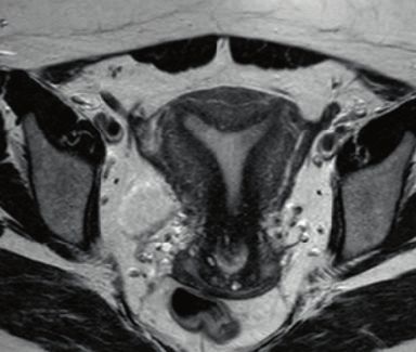
CONTENTS
TERMINOLOGY: CONTRAST AGENTS AND CONTRAST MEDIA

PREFACE                                                           1
AUTHOR ACKNOWLEDGEMENTS                                           2
CONTENTS                                                          3

1. TECHNICAL INFORMATION                                          4
2. DISEASE SPECIFIC INVESTIGATIONS                            12
   a. Cervical Cancer                                         12
       i. Primary Staging                                     12
       ii. Follow up and Investigation of Recurrence          16
  b. Endometrial Cancer                                       19
  c. Vaginal Masses                                           25
  d. Characterisation of Adnexal Mass                         28
  e. Staging and Follow-up of Ovarian Cancer                  32
  f. Endometriosis                                            38
  g. Leiomyoma                                                45
  h. Female Tract Congenital Abnormalities                    51
  i. Gynaecologic Emergencies                                 55
  j. Pelvic Floor Imaging                                     61
  k. Placental Disease                                        65
  l. Fistula                                                  75

                                                       CONTENTS       3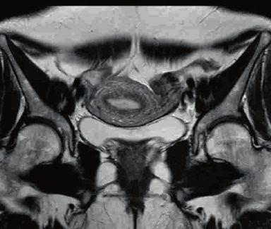
1. TECHNICAL INFORMATION
    Authors: Rita Lucas and Teresa Margarida Cunha

    Abbreviations
            MR – magnetic resonance
            IM – intramuscular
            IV – intravenous
            TR – time to repetition
            TE – time to echo
            DWI – diffusion weighted images
            FS – fat saturation
            FOV – field of view
            SS – single shot
            FSE – fast spin echo
            ms – milliseconds
    The MRI protocol should always be tailored to the main indication for
    pelvic MRI. These are general recommendations for gynecological studies.

    1. Patient preparation
            • The patients are recommended to fast for four hours and, if
              possible, asked to empty their bladder about one hour prior
              to the examination in order to obtain a moderately full bladder
              during the exam.
            • Optional: Rectal enema to clean the bowel, one the day
              before the exam and other on the morning of the exam.
            • Avoid vaginal tampons.
            OPTIONAL:
            • Antispasmodic drugs to reduce bowel motion: 40 mg of
              butylscopolamine IM/IV before the exam or 0.5-1.0 mg IV/
              IM of glucagon, taking into account hospital availability and
              patient individual contraindications (e.g. myasthenia gravis,
              megacolon or narrow angle glaucoma).
            • In the evaluation of deep pelvic endometriosis, ultrasound gel
              may be instilled into vagina (about 60 mL) and into the rectum
              (about 200mL).
            • Also in cases of vaginal pathology or congenital uterine
              abnormalities vaginal opacification with ultrasound gel may be
              considered

4    1. Technical Information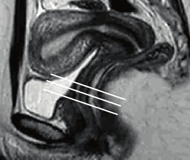
2. Patient Positioning
       • The patient is usually scanned in the supine position.
       OPTIONAL:
       • To reduce anxiety and claustrophobia, if there is excessive
         patient movement, the patient may be studied in prone
         position.

3. Technical requirements
Dedicated phase-array surface abdominopelvic coil both 1.5T and 3.0T
       • Pelvic phased array coils are recommended at both 1.5T and
         3.0T to increase signal-to-noise ratio (SNR).
       • Saturation bands: anterior and superior.

4. MRI Protocol
Examples of suitable sequences and imaging parameters

Sequence             Plan           TR/TE            FOV

T2W SSFSE or
                                                     Covering the whole
HASTE (Localizer Coronal            3,4/ 1,71ms
                                                     abdominopelvic area
sequence)
       • To localize and plan the dedicated sequences.

                                         Slice
                                            Inter-
Sequence     Plan           TR/TE        thick-
                                            section FOV
                                         ness
                                            gap
                                            0.4-
T2W FSE      Axial          4000/90ms 4-5mm         Whole pelvis
                                            0.5mm
                                                    From one hip
T2W FSE      Sagittal       4000/90ms 4mm   0.4mm
                                                    to another
T2W FSE      Coronal        4000/90ms 4mm           0.4mm    Whole pelvis
                                                             Small,
             Axial of                                        dedicated to
T2W FSE                 5000/110ms 4mm              0.4mm
             the cervix                                      the area of
                                                             interest

                                                  1. Technical Information   5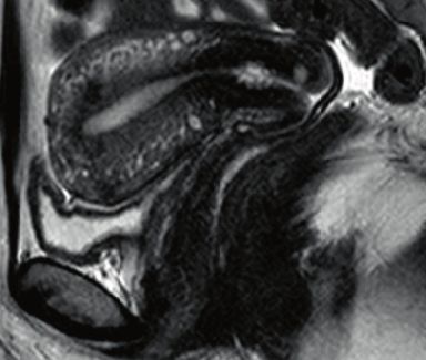
Small,
               Axial of                                     dedicated to
    T2W FSE               4000/90ms 4mm            0.4mm
               the uterus                                   the area of
                                                            interest
                                                            Small,
               Sagittal of                                  dedicated to
    T2W FSE                4000/90ms 4mm           0.4mm
               the uterus                                   the area of
                                                            interest
               Coronal of
                                                            Small,
               the uterus
                                                            dedicated to
    T2W FSE    or axial   4000/90ms 4mm            0.4mm
                                                            the area of
               of the
                                                            interest
               ovaries
                                                            Small,
               Oblique
                                                            dedicated to
    T2W FSE    (endome-     5000/110ms 3mm         0.3mm
                                                            the area of
               triosis)
                                                            interest
                                                            Small,
               Axial of                                     dedicated to
    T2W FSE               4000/90ms 4mm            0.4mm
               the vagina                                   the area of
                                                            interest
                                                            Large from
                                                            renal hilum
                                                            to pubic
    T2W FSE    Axial        4000/90ms 5-6mm 1mm             symphysis/
                                                            from
                                                            diaphragm to
                                                            iliac crests
          • T2W sequences yield the majority of information.
          • Sequences are oriented in relation to uterus axis for uterine
            pathology, otherwise in relation to the pelvis:
          • Perpendicular to endocervical canal longitudinal axis to
            evaluate the cervix.
          • Perpendicular to the uterine body longitudinal axis to evaluate
            the myometrial endometrial interface (eg. in endometrial
            cancers).

6   1. Technical Information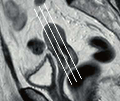
• In cases of congenital uterine abnormalities or benign uterine
       pathology additional oblique acquisitions may be necessary
       depending on uterine topography (sagittal/coronal oblique
       or parallel to uterine body and axial oblique perpendicular to
       uterine body).
     • When doubt remain whether a mass originates from the
       ovaries or from the uterus, an additional acquisition might
       help, oriented in the ovarian axial plan, which corresponds to
       a plan parallel to the endometrial cavity (coronal plan of the
       uterine body).
     • In cases of deep pelvic endometriosis additional oblique
       acquisitions may also be necessary depending on uterine
       topography (particularly an axial oblique plane perpendicular
       to uterine cervix).
     • The sequences with big FOV are required to check the
       retroperitoneal lymph nodes and to evaluate the kidneys.

                                    Slice    Inter-
Sequence   Plan       TR/TE         thick-   section FOV
                                    ness     gap
T1W FSE
without    Axial      648/10ms      5mm      0.5mm     Whole pelvis
FS
T1W FSE
           Axial      648/10ms      5mm      0.5mm     Whole pelvis
with FS
           Coronal of
                                                       Small,
           the uterus
T1W FSE                                                dedicated to
           or axial   648/10ms      4mm      0.4mm
with FS                                                the area of
           of the
                                                       interest
           ovaries
     • The fat-saturated T1W sequence is required to distinguish
       fat from haemorrhage in lesions with high signal intensity on
       T1W images.

                                          1. Technical Information      7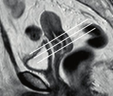
Slice    Inter-
    Sequence   Plan         TR/TE         thick-   section FOV
                                          ness     gap
    T1W GRE
               Axial        5,5/2,7ms     2mm      1mm       Whole pelvis
    3D FS
    T1W GRE
               Sagittal     5,5/2,7ms     2mm      1mm       Whole pelvis
    3D FS
    T1W GRE    Axial of
                          5,5/2,7ms       2mm      1mm       Whole pelvis
    3D FS      the uterus
               Coronal of
               the uterus
    T1W GRE
               or axial   5,5/2,7ms       2mm      1mm        Whole pelvis
    3D FS
               of the
               ovaries
          • The standard gadolinium contrast medium dose for soft
            tissue imaging is 0.1mmol/kg body weight, followed by a
            saline flush of 20 ml.
          • One acquisition before contrast administration and five
            additional sequential acquisitions post-contrast injection
            acquisitions with 20 seconds of delay between them, until
            150 seconds.
          • The most adequate plan to evaluate enhancement in the
            cervix is the sagittal and usually a late phase acquisition is
            done in the axial oblique plan (perpendicular to uterine cervix).
          • The most adequate plan to evaluate the enhancement of
            the myometrium is in the axial oblique plan (perpendicular to
            uterine body).
          • For ovarian lesions as stated before, an acquisition in the
            ovarian axial plan might be necessary.

8   1. Technical Information
Slice Inter-
Sequence Plan TR/TE thick- section FOV
ness gap
Axial
Equal to T2W
DWI of the 3100/53ms 6mm 1mm
for abdomen
abdomen
Axial of 0.4- Equal to T2W
DWI 3100/53ms 4-5mm
the pelvis 0.5mm for pelvis
Equal to
T2W axial of
Axial of 0.4-
DWI 3100/53ms 4-5mm the cervix
the cervix 0.5mm
(cervical
cancer)
Equal to
Axial
T2W axial of
of the 0.4-
DWI 3100/53ms 4-5mm the uterus
uterine 0.5mm
(endometrial
body
cancer)
• Abdomen (b-value – 0, 500 and 1000).
• Pelvis (b-value – 0, 200 and 1000).
1. Technical Information 9Schematic representation of specific MRIplanes orientation:
Axial of the
cervix
Axial of the
uterus
Coronal of the
uterus or axial of
the ovaries
Sagittal of the
uterus
Axial of the
vagina
10 1. Technical InformationTECHNICAL ASPECTS OF CT OF THE FEMALE PELVIS
Abbreviations
CT – Computed Tomography
1. Patient preparation
• The patient is generally recommended to fast for at least 4
hours and empty the bladder aound hour before examination.
• Oral administration of 1 litre of iodinated contrast media or
water (depending on clinical scenario) over a period of 1 hour
before the examination.
• Avoid vaginal tampon.
OPTIONAL:
• Vaginal or rectal contrast opacification may be necessary.
2. Patient Positioning
• The patient should be supine.
3. Technical requirements
• At least a 64-row CT is advisable (about 1 mm slice thickness
to allow quality multiplanar reformations).
• Acquisition before (e.g. for ovarian cancer staging) and 70-90
seconds post-injection of iodinated contrast media (100–
150mL of intravenous iodinated contrast medium injected at
a rate of 3–4mL/second).
1. Technical Information 112. DISEASE SPECIFIC INVESTIGATIONS
a. Cervical Cancer
i. Primary Staging
Author: Rosemarie Forstner
1. Clinical background
Cervical cancer is the fourth most frequent cancer in women
worldwide. The large majority of cases (around 85%) occur in the less
developed regions. There were an estimated 266,000 deaths from
cervical cancer worldwide in 2012. Almost nine out of ten (87%) deaths
occur in the less developed regions [1].
Cervical carcinoma spreads by direct tumour invasion through the
stroma into the parametria towards the pelvic wall. Spread may also
occur via the uterosacral ligaments to the pelvic sidewall; upward into
the corpus of the uterus or downward into the vagina; and lymphatic
spread to the paracervical, parametrial and presacral lymph nodes,
and then to the external iliac (obturator), internal iliac and common
iliac nodes. Retroperitoneal and supraclavicular nodal involvement is
only seen in advanced disease. Spread to the lungs, bone and liver is
unusual at initial presentation.
Tumours in early stages, with less than 4 cm and without parametrial
involvement can be treated with surgery [2]. In large tumours (> 4 cm),
or tumours in advanced stages or in the absence of surgical conditions
the patients are treated with chemoradiotherapy (CRT) [3]. Young
women with small tumours who want to preserve fertility may choose
to perform a trachelectomy (only in tumours less than 2 cm in greatest
diameter, located ≥ 1 cm from the internal os and if the cervical length
is ≥ 2.5 cm in greater sagittal axis).
2. Imaging techniques
MRI is the preferred imaging technique for evaluation of the cervix
and staging of abdominal and pelvic spread of disease (see protocol
chapter). CT of the chest, abdomen and pelvis maybe be undertaken for
complete staging.
12 2. Disease Specific Investigations3. Diagnosis: Key points
• Cervical carcinoma is typically moderately hyperintense on T2-WI,
regardless of histological subtype, so this is the key sequence in
tumour evaluation.
• These tumours may be exophytic or endocervical („barrel-shaped“) [4].
• Vaginal invasion is assumed when there is disruption of the hypointense
vaginal wall by intermediate / high intensity tumour on T2-WI.
• Evaluation in the oblique plane perpendicular to the cervical canal is
mandatory to detect parametrial invasion.
• When there is preservation of the hypointense ring of cervical stroma
and the hypointense stroma is> 3 mm thick (signal „hypointense ring“),
parametrial invasion can be excluded with a specificity of 96-99% [5].
• Parametrial invasion is implied if disruption of the hypointense
signal of cervical stroma, evident intermediate/high intensity tumour
extension to the adjacent parametrial tissue or invasion of the
vesicouterine ligament.
• Extension into the lower third of the vagina is considered when the
tumour extends below the plane of the bladder floor level, behind the
urethra.
• Invasion of the pelvic wall is considered when the distance between
the tumour and the pelvic wall (internal obturator muscle, levator ani,
piriformis or iliac vessels) is < 3 mm [6].
• The preservation of a fat plane between the wall of the bladder /
rectum and the tumour is a good radiology criterion for exclusion of
invasion of these structures [4]. Rectum or bladder wall invasion is
best evaluated in contrast enhanced sequences.
• The identification of suspicious lymph nodes is based on dimensional
and morphological criteria: pelvic lymph nodes > 8 mm, abdominal
lymph nodes > 10 mm and inguinal lymph nodes > 15 mm of short
axis in the axial plane, and also lymph nodes with irregular contour or
with evidence of necrosis.
• In DWI the apparent diffusion coefficients (ADCs) calculated for
cervical tumours are lower than those of normal cervical stroma,
increasing the contrast between normal tissue and the tumour,
what can be particularly useful in the case of small tumours.
Nodal metastases also exhibit decreased ADC values allowing the
identification of pathological nodes with only 5 mm.
• It is recommended to compare the signal intensity in DWI with high b
value (b1000) with the signal from the node on T2-WI with the signal
intensity of the primary tumour [7].
2. Disease Specific Investigations 134. Staging
Cervical carcinoma staging remains clinical according to the guidelines
of the International Federation of Gynaecology and Obstetrics (FIGO),
to ensure uniformity of criteria between countries with different
resources. Imaging evaluation is widely recommended, when available,
in order to increase the accuracy of clinical staging and support
therapeutic decision, with the main purpose of identifying the patients
that are surgery candidates.
5. Follow-up
MRI is also recommended for the assessment of tumour response,
surveillance of possible therapeutic complications and detection of
recurrence. See separate chapter on follow-up.
6. Pitfalls
• Limitations in determining the parametrial invasion by MRI:
microscopic parametrial invasion cannot be ruled out and on the
other hand, linear extensions to the parametrial fat can only be due to
inflammatory phenomena [8].
• Mucosal nodularity depicted on T2-WI may be due to bullous
oedema (swelling areas of focal bladder epithelium associated with
inflammation / chronic irritation phenomena)[9].
• The presence of inflammation/swelling might be responsible for tumour
over-estimation on T2-WI and following intervention (such as a cone
biopsy), changes can arise at the site of the biopsy that can be mistaken
for the primary tumour. It is recommended that an interval of at least
one week to ten days be allowed between the biopsy and MRI [4].
• Even in the absence of dimensional and morphological criteria, nodal
microscopic disease cannot be ruled out. On ADC maps, there is a
considerable overlap between the values calculated for metastatic
and reactive nodes [10].
7. Radiology Report Checklist
• lesion size (measured in at least two planes)
• extent of the tumour (to the isthmus, body of the uterus or vagina)
• parametrial extension
• invasion of the bladder, rectum or pelvic wall
• pelvic inguinal and para-aortic lymph nodes with dimensional or
morphological criteria of suspicion
• presence of hydronephrosis
14 2. Disease Specific Investigations• distant metastasis
• The FIGO stage should not be mentioned in the report unless this is
the standard practice used by the local tumour board.
References:
[1] International Agency for Research on Cancer. GLOBOCAN 2012:
estimated cancer incidence, mortality and prevalence worldwide in
2012. Lyon, France: IARC; 2013 Dec Available from: http://globocan.iarc.
fr/.
[2] Small W. Jr, Strauss J.B., Jhingran A., Yashar C.M., Gaffney D.K.,
Cardenes H.R. et al. Expert Panel on Radiation Oncology–Gynecology.
ACR Appropriateness Criteria® definitive therapy for early stage cervical
cancer. Am J Clin Oncol. 2012 Aug;35(4):399-405.
[3] Koh W.J., Greer B.E., Abu-Rustum N.R., Apte S.M., Campos S.M.,
Chan J. et al. Cervical cancer. J Natl Compr Canc Netw 2013 Mar 1;
11(3):320-43.
[4] Engin G. Cervical cancer: MR imaging findings before, during, and
after radiation therapy. Eur Radiol 2006 Feb; 16(2):313-24.
[5] Sala E., Rockall A.G., Freeman S.J., Mitchell D.G:, Reinhold C. The
Added Role of MR Imaging in Treatment Stratification of Patients with
Gynecologic Malignancies: What the Radiologist Needs to Know.
Radiology 2013; 266:717-40.
[6] Hricak H., Yu K.K. Radiology in invasive cervical cancer. Am J
Roentgenol 1996; 167:1101-8.
[7] Whittaker C.S., Coady A., Culver L., Rustin G., Padwick M., Padhani
AR. Diffusion-weighted MR imaging of female pelvic tumours: a
pictorial review. Radiographics 2009 May-Jun; 29(3):759-74.
[8] Sahdev A., Sohaib S.A., Wenaden A.E., Shepherd J.H., Reznek
R.H. The performance of magnetic resonance imaging in early cervical
carcinoma: a long-term experience. Int J Gynecol Cancer 2007 May-
Jun; 17(3):629-36.
[9] Rockall A.G., Ghosh S., Alexander-Sefre F., Babar S., Younis M.T., Naz
S. et al. Can MRI rule out bladder and rectal invasion in cervical cancer
to help select patients for limited EUA? Gynecol Oncol 2006 May;
101(2):244-9.
[10] Siegel C.L., Andreotti R.F., Cardenes H.R., Brown D.L., Gaffney
D.K., Horowitz N.S. et al. ACR Appropriateness Criteria® pretreatment
planning of invasive cancer of the cervix. Am Coll Radiol 2012 Jun;
9(6):395-402.
2. Disease Specific Investigations 15ii. Follow up and Investigation of Recurrence
Authors: Aki Kido and Rosemarie Forstner
1. Clinical background
One of the purpose of follow-up is early detection of recurrence disease
that should be more likely to be effective to treatment, may resulting
in improvement the clinical outcome of patient with relapsing cervical
cancer (1). On present, posttreatment surveillance program differs
widely among different countries and among different institutions (1).
Patients may be imaged due to symptoms (such as vaginal bleeding or
pelvic pain) or in some cases, there may be regular surveillance (such
as following fertility preserving trachelectomy).
2. Imaging
Tumours undergoing CRT respond by decreasing their size and the
signal intensity in T2. However in the first 3 months after CRT high
signal intensity of the cervical stroma can be related to residual tumour
but also to inflammation or fibrosis. Response to treatment may also be
evaluated using FDG-PET/CT (Grigsby et al).
The majority of recurrences occur within 2 years of treatment (62-89%)
(2)(10). Then, NCCN guidelines recommended history and physical
examination every 3–4 months for the first 2 years, every 6 months for
the next 3 years for high risk patients. For low risk patients, they are
suggested every 6 monthgs for the first 2 years and yearly for thenext
3 years. Many institution continue follow-up until 10 years by annual
examination after 5 years (2).
As for the surveillance strategy, physical examination and vaginal
vault cytology were the most common methods and accepted (2, 3).
Physical examination indicated the highest detection rate compared
with cytologic evaluation and imaging modalities (3).The role of CT and
ultrasound has not been constructed even if a relatively high number of
asymptomatic patients had been already diagnosed by these methods.
Imaging has suggested for surveillance in the asymptomatic patient (3)
Imaging techniques are influenced by the most frequent location of
recurrence, which is vaginal vault or central pelvis(19-57%) followed
by pelvic wall (2,4). MR may be superior to CT for detecting recurrent
tumor after surgery or radiation treatment because of its usefulness in
the detection of disease recurrence due to its high-contrast resolution.
16 2. Disease Specific InvestigationsRecurrent tumor demostrate heterogenous high signal intensity on T2-
WI, and enhancement degree is variable (5). MRI may be more accurate
than CT for evaluating tumor invasion of the bladder, rectum and pelvic
wall (6,7). Evidence of additional use of DWI has not been constructed.
3. Diagnosis
Recurrence of carcinoma of the cervix usually is heterogeneous
hyperintense on T2 and demonstrates inhomogeneous contrast uptake.
It may occur locally (in the cervix, vaginal vault, parametria or pelvic
wall), in lymph nodes or as distant metastases (especially bone or lung,
less frequently in the liver).
Parametrial recurrence may also be hard to differentiate from residual
fibrosis, however fibrosis tends to remain stable on subsequent studies
[Engin G 2006].
The frequency of distant metastasis varies depending on the report,
ranging 15-61%. Among the patients with distant relapses, almost half
of them had lung metastases or an hepatic recurrence. In addition,
58% of recurrent patients had multiple localizations (2,4). Considering
the freqnecy of distant and multiple site recurrences, CT or 18FDG PET-
CT can cover the every site. Recently, 18FDG PET-CT has recognized as
useful modality in identifying patients ealier in the recurrence process,
because of its high accuracy in evaluating tumor recurrence, though it
is currently ongoing for constructing the evidence (2). Regarding lymph
node metastases, fundamental assessment using size criteia shows
limited evaluation value. Central lymph node necrosis is suggesteed as
a helpful finding for differentiating metastatic nodes from nonmetastatic
nodes (8). In addition, combined usage of DWI and T2WI is suggested
for the improved detectability of LNs.
2. Disease Specific Investigations 17References:
(1) Zanagnolo V, Minig LA, Gadducci A, Maggino T, Sartori E, Zola
P, Landoni F.Surveillance procedures for patients for cervical
carcinoma: a review of the literature.Int J Gynecol Cancer. 2009
Apr;19(3):306-13.
(2) Elit L, Fyles AW, Devries MC, Oliver TK, Fung-Kee-Fung M; Cancer
Disease Site Group. Follow-up for women after treatment for
cervical cancer: a systematic review.Gynecology Gynecol Oncol.
2009 Sep;114(3):528-35
(3) Salani R, Backes FJ, Fung MF, Holschneider CH, Parker LP, Bristow
RE, Goff BA.Posttreatment surveillance and diagnosis of recurrence
in women with gynecologic malignancies: Society of Gynecologic
Oncologists recommendations.Am J Obstet Gynecol. 2011
Jun;204(6):466-78.
(4) Zola P, Fuso L, Mazzola S, Piovano E, Perotto S, Gadducci A, Galletto
L, Landoni F, Maggino T, Raspagliesi F, Sartori E, Scambia G.Could
follow-up different modalities play a role in asymptomatic cervical
cancer relapses diagnosis? An Italian multicenter retrospective
analysis.Gynecol Oncol. 2007 Oct;107(1 Suppl 1):S150-4.
(5) Engin G. Cervical cancer: MR imaging findings before, during, and
after radiation therapy. Eur Radiol 2006 Feb; 16(2):313-24.
(6) Yamashita Y, Harada M, Torashima M, Takahashi M, Miyazaki
K, Tanaka N, Okamura H.Dynamic MR imaging of recurrent
postoperative cervical cancer. J Magn Reson Imaging. 1996 Jan-
Feb;6(1):167-71.
(7) Kim SH, Han MC.Invasion of the urinary bladder by uterine cervical
carcinoma: evaluation with MR imaging. AJR Am J Roentgenol.
1997 Feb;168(2):393-7.
(8) Yang WT, Lam WW, Yu MY, Cheung TH, Metreweli C. Comparison
of dynamic helical CT and dynamic MR imaging in the evaluation of
pelvic lymph nodes in cervical carcinoma. AJR Am J Roentgenol.
2000 Sep;175(3):759-66.
(9) Engin G. Cervical cancer: MR imaging findings before, during, and
after radiation therapy. Eur Radiol 2006 Feb; 16(2):313-24.
(10) Babar S, Goode A, Rockall A, Reznek RH. Magnetic resonance
imaging patterns of recurrent cervical carcinoma. European
Radiology 2004;14(Suppl 2):429.
18 2. Disease Specific Investigationsb. Endometrial Cancer
Authors: Stephanie Nougaret and Evis Sala
Clinical Background
Endometrial cancer is the fourth most common cancer in women in
Europe, and the tenth most common cancer overall (1). More than 90%
of cases occur in women older than 50 years of age. The incidence of
the disease is rising because the population is aging and the prevalence
of diabetes and obesity is increasing. The prognosis depends on factors
including stage, depth of myometrial invasion, lymphovascular invasion,
histologic grade, and nodal status (2). The major clinical challenges are
the optimal selection of patients at high risk for advanced disease who
would benefit from more extensive surgical procedures (i.e, lymph node
dissection), and the avoidance of overtreatment in patients at low risk.
1. Who should be imaged and why?
Endometrial carcinoma is surgically staged according to the joint 2010
FIGO/TNM classification system. Thus, imaging serves as an adjunct in
treatment stratification (3).
Total hysterectomy with bilateral salpingo-oophorectomy with pelvic and
para-aortic lymph node dissection is the standard staging procedure for
endometrial carcinoma. Staging is typically performed with laparoscopy.
Decisions about adjuvant therapy for endometrial carcinoma are based
upon clinicopathologic factors (i.e.: grade, FIGO stage).
However, most patients present with FIGO stage I disease and are at
low risk for lymph node metastases. There is an ongoing controversy
regarding the clinical benefit of lymphadenectomy in early-stage EC.
Lymphadenectomy allows complete surgical staging and facilitates
adjuvant treatment selection, potentially reducing the morbidity of
unnecessary radiation therapy. However, lymphadenectomy carries
a 7–10% risk of lymphocele development and a 23% risk of lower-
extremity lymphedema. Several recent large prospective trials showed
no survival benefit after lymphadenectomy in patients with early-stage
grade 1 and 2 endometrioid adenocarcinoma. Therefore, in patients
with clinical stage I disease, the need for lymphadenectomy may
be determined based on the presence of risk factors that increase
the likelihood of finding lymph node metastases and subsequent
recurrence. Accordingly to the European Society of Medical Oncology,
lymphadenectomy is not recommended in the low-risk group,i.e. stage
I grade 1 or 2 endometrioid adenocarcinoma with less than 50% MI.
2. Disease Specific Investigations 19Lymphadenectomy is suggested or recommended for all other patients
with newly diagnosed EC.
In this schema, preoperative information regarding the depth of MI and
histologic subtype is essential to tailor the surgical. In this setting, MRI
can assess the depth of myometrial invasion while histologic type and
grade may be determined by endometrial sampling.
Other indications for MRI include:
• Young patients with low-grade endometrial cancer who wish
to preserve fertility to exclude myometrial invasion in
• Neoadjuvant chemoradiotherapy planning in patients with
surgical contraindication due to medical comorbidities or
extrauterine tumor extension.
• Distinction of cervical or endometrial origin of uterine cancer
in cases of biopsy-proved adenocarcinoma, especially when
there is involvement of both the cervix and lower uterine
segment (4).
2. Diagnosis
The diagnosis of endometrial cancer is established by histological
assessment. MRI may be used to differentiate cancers of cervical from
those of uterine origin in equivocal cases.
3. Staging, treatment planning and imaging techniques
3.1 Recommended MRI protocol
In 2018, updated guidelines on endometrial cancer MRI staging were
published (5) and proposed a dedicated MRI protocol which include briefly:
¡¡ Small FOV sagittal pelvis: T2WI
¡¡ High-spatial-resolution small-field-of-view axial oblique
perpendicular to uterus corpus: T2WI (for accurate evaluation
of depth of myometrial invasion)
¡¡ Large FOV up to renal hilum: T1WI or T2WI (for lymph nodes
and hydronephrosis)
¡¡ DWI in axial oblique to match the T2WI
¡¡ DWI axial large FOV to match the large FOV axial T2WI in
case of Grade 3 endometriod adenocarcinomas or non-
endometriod carcinomas.
¡¡ Contrast-enhanced images acquired at 2min30 for best
contrast between the tumor and the myometrium. The 2min
30 images can be obtained either as a DCE-MRI acquision or
a single phase axial oblique acquision
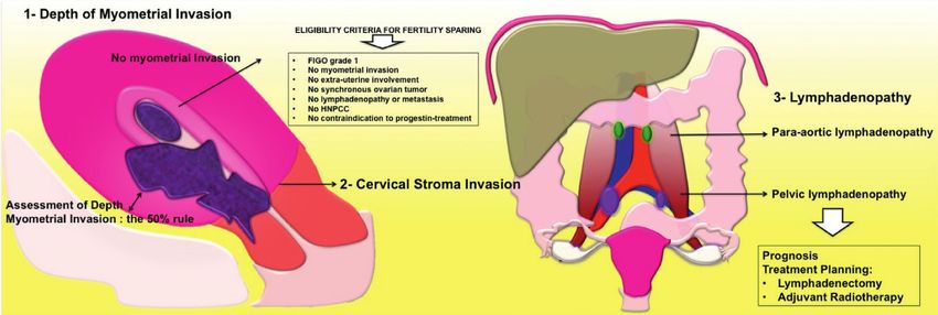
20 2. Disease Specific Investigations3.2 Key points (FIGURE 1):
• DEPTH OF MYOMETRIAL INVASION:
¡¡ Evaluation in the oblique plane perpendicular to the
endometrial cavity is mandatory to correctly assess depth of
myometrial invasion (5,6,7)
¡¡ Endometrial cancer is usually isointense to the myometrium
on T1WI and hyperintense to the myometrium on T2WI.
¡¡ On contrast-enhanced T1WI, the tumor usually enhances
homogeneously and more slowly and less avidly than the
adjacent myometrium.
¡¡ On DWI, the tumor is of high signal intensity with restricted
diffusion on the ADC maps (7).
¡¡ An intact junctional zone and a smooth band of early
subendometrial enhancement exclude deep myometrial
invasion.
¡¡ In stage IB, the tumor extends > 50% into the myometrium,
with associated disruption or irregularity of the junctional
zone and subendometrial enhancement. The presence of
low-signal-intensity tumor within the outer myometrium or
beyond indicates deep myometrial invasion (5,6,7)
• EXTRA-UTERINE EXTENSION:
¡¡ On T2WI, cervical stromal invasion is diagnosed by
intermediate to high-signal-intensity tumor disrupting the
normal low-signal-intensity cervical stroma.
¡¡ Disruption of the low-signal-intensity of the uterine serosa
and/or irregular uterine contour on T2WI, loss of the normal
rim of highly enhancing myometrium on DCE indicate serosal
involvement.
¡¡ DWI can aid detection of adnexa deposit, especially in high-
grade endometrioid, clear cell, or serous papillary tumors.
¡¡ Bullous edema of the bladder maybe a sign of tumor in the
subserosal or muscular layer of the bladder, but this sign
alone is not sufficient for diagnosis of stage IVA disease. On
T2WI, bladder/rectal involvement is diagnosed when tumor
abuts or indents the bladder/rectum over a significant area,
tumor interrupts the low signal intensity of the bladder/rectal
muscular layer or tumor invades the bladder/rectal muscular
wall and tumor nodules are seen in the mucosal layer.
2. Disease Specific Investigations 213.3 Pitfalls
• Overestimation of depth of myometrial invasion may be
caused by leiomyomas, adenomyosis and cornual tumor
location.
• Causes of false-positive cases for malignancy on DWI are
secretory and hyperplastic endometrium with consequently
low ADC values. Blood product retention also demonstrates
low ADC; T1W sequence can help to confirm presence of
blood products.
• Lymph nodes can be easily detected on DW-MRI as high
signal intensity ovoid structures. Attempts to increase
diagnostic accuracy between malignant and benign lymph
nodes based on ADC have been made. All published studies
showed a certain degree of overlap between benign and
malignant pelvic lymph nodes; none were able to define the
negative predictive value of DWI.
3.4 Radiology Report Checklist (FIGURE 1)
• The radiology report must be interpreted in conjunction with
tumor grade and type. Indeed, grade 3 tumor and serous/
clear cell adenocarcinomas have a more aggressive behavior
and the probability of advanced disease is higher (8,9).
• Tumor size
• Depth of myometrial invasion (8,9).
• Cervical stromal invasion (8,9).
• Vagina, adnexa and serosal invasion (8,9).
• Invasion of the bladder, rectum or pelvic wall (8,9).
• Pelvic, inguinal and para-aortic lymph nodes with dimensional
or morphological criteria of suspicion (8,9).
• Presence of hydronephrosis (8,9).
• Distant metastasis (8,9).
• Assessment of FIGO staging on MRI
22 2. Disease Specific InvestigationsReferences:
1. Ferlay J, Steliarova-Foucher E, Lortet-Tieulent J, et al.Cancer
incidence and mortality patterns in Europe: Estimates for 40
countries in 2012. European Journal of Cancer (2013) 49, 1374-1403
2. Larson DM, Connor GP, Broste SK, Krawisz BR, Johnson KK.
Prognostic signifi cance of gross myometrial invasion with
endometrial cancer. Obstet Gynecol 1996;88(3):394– 398.
3. Sala E., Rockall A.G., Freeman S.J., Mitchell D.G:, Reinhold C.
The Added Role of MR Imaging in Treatment Stratification of
Patients with Gynecologic Malignancies: What the Radiologist
Needs to Know. Radiology 2013; 266:717-40.
4. Vargas HA, Akin O, Zheng J, et al.. The value of MR imaging
when the site of uterine cancer origin is uncertain. Radiology
2011;258(3):785–792.
5. Nougaret S, Horta M, Sala E et al. Endometrial Cancer MRI
staging: Updated Guidelines of the European Society of Urogenital
Radiology. Eur Radiol 2018 Ehead epub of print.
6. Frei KA, Kinkel K, Bonél HM, Lu Y, Zaloudek C, Hricak H. Prediction
of deep myometrial invasion in patients with endometrial cancer:
clinical utility of contrast-enhanced MR imaging-a metaanalysis and
Bayesian analysis. Radiology 2000;216(2):444–449.
7. Beddy P, Moyle P, Kataoka M, et al.. Evaluation of depth of
myometrial invasion and overall staging in endometrial cancer:
comparison of diffusion-weighted and dynamic contrast-enhanced
MR imaging. Radiology 2012;262(2):530–537.
8. Sala E, Rockall A, Kubik-Huch RA. Advances in magnetic resonance
imaging of endometrial cancer. Eur Radiol 2011;21(3):468– 473
9. Kinkel K, Forstner R, Danza FM, et al. Staging of endometrial
cancer with MRI: guidelines of the European Society of Urogenital
Imaging. Eur Radiol 2009;19(7):1565–1574.
2. Disease Specific Investigations 23FIGURE 1 - Key points to analyse on MRI 24 2. Disease Specific Investigations
c. Vaginal Masses
Authors: Laura Brunesch and Celine Alt

1. Clinical background:
Primary cancer of the vagina represents only around 3% of all
gynecologic cancers. Squamous cell carcinoma is the most common
histologic type (90%), it is frequently associated with high-risk human
papillomavirus infection (especially type 16) and spreads early into
adjacent organs. Secondary involvement of the vagina by neoplasms
of adjacent organs or metastasis is more common [1]computed
tomography (CT. Lymphatic spread from upper vaginal neoplasms is to
internal and external iliac nodes initially, whereas lesions of the lower
third of the vagina initially spread to inguinal and femoral nodes [2].

2. Imaging:
The role of imaging in vaginal masses is the report of the localization,
shape, size and extension of the lesion and to exclude differential
diagnoses [3]. If malignancy is proven, imaging is used for primary
staging, therapy control or detection of recurrence.

Imaging techniques:
MRI of the pelvis is the modality of choice for local evaluation due
to an excellent soft tissue contrast and the possibility to differentiate
between primary vaginal location or invasion from surrounding organs
[4]. It is also useful for the evaluation of local recurrence [5].
Instillation of 60ml of ultrasound gel into the vagina is helpful. T2W
images of the pelvis in axial, sagittal and axial oblique plane (to the
vagina), T1W images of the pelvis in axial plane and DWI of the pelvis in
axial plane should at least be performed. Pre and dynamic T1W contrast
enhanced imaging of the pelvis in the axial plane with fat saturation,
and T1W post contrast sequence of the pelvis with fat saturation in
sagittal plane may optionally be useful.
Regarding patient’s positioning, technical requirements and detailed
information of MR sequences see „Technical aspects of MRI of the
female pelvis“ chapter.

                                     2. Disease Specific Investigations     25
Helical CT of the chest, abdomen and pelvis after IV contrast
administration is used to detect distant metastasis and nodal spread
[5]. Sagittal and coronal reconstructions should be performed. CT is not
the modality of choice for local staging. For more detailed information
about the technical aspects of CT, please see the „Technical aspects of
CT of the female pelvis“ chapter.
Sonography can be of use to evaluate inguinal groin nodes and to guide
their biopsy if suspicious [6].
3. Diagnosis:
Vaginal cancer is almost always diagnosed clinically by biopsy. It may
be asymptomatic in early stages, whereas in advanced stages, it
may present with pelvic pain or bloody vaginal discharge. Differential
diagnoses for vaginal masses may be leiomyoma, retention cyst,
endometriosis, Gartner cyst, lymphoma, metastasis, melanoma or
sarcoma [7].
4. Staging:
Staging for vaginal cancer is performed according to the current version
of the FIGO-classification or the UICC-criteria. Only lesions that do not
involve the cervix or the vulva are defined as primary vaginal cancer.
5. Follow-up:
Recurrence of squamous cell carcinoma mostly occurs in the first
two years after treatment, therefore close clinical follow-up is
recommended. MR is used for the evaluation of suspected local
recurrences. A scheduled follow-up with CT is reasonable for patients
with initially bulky or advanced disease. The application of PET may be
helpful, but up to now, only few data are available [8, 9].
26 2. Disease Specific InvestigationsReferences:
1. Walker DK, Salibian R a., Salibian a. D, et al. (2011) Overlooked
Diseases of the Vagina: A Directed Anatomic-Pathologic Approach
for Imaging Assessment. Radiographics 31:1583–1598. doi:
10.1148/rg.316115531
2. Rajaram S, Maheshwari A, Srivastava A (2015) Staging for vaginal
cancer. Best Pract Res Clin Obstet Gynaecol 29:822–832. doi:
10.1016/j.bpobgyn.2015.01.006
3. Elsayes KM, Narra VR, Dillman JR, et al. (2007) Vaginal
masses: magnetic resonance imaging features with pathologic
correlation. Acta Radiol 48:921–933. doi: 782896630
[pii]10.1080/02841850701552926
4. Gardner CS, Sunil J, Klopp AH, et al. (2015) Primary vaginal cancer:
role of MRI in diagnosis, staging and treatment. Br J Radiol
88:20150033. doi: 10.1259/bjr.20150033
5. Lee LJ, Jhingran A, Kidd E, et al. (2013) Acr appropriateness Criteria
management of vaginal cancer. Oncology (Williston Park) 27:1166–73.
6. Esen G (2006) Ultrasound of superficial lymph nodes. Eur J Radiol
58:345–359. doi: 10.1016/j.ejrad.2005.12.039
7. Zaspel U, Hamm B (2007) Vagina. MRI CT Female Pelvis
8. Kitajima K, Suenaga Y, Ueno Y, et al. (2014) Value of fusion of PET
and MRI in the detection of intra-pelvic recurrence of gynecological
tumor: Comparison with 18F-FDG contrast-enhanced PET/CT and
pelvic MRI. Ann Nucl Med 28:25–32. doi: 10.1007/s12149-013-0777-6
9. Robertson NL, Hricak H, Sonoda Y, Sosa RE, Benz M, Lyons G,
Abu-Rustum NR, Sala E VH (2015) The impact of FDG-PET/CT in the
management of patients with vulvar and vaginal cancer. Gynecol
Oncol http://dx.doi.org/10.1016/j.ygyno.2016.01.011.
2. Disease Specific Investigations 27d. Characterisation of Adnexal Mass
Auhtors: Olivera Nikolic, Milagros Otero-Garcia and Laure Fournier
CLINICAL BACKGROUND
It is of great clinical importance to determine the nature of
sonographically indeterminate adnexal mass. The extent of necessary
surgery and who should perform it strongly depend on this judgment.
Benign masses may be managed conservatively or resected under
the care of a general gynaecologist, while malignant adnexal masses
require radical citoreductive surgery by a specialist surgeon with
expertise in gynaecological oncology (1, 2, 3).
1 - Who should be imaged and why?
Women with clinically suspected adnexal masses, especially if the
masses are combined with high serum level of CA-125.
2 - Imaging techniques
• US, Color Doppler (CD)
• Magnetic resonance (MR)
US-the first-line imaging study of women suspected to have an
adnexal mass (4). US is used to place adnexal mass into one of three
categories: 1. a benign mass, 2. a malignant mass, 3. an indeterminate
mass, using IOTA simple rules. The US indeterminate adnexal
mass is defined as the complex one, which even after including CD
assessment, cannot be confidentaly placed into either the benign or
malignant category; or the one for which the site of origin, from the
ovary, uterus or another pelvic structure has to be established (1).
28 2. Disease Specific InvestigationsUS PROTOCOL
• Transabdominal US (full bladder, in case of overdistension
imaging may be repeated after partial bladder emptying)
• Transvaginal sonogram (urinary bladder is preferably empty)
¡¡ When evaluating the adnexa, the first and most important
step is the identification of the ovaries, for assessing
the presence of adnexal pathology. The ovary should be
measured in three dimensions (width, length and depth) on
views obtained in two ortogonal planes.
¡¡ Adnexal region should be surveyed for masses and dilated
tubular structures
¡¡ If adnexal abnormality is noted, its relationship with the
ovaries and uterus should be evaluated (the size and US
characteristics of the adnexal mass should be documented).
¡¡ Spectral, CD and/or power Doppler (PD) US are the indicators
of the vascular characteristics of adnexal masses (5).
MRI
For women with indeterminate adnexal masses MR imaging is the
method of choice (6,7). In these women MRI imaging can reduce the
number of unnecessary surgery for benign lesions and the risk of
missing malignant lesions.
• Imaging: 1. T2W Sag of the pelvis, 2. T1W and T2W
sequences covering the mass in the same ortogonal plane
(axial or coronal) with similar slice thickness, 3. DWI Axial,
4. T1W DCE study
Option 1: If mass demonstrates high signal intensity on
T1WI→ Axial FSE T1W FS
Option 2: If doubts whether mass belongs to ovary or uterus
¡¡ Axial plan of the ovary FSE T1W FS
¡¡ Axial plan of the ovary FSE T2WI
¡¡ Axial plan of the ovary 3D T1W FS
Note: Axial plane of the ovary corresponds to the parallel plan of the
endometrial cavity (perfect coronal plane of the body of the uterus).
2. Disease Specific Investigations 29Table 1. Dominant signal characteristic of indeterminate adnexal
masses (1, 2, 3)
T1 ‘bright’ T2 solid Cystic-solid
Mature teratoma Leiomyoma Cystadenoma
Haemorrhagic cyst Fibroma/thecoma Cystadenofibroma
Endometrioma Struma ovarii Borderline tumour
Mucinous
Primary cancer Primary cancer
cystadenoma
Melanoma metastasis Metastasis Metastasis
Hydrosalpinx
Abscess
Staging and follow up (see chapter ovarian cancer)
Pitfalls
• Most indeterminate adnexal masses result from common
benign conditions.
• MR imaging has been shown to be highly accurate in
characterising indeterminate adnexal lesions on US.
• MR imaging scoring system with addition of functional
imaging techniques, including perfusion and diffusion-
weighted sequences has allowed new criteria to be added to
conventional MRI in characterization of indeterminate adnexal
masses at US (8).
Tips and tricks
• US demonstration of a solid component within a cystic mass
is the most important predictor of malignancy (9).
• MR imaging allows identification of blood products within
hemorrhagic masses that may mimic solid tumor at US.
• Fat-suppressed T1-weighted MR images may reveal small
amounts of fat, which allows diagnosis of a mature teratoma.
• Contrast-enhanced T1-weighted MR imaging depicts features
of malignancy such as enhancing mural nodules and/or
enhancing solid areas with or without necrosis (2).
30 2. Disease Specific InvestigationsReferences:
1. Spencer JA, Forstner R, Cunha TM, Kinkel K. ESUR guidelines for
MR imaging of the sonographically indeterminate adnexal mass: an
algorithimic approach. Eur Radiol 2010; 20: 25-35.
2. Spencer JA, Ghattamaneni S. MR imaging of the sonographically
indeterminate adnexal mass. Radiology 2010; 256: 677-694.
3. Forstner R et al. ESUR recommendations for MR imaging of the
sonographically indeterminate adnexal mass: an update. Eur Radiol
2017; 27: 2248-2257.
4. Thomassin-Nagarra I et al. Characterization of complex adnexal
masses: value of adding perfusion and diffusion-weighted MR
imaging to conventional MR imaging. Radiology 2011; 258 (3): 793-
803.
5. AIUM, ACR, ACOG, SPR, SRU. AIUM practice guideline for the
performance of ultrasound of the female pelvis. J Ultrasound Med
2014; 33:1122-1130.
6. Adusumilli et al. MRI of sonographically indeterminate adnexal
masses. AJR 2006;187:732-740.
7. Sohaib SAA, Sahdev A, Trappen PV, Jacobs IJ, Reznek RH.
Characterization of adnexal mass lesions on MR imaging. AJR
2003; 180: 1297-1304.
8. Thomassin-Nagarra I et al. Adnexal Masses: Development and
Preliminary Validation of an MR Imaging Scoring System. Radiology
2013; 267: 432–443.
9. Brown DL, Dudiak KM, Laing FC. Adnexal masses: US
characterization and reporting. Radiology 2010; 254: 342-354.
2. Disease Specific Investigations 31e. Staging and Follow-up of Ovarian Cancer
Authors: Milagros Otero-Garcia, Olivera Nikolic and Laure Fournier
Clinical background
Ovarian cancer (OC) is a genetically heterogeneous disease with a
poor prognosis, mostly represented by epithelial cancers (95%) among
which high-grade serous OC are the most frequent. Fallopian tube
cancer and primary peritoneal cancer are included in the same staging
system by the International Federation of Gynecology and Obstetrics
(FIGO). The extent and anatomic location of peritoneal spread dictates
the choice between cytoreductive primary surgery versus neoadjuvant
chemotherapy. The accurate mapping of the disease using imaging
plays a crucial role in treatment selection and thus directly influences
patient outcome (1, 2).
Imaging
1-Who should be imaged and why?
Women with diagnosed (or highly suspected) OC for staging and
follow‑up.
Clinical Features:
In its early stages, OC is often asymptomatic. Symptomatic OC is more
frequently associated with advanced disease. Large adnexal masses
may be palpable at abdominal/pelvic examination. Other symptoms
include: bloating, pelvic or abdominal pain, difficulty eating or feeling full
quickly, and urinary symptoms (2). Serum tumor markers may be helpful
in making the diagnosis (3).
CT or US-guided Fine needle aspiration (FNA) or percutaneous
core biopsy
They should only be performed in patients with peritoneal
dissemination who are not surgical candidates, to rule out other
cancers (bowel, pancreatic, lymphoma...).
32 2. Disease Specific Investigations2-Imaging techniques
Ultrasound and MRI are used to detect and characterize adnexal
masses (see adnexal mass chapter). Contrast enhanced CT, MRI and
PET/CT are used for staging and follow-up (4).
For contrast enhanced CT technical details see table below.
3-Diagnosis
US is the first diagnostic tool to detect an ovarian mass. If the mass is
indeterminate at US, an MRI should be performed (see adnexal mass
chapter) (4).
4-Staging
Contrast enhanced CT: identifies eligible patients for complete
cytoreductive surgery, establishes a precise mapping of peritoneal
lesions, and anticipates possible surgical difficulties (5, 6).
MRI: Per-lesion sensitivity is higher for MRI (particularly using DW
sequences) for implants smaller than 1 cm, in anatomic areas where
small tumor implants are adjacent to tissues with similar signal
intensity, in detecting small peritoneal implants, and investigating
bladder or rectal involvement (7).
PET/CT: has high sensitivity in identifying positive lymph nodes larger than
7 mm, distant metastases, and in identifying patients not candidates for
an optimal debulking and who may need preoperative chemotherapy (8).
2. Disease Specific Investigations 33Table: primary OC staging with contrast enhanced CT
Key points of radiology report
FIGO stage I: (diagnosis using MRI, see adnexal mass chapter)
Disease limited to
the adnexa
FIGO stage II: - Rectosigmoid involvement
Disease spreading - Pelvic lymph nodes 10 mm short axis
to the pelvis or
Possibly non-resectable
primary peritoneal
cancer - Bladder (MRI > CT), trigone +++
- Pelvic wall
FIGO stage III: - Amount of ascites
Disease spreading - Retroperitoneal lymph nodes 10 mm short axis
to peritoneum
Possibly non-resectable
outside the pelvis
or retroperitoneal - “Liver hilum” i.e. porta hepatis, gastrohepatic
lymph nodes ligament, intersegmental fissure, gallbladder
fossa
- Lesser sac
- Mesenteric involvement
- Bowel loops (requiring > 3 resections)
- Supra-renal retroperitoneal lymph nodes 10 mm
short axis
FIGO stage IV: - Umbilical deposit,
Parenchymal - Inguinal lymph nodes,
metastasis or
- Pleural effusion, thickening
extra-abdominal
dissemination - Cardiophrenic lymph node 5 mm short axis
Possibly non-resectable
- Parenchymal hepatic or splenic metastasis
Complications - Intestinal obstruction
- Hydronephrosis
- Obstruction or venous thrombosis
34 2. Disease Specific Investigations5-Follow-up
Imaging will be performed if patients present symptoms or an elevation
of tumor markers.
Contrast enhanced CT: is the standard imaging technique for
evaluation of suspected recurrence of OC. The CT protocol is the same
as staging protocol.
PET/CT: has the greatest utility in those patients with rising CA 125
levels and negative conventional imaging results.
6-Pitfalls
One of the most frequent mistakes is erroneously reporting deposits on
the liver visceral peritoneum as parenchymal metastases, particularly
when there is important scalloping. There are significant challenges in
detecting the extent of small volume peritoneal disease.
7-Tips and tricks
Read the CT by posting transverse and coronal reconstructions side by
side.
Start reading by looking for the most frequent locations of peritoneal
deposits, namely: greater omentum, right subphrenic space (diaphragm
and liver surface) and paracolic gutters.
2. Disease Specific Investigations 358-Key references
1.- Prat, J. and F.C.o.G. Oncology: Staging classification for cancer of
the ovary, fallopian tube, and peritoneum. Int J Gynaecol Obstet
124: p. 1-5, 2014
2.- (NCCN Guidelines ®) Ovarian Cancer. 20th Edition (2015).
www.trikobe.org/nccn/guideline/gynecological/english/ovarian.pdf.
Accessed December 27, 2015.
3.- Montagnana M, Danese E, Ruzzenente O, Bresciani V, Nuzzo T,
Salvagno GL, Franchi M, Lippi G, Guidi GC. The ROMA (Risk of
Ovarian Malignancy Algorithm) for estimating the risk of epithelial
ovarian cancer in women presenting with pelvic mass: is it really
useful?. Clin Chem Lab Med 2011 Mar;49(3):521-5.
4.- Mohaghegh P, Rockall AG. Imaging Strategy for Early Ovarian
Cancer: Characterization of Adnexal Masses with Conventional and
Advanced Imaging Techniques. Radiographics 2012; 32:1751-1773.
5.- Donald G. Mitchell, Marcia C. Javitt, Phyllis Glanc, Genevieve
L. Bennett, Douglas L. Brown, Theodore Dubinsky, Mukesh
G.Harisinghani, Robert D. Harris, Neil S. Horowitz, Pari V.
Pandharipande, Harpreet K. Pannu, Ann E. Podrasky, Henry D.
Royal, Thomas D. Shipp, Cary Lynn Siegel, Lynn Simpson, Jade J.
Wong-You-Cheong, Carolyn M. Zelop. ACR Appropriateness Criteria
staging and follow-up of ovarian cancer. J Am Coll Radiol. 2013
Nov;10(11):822-7. doi: 10.1016/j.jacr.2013.07.017.
6.- Atul B. Shinagare, Ailbhe C. O’Neill, SuChun Cheng, Bhanusupriya
Somarouthu, Sree H. Tirumani, Mizuki Nishino, Annick D. Van den
Abbeele, Nikhil H. Ramaiya. Advanced High-Grade Serous Ovarian
Cancer: Frequency and Timing of Thoracic Metastases and
the Implications for Chest Imaging Follow-up. Radiology 2015;
277:733–740.
7.- Michielsen K, Vergote I, Op de Beeck K, Amant F, Leunen K,
Moerman P, Deroose C, Souverijns G, Dymarkowski S, De Keyzer F,
Vandecaveye V. Whole-body MRI with diffusion-weighted sequence
for staging of patients with suspected ovarian cancer: a clinical
feasibility study in comparison to CT and FDG-PET/CT. Eur Radiol.
2014 Apr;24(4):889-901.
8.- Priyanka Prakash, Carmel G. Cronin, Michael A. Blake. Role of PET/
CT in Ovarian Cancer. AJR 2010; 194:W464–W470.
36 2. Disease Specific InvestigationsRecommended CT protocol:
Preparation Oral contrast: to drink 30-60 min before CT study
(unless signs of bowel obstruction):
negative (water) or positive (gastrografin®)
i.v. contrast 300-350 mg I/mL, 100-120 mL/ 2.5-3.5 mL/s
Acquisition Portal phase (60-80s post injection):
Lung bases ( 4-5 cm above right diaphragm)
to pubic symphysis
Chest (arterial phase (35s) or portal phase):
optional
Post-processing Transverse, coronal and sagittal MPR:
2-3mm thickness, soft kernel
Lung kernel reconstruction
(1mm slices and MIP reconstructions)
2. Disease Specific Investigations 37f. Endometriosis
Author: Nishat Bharwani
Clinical background
Endometriosis is a common gynaecological condition that is defined as
functional ectopic endometrial glands and stroma outside the uterine
cavity. It predominantly affects women of reproductive age (between
25 and 40 years) with a prevalence of approximately 10%. The aetiology
and pathogenesis remain unknown although several theories have been
proposed including metastatic, metaplastic and induction theories.
There is a wide spectrum of clinical presentations ranging from
asymptomatic to women with non-specific pelvic pain, dysmenorrhoea,
dyspareunia and infertility. Symptoms do not always correlate with the
extent or severity of disease, but depend primarily on the site of the
endometriotic deposit(s). Sequelae of endometriosis can include deep
pelvic fibrosis and rarely an endometriosis-associated neoplasm.
Endometriosis involves the ovaries in up to 60% of patients. Extra-
ovarian pelvic locations include the uterine ligaments, posterior
cul de sac (pouch of Douglas), pelvic peritoneum, fallopian tubes,
rectosigmoid, anterior cul de sac (vesicouterine pouch), laparotomy
scars and the urinary bladder in decreasing frequency. Extra-peritoneal
sites such as lung, pleura and central nervous system have been
reported but are extremely rare.
The treatment options include conservative management, medical
therapy, surgery or a combination of these options. Presenting
symptoms and the desire to preserve fertility play an important role in
the choice of the most appropriate treatment strategy.
38 2. Disease Specific InvestigationsImaging
Indication:
Imaging is performed where there is a clinical suspicion of
endometriosis but equivocal clinical history and examination.
In addition magnetic resonance imaging (MRI) is indicated where
an adnexal lesion remains indeterminate on US or if there is clinical
suspicion of malignant transformation.
Imaging techniques:
Pelvic ultrasound (US) is performed as the first line imaging modality
however MRI is increasingly performed as an additional investigation in
complex cases and for surgical planning.
Computed tomography (CT) plays a limited role in the evaluation of
endometriosis.
There is no role for plain radiography or nuclear medicine studies.
Diagnosis:
The gold standard technique for the diagnosis of pelvic endometriosis
is laparoscopy with biopsy of the suspicious lesions for histologic
confirmation of the presence of ectopic endometrial glands.
There are three forms of pelvic endometriosis:
i. Ovarian endometrioma (most common location)
ii. Superficial peritoneal lesions which are non-invasive
iii. Deep pelvic endometriosis (DPE) where endometrial glands and
stroma invade at least 5mm below the peritoneal surface.
As the majority of patients have either no or non-specific symptoms,
diagnosis can be challenging and imaging (particularly transvaginal
US and MRI) is increasingly used as a non-invasive tool for initial
assessment.
2. Disease Specific Investigations 39Ultrasound (US)
• 1st line imaging modality
• Both transabdominal US and TVUS should be performed
• Positives: Easily accessible, cost-effective and widely available
• Negatives: highly operator dependent, limited field of view
• Local tenderness provoked by gentle pressure form the TVUS
probe can be used to guide assessment
• Use of bowel preparation and rectal water can improve
accuracy for detection of DPE and intestinal lesions
• Typical appearances:
¡¡ Ovarian endometrioma: persistent cystic lesion with
diffuse low level internal echoes, multilocularity and
hyperechoic foci within the wall. However appearances
can be quite variable.
¡¡ DPE: endometriotic plaques are generally hypoechoic
when compared to myometrium and often contain
multiple bright foci or small cystic areas. Margins are
often ill-defined and there is limited movement of organs
with gentle probe pressure.
Magnetic Resonance Imaging (MRI)
• Utilised where clinical examination and US are inconclusive
and also for pre-surgical planning.
• Imaging can be performed at 1.5T or 3.0T
• Pelvic phased array coil recommended
• Patient preparation:
¡¡ Fast 3-6 hours prior to the examination
¡¡ Bowel preparation and abdominal strapping/contention
are recommended by several centres
¡¡ Use of an anti-peristaltic agent to limit bowel motion
artefact is advocated unless contra-indicated
¡¡ Vaginal and/or rectal opacification are optional and have
been reported to increase accuracy in some cases
40 2. Disease Specific InvestigationsYou can also read