Third universal definition of myocardial infarction
←
→
Page content transcription
If your browser does not render page correctly, please read the page content below
European Heart Journal (2012) 33, 2551–2567                               EXPERT CONSENSUS DOCUMENT
               doi:10.1093/eurheartj/ehs184

Third universal definition of myocardial infarction
Kristian Thygesen, Joseph S. Alpert, Allan S. Jaffe, Maarten L. Simoons,
Bernard R. Chaitman and Harvey D. White: the Writing Group on behalf of the Joint
ESC/ACCF/AHA/WHF Task Force for the Universal Definition of Myocardial
Infarction

Authors/Task Force Members Chairpersons: Kristian Thygesen (Denmark)*,
Joseph S. Alpert, (USA)*, Harvey D. White, (New Zealand)*, Biomarker
Subcommittee: Allan S. Jaffe (USA), Hugo A. Katus (Germany), Fred S. Apple (USA),
Bertil Lindahl (Sweden), David A. Morrow (USA), ECG Subcommittee:
Bernard R. Chaitman (USA), Peter M. Clemmensen (Denmark), Per Johanson
(Sweden), Hanoch Hod (Israel), Imaging Subcommittee: Richard Underwood (UK),
Jeroen J. Bax (The Netherlands), Robert O. Bonow (USA), Fausto Pinto (Portugal),
Raymond J. Gibbons (USA), Classification Subcommittee: Keith A. Fox (UK), Dan Atar
(Norway), L. Kristin Newby (USA), Marcello Galvani (Italy), Christian W. Hamm
(Germany), Intervention Subcommittee: Barry F. Uretsky (USA), Ph. Gabriel Steg
(France), William Wijns (Belgium), Jean-Pierre Bassand (France), Phillippe Menasché
(France), Jan Ravkilde (Denmark), Trials & Registries Subcommittee:
E. Magnus Ohman (USA), Elliott M. Antman (USA), Lars C. Wallentin (Sweden),
Paul W. Armstrong (Canada), Maarten L. Simoons (The Netherlands), Heart Failure
Subcommittee: James L. Januzzi (USA), Markku S. Nieminen (Finland),
Mihai Gheorghiade (USA), Gerasimos Filippatos (Greece), Epidemiology
Subcommittee: Russell V. Luepker (USA), Stephen P. Fortmann (USA),
Wayne D. Rosamond (USA), Dan Levy (USA), David Wood (UK), Global Perspective
Subcommittee: Sidney C. Smith (USA), Dayi Hu (China), José-Luis Lopez-Sendon
(Spain), Rose Marie Robertson (USA), Douglas Weaver (USA), Michal Tendera
(Poland), Alfred A. Bove (USA), Alexander N. Parkhomenko (Ukraine),
Elena J. Vasilieva (Russia), Shanti Mendis (Switzerland).
ESC Committee for Practice Guidelines (CPG): Jeroen J. Bax, (CPG Chairperson) (Netherlands),
Helmut Baumgartner (Germany), Claudio Ceconi (Italy), Veronica Dean (France), Christi Deaton (UK),
Robert Fagard (Belgium), Christian Funck-Brentano (France), David Hasdai (Israel), Arno Hoes (Netherlands),
Paulus Kirchhof (Germany/UK), Juhani Knuuti (Finland), Philippe Kolh (Belgium), Theresa McDonagh (UK),
Cyril Moulin (France), Bogdan A. Popescu (Romania), Željko Reiner (Croatia), Udo Sechtem (Germany),
Per Anton Sirnes (Norway), Michal Tendera (Poland), Adam Torbicki (Poland), Alec Vahanian (France),
Stephan Windecker (Switzerland).

* Corresponding authors/co-chairpersons: Professor Kristian Thygesen, Department of Cardiology, Aarhus University Hospital, Tage-Hansens Gade 2, DK-8000 Aarhus C,
Denmark. Tel: +45 7846-7614; fax: +45 7846-7619: E-mail: kristhyg@rm.dk. Professor Joseph S. Alpert, Department of Medicine, Univ. of Arizona College of Medicine, 1501
N. Campbell Ave., P.O. Box 245037, Tucson AZ 85724, USA, Tel: +1 520 626 2763, Fax: +1 520 626 0967, Email: jalpert@email.arizona.edu. Professor Harvey D. White,
Green Lane Cardiovascular Service, Auckland City Hospital, Private Bag 92024, 1030 Auckland, New Zealand. Tel: +64 9 630 9992, Fax: +64 9 630 9915, Email: harveyw@
adhb.govt.nz.
& The European Society of Cardiology, American College of Cardiology Foundation, American Heart Association, Inc., and the World Heart Federation 2012. For permissions
please email: journals.permissions@oup.com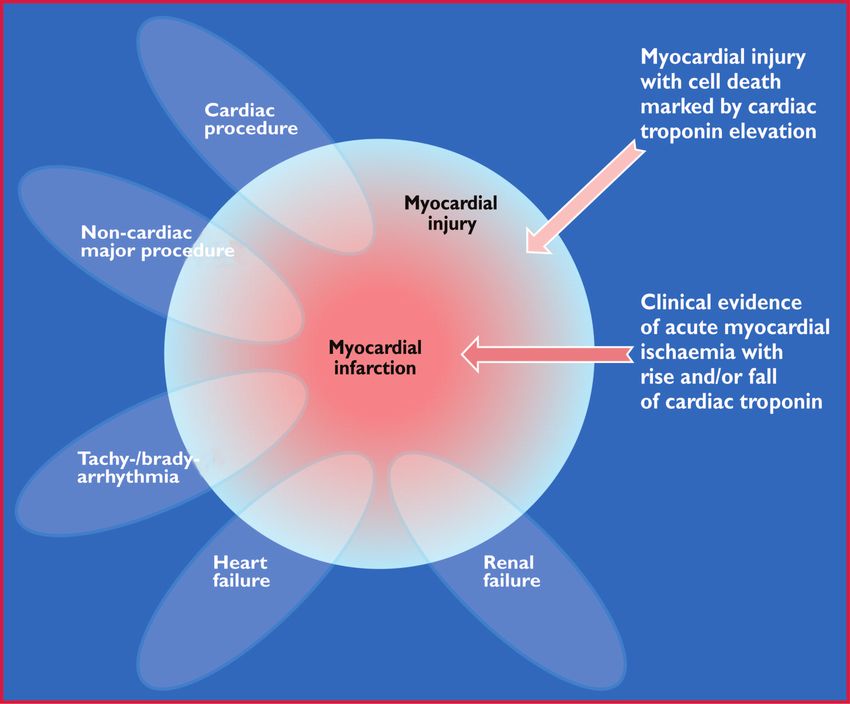
2552                                                                                                                         Expert Consensus Document

Document Reviewers: Joao Morais, (CPG Review Co-ordinator) (Portugal), Carlos Aguiar (Portugal),
Wael Almahmeed (United Arab Emirates), David O. Arnar (Iceland), Fabio Barili (Italy), Kenneth D. Bloch (USA),
Ann F. Bolger (USA), Hans Erik Bøtker (Denmark), Biykem Bozkurt (USA), Raffaele Bugiardini (Italy),
Christopher Cannon (USA), James de Lemos (USA), Franz R. Eberli (Switzerland), Edgardo Escobar (Chile),
Mark Hlatky (USA), Stefan James (Sweden), Karl B. Kern (USA), David J. Moliterno (USA), Christian Mueller
(Switzerland), Aleksandar N. Neskovic (Serbia), Burkert Mathias Pieske (Austria), Steven P. Schulman (USA),
Robert F. Storey (UK), Kathryn A. Taubert (Switzerland), Pascal Vranckx (Belgium), Daniel R. Wagner (Luxembourg)

The disclosure forms of the authors and reviewers are available on the ESC website www.escardio.org/guidelines

Online publish-ahead-of-print 24 August 2012

Table of Contents                                                                         Abbreviations and acronyms
Abbreviations and acronyms . . . . . . . . . . . . . . . . . . . . . . . . 2552
     Definition of myocardial infarction . . . . . . . . . . . . . . . . . 2553           ACCF      American College of Cardiology Foundation
     Criteria for acute myocardial infarction . . . . . . . . . . . . . . 2553            ACS       acute coronary syndrome
     Criteria for prior myocardial infarction . . . . . . . . . . . . . . 2553            AHA       American Heart Association
Introduction . . . . . . . . . . . . . . . . . . . . . . . . . . . . . . . . . . 2553     CAD       coronary artery disease
Pathological characteristics of myocardial ischaemia and infarction 2554                  CABG      coronary artery bypass grafting
Biomarker detection of myocardial injury with necrosis . . . . . . 2554                   CKMB      creatine kinase MB isoform
Clinical features of myocardial ischaemia and infarction . . . . . . 2555                 cTn       cardiac troponin
Clinical classification of myocardial infarction . . . . . . . . . . . . . 2556           CT        computed tomography
     Spontaneous myocardial infarction (MI type 1) . . . . . . . . . 2556                 CV        coefficient of variation
     Myocardial infarction secondary to an ischaemic imbalance                            ECG       electrocardiogram
     (MI type 2) . . . . . . . . . . . . . . . . . . . . . . . . . . . . . . . . 2556     ESC       European Society of Cardiology
     Cardiac death due to myocardial infarction (MI type 3) . . . 2557                    FDG       fluorodeoxyglucose
     Myocardial infarction associated with revascularization                              h         hour(s)
     procedures (MI types 4 and 5) . . . . . . . . . . . . . . . . . . . 2557             HF        heart failure
Electrocardiographic detection of myocardial infarction . . . . . . 2557                  LBBB      left bundle branch block
Prior myocardial infarction . . . . . . . . . . . . . . . . . . . . . . . . . 2558        LV        left ventricle
Silent myocardial infarction . . . . . . . . . . . . . . . . . . . . . . . . . 2559       LVH       left ventricular hypertrophy
Conditions that confound the ECG diagnosis of myocardial                                  MI        myocardial infarction
infarction . . . . . . . . . . . . . . . . . . . . . . . . . . . . . . . . . . . . 2559   mIBG      meta-iodo-benzylguanidine
Imaging techniques . . . . . . . . . . . . . . . . . . . . . . . . . . . . . . 2559       min       minute(s)
     Echocardiography . . . . . . . . . . . . . . . . . . . . . . . . . . . . 2559        MONICA    Multinational MONItoring of trends and determinants
     Radionuclide imaging . . . . . . . . . . . . . . . . . . . . . . . . . . 2559                  in CArdiovascular disease)
     Magnetic resonance imaging . . . . . . . . . . . . . . . . . . . . . 2560            MPS       myocardial perfusion scintigraphy
     Computed tomography . . . . . . . . . . . . . . . . . . . . . . . . 2560             MRI       magnetic resonance imaging
     Applying imaging in acute myocardial infarction . . . . . . . . . 2560               mV        millivolt(s)
     Applying imaging in late presentation of myocardial infarction 2560                  ng/L      nanogram(s) per litre
Diagnostic criteria for myocardial infarction with PCI (MI type 4) 2560                   Non-Q MI non-Q wave myocardial infarction
Diagnostic criteria for myocardial infarction with CABG                                   NSTEMI    non-ST-elevation myocardial infarction
(MI type 5) . . . . . . . . . . . . . . . . . . . . . . . . . . . . . . . . . . . 2561    PCI       percutaneous coronary intervention
Assessment of MI in patients undergoing other cardiac                                     PET       positron emission tomography
procedures . . . . . . . . . . . . . . . . . . . . . . . . . . . . . . . . . . . 2561     pg/mL     pictogram(s) per millilitre
Myocardial infarction associated with non-cardiac procedures . . 2562                     Q wave MI Q wave myocardial infarction
Myocardial infarction in the intensive care unit . . . . . . . . . . . . 2562             RBBB      right bundle branch block
Recurrent myocardial infarction . . . . . . . . . . . . . . . . . . . . . . 2562          sec       second(s)
Reinfarction . . . . . . . . . . . . . . . . . . . . . . . . . . . . . . . . . . . 2562   SPECT     single photon emission computed tomography
Myocardial injury or infarction associated with heart failure . . . 2562                  STEMI     ST elevation myocardial infarction
Application of MI in clinical trials and quality assurance                                ST– T     ST-segment – T wave
programmes . . . . . . . . . . . . . . . . . . . . . . . . . . . . . . . . . . 2563       URL       upper reference limit
Public policy implications of the adjustment of the MI definition 2563                    WHF       World Heart Federation
Global perspectives of the definition of myocardial infarction . . 2564                   WHO       World Health Organization
Conflicts of interest . . . . . . . . . . . . . . . . . . . . . . . . . . . . . 2564
Acknowledgements . . . . . . . . . . . . . . . . . . . . . . . . . . . . . . 2564
References . . . . . . . . . . . . . . . . . . . . . . . . . . . . . . . . . . . 2564
Expert Consensus Document 2553
Definition of myocardial infarction
Criteria for acute myocardial infarction
The term acute myocardial infarction (MI) should be used when there is evidence of myocardial necrosis in a clinical setting consistent with acute myocardial
ischaemia. Under these conditions any one of the following criteria meets the diagnosis for MI:
• Detection of a rise and/or fall of cardiac biomarker values [preferably cardiac troponin (cTn)] with at least one value above the 99th percentile upper
reference limit (URL) and with at least one of the following:
Symptoms of ischaemia.
New or presumed new significant ST-segment–T wave (ST–T) changes or new left bundle branch block (LBBB).
Development of pathological Q waves in the ECG.
Imaging evidence of new loss of viable myocardium or new regional wall motion abnormality.
Identification of an intracoronary thrombus by angiography or autopsy.
• Cardiac death with symptoms suggestive of myocardial ischaemia and presumed new ischaemic ECG changes or new LBBB, but death occurred before cardiac
biomarkers were obtained, or before cardiac biomarker values would be increased.
• Percutaneous coronary intervention (PCI) related MI is arbitrarily defined by elevation of cTn values (>5 x 99th percentile URL) in patients with normal
baseline values (≤99th percentile URL) or a rise of cTn values >20% if the baseline values are elevated and are stable or falling. In addition, either (i) symptoms
suggestive of myocardial ischaemia or (ii) new ischaemic ECG changes or (iii) angiographic findings consistent with a procedural complication or (iv) imaging
demonstration of new loss of viable myocardium or new regional wall motion abnormality are required.
• Stent thrombosis associated with MI when detected by coronary angiography or autopsy in the setting of myocardial ischaemia and with a rise and/or fall of
cardiac biomarker values with at least one value above the 99th percentile URL.
• Coronary artery bypass grafting (CABG) related MI is arbitrarily defined by elevation of cardiac biomarker values (>10 x 99th percentile URL) in patients
with normal baseline cTn values (≤99th percentile URL). In addition, either (i) new pathological Q waves or new LBBB, or (ii) angiographic documented new
graft or new native coronary artery occlusion, or (iii) imaging evidence of new loss of viable myocardium or new regional wall motion abnormality.
Criteria for prior myocardial infarction
Any one of the following criteria meets the diagnosis for prior MI:
• Pathological Q waves with or without symptoms in the absence of non-ischaemic causes.
• Imaging evidence of a region of loss of viable myocardium that is thinned and fails to contract, in the absence of a non-ischaemic cause.
• Pathological findings of a prior MI.
Introduction significantly improved, resulting in less myocardial injury and necro-
Myocardial infarction (MI) can be recognised by clinical features, in- sis, in spite of a similar clinical presentation. Moreover, it appears
cluding electrocardiographic (ECG) findings, elevated values of bio- necessary to distinguish the various conditions which may cause
chemical markers (biomarkers) of myocardial necrosis, and by MI, such as ‘spontaneous’ and ‘procedure-related’ MI. Accordingly,
imaging, or may be defined by pathology. It is a major cause of physicians, other healthcare providers and patients require an
death and disability worldwide. MI may be the first manifestation up-to-date definition of MI.
of coronary artery disease (CAD) or it may occur, repeatedly, in In 2000, the First Global MI Task Force presented a new
patients with established disease. Information on MI rates can definition of MI, which implied that any necrosis in the setting of
provide useful information regarding the burden of CAD within myocardial ischaemia should be labelled as MI.1 These principles
and across populations, especially if standardized data are collected were further refined by the Second Global MI Task Force,
in a manner that distinguishes between incident and recurrent leading to the Universal Definition of Myocardial Infarction
events. From the epidemiological point of view, the incidence of Consensus Document in 2007, which emphasized the different
MI in a population can be used as a proxy for the prevalence of conditions which might lead to an MI.2 This document, endorsed
CAD in that population. The term ‘myocardial infarction’ may by the European Society of Cardiology (ESC), the American
have major psychological and legal implications for the individual College of Cardiology Foundation (ACCF), the American Heart
and society. It is an indicator of one of the leading health problems Association (AHA), and the World Heart Federation (WHF), has
in the world and it is an outcome measure in clinical trials, obser- been well accepted by the medical community and adopted by
vational studies and quality assurance programmes. These studies the WHO.3 However, the development of even more sensitive
and programmes require a precise and consistent definition of MI. assays for markers of myocardial necrosis mandates further
In the past, a general consensus existed for the clinical syndrome revision, particularly when such necrosis occurs in the setting of
designated as MI. In studies of disease prevalence, the World the critically ill, after percutaneous coronary procedures or after
Health Organization (WHO) defined MI from symptoms, ECG cardiac surgery. The Third Global MI Task Force has continued
abnormalities and cardiac enzymes. However, the development the Joint ESC/ACCF/AHA/WHF efforts by integrating these
of ever more sensitive and myocardial tissue-specific cardiac insights and new data into the current document, which now
biomarkers and more sensitive imaging techniques now allows recognizes that very small amounts of myocardial injury or necrosis
for detection of very small amounts of myocardial injury or necro- can be detected by biochemical markers and/or imaging.
sis. Additionally, the management of patients with MI has2554 Expert Consensus Document
Pathological characteristics kinase (CKMB) are increased.2 Cardiac troponin I and T are com-
ponents of the contractile apparatus of myocardial cells and are
of myocardial ischaemia expressed almost exclusively in the heart. Although elevations of
and infarction these biomarkers in the blood reflect injury leading to necrosis
of myocardial cells, they do not indicate the underlying mechan-
MI is defined in pathology as myocardial cell death due to pro- ism.5 Various possibilities have been suggested for release of struc-
longed ischaemia. After the onset of myocardial ischaemia, histo- tural proteins from the myocardium, including normal turnover of
logical cell death is not immediate, but takes a finite period of myocardial cells, apoptosis, cellular release of troponin degradation
time to develop—as little as 20 min, or less in some animal products, increased cellular wall permeability, formation and
models.4 It takes several hours before myocardial necrosis can release of membranous blebs, and myocyte necrosis.6 Regardless
be identified by macroscopic or microscopic post-mortem exam- of the pathobiology, myocardial necrosis due to myocardial ischae-
ination. Complete necrosis of myocardial cells at risk requires at mia is designated as MI.
least 2–4 h, or longer, depending on the presence of collateral cir- Also, histological evidence of myocardial injury with necrosis
culation to the ischaemic zone, persistent or intermittent coronary may be detectable in clinical conditions associated with predomin-
arterial occlusion, the sensitivity of the myocytes to ischaemia, pre- antly non-ischaemic myocardial injury. Small amounts of myocar-
conditioning, and individual demand for oxygen and nutrients.2 The dial injury with necrosis may be detected, which are associated
entire process leading to a healed infarction usually takes at least with heart failure (HF), renal failure, myocarditis, arrhythmias, pul-
5 –6 weeks. Reperfusion may alter the macroscopic and micro- monary embolism or otherwise uneventful percutaneous or surgi-
scopic appearance. cal coronary procedures. These should not be labelled as MI or a
complication of the procedures, but rather as myocardial injury, as
Biomarker detection of illustrated in Figure 1. It is recognized that the complexity of clinical
circumstances may sometimes render it difficult to determine
myocardial injury with necrosis where individual cases may lie within the ovals of Figure 1. In this
Myocardial injury is detected when blood levels of sensitive and setting, it is important to distinguish acute causes of cTn elevation,
specific biomarkers such as cTn or the MB fraction of creatine which require a rise and/or fall of cTn values, from chronic
Figure 1 This illustration shows various clinical entities: for example, renal failure, heart failure, tachy- or bradyarrhythmia, cardiac or non-
cardiac procedures that can be associated with myocardial injury with cell death marked by cardiac troponin elevation. However, these entities
can also be associated with myocardial infarction in case of clinical evidence of acute myocardial ischaemia with rise and/or fall of cardiac
troponin.Expert Consensus Document 2555
elevations that tend not to change acutely. A list of such clinical cir- of variation (CV) at the 99th percentile URL for each assay, should
cumstances associated with elevated values of cTn is presented in be defined as ≤10%. Better precision (CV ≤10%) allows for more
Table 1. The multifactorial contributions resulting in the myocardial sensitive assays and facilitates the detection of changing values.13
injury should be described in the patient record. The use of assays that do not have optimal precision (CV .10%
The preferred biomarker—overall and for each specific category at the 99th percentile URL) makes determination of a significant
of MI—is cTn (I or T), which has high myocardial tissue specificity change more difficult but does not cause false positive results.
as well as high clinical sensitivity. Detection of a rise and/or fall of Assays with CV .20% at the 99th percentile URL should not be
the measurements is essential to the diagnosis of acute MI.7 An used.13 It is acknowledged that pre-analytic and analytic problems
increased cTn concentration is defined as a value exceeding the can induce elevated and reduced values of cTn.10,11
99th percentile of a normal reference population [upper reference Blood samples for the measurement of cTn should be drawn on
limit (URL)]. This discriminatory 99th percentile is designated as first assessment and repeated 3 –6 h later. Later samples are
the decision level for the diagnosis of MI and must be determined required if further ischaemic episodes occur, or when the timing
for each specific assay with appropriate quality control in each of the initial symptoms is unclear.14 To establish the diagnosis of
laboratory.8,9 The values for the 99th percentile URL defined by MI, a rise and/or fall in values with at least one value above the de-
manufacturers, including those for many of the high-sensitivity cision level is required, coupled with a strong pre-test likelihood.
assays in development, can be found in the package inserts for The demonstration of a rising and/or falling pattern is needed to
the assays or in recent publications.10,11,12 distinguish acute- from chronic elevations in cTn concentrations
Values should be presented as nanograms per litre (ng/L) or that are associated with structural heart disease.10,11,15 – 19 For
picograms per millilitre (pg/mL) to make whole numbers. Criteria example, patients with renal failure or HF can have significant
for the rise of cTn values are assay-dependent but can be defined chronic elevations in cTn. These elevations can be marked, as
from the precision profile of each individual assay, including high- seen in many patients with MI, but do not change acutely.7
sensitivity assays.10,11 Optimal precision, as described by coefficient However, a rising or falling pattern is not absolutely necessary to
make the diagnosis of MI if a patient with a high pre-test risk of
MI presents late after symptom onset; for example, near the
peak of the cTn time-concentration curve or on the slow-declining
Table 1 Elevations of cardiac troponin values because
portion of that curve, when detecting a changing pattern can be
of myocardial injury
problematic. Values may remain elevated for 2 weeks or more fol-
lowing the onset of myocyte necrosis.10
Injury related to primary myocardial ischaemia
Sex-dependent values may be recommended for high-sensitivity
Plaque rupture troponin assays.20,21 An elevated cTn value (.99th percentile
Intraluminal coronary artery thrombus formation
URL), with or without a dynamic pattern of values or in the
Injury related to supply/demand imbalance of absence of clinical evidence of ischaemia, should prompt a search
myocardial ischaemia
for other diagnoses associated with myocardial injury, such as myo-
Tachy-/brady-arrhythmias carditis, aortic dissection, pulmonary embolism, or HF. Renal failure
Aortic dissection or severe aortic valve disease and other more non-ischaemic chronic disease states, that can be
Hypertrophic cardiomyopathy
Cardiogenic, hypovolaemic, or septic shock associated with elevated cTn levels, are listed in Table 1.10,11
Severe respiratory failure If a cTn assay is not available, the best alternative is CKMB
Severe anaemia (measured by mass assay). As with troponin, an increased CKMB
Hypertension with or without LVH value is defined as a measurement above the 99th percentile
Coronary spasm
Coronary embolism or vasculitis
URL, which is designated as the decision level for the diagnosis
Coronary endothelial dysfunction without significant CAD of MI.22 Sex-specific values should be employed.22
Injury not related to myocardial ischaemia
Cardiac contusion, surgery, ablation, pacing, or defibrillator shocks
Rhabdomyolysis with cardiac involvement
Clinical features of myocardial
Myocarditis
Cardiotoxic agents, e.g. anthracyclines, herceptin
ischaemia and infarction
Onset of myocardial ischaemia is the initial step in the develop-
Multifactorial or indeterminate myocardial injury
ment of MI and results from an imbalance between oxygen
Heart failure
supply and demand. Myocardial ischaemia in a clinical setting can
Stress (Takotsubo) cardiomyopathy
Severe pulmonary embolism or pulmonary hypertension usually be identified from the patient’s history and from the
Sepsis and critically ill patients ECG. Possible ischaemic symptoms include various combinations
Renal failure of chest, upper extremity, mandibular or epigastric discomfort
Severe acute neurological diseases, e.g. stroke, subarachnoid
(with exertion or at rest) or an ischaemic equivalent such as dys-
haemorrhage
Infiltrative diseases, e.g. amyloidosis, sarcoidosis pnoea or fatigue. The discomfort associated with acute MI usually
Strenuous exercise lasts .20 min. Often, the discomfort is diffuse—not localized, nor
positional, nor affected by movement of the region—and it may be
accompanied by diaphoresis, nausea or syncope. However, these2556 Expert Consensus Document
symptoms are not specific for myocardial ischaemia. Accordingly, Spontaneous myocardial infarction
they may be misdiagnosed and attributed to gastrointestinal, (MI type 1)
neurological, pulmonary or musculoskeletal disorders. MI may
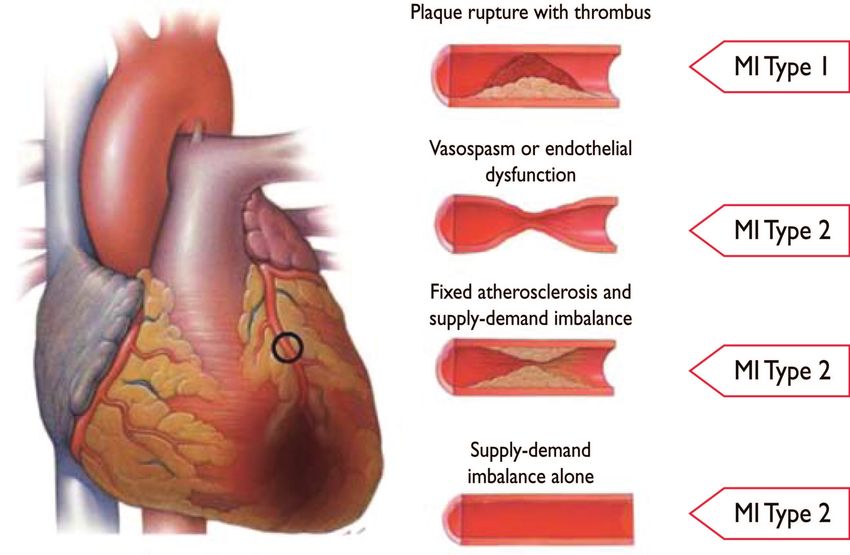
This is an event related to atherosclerotic plaque rupture, ul-
occur with atypical symptoms—such as palpitations or cardiac
ceration, fissuring, erosion, or dissection with resulting intralum-
arrest—or even without symptoms; for example in women, the
inal thrombus in one or more of the coronary arteries, leading
elderly, diabetics, or post-operative and critically ill patients.2
to decreased myocardial blood flow or distal platelet emboli
Careful evaluation of these patients is advised, especially when
with ensuing myocyte necrosis. The patient may have under-
there is a rising and/or falling pattern of cardiac biomarkers.
lying severe CAD but, on occasion (5 to 20%), non-obstructive
or no CAD may be found at angiography, particularly in
Clinical classification of women.23 – 25
myocardial infarction
For the sake of immediate treatment strategies, such as reperfusion
therapy, it is usual practice to designate MI in patients with chest Myocardial infarction secondary
discomfort, or other ischaemic symptoms that develop ST eleva- to an ischaemic imbalance (MI type 2)
tion in two contiguous leads (see ECG section), as an ‘ST elevation In instances of myocardial injury with necrosis, where a condition
MI’ (STEMI). In contrast, patients without ST elevation at presenta- other than CAD contributes to an imbalance between myocardial
tion are usually designated as having a ‘non-ST elevation MI’ oxygen supply and/or demand, the term ‘MI type 2’ is employed
(NSTEMI). Many patients with MI develop Q waves (Q wave (Figure 2). In critically ill patients, or in patients undergoing major
MI), but others do not (non-Q MI). Patients without elevated (non-cardiac) surgery, elevated values of cardiac biomarkers may
biomarker values can be diagnosed as having unstable angina. In appear, due to the direct toxic effects of endogenous or
addition to these categories, MI is classified into various types, exogenous high circulating catecholamine levels. Also coronary
based on pathological, clinical and prognostic differences, along vasospasm and/or endothelial dysfunction have the potential to
with different treatment strategies (Table 2). cause MI.26 – 28
Table 2 Universal classification of myocardial infarction
Type 1: Spontaneous myocardial infarction
Spontaneous myocardial infarction related to atherosclerotic plaque rupture, ulceration, fissuring, erosion, or dissection with resulting intraluminal thrombus in
one or more of the coronary arteries leading to decreased myocardial blood flow or distal platelet emboli with ensuing myocyte necrosis. The patient may have
underlying severe CAD but on occasion non-obstructive or no CAD.
Type 2: Myocardial infarction secondary to an ischaemic imbalance
In instances of myocardial injury with necrosis where a condition other than CAD contributes to an imbalance between myocardial oxygen supply and/or
demand, e.g. coronary endothelial dysfunction, coronary artery spasm, coronary embolism, tachy-/brady-arrhythmias, anaemia, respiratory failure, hypotension,
and hypertension with or without LVH.
Type 3: Myocardial infarction resulting in death when biomarker values are unavailable
Cardiac death with symptoms suggestive of myocardial ischaemia and presumed new ischaemic ECG changes or new LBBB, but death occurring before blood
samples could be obtained, before cardiac biomarker could rise, or in rare cases cardiac biomarkers were not collected.
Type 4a: Myocardial infarction related to percutaneous coronary intervention (PCI)
Myocardial infarction associated with PCI is arbitrarily defined by elevation of cTn values >5 x 99th percentile URL in patients with normal baseline values (£99th
percentile URL) or a rise of cTn values >20% if the baseline values are elevated and are stable or falling. In addition,either (i) symptoms suggestive of myocardial
ischaemia, or (ii) new ischaemic ECG changes or new LBBB, or (iii) angiographic loss of patency of a major coronary artery or a side branch or persistent slow-
or no-flow or embolization, or (iv) imaging demonstration of new loss of viable myocardium or new regional wall motion abnormality are required.
Type 4b: Myocardial infarction related to stent thrombosis
Myocardial infarction associated with stent thrombosis is detected by coronary angiography or autopsy in the setting of myocardial ischaemia and with a rise and/
or fall of cardiac biomarkers values with at least one value above the 99th percentile URL.
Type 5: Myocardial infarction related to coronary artery bypass grafting (CABG)
Myocardial infarction associated with CABG is arbitrarily defined by elevation of cardiac biomarker values >10 x 99th percentile URL in patients with normal
baseline cTn values (£99th percentile URL). In addition, either (i) new pathological Q waves or new LBBB, or (ii) angiographic documented new graft or new
native coronary artery occlusion, or (iii) imaging evidence of new loss of viable myocardium or new regional wall motion abnormality.Expert Consensus Document 2557
Figure 2 Differentiation between myocardial infarction (MI) types 1 and 2 according to the condition of the coronary arteries.
Cardiac death due to myocardial promptly (i.e. target within 10 min) after clinical presentation.2
infarction (MI type 3) Dynamic changes in the ECG waveforms during acute myocardial
ischaemic episodes often require acquisition of multiple ECGs, par-
Patients who suffer cardiac death, with symptoms suggestive of myo-
ticularly if the ECG at initial presentation is non-diagnostic. Serial
cardial ischaemia accompanied by presumed new ischaemic ECG
recordings in symptomatic patients with an initial non-diagnostic
changes or new LBBB—but without available biomarker values—
ECG should be performed at 15-30 min intervals or, if available,
represent a challenging diagnostic group. These individuals may die
continuous computer-assisted 12-lead ECG recording. Recur-
before blood samples for biomarkers can be obtained, or before ele-
rence of symptoms after an asymptomatic interval are an indica-
vated cardiac biomarkers can be identified. If patients present with
tion for a repeat tracing and, in patients with evolving ECG
clinical features of myocardial ischaemia, or with presumed new is-
abnormalities, a pre-discharge ECG should be acquired as a base-
chaemic ECG changes, they should be classified as having had a
line for future comparison. Acute or evolving changes in the ST–
fatal MI, even if cardiac biomarker evidence of MI is lacking.
T waveforms and Q waves, when present, potentially allow the
Myocardial infarction associated clinician to time the event, to identify the infarct-related artery,
to estimate the amount of myocardium at risk as well as progno-
with revascularization procedures
sis, and to determine therapeutic strategy. More profound
(MI types 4 and 5) ST-segment shift or T wave inversion involving multiple leads/ter-
Periprocedural myocardial injury or infarction may occur at some ritories is associated with a greater degree of myocardial ischae-
stages in the instrumentation of the heart that is required during mia and a worse prognosis. Other ECG signs associated with
mechanical revascularization procedures, either by PCI or by cor- acute myocardial ischaemia include cardiac arrhythmias, intraven-
onary artery bypass grafting (CABG). Elevated cTn values may be tricular and atrioventricular conduction delays, and loss of pre-
detected following these procedures, since various insults may cordial R wave amplitude. Coronary artery size and distribution
occur that can lead to myocardial injury with necrosis.29 – 32 It is of arterial segments, collateral vessels, location, extent and sever-
likely that limitation of such injury is beneficial to the patient: ity of coronary stenosis, and prior myocardial necrosis can all
however, a threshold for a worsening prognosis, related to an impact ECG manifestations of myocardial ischaemia.36 Therefore
asymptomatic increase of cardiac biomarker values in the the ECG at presentation should always be compared to prior
absence of procedural complications, is not well defined.33 – 35 Sub- ECG tracings, when available. The ECG by itself is often insuffi-
categories of PCI-related MI are connected to stent thrombosis cient to diagnose acute myocardial ischaemia or infarction,
and restenosis that may happen after the primary procedure. since ST deviation may be observed in other conditions, such
as acute pericarditis, left ventricular hypertrophy (LVH), left
Electrocardiographic detection bundle branch block (LBBB), Brugada syndrome, stress cardiomy-
opathy, and early repolarization patterns.37 Prolonged new
of myocardial infarction ST-segment elevation (e.g. .20 min), particularly when asso-
The ECG is an integral part of the diagnostic work-up of patients ciated with reciprocal ST-segment depression, usually reflects
with suspected MI and should be acquired and interpreted acute coronary occlusion and results in myocardial injury with2558 Expert Consensus Document
Electrocardiographic evidence of myocardial ischaemia in the dis-
Table 3 ECG manifestations of acute myocardial tribution of a left circumflex artery is often overlooked and is
ischaemia (in absence of LVH and LBBB) best captured using posterior leads at the fifth intercostal space
(V7 at the left posterior axillary line, V8 at the left mid-scapular
ST elevation line, and V9 at the left paraspinal border). Recording of these
New ST elevation at the J point in two contiguous leads with the leads is strongly recommended in patients with high clinical suspi-
cut-points: ≥0.1 mV in all leads other than leads V2–V3 where the cion for acute circumflex occlusion (for example, initial ECG non-
following cut points apply: ≥0.2 mV in men ≥40 years; ≥0.25 mV in men
diagnostic, or ST-segment depression in leads V1 – 3).41 A cut-point
1.
(posterior infarction), especially when the terminal T wave is posi-
tive (ST elevation equivalent), however this is non-specific.41 – 43 In
patients with inferior and suspected right ventricular infarction,
right pre-cordial leads V3R and V4R should be recorded, since
necrosis. As in cardiomyopathy, Q waves may also occur due to ST elevation ≥0.05 mV (≥0.1 mV in men ,30 years old) provides
myocardial fibrosis in the absence of CAD. supportive criteria for the diagnosis.42
ECG abnormalities of myocardial ischaemia or infarction may be During an episode of acute chest discomfort, pseudo-
inscribed in the PR segment, the QRS complex, the ST-segment or normalization of previously inverted T waves may indicate
the T wave. The earliest manifestations of myocardial ischaemia are acute myocardial ischaemia. Pulmonary embolism, intracranial
typically T wave and ST-segment changes. Increased hyperacute T processes, electrolyte abnormalities, hypothermia, or peri-/myo-
wave amplitude, with prominent symmetrical T waves in at least carditis may also result in ST– T abnormalities and should be con-
two contiguous leads, is an early sign that may precede the eleva- sidered in the differential diagnosis. The diagnosis of MI is more
tion of the ST-segment. Transient Q waves may be observed difficult in the presence of LBBB.44,45 However, concordant
during an episode of acute ischaemia or (rarely) during acute MI ST-segment elevation or a previous ECG may be helpful to de-
with successful reperfusion. Table 3 lists ST– T wave criteria for termine the presence of acute MI in this setting. In patients
the diagnosis of acute myocardial ischaemia that may or may not with right bundle branch block (RBBB), ST–T abnormalities in
lead to MI. The J point is used to determine the magnitude of leads V1 –V3 are common, making it difficult to assess the pres-
the ST-segment shift. New, or presumed new, J point elevation ence of ischaemia in these leads: however, when new ST eleva-
≥0.1mV is required in all leads other than V2 and V3. In healthy tion or Q waves are found, myocardial ischaemia or infarction
men under age 40, J-point elevation can be as much as 0.25 mV should be considered.
in leads V2 or V3, but it decreases with increasing age. Sex differ-
ences require different cut-points for women, since J point eleva-
tion in healthy women in leads V2 and V3 is less than in men.38 Prior myocardial infarction
‘Contiguous leads’ refers to lead groups such as anterior leads
(V1 –V6), inferior leads (II, III, aVF) or lateral/apical leads (I, aVL). As shown in Table 4, Q waves or QS complexes in the absence of
Supplemental leads such as V3R and V4R reflect the free wall of QRS confounders are pathognomonic of a prior MI in patients with
the right ventricle and V7 – V9 the infero-basal wall. ischaemic heart disease, regardless of symptoms.46,47 The specifi-
The criteria in Table 3 require that the ST shift be present in two city of the ECG diagnosis for MI is greatest when Q waves
or more contiguous leads. For example, ≥0.2 mV of ST elevation occur in several leads or lead groupings. When the Q waves are
in lead V2, and ≥0.1 mV in lead V1, would meet the criteria of two associated with ST deviations or T wave changes in the same
abnormal contiguous leads in a man .40 years old. However, leads, the likelihood of MI is increased; for example, minor Q
≥0.1mV and ,0.2mV of ST elevation, seen only in leads V2-V3
in men (or ,0.15mV in women), may represent a normal
finding. It should be noted that, occasionally, acute myocardial is- Table 4 ECG changes associated with prior
chaemia may create sufficient ST-segment shift to meet the criteria myocardial infarction
in one lead but have slightly less than the required ST shift in a con-
tiguous lead. Lesser degrees of ST displacement or T wave inver- Any Q wave in leads V2–V3 ≥0.02 sec or QS complex in leads V2 and V3.
sion do not exclude acute myocardial ischaemia or evolving MI, Q wave ≥0.03 sec and ≥0.1 mV deep or QS complex in leads I,
since a single static recording may miss the more dynamic ECG II, aVL, aVF or V4–V6 in any two leads of a contiguous lead grouping
changes that might be detected with serial recordings. ST elevation (I, aVL; V1–V6; II, III, aVF).a
or diagnostic Q waves in contiguous lead groups are more specific R wave ≥0.04 sec in V1–V2 and R/S ≥1 with a concordant positive
than ST depression in localizing the site of myocardial ischaemia T wave in absence of conduction defect.
or necrosis.39,40 Supplemental leads, as well as serial ECG record-
ings, should always be considered in patients that present a
The same criteria are used for supplemental leads V7 –V9.
with ischaemic chest pain and a non-diagnostic initial ECG.41,42Expert Consensus Document 2559
waves ≥0.02 sec and ,0.03 sec that are ^ 0.1 mV deep are ventricular hypertrophy, myocarditis, acute cor pulmonale, or
suggestive of prior MI if accompanied by inverted T waves in the hyperkalaemia may be associated with Q waves or QS complexes
same lead group. Other validated MI coding algorithms, such as in the absence of MI. ECG abnormalities that mimic myocardial
the Minnesota Code and WHO MONICA, have been used in ischaemia or MI are presented in Table 5.
epidemiological studies and clinical trials.3
Imaging techniques
Silent myocardial infarction Non-invasive imaging plays many roles in patients with known or
Asymptomatic patients who develop new pathologic Q wave suspected MI, but this section concerns only its role in the diagno-
criteria for MI detected during routine ECG follow-up, or reveal sis and characterisation of MI. The underlying rationale is that re-
evidence of MI by cardiac imaging, that cannot be directly attribu- gional myocardial hypoperfusion and ischaemia lead to a cascade
ted to a coronary revascularization procedure, should be termed of events, including myocardial dysfunction, cell death and healing
‘silent MI’.48 – 51 In studies, silent Q wave MI accounted for 9 – by fibrosis. Important imaging parameters are therefore perfusion,
37% of all non-fatal MI events and were associated with a signifi- myocyte viability, myocardial thickness, thickening and motion, and
cantly increased mortality risk.48,49 Improper lead placement or the effects of fibrosis on the kinetics of paramagnetic or radio-
QRS confounders may result in what appear to be new Q waves opaque contrast agents.
or QS complexes, as compared to a prior tracing. Thus, the diag- Commonly used imaging techniques in acute and chronic infarc-
nosis of a new silent Q wave MI should be confirmed by a repeat tion are echocardiography, radionuclide ventriculography, myocar-
ECG with correct lead placement, or by an imaging study, and by dial perfusion scintigraphy (MPS) using single photon emission
focussed questioning about potential interim ischaemic symptoms. computed tomography (SPECT), and magnetic resonance imaging
(MRI). Positron emission tomography (PET) and X-ray computed
tomography (CT) are less common.52 There is considerable
Conditions that confound the ECG overlap in their capabilities and each of the techniques can, to a
greater or lesser extent, assess myocardial viability, perfusion,
diagnosis of myocardial infarction and function. Only the radionuclide techniques provide a direct as-
A QS complex in lead V1 is normal. A Q wave ,0.03 sec and sessment of myocyte viability, because of the inherent properties
,25% of the R wave amplitude in lead III is normal if the frontal of the tracers used. Other techniques provide indirect assessments
QRS axis is between -308 and 08. A Q wave may also be normal of myocardial viability, such as contractile response to dobutamine
in aVL if the frontal QRS axis is between 608 and 908. Septal Q by echocardiography or myocardial fibrosis by MR.
waves are small, non-pathological Q waves ,0.03 sec and
,25% of the R-wave amplitude in leads I, aVL, aVF, and V4 –V6. Echocardiography
Pre-excitation, obstructive, dilated or stress cardiomyopathy, The strength of echocardiography is the assessment of cardiac
cardiac amyloidosis, LBBB, left anterior hemiblock, LVH, right structure and function, in particular myocardial thickness, thicken-
ing and motion. Echocardiographic contrast agents can improve
visualisation of the endocardial border and can be used to assess
Table 5 Common ECG pitfalls in diagnosing myocardial perfusion and microvascular obstruction. Tissue
myocardial infarction Doppler and strain imaging permit quantification of global and re-
gional function.53 Intravascular echocardiographic contrast agents
False positives have been developed that target specific molecular processes,
but these techniques have not yet been applied in the setting
• Early repolarization
• LBBB of MI.54
• Pre-excitation
• J point elevation syndromes, e.g. Brugada syndrome Radionuclide imaging
• Peri-/myocarditis Several radionuclide tracers allow viable myocytes to be
• Pulmonary embolism
• Subarachnoid haemorrhage imaged directly, including the SPECT tracers thallium-201,
• Metabolic disturbances such as hyperkalaemia technetium-99m MIBI and tetrofosmin, and the PET tracers
• Cardiomyopathy F-2-fluorodeoxyglucose (FDG) and rubidium-82.18,52 The strength
• Lead transposition of the SPECT techniques is that these are the only commonly avail-
• Cholecystitis
• Persistent juvenile pattern able direct methods of assessing viability, although the relatively low
• Malposition of precordial ECG electrodes resolution of the images leaves them at a disadvantage for detecting
• Tricyclic antidepressants or phenothiazines small areas of MI. The common SPECT radiopharmaceuticals are also
False negatives tracers of myocardial perfusion and the techniques thereby readily
detect areas of MI and inducible perfusion abnormalities. ECG-gated
• Prior MI with Q-waves and/or persistent ST elevation
• Right ventricular pacing imaging provides a reliable assessment of myocardial motion, thick-
• LBBB ening and global function. Evolving radionuclide techniques that are
relevant to the assessment of MI include imaging of sympathetic in-
nervation using iodine-123-labelled meta-iodo-benzylguanidine2560 Expert Consensus Document
(mIBG),55 imaging of matrix metalloproteinase activation in ventricu- rupture, acute ventricular septal defect, and mitral regurgitation
lar remodelling,56,57 and refined assessment of myocardial secondary to papillary muscle rupture or ischaemia.
metabolism.58 Radionuclide imaging can be used to assess the amount of myo-
cardium that is salvaged by acute revascularization.64 Tracer is
Magnetic resonance imaging injected at the time of presentation, with imaging deferred until
The high tissue contrast of cardiovascular MRI provides an accur- after revascularization, providing a measure of myocardium at
ate assessment of myocardial function and it has similar capability risk. Before discharge, a second resting injection provides a
to echocardiography in suspected acute MI. Paramagnetic contrast measure of final infarct size, and the difference between the two
agents can be used to assess myocardial perfusion and the increase corresponds to the myocardium that has been salvaged.
in extracellular space that is associated with the fibrosis of prior MI.
These techniques have been used in the setting of acute MI,59,60 Applying imaging in late presentation
and imaging of myocardial fibrosis by delayed contrast enhance- of myocardial infarction
ment is able to detect even small areas of subendocardial MI. It In case of late presentation after suspected MI, the presence of re-
is also of value in detecting myocardial disease states that can gional wall motion abnormality, thinning or scar in the absence of
mimic MI, such as myocarditis.61. non-ischaemic causes, provides evidence of past MI. The high reso-
lution and specificity of late gadolinium enhancement MRI for the
Computed tomography detection of myocardial fibrosis has made this a very valuable tech-
Infarcted myocardium is initially visible as a focal area of decreased nique. In particular, the ability to distinguish between subendocar-
left ventricle (LV) enhancement, but later imaging shows hyper- dial and other patterns of fibrosis provides a differentiation
enhancement, as with late gadolinium imaging by MRI.62 This between ischaemic heart disease and other myocardial abnormal-
finding is clinically relevant because contrast-enhanced CT may ities. Imaging techniques are also useful for risk stratification after a
be performed for suspected pulmonary embolism and aortic dis- definitive diagnosis of MI. The detection of residual or remote is-
section—conditions with clinical features that overlap with those chaemia and/or ventricular dysfunction provides powerful indica-
of acute MI—but the technique is not used routinely. Similarly, tors of later outcome.
CT assessment of myocardial perfusion is technically feasible but
not yet fully validated.
Diagnostic criteria for myocardial
Applying imaging in acute myocardial infarction with PCI (MI type 4)
infarction Balloon inflation during PCI often causes transient ischaemia,
Imaging techniques can be useful in the diagnosis of acute MI whether or not it is accompanied by chest pain or ST– T changes.
because of their ability to detect wall motion abnormalities or Myocardial injury with necrosis may result from recognizable peri-
loss of viable myocardium in the presence of elevated cardiac bio- procedural events—alone or in combination—such as coronary
marker values. If, for some reason, biomarkers have not been mea- dissection, occlusion of a major coronary artery or a side-branch,
sured or may have normalized, demonstration of new loss of disruption of collateral flow, slow flow or no-reflow, distal emboliza-
myocardial viability in the absence of non-ischaemic causes tion, and microvascular plugging. Embolization of intracoronary
meets the criteria for MI. Normal function and viability have a thrombus or atherosclerotic particulate debris may not be prevent-
very high negative predictive value and practically exclude acute able, despite current anticoagulant and antiplatelet adjunctive
MI.63 Thus, imaging techniques are useful for early triage and dis- therapy, aspiration or protection devices. Such events induce inflam-
charge of patients with suspected MI. However, if biomarkers mation of the myocardium surrounding islets of myocardial necro-
have been measured at appropriate times and are normal, this sis.65 New areas of myocardial necrosis have been demonstrated
excludes an acute MI and takes precedence over the imaging by MRI following PCI.66
criteria. The occurrence of procedure-related myocardial cell injury with
Abnormal regional myocardial motion and thickening may be necrosis can be detected by measurement of cardiac biomarkers
caused by acute MI or by one or more of several other conditions, in- before the procedure, repeated 3– 6 h later and, optionally,
cluding prior MI, acute ischaemia, stunning or hibernation. Non- further re-measurement 12 h thereafter. Increasing levels can
ischaemic conditions, such as cardiomyopathy and inflammatory or only be interpreted as procedure-related myocardial injury if the
infiltrative diseases, can also lead to regional loss of viable myocardium pre-procedural cTn value is normal (≤99th percentile URL) or if
or functional abnormality. Therefore, the positive predictive value of levels are stable or falling.67,68 In patients with normal pre-
imaging for acute MI is not high unless these conditions can be procedural values, elevation of cardiac biomarker values above
excluded, and unless a new abnormality is detected or can be pre- the 99th percentile URL following PCI are indicative of
sumed to have arisen in the setting of other features of acute MI. procedure-related myocardial injury. In earlier studies, increased
Echocardiography provides an assessment of many non- values of post-procedural cardiac biomarkers, especially CKMB,
ischaemic causes of acute chest pain, such as peri-myocarditis, were associated with impaired outcome.69,70 However, when
valvular heart disease, cardiomyopathy, pulmonary embolism or cTn concentrations are normal before PCI and become abnormal
aortic dissection.53 It is the imaging technique of choice for detect- after the procedure, the threshold above the 99th percentile
ing complications of acute MI, including myocardial free wall URL—whereby an adverse prognosis is evident—is not wellExpert Consensus Document 2561
defined71 and it is debatable whether such a threshold even In patients with normal values before surgery, any increase of
exists.72 If a single baseline cTn value is elevated, it is impossible cardiac biomarker values after CABG indicates myocardial necro-
to determine whether further increases are due to the procedure sis, implying that an increasing magnitude of biomarker concentra-
or to the initial process causing the elevation. In this situation, it tions is likely to be related to an impaired outcome. This has been
appears that the prognosis is largely determined by the pre- demonstrated in clinical studies employing CKMB, where eleva-
procedural cTn level.71 These relationships will probably become tions 5, 10 and 20 times the URL after CABG were associated
even more complex for the new high-sensitivity troponin assays.70 with worsened prognosis; similarly, impaired outcome has been
In patients undergoing PCI with normal (≤99th percentile URL) reported when cTn values were elevated to the highest quartile
baseline cTn concentrations, elevations of cTn .5 x 99th percentile or quintile of the measurements.79 – 83
URL occurring within 48 h of the procedure—plus either (i) evi- Unlike prognosis, scant literature exists concerning the use of
dence of prolonged ischaemia (≥20 min) as demonstrated by pro- biomarkers for defining an MI related to a primary vascular event
longed chest pain, or (ii) ischaemic ST changes or new pathological in a graft or native vessel in the setting of CABG. In addition, when
Q waves, or (iii) angiographic evidence of a flow limiting complica- the baseline cTn value is elevated (.99th percentile URL), higher
tion, such as of loss of patency of a side branch, persistent slow-flow levels of biomarker values are seen post-CABG. Therefore, bio-
or no-reflow, embolization, or (iv) imaging evidence of new loss of markers cannot stand alone in diagnosing MI in this setting. In
viable myocardium or new regional wall motion abnormality—is view of the adverse impact on survival observed in patients with
defined as PCI-related MI (type 4a). This threshold of cTn values significant elevation of biomarker concentrations, this Task Force
.5 x 99th percentile URL is arbitrarily chosen, based on clinical suggests, by arbitrary convention, that cTn values .10 x 99th per-
judgement and societal implications of the label of peri-procedural centile URL during the first 48 h following CABG, occurring from a
MI. When a cTn value is ≤5 x 99th percentile URL after PCI and normal baseline cTn value (≤99th percentile URL). In addition,
the cTn value was normal before the PCI—or when the cTn value either (i) new pathological Q waves or new LBBB, or (ii) angiogra-
is .5 x 99th percentile URL in the absence of ischaemic, angiographic phically documented new graft or new native coronary artery oc-
or imaging findings—the term ‘myocardial injury’ should be used. clusion, or (iii) imaging evidence of new loss of viable myocardium
If the baseline cTn values are elevated and are stable or falling, or new regional wall motion abnormality, should be considered as
then a rise of .20% is required for the diagnosis of a type 4a diagnostic of a CABG-related MI (type 5). Cardiac biomarker
MI, as with reinfarction. Recent data suggest that, when PCI is release is considerably higher after valve replacement with
delayed after MI until biomarker concentrations are falling or CABG than with bypass surgery alone, and with on-pump CABG
have normalized, and elevation of cardiac biomarker values then compared to off-pump CABG.84 The threshold described above
reoccurs, this may have some long-term significance. However, is more robust for isolated on-pump CABG. As for PCI, the exist-
additional data are needed to confirm this finding.73 ing principles from the universal definition of MI should be applied
A subcategory of PCI-related MI is stent thrombosis, as docu- for the definition of MI .48 h after surgery.
mented by angiography and/or at autopsy and a rise and/or fall
of cTn values .99th percentile URL (identified as MI type 4b). In
order to stratify the occurrence of stent thrombosis in relation Assessment of MI in patients
to the timing of the PCI procedure, the Academic Research Con- undergoing other cardiac
sortium recommends temporal categories of ‘early’ (0 to 30 days),
‘late’ (31 days to 1 year), and ‘very late’ (.1 year) to distinguish
procedures
likely differences in the contribution of the various pathophysio- New ST–T abnormalities are common in patients who undergo
logical processes during each of these intervals.74 Occasionally, cardiac surgery. When new pathological Q waves appear in different
MI occurs in the clinical setting of what appears to be a stent territories than those identified before surgery, MI (types 1 or 2)
thrombosis: however, at angiography, restenosis is observed should be considered, particularly if associated with elevated
without evidence of thrombus (see section on clinical trials). cardiac biomarker values, new wall motion abnormalities or
haemodynamic instability.
Novel procedures such as transcatheter aortic valve implant-
Diagnostic criteria for myocardial ation (TAVI) or mitral clip may cause myocardial injury with necro-
sis, both by direct trauma to the myocardium and by creating
infarction with CABG (MI type 5) regional ischaemia from coronary obstruction or embolization. It
During CABG, numerous factors can lead to periprocedural myo- is likely that, similarly to CABG, the more marked the elevation
cardial injury with necrosis. These include direct myocardial of the biomarker values, the worse the prognosis—but data on
trauma from (i) suture placement or manipulation of the heart, (ii) that are not available.
coronary dissection, (iii) global or regional ischaemia related to inad- Modified criteria have been proposed for the diagnosis of peri-
equate intra-operative cardiac protection, (iv) microvascular events procedural MI ≤72 h after aortic valve implantation.85 However,
related to reperfusion, (v) myocardial injury induced by oxygen free given that there is too little evidence, it appears reasonable to
radical generation, or (vi) failure to reperfuse areas of the myocar- apply the same criteria for procedure-related MI as stated above
dium that are not subtended by graftable vessels.75 – 77 MRI studies for CABG.
suggest that most necrosis in this setting is not focal but diffuse Ablation of arrhythmias involves controlled myocardial injury
and localized in the subendocardium.78 with necrosis, by application of warming or cooling of the tissue.2562 Expert Consensus Document
The extent of the injury with necrosis can be assessed by cTn Reinfarction
measurement: however, an elevation of cTn values in this
context should not be labelled as MI. The term ‘reinfarction’ is used for an acute MI that occurs within
28 days of an incident- or recurrent MI.3 The ECG diagnosis of sus-
pected reinfarction following the initial MI may be confounded by
Myocardial infarction associated the initial evolutionary ECG changes. Reinfarction should be con-
with non-cardiac procedures sidered when ST elevation ≥0.1 mV recurs, or new pathognomon-
ic Q waves appear, in at least two contiguous leads, particularly
Perioperative MI is the most common major perioperative vascular when associated with ischaemic symptoms for 20 min or longer.
complication in major non-cardiac surgery, and is associated with a Re-elevation of the ST-segment can, however, also be seen in
poor prognosis.86,87 Most patients who have a perioperative MI threatened myocardial rupture and should lead to additional diag-
will not experience ischaemic symptoms. Nevertheless, asymp- nostic workup. ST depression or LBBB alone are non-specific find-
tomatic perioperative MI is as strongly associated with 30-day mor- ings and should not be used to diagnose reinfarction.
tality, as is symptomatic MI.86 Routine monitoring of cardiac In patients in whom reinfarction is suspected from clinical signs
biomarkers in high-risk patients, both prior to and 48–72 h after or symptoms following the initial MI, an immediate measurement
major surgery, is therefore recommended. Measurement of high- of cTn is recommended. A second sample should be obtained
sensitivity cTn in post-operative samples reveals that 45% of 3–6 h later. If the cTn concentration is elevated, but stable or de-
patients have levels above the 99th percentile URL and 22% have creasing at the time of suspected reinfarction, the diagnosis of rein-
an elevation and a rising pattern of values indicative of evolving farction requires a 20% or greater increase of the cTn value in the
myocardial necrosis.88 Studies of patients undergoing major non- second sample. If the initial cTn concentration is normal, the cri-
cardiac surgery strongly support the idea that many of the infarc- teria for new acute MI apply.
tions diagnosed in this context are caused by a prolonged imbal-
ance between myocardial oxygen supply and demand, against a
background of CAD.89,90 Together with a rise and/or fall of cTn
values, this indicates MI type 2. However, one pathological study
Myocardial injury or infarction
of fatal perioperative MI patients showed plaque rupture and plate- associated with heart failure
let aggregation, leading to thrombus formation, in approximately
Depending on the assay used, detectable-to-clearly elevated cTn
half of such events;91 that is to say, MI type 1. Given the differences
values, indicative of myocardial injury with necrosis, may be seen
that probably exist in the therapeutic approaches to each, close
in patients with HF syndrome.96 Using high-sensitivity cTn assays,
clinical scrutiny and judgement is needed.
measurable cTn concentrations may be present in nearly all
patients with HF, with a significant percentage exceeding the 99th
Myocardial infarction in the percentile URL, particularly in those with more severe HF syn-
drome, such as in acutely decompensated HF.97
intensive care unit Whilst MI type 1 is an important cause of acutely decompen-
Elevations of cTn values are common in patients in the intensive sated HF—and should always be considered in the context of an
care unit and are associated with adverse prognosis, regardless acute presentation—elevated cTn values alone, in a patient with
of the underlying disease state.92,93 Some elevations may reflect HF syndrome, do not establish the diagnosis of MI type 1 and
MI type 2 due to underlying CAD and increased myocardial may, indeed, be seen in those with non-ischaemic HF. Beyond
oxygen demand.94 Other patients may have elevated values of MI type 1, multiple mechanisms have been invoked to explain
cardiac biomarkers, due to myocardial injury with necrosis measurable-to-pathologically elevated cTn concentrations in
induced by catecholamine or direct toxic effect from circulating patients with HF.96,97 For example, MI type 2 may result from
toxins. Moreover, in some patients, MI type 1 may occur. It is increased transmural pressure, small-vessel coronary obstruction,
often a challenge for the clinician, caring for a critically ill patient endothelial dysfunction, anaemia or hypotension. Besides MI type
with severe single organ or multi-organ pathology, to decide on 1 or 2, cardiomyocyte apoptosis and autophagy due to wall
a plan of action when the patient has elevated cTn values. If and stretch has been experimentally demonstrated. Direct cellular
when the patient recovers from the critical illness, clinical judge- toxicity related to inflammation, circulating neurohormones, infil-
ment should be employed to decide whether—and to what trative processes, as well as myocarditis and stress cardiomyop-
extent—further evaluation for CAD or structural heart disease athy, may present with HF and abnormal cTn measurement.97
is indicated.95 Whilst prevalent and complicating the diagnosis of MI, the pres-
ence, magnitude and persistence of cTn elevation in HF is increas-
ingly accepted to be an independent predictor of adverse
Recurrent myocardial infarction outcomes in both acute and chronic HF syndrome, irrespective
‘Incident MI’ is defined as the individual’s first MI. When features of of mechanism, and should not be discarded as ‘false positive’.97,98
MI occur in the first 28 days after an incident event, this is not In the context of an acutely decompensated HF presentation,
counted as a new event for epidemiological purposes. If character- cTn I or T should always be promptly measured and ECG
istics of MI occur after 28 days following an incident MI, it is con- recorded, with the goal of identifying or excluding MI type 1 as
sidered to be a recurrent MI.3 the precipitant. In this setting, elevated cTn values should beYou can also read