Neural stem cells: ready for therapeutic applications?
←
→
Page content transcription
If your browser does not render page correctly, please read the page content below

Casarosa et al. Molecular and Cellular Therapies 2014, 2:31
http://www.molcelltherapies.com/content/2/1/31

 REVIEW                                                                                                                                              Open Access

Neural stem cells: ready for therapeutic
applications?
Simona Casarosa, Yuri Bozzi and Luciano Conti*

  Abstract
  Neural stem cells (NSCs) offer a unique and powerful tool for basic research and regenerative medicine. However,
  the challenges that scientists face in the comprehension of the biology and physiological function of these cells are
  still many. Deciphering NSCs fundamental biological aspects represents indeed a crucial step to control NSCs fate
  and functional integration following transplantation, and is essential for a safe and appropriate use of NSCs in
  injury/disease conditions. In this review, we focus on the biological properties of NSCs and discuss how these cells
  may be exploited to provide effective therapies for neurological disorders. We also review and discuss ongoing
  NSC-based clinical trials for these diseases.
  Keywords: Neural stem cells, Pluripotent stem cells, Cell therapy, Neuron, Neurodegenerative diseases,
  Neurodevelopmental disorders

Review                                                                                   agreement in the treatment of epidermal and corneal
Introduction                                                                             disorders, applications for diseases affecting the nervous
Conventional pharmacological treatments for most neuro-                                  system yet represent a pioneering field, being in the early
degenerative conditions relieve some symptoms but rarely                                 phases of clinical scrutiny. What is still missing to effect-
vary the course of the disease or halt its progression. Graft-                           ively translate NSC research into clinical applications? Al-
ing of human fetal tissue has provided a proof of concept                                though important scientific progresses in the field have
for cell therapy approaches to neurodegenerative diseases                                been achieved, we still lack a profound understanding of
in a number of clinical studies, including treatment of                                  the basic biology of NSCs and how to manipulate these
Parkinson’s and Huntington’s disease patients [1]. Nonethe-                              cells to provide reliable, safe and effective outcomes in
less, this does not represent a practical route for large-scale                          cell-replacement approaches.
therapeutic applications due to limited availability and                                   NSCs are immature cells present in the developing and
quality of human fetal tissue, as well as for ethical                                    adult Central Nervous System (CNS). Typically, NSCs are
considerations.                                                                          defined by three cardinal characteristics: self-renewal po-
   To this regard, in the last years, great media consider-                              tential, neural tripotency (i.e., the capability to give rise to
ation has brought neural stem cell (NSC) research into the                               all of the major neural lineages: neurons, astrocytes and
spotlight. Most of this attention has been raised by the                                 oligodendrocytes) and competence for in vivo regener-
stimulating prospects of NSCs application for cell replace-                              ation (Figure 1; [2]). They have the potential to generate
ment therapies for neurological disorders, engendering                                   both neurons and glia of the developing brain and they
hopes and expectations in the public and, particularly, in                               also account for the limited regenerative potential in the
patients. Despite the evident benefits pledged by the NSC                                adult brain. In the adult CNS, NSCs reside in defined
field and some encouraging preliminary studies in animal                                 regions (“neurogenic niches”) that sustain their multi-
models, there still remains a gap between theory and                                     potency and regulate the balance between symmetrical
practice. Indeed, while stem cell-based therapies are the                                self-renewal and fate-committed asymmetric divisions [3].
current standard of care for blood tumors and are gaining                                  Several studies have shown that NSCs can be extracted
                                                                                         from neural tissue or generated from pluripotent cellular
* Correspondence: luciano.conti@unitn.it                                                 sources, genetically manipulated and differentiated in vitro
Center for Integrative Biology, Università degli Studi di Trento, Via                    [2]. In the past two decades, a variety of protocols for NSCs
Sommarive 9, Povo-Trento 38123, Italy

                                           © 2014 Casarosa et al.; licensee BioMed Central Ltd. This is an Open Access article distributed under the terms of the Creative
                                           Commons Attribution License (http://creativecommons.org/licenses/by/4.0), which permits unrestricted use, distribution, and
                                           reproduction in any medium, provided the original work is properly credited. The Creative Commons Public Domain
                                           Dedication waiver (http://creativecommons.org/publicdomain/zero/1.0/) applies to the data made available in this article,
                                           unless otherwise stated.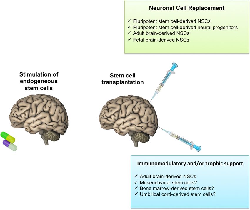
Casarosa et al. Molecular and Cellular Therapies 2014, 2:31                                                           Page 2 of 17
http://www.molcelltherapies.com/content/2/1/31

 Figure 1 Cardinal neural stem cell properties.

purification, generation and expansion in floating or adher-         adult stages. Besides their ability to divide asymmetric-
ent conditions have been described. However, beside these            ally and to serve as progenitors of neurons and glia, RG
important progresses, the identification of the best sources         cells constitute a scaffold on which neurons migrate in
for NSCs derivation and the optimization of approaches to            the developing brain. RG differentiation potential is less
stably proliferate them clonally in vitro still represents a         extensive compared to that of NEPs. Along with RG, an-
major goal of NSC research.                                          other population of immature neural cells is constituted by
   Yet, for a realistic exploitation of NSCs for cell therapies,     Basal Progenitors (BPs) [8]. They are generated at early
clinically-suitable NSC systems should hold specific key             phases of development by NEPs and at later stages by RG.
properties including (i) standardized production and scal-           BPs mostly undergo one or two rounds of division, gener-
ability to good medical practice (GMP), (ii) karyotypic              ating one or two pairs of neurons. Hence, BPs may be
stability, (iii) ability to correctly integrate in the host tissue   considered neurogenic transit-amplifying progenitors that
and (iv) differentiate into the required functional neural           specifically increase the production of neurons during re-
cells. In addition, NSCs should exhibit a reproducible, pre-         stricted developmental time periods in definite brain areas
dictable and safe behavior following in vivo injection.              (i.e. cerebral cortex).
                                                                        At the end of neurogenesis (roughly at birth in mice),
NSCs in brain organogenesis and homeostasis                          neurogenic RG cells are exhausted and residual RG cells
In the developing and adult CNS, different NSC popu-                 are converted into a unique astrocyte-like subpopulation
lations dynamically appear following predetermined                   [9]. This population will make up the NSC pool of the
spatio-temporal developmental programs. Molecular and                adult brain, endorsed with neurogenesis and gliogenesis
biological characteristics of NSCs greatly vary depending            maintainance throughout adult life.
on the region and developmental stage considered [4].                   The concept that the adult brain retains the ability to
  Development of the vertebrate CNS starts with neural               self-renew some of its neurons has been broadly recog-
plate folding to originate the neural tube, consisting of            nized in the last two decades and has represented a break-
radially elongated neuroepithelial cells (NEPs) [5]. NEPs            through in neurosciences. Pioneering studies from Altman
develop definite identities and different fates depending            and Das already reported the generation of new neurons
on their positions along the rostrocaudal (R-C) and dorso-           in a variety of structures in the adult rat and cat includ-
ventral (D-V) axes of the neural tube. Patterning along the          ing the olfactory bulb, hippocampus, and cerebral cor-
R-C axis leads to the initial distinction into prosencephalon,       tex [10]. However, their results were widely neglected until
mesencephalon, rhombencephalon and spinal cord territor-             the early 1990s, when the formation of new neurons in
ies. NEPs are accountable for the first wave of neurogenesis         adult rodent brain was clearly demonstrated [11,12]. This
in the neural tube. As development proceeds, NEPs convert            led to the identification of the germinal zones of the adult
themselves into another transitory NSC type, the so-called           brain. These are specialized niches located in the subven-
“radial glia” (RG) [6,7]. This rapidly constitutes the main          tricular zone (SVZ) of the lateral ventricle wall and in the
progenitor cell population in mid/late development and               subgranular zone (SGZ) of the dentate gyrus of the hippo-
early postnatal life while disappearing at late postnatal and        campus [3]. Whether NSCs reside in other regions of the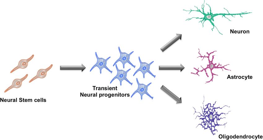
Casarosa et al. Molecular and Cellular Therapies 2014, 2:31                                                        Page 3 of 17
http://www.molcelltherapies.com/content/2/1/31

adult mammalian brain is still disputed. Neuroblasts pro-         years, there are still many major gaps regarding their
duced in the rodent SVZ migrate to the olfactory bulb fol-        in vivo control.
lowing the rostral migratory stream (RMS), an anatomic
structure well characterized in the rodent brain. The NSCs        NSCs for cell replacement approaches: requirements &
located in the SVZ, also called type B cells, generate actively   available in vitro systems
dividing intermediate cells, named type C cells, which            A large number of studies have explored grafting behavior
further divide giving rise to neuroblasts, referred to as type    of several NSCs typologies (and their progeny) in a variety
A cells that migrate away from the SVZ. These migrating           of preclinical studies and in some clinical investigations.
neuroblasts are organized in chains that connect the SVZ          Nevertheless, NSCs used for clinical applications should
to the olfactory bulb (constituting the RMS) where they           be safe, effective and accessible in large amount in GMP
gradually mature into functional GABAergic granule                conditions. A variety of different sources for NSCs have
neurons. Fate-mapping studies actually reveal that type B         been tested, including fetal- and adult CNS-derived NSCs,
cells are not developmentally restricted to neuronal lineages     neural progenitors derived from pluripotent cells, and
but can give rise also to glial progenies, suggesting they are    a range of non-neural stem cells, such as mesenchymal
authentic tripotent NSCs. The second germinal zone of the         (MSCs) and bone marrow-derived (BMDSCs) stem cells.
adult mammalian brain is the dentate gyrus of the hippo-          With these issues in mind, it should be remarked that up
campus. Astrocyte-like NSCs, called type I progenitors,           to now an ideal NSC system is not yet available to the
have been identified within the SGZ facing the dentate            clinic. Here, we will restrict our discussion to NSCs de-
gyrus hilus. They share several properties with the type          rived from neural tissue and from pluripotent stem cells.
B cells of the adult SVZ, although they apparently exhibit a      Advantages and disadvantages of each source and recent
narrower developmental potential. Type I progenitors likely       experimental evidence that highlight their potential use
divide asymmetrically to produce immature proliferating           for clinical applications will be presented.
progenitors, type II cells. These gradually differentiate into
migrating neuroblasts that travel into the granule cell           Fetal- and adult-derived NSCs
layer of the dentate gyrus, where they progressively mature       The isolation of NSCs from their natural niches and their
into functional granule neurons. Differently from the type        expansion in culture have been challenging issues, because
B cells of the SVZ, the progeny of type I progenitors does        the requirements to maintain these cells in their physio-
not migrate long distances, but remains localized in clus-        logical state are yet poorly understood. In the early ‘90s,
ters closely connected to the parent cell. Additionally, hip-     the identification of EGF and FGF-2 as key mitogens
pocampal NSCs appear to be developmentally restricted             for NSCs led to set up culture conditions that support
to become granule neurons; currently, there is no evidence        extended cell division of cells with NSCs properties [11,12].
that type I progenitors can generate mature glial deriva-         Since then, several studies reported that NSCs can be
tives in vivo.                                                    isolated from various regions of rodent (mouse and rat)
   The discovery of NSCs and neurogenesis in the adult            and human brain at several developmental stages as well as
mammalian CNS has tremendously changed our view of                from germinative areas of the adult brain. A widely used
the plasticity and function of the brain. This has prompted       method is to culture NSCs as neurospheres (Figure 2; [15]).
excitement for the possible exploitement of intrinsic             These are free-floating aggregates of neural progenitors,
neurogenic activity to cure brain diseases and rescue brain       each, in theory, deriving from a single NSC. Their gen-
function after injury. Mobilization of endogenous NSCs            eration relies on neural tissue micro-dissection followed
has thus emerged as a potential therapeutic approach for          by exposure to defined mitogen-supplemented media.
neural repair. It is known that brain injury promotes the         In such a procedure, primary cells are plated in low-
proliferation of adjacent NSCs, generating new astrocytes         attachment culture flasks in serum-free media supple-
and neurons [13]. For example, focal ischemia transiently         mented with EGF and/or FGF-2. In these conditions,
induces forebrain SVZ cell proliferation and neurogenesis.        differentiating or differentiated cells are supposed to die,
The NSCs in the SVZ and DG are also stimulated after              whereas NSCs respond to mitogens, divide and form float-
traumatic brain injury or seizures [14], suggesting that          ing aggregates (primary neurospheres) that can be dissoci-
adult neurogenesis may play a role in self-recovery               ated and re-plated to generate secondary neurospheres.
mechanisms of the brain. However, the amount of                   This procedure can be sequentially repeated several times
spontaneously produced neuroblasts after brain injury             to expand a NSC population.
is highly limited, and their survival and differentiation           Complementary to neurosphere culture is adherent cul-
into mature neurons are far from obtaining regenera-              ture, in which cells are more easily monitored and have
tive effects.                                                     better access to growth factors (Figure 2). In the last dec-
   It should be emphasized that, although our understand-         ade, several groups reported the generation and expansion
ing of NSCs has increased dramatically over the past few          of adherent NSC lines from neural tissue of rodent and
Casarosa et al. Molecular and Cellular Therapies 2014, 2:31                                                          Page 4 of 17
http://www.molcelltherapies.com/content/2/1/31

 Figure 2 Sources and in vitro growth protocols for neural stem cell generation and expansion.

human origin. According to this procedure, NSCs can be               hampered by culture conditions, especially for human
competently expanded as adherent clonal homogeneous                  NSCs. Nonetheless, major differences between fetal- and
NSC lines by exposure to specific mitogens, such as EGF              adult-derived human NSCs have been reported in terms
and/or FGF2 [16,17]. In these conditions, cells divide sym-          of both biological and molecular properties. Fetal-derived
metrically, retaining their tripotential differentiation cap-        human NSCs generally exhibit a shorter doubling time, a
acity. Adherent culture regimens have been shown to allow            more extensive expansion potential in vitro and better
for cultures with less differentiated cells compared to              integrative potential following grafting in animal models
the neurosphere assay, where cell–cell contacts and non-             [18-21]. Noteworthy, substantial differences have been also
uniform mitogens exposure is thought to stimulate differ-            described when comparing human NSCs derived from dif-
entiation programs [2].                                              ferent brain areas of the same fetus [22].
  Although several studies have attempted to provide
comparisons between fetal- and adult-derived NSCs, sys-              NSCs from pluripotent stem cells
tematic side-by-side analyses are still few and do not allow         Neuralization protocols applied to mouse and human
to draw any solid conclusions. In fact, results might be             pluripotent stem cells, including embryonic stem cells
Casarosa et al. Molecular and Cellular Therapies 2014, 2:31                                                         Page 5 of 17
http://www.molcelltherapies.com/content/2/1/31

(ESCs) and induced pluripotent stem cells (iPSCs), allow         ESC/iPSCs-derived cells are under scrutiny. In this view,
for the generation of NSCs populations. ESCs are derived         recent studies have reported the direct conversion of
from the inner cell mass (ICM) of blastocyst stage mamma-        adult somatic cells into NSCs, thus opening a new path to
lian embryos [23,24]. They are characterized by an intrinsic     generate NSCs without contamination of undifferentiated
capacity for self-renewal and the ability to generate all        pluripotent stem cells [34].
cell types derived from the three embryonic germ layers            The ability to generate patient-specific iPSCs and NSCs
(pluripotency). In the last years, the advent of iPSC technol-   clearly provides enormous prospective for future personal-
ogy has completely revolutioned the “pluripotency” field,        ized medicine, although too little is yet known about these
avoiding the requirement of embryos as source of rodent          cells to make any firm prediction.
and, most importantly, of human pluripotent stem cells.
Moreover, the use of iPSC opens new possibilities for            Possible therapeutic actions of grafted NSCs in different
studies of human development and disorders, further              neurodegenerative conditions
increasing the potential biomedical applications of this         Although the capacity of NSCs to divide and appropriately
type of cells [25]. iPSCs are the product of a reprogram-        differentiate in vitro has attracted much attention for clin-
ming procedure that allows the conversion of somatic             ical translation, it does not assure that these cells func-
cells directly into pluripotent cells [26,27]. This tech-        tionally incorporate into the recipient tissue and produce
nology is straightforward, robust and since its discovery        efficient restoration of compromised functions after graft-
has been implemented in terms of efficiency and repro-           ing. In order to generate therapeutic benefits in specific
ducibility. iPSCs closely resemble ESCs with respect to          neurological diseases, grafted cells have to accomplish a
expression of pluripotency markers, self-renewal poten-          certain grade of morphological, anatomical and functional
tial, and multilineage differentiation potential. Both murine    integration into the impaired host CNS tissue.
and human pluripotent stem cells can be exposed to neur-            Neurodegenerative disorders embody a heterogeneous
alizing in vitro protocols to generate a large amount of         collection of chronic and progressive diseases charac-
NSCs or progenitor cells. In these conditions, pluripotent       terized by distinct aetiologies, anatomical impairments
stem cells undergo progressive lineage restrictions similar      and symptoms [35]. Some of these disorders, such as
to those observed during normal fetal development, lead-         Huntington’s disease (HD), are acquired in an entirely gen-
ing to the generation of a range of distinct neural precur-      etic manner. Alzheimer’s disease (AD), amyotrophic lateral
sor populations [2].                                             sclerosis (ALS), and Parkinson’s disease (PD) mainly occur
   Generally, two main procedures to generate NSCs from          sporadically, although familiar forms caused by inheritance
pluripotent stem cells have been developed. The first            of gene mutations are known. On the other hand, the CNS
strategy relies on the formation of embryoid bodies (EBs),       can also be affected by other non-degenerative conditions,
three-dimensional (3D) aggregates. EBs recapitulate many         such as spinal cord injury and stroke, with no genetic herit-
aspects of cell differentiation occurring during early mam-      able components.
malian embryogenesis and give rise to cells of the three            By virtue of this extreme heterogeneity, different spe-
germ layers, including neural cells. EBs dissociated and         cific requirements should be envisaged when considering
plated in adhesion on coated plastic surfaces in defined         cell replacement as a possible therapeutic strategy. We can
media will produce rosette-like neural cells corresponding       distinguish between (i) “neuronal” CNS degenerative disor-
to the NEPs of the developing brain [28,29]. This NSC            ders caused by a prominent loss of specific neuronal popula-
population can be subsequently enriched, although no effi-       tions and (ii) “non neuronal” CNS degenerative conditions
cient methods for their extensive expansion have been            characterized by loss of non neuronal elements.
reported. EB-independent procedures based on adherent               In the case of neuronal degeneration, the success of cell
monolayer protocols have been also described [30,31].            replacement strictly depends on the complexity and accur-
   Electrophysiology studies have shown that pluripotent-        acy of the pattern of connectivity that needs to be restored.
derived NSCs efficiently generate fully mature neurons           In PD, affected dopaminergic neurons in the substantia
in vitro, as well as functionally integrated neurons after       nigra (SN) exert a modulatory action on striatal target cir-
transplantation in the mammalian CNS [32,33]. Never-             cuits mostly through the release of dopamine. This system
theless, major limitations to therapeutic applications of        is defined as “paracrine” and even a partial pattern repair
pluripotent-derived NSCs are represented by safety con-          may lead to a significant functional recovery in such condi-
cerns and caveats about their clinical-grade production.         tions. Indeed, in PD the donor cells can be transplanted
Indeed, grafted pluripotent cells can form teratomas, im-        directly into the target region to circumvent the problem
plying that in a clinical setting residual undifferenti-         of long-distance neuritic growth in the adult CNS. Despite
ated pluripotent stem cells should be excluded from              the ectopic location, if grafted cells are able to re-establish
the cell preparation before grafting. Protocols for avoiding     a regulated and efficient release of dopamine, they can lead
teratocarcinoma formation in vivo after transplantation of       to a clinically relevant functional recovery. However, cell-
Casarosa et al. Molecular and Cellular Therapies 2014, 2:31                                                               Page 6 of 17
http://www.molcelltherapies.com/content/2/1/31

based treatment strategies are extremely difficult for other           in preclinical studies. Moreover, a few attempts are already
diseases such as HD, ALS, trauma, stroke, and AD, which                being made to translate these discoveries into the clinical
are characterized by the need of a complex pattern repair.             setting. Currently, the registry and results database of
   Differently, “non neuronal” CNS degenerative syndromes              publicly and privately supported clinical studies with
such as multiple sclerosis (MS) characterized by severe in-            human participants conducted around the world (http://
flammation, oligodendroglial degeneration and axonal de-               www.clinicaltrial.gov/) reports 880 international clinical tri-
myelination, represent a good target for cell replacement,             als employing the use of stem cells for treatment of patients
due to their limited requirements for pattern repair [36]. In          affected by several CNS disorders (query terms: “stem cells
MS, grafted cells should produce oligodendrocytes able to              AND nervous system”) among which 89 are testing NSC
restore axonal myelination in order to lead to a functional            injection approaches (query terms: “neural stem cell AND
rescue.                                                                injection AND nervous system disease”). If restricting
   It should also be emphasized that although pattern re-              the search to non-tumor diseases, the database returns
pair is critical to obtain permanent efficacy in the brain,            51 studies (query terms: “neural stem cell AND injection
cells transplanted into the brain may also be beneficial               AND nervous system disease NOT tumor”) with only 27
via the release of molecules that may either stimulate the             of these currently open. Interestingly, if we analyze these
regenerative potential of local cells (where present) or               results more carefully, only 5 studies are actually open to
increase the survival of the remaining host elements,                  explore the potential clinical relevance of NSCs while the
thus slowing disease progression (Figure 3; [37]). Also,               remaining are testing the regenerative potential of mesen-
immunomodulatory activity of the grafted cells could                   chymal stem cells whose actual uselfness in brain diseases
be of benefit in diseases such as MS where a prominent                 is far from being a solid preclinical reality.
disease-associated inflammation contributes to the estab-                 To date, few Phase I and II clinical studies employing
lishment and progression of the disease (Figure 3).                    NSCs (a dozen in highly debilitating CNS disorders; de-
                                                                       tails reported in Table 1) have been performed, with the
Current clinical trials involving NSCs for                             main objective to demonstrate safety and practicability and
neurodegenerative diseases                                             to explore the potential effectiveness of the treatments.
A large number of studies have explored the grafting be-               These clinical trials are testing only few NSCs products,
havior of several NSCs typologies (and their progenies)                mainly consisting of fetal-tissue-derived allogenic NSCs.

 Figure 3 Therapeutical strategies for neural stem cells exploitation in CNS diseases.
http://www.molcelltherapies.com/content/2/1/31
Casarosa et al. Molecular and Cellular Therapies 2014, 2:31
Table 1 Clinical trials involving NSCs for neurodegenerative diseases
NCT number Title Recruitment Conditions Interventions Sponsor/Collaborators Phases Enrollment Start Completion
date date
NCT00337636 Study of HuCNS-SC Cells in Completed Neuronal Ceroid Biological: HuCNS-SC StemCells, Inc. Phase 6 May September
Patients With Infantile or Late Lipofuscinosis 1 2006 2009
Infantile Neuronal Ceroid
Lipofuscinosis (NCL)
NCT01005004 Study of Human Central Completed Pelizaeus-Merzbacher Biological: HuCNS-SC StemCells, Inc. Phase 4 November December
Nervous System (CNS) Stem Disease cells implantation 1 2009 2012
Cells Transplantation in
Pelizaeus-Merzbacher Disease
(PMD) Subjects
NCT01151124 Pilot Investigation of Stem Active, not Stroke Biological: CTX0E03 ReNeuron Limited Phase 12 June March
Cells in Stroke recruiting neural stem cells 1 2010 2015
implantation
NCT01217008 Safety Study of GRNOPC1 Completed Spinal Cord Injury Biological: hES-derived Asterias Biotherapeutics, Inc. Phase 5 October July
in Spinal Cord Injury GRNOPC1 implantation 1 2010 2013
NCT01238315 Safety and Efficacy Study of Withdrawn Neuronal Ceroid Biological: HuCNS-SC StemCells, Inc. Phase 0 November April
HuCNS-SC in Subjects With Lipofuscinosis cells implantation 1 2010 2011
Neuronal Ceroid Lipofuscinosis
NCT01321333 Study of Human Central Active, not Thoracic Spinal Cord Biological: HuCNS-SC StemCells, Inc. Phase 12 March December
Nervous System Stem Cells recruiting Injury|Spinal Cord cells implantation 1-2 2011 2015
(HuCNS-SC) in Patients With Injury|Spinal Cord Injury
Thoracic Spinal Cord Injury Thoracic|Spinal Cord
Trauma
NCT01348451 Human Spinal Cord Derived Active, not Amyotrophic Lateral Biological: human Neuralstem Inc. Phase 18 January August
Neural Stem Cell Transplantation recruiting Sclerosis neural spinal cord 1 2009 2014
for the Treatment of Amyotrophic stem cells implantation
Lateral Sclerosis
NCT01640067 Human Neural Stem Cell Recruiting Amyotrophic Lateral Biological: Human Azienda Ospedaliera Santa Phase 18 December September
Transplantation in Amyotrophic Sclerosis Neural Stem Cells Maria, Terni, Italy|Azienda 1 2011 2016
Lateral Sclerosis (ALS) implantation Ospedaliero Universitaria
Maggiore della Carita|Università
di Padova Italy
NCT01725880 Long-Term Follow-Up of Enrolling by Spinal Cord Injury Observation StemCells, Inc. Phase 12 November March
Transplanted Human Central invitation 1-2 2012 2019
Nervous System Stem Cells
(HuCNS-SC) in Spinal Cord
Trauma Subjects
NCT01730716 Dose Escalation and Safety Enrolling by Amyotrophic Lateral Biological: Human Neuralstem Inc. Phase 18 May April
Study of Human Spinal Cord invitation Sclerosis spinal cord stem 2 2013 2014
Derived Neural Stem Cell cell implantation
Transplantation for the
Page 7 of 17
Treatment of Amyotrophic
Lateral Sclerosishttp://www.molcelltherapies.com/content/2/1/31
Casarosa et al. Molecular and Cellular Therapies 2014, 2:31
Table 1 Clinical trials involving NSCs for neurodegenerative diseases (Continued)
NCT01772810 Safety Study of Human Not yet Spinal Cord Injury (SCI) Biological: Human Neuralstem Inc. Phase 8 May March
Spinal Cord-derived recruiting spinal cord stem 1 2014 2016
Neural Stem Cell cells implantation
Transplantation for
the Treatment of Chronic SCI
NCT02117635 Pilot Investigation of Stem Not yet Ischaemic Stroke| Biological: CTX DP ReNeuron Phase 41 June June
Cells in Stroke Phase II Efficacy recruiting Cerebral Infarction| Limited 2 2014 2017
Hemiparesis|Arm
Paralysis
Page 8 of 17Casarosa et al. Molecular and Cellular Therapies 2014, 2:31 Page 9 of 17 http://www.molcelltherapies.com/content/2/1/31 Among them, HuCNS-SC, a StemCells Inc. (Palo Alto, CA) neurodegenerative disease with no safety concerns attrib- product candidate, is a purified population of allogenic uted to the grafts. Examination of the brains from three NSCs derived from human fetal (16–20 weeks) brain tis- patients who expired due to causes related to the under- sue, sorted using the CD133 marker and expanded in cul- lying disease showed evidence of engraftment, migration ture as neurospheres. These cells are routinely stored in a and long-term survival of the HuCNS-SC cells following frozen state and reanimated before transplantation [38,39]. transplantation and the planned cessation of immunosup- In May 2006 a clinical trial (the first using human NSCs) pression. Grafted cells provided widespread global replace- was approved at Oregon Health and Science University ment enzyme and bystander neuroprotection. Although (OHSU, Portland, OR, USA) for the use of these cells for these results look very promising, more detailed analyses lysosomal storage diseases (ClinicalTrials.gov identifier, and a longer patients’ follow up will be fundamental to draw NCT00337636), rare (one in 100,000 children) fatal auto- more solid conclusions. In 2009, the Company completed somal recessive progressive neurological disorders caused the Phase I safety study and in October 2010 embarked on a by mutations in enzymes that ultimately lead to accumula- Phase Ib safety and efficacy trial (clinicaltrials.gov identifier tion of neurotoxic lipofuscins. Patients generally lose their no. NCT01238315) in 6 children with less advanced Batten’s vision, develop seizures and dementia, and die before their disease and therefore most likely to benefit from a timely teens. In this application NSCs serve as “Trojan Horses” neural stem cell transplant. However, the study was discon- to deliver enzymes to other cells within the brain, a con- tinued because of the failure to enroll patients meeting the cept established in rodent models of enzyme deficiencies. study criteria (out of 22 initial prospects, none of them met Preclinical studies indicated that intracerebral grafts into the entry criteria). animal model of infantile neuronal ceroid lipofuscinosis A second open-label phase I clinical trial (ClinicalTrials. (NCL, Batten’s disease) integrate into the host brain, gov NCT01005004) has been sponsored by StemCell produce and release the defective enzyme resulting in Inc. at the University of California, San Francisco (UCSF) protection of affected neurons. In this open-label, dose- using HuCNS-SC brain transplantation for Pelizaeus- escalating phase I trial, single donor HuCNS-SC were Merzbacher disease (PMD). PMD is a X-linked congenital injected into 6 patients with either infantile or late infant- leukodystrophy characterized by defective myelination [41]. ile NCL. Enrollment in the trial was limited to patients in Preclinical studies in animal models showed that intracere- the advanced stages of the disease (patients with signifi- bral injection of HuCNS-SC produced oligodendrocytes cant neurological and cognitive impairment, whose devel- leading to remylination of neurons affected by the mutated opmental age was demonstrated to be less than two-thirds gene for PMD [42]. This phase I clinical trial has enrolled of their chronological age). Two dose levels were adminis- four young children with an early severe form of PMD. tered with the first 3 patients receiving a target dose of Each patient received a total brain dose of 300 million cells approximately 500 million cells and the other 3 patients through two injections into the frontal white matter area receiving a target dose of approximately 1 billion cells. of each hemisphere. Immunosuppression was administered The cells were grafted directly into each patient’s brain for 9 months following transplantation. The patients have (injections were performed into eight areas of each been followed for twelve months after transplantation; dur- child’s brain) and patients received immunosuppression ing this period they underwent regular neurological as- for 12 months after transplantation. The grafting pro- sessments and MRI analyses. Data regarding this clinical cedure and combination with prolonged immune sup- trial have been published in October 2012, indicating a pression were both well tolerated thus showing a favorable good safety profile for the HuCNS-SC cells and the trans- safety profile with transplanted cells, neurosurgical proced- plantation procedure [43]. Clinical assessment revealed ure, and immunosuppression regimen. In addition, trans- small gains in motor and cognitive function in three of planted cells showed long-term survival. Five patients (one the four patients; the fourth patient remained clinically died for disease) completed the 12-month Phase I study stable. Moreover, MRI inspections suggest myelination and were subsequently enrolled in a subsequent four-year, in the region of the transplantation, which progressed long-term observational study, with three of the five sur- over time and persisted after the withdrawal of immuno- viving to the end of the four-year study [40]. Three of the suppression at nine months. Upon completion of the Phase six patients transplanted with HuCNS-SC cells have now I trial, all four patients have been enrolled into a 4-year survived more than five years post-transplant, and it is long-term follow up study. noteworthy that each have stable quality-of-life measures, StemCells Inc. has also sponsored a Phase I/II clinical considering that they suffer a progressive neurodegenera- trial (clinicaltrials.gov identifier no. NCT01321333) at the tive disorder. Assessment of the patients’ cognitive and University Hospital Balgrist (Zurich, Switzerland) for assay- neurological function revealed stable scores in some tests, ing safety and preliminary efficacy of intramedullary spinal but the clinical outcomes were generally consistent with cord transplantation of human HuCNS-SC neurospheres the expected course of impairment associated with this in subjects with thoracic (T2-T11) spinal cord trauma. The
Casarosa et al. Molecular and Cellular Therapies 2014, 2:31                                                         Page 10 of 17
http://www.molcelltherapies.com/content/2/1/31

study began in March 2011 and includes 12 subjects who             with some arm dysfunction and received grafts on one
suffered spinal cord injury (SCI) in the 3 to 12 months            side of their neck. The last group (Cohort E) included
prior to cell transplantation. Each subject received a total       ALS patients still ambulatory, who received both injec-
fixed dose of approximately 20 million cells injected dir-         tions on one side of their neck and injections on both
ectly into the thoracic spinal cord near the injury. The first     sides of their lower spine. The last patient in this Phase
patients have been transplanted with no safety concerns            I trial was treated in August 2012 and the trial was con-
arising concerning the surgery and the cellular transplant         cluded in February 2013. The clinical assessments on
[44]. This trial is estimated to end in March 2016. In No-         the first 12 grafted patients demonstrated no evidence
vember 2012 the consequential long-term follow up of the           of acceleration of disease progression with the planned
12 patients subjected to HuCNS-SC transplantation has              18 months post-transplantation follow up [46]. In April
started and it will last until March 2018 (clinicaltrials.gov      2013, following conclusion of its Phase I FDA-approved
identifier no. NCT01725880).                                       trial, Neuralstem received approval to start a Phase II dose
   Recently, also Neuralstem Inc. has received FDA ap-             escalation and safety trial (clinicaltrials.gov identifier no.
proval to initiate a Phase I safety trial using proprietary        NCT01730716) to be performed in three centers: Emory
NSCs for chronic spinal cord injury (clinicaltrials.gov            University Hospital in Atlanta, Georgia, site of Phase I;
identifier no. NCT01772810). The cell line used in this            ALS Clinic at the University of Michigan Health System,
trial (NSI-566RSC) is derived from the cervical and upper          in Ann Arbor, Michigan, and Massachusetts General Hos-
thoracic regions of the spinal cord from a single 8-week           pital in Boston. This Phase II trial is designed to treat up
human fetus after an elective abortion [45]. The cells are         to 15 ambulatory patients, in five different dosing cohorts,
serially expanded as monolayer in serum-free media sup-            advancing up to a maximum of 40 injections, and 400,000
plemented with FGF-2 as the sole mitogen to maintain               cells per injection based on safety. The first 12 patients
proliferation and prevent differentiation. This trial will         will receive injections in the cervical region of the spinal
enroll up to eight SCI patients with thoracic lesions (T2-         cord only, where the stem cells could help preserve
T12) one to two years post-injury with no motor or sen-            breathing function. The final three patients will receive
sory function in the relevant segments at and below injury         both cervical and lumbar injections.
(complete paralysis). All patients in the trial will receive          Another ALS Phase I clinical trial using allogenic fetal
six injections close to injury site with the first four patients   brain-derived human NSCs has been approved to the
receiving 100,000 cells per injection and the second four          Azienda Ospedaliera Santa Maria (Terni, Italy) in June
patients 200,000 cells per injection. The patients will            2012. This study includes a total of 18 ALS patients
also receive immunosuppressive therapy, which will last            that will be treated with intraspinal implanted allogeneic
for three months, as tolerated. Late this year, Neuralstem         foetal-derived neurospheres (clinicaltrials.gov identifier no.
Inc. will also start an acute spinal cord injury trial in Seoul    NCT01640067).
in collaboration with their South Korean partner CJ                   Fetal NSCs are also being used for treatment of disabled
CheilJedang.                                                       ischemic stroke patients by the company ReNeuron Lim-
   NSI-566 NSCs have also been used in clinical trials             ited (UK). For this clinical application, ReNeuron uses
for the treatment of ALS (Lou Gehrig’s disease). In 2009           its proprietary allogenic foetal-derived brain human NSCs
Neuralstem Inc. sponsored the first NSC-based phase                (CTX0E03), which were derived from human fetal brain
I safety trial in ALS (clinicaltrials.gov identifier no.           tissue following genetic modification with a conditional
NCT01348451) at the Emory University School of Medicine            immortalizing gene, c-mycER [47]. This transgene gener-
(Atlanta, GA, USA). The trial enrolled 18 patients divided         ates a fusion protein that stimulates cell proliferation in
in 5 groups characterized by slightly different inclusion cri-     the presence of a synthetic drug, 4-hydroxy-tamoxifen
teria and receiving different procedures in terms of site and      (4-OHT). The cell line is clonal, expands rapidly in culture
number of injections (number of cells/injection: 100,000).         and has a normal human karyotype (46 XY). In the absence
The first group (cohort A) included patients with advanced         of growth factors and 4-OHT, the cells undergo growth
ALS who are unable to walk, and received transplant injec-         arrest and differentiate into neurons and astrocytes.
tions in one or both sides of the lower spinal cord. The trial     The Phase I clinical trial (clinicaltrials.gov identifier no.
then progressed to patients who were still ambulatory. The         NCT01151124) started on June 2012 at the Glasgow
first three of these patients (Cohort B) received five unilat-     Southern General Hospital (Glasgow, Scotland). The study
eral injections while the next three patients (Cohort C) re-       is designed to test the safety of CTX0E03 NSCs product
ceived ten bilateral injections in the same lumbar region.         by direct single dose injection into the damaged brains of
In 2011 Neuralstem received approval to move into the              12 male patients 60 years of age or over who remain mod-
cervical (upper back) stage of the trial. In this stage,           erately to severely disabled 6 months to 5 years following
two groups of three patients each were included. The               an ischemic stroke. The trial will consider four ascending
first three of these patients (Cohort D) were ambulatory           doses of CTX0E03 cells (4 dosage groups of three patients
Casarosa et al. Molecular and Cellular Therapies 2014, 2:31                                                       Page 11 of 17
http://www.molcelltherapies.com/content/2/1/31

at each dose level receiving 2 million, 5 million, 10 million    effects have also been reported after transplantation of
or 20 million cells). Clinical outcomes will be measured         different types of stem cells. Grafting of human umbilical
over 24 months and patients will be invited to participate       cord stem cells [53] or genetically-engineered bone marrow
in a long-term follow-up trial for a further 8 years.            mesenchymal stem cells [54] have been shown to amelior-
  To date, only one study has exploited the use of human         ate seizures in the pilocarpine rat model of TLE.
ES-derived neural cells for the treatment of CNS injury in          Currently, research performed on animal models does
a clinical setting. In 2010, Geron Corporation started a         not provide a rationale for the use of stem cells in neu-
Phase I Safety clinical trial with human ES cell-derived         rodevelopmental disorders, with the only exception of
oligodendrocyte progenitors (GRNOPC1 cells) [48-50]              epilepsy. Indeed, massive cell loss is detected in the epi-
in patients with neurologically complete, subacute spinal        leptic brain, thus offering a rationale for cell replacement
cord injury (ClinicalTrials.gov identifier: NCT01217008).        therapies. On the contrary, current research does not
The study was designed to test the safety of the Geron           support the idea that stem cell transplantation may work
GRNOPC1 cell product by direct single dose (2 million            in the case of other neurodevelopmental disorders such as
cells) injection into the damaged spinal cord of five patients   autism, since no massive cell loss is observed in the brain
with neurologically complete spinal cord injuries (between       of autistic patients. Accordingly, a Phase I trial to test the
7 and 14 days post injury). The enthusiasm about this study      effect of stem cells transplantation in 20 patients with
was abated only a year later, when Geron suddenly and sur-       temporal lobe epilepsy is currently ongoing (clinicaltrials.
prisingly stopped the trial and decided to give up on the        gov identifier no. NCT00916266). The study, authorized
stem cell division. In 2013 Asterias Biotherapeutics, Inc., a    on July 2008 to the Instituto do Cerebro de Brasilia (Brazil),
subsidiary of BioTime, purchased Geron’s stem cell division      has the aim to evaluate autologous bone marrow derived
and announced that it will resume the spinal cord trial.         stem cells (BMDSCs) transplantation as a safe and poten-
                                                                 tially beneficial treatment for patients with temporal lobe
Is there a rationale for stem cell-based treatments for          refractory epilepsy. As primary outcomes were considered
neurodevelopmental disorders?                                    evaluation of seizure frequency, hippocampal volume and
An increasing number of recent studies indicate that             cognitive performance. Although the study should have
neurological and neuropsychiatric disorders including epi-       been completed in June 2012, no results are currently
lepsy, autism and schizophrenia may arise from altered           available. Beyond the lack of solid results, it should be
brain development. According to this view, neurodevelop-         emphasized that the rationale for using BMDSCs trans-
mental disorders may emerge from altered neurogenetic            plantation in TLE patients is rather inappropriate, since
processes that lead to misplacement or loss of neurons           these cells can not replace neural cell lost in these pa-
and their connections in the postnatal brain. It is therefore    tients (see also below, “Clinical testing of non-neural cells
not surprising that, in recent years, much attention has         for CNS diseases”).
been paid to the possible use of NSCs to understand (and            More surprisingly, one completed and five ongoing tri-
possibly treat) these pathologies. Currently, NSCs trans-        als have been approved to test the efficacy of stem cell
plantation strategies have been essentially tested in rodent     transplantation in patients affected by autism spectrum
models of temporal lobe epilepsy (TLE). In both humans           disorders (query terms: “stem cells AND autism”) (details
and rodents, TLE is accompanied by massive cell loss in          are reported in Table 2). None of these studies is actually
the hippocampus and limbic areas [51]. In the past ten           injecting NSCs or pluripotent cells derivatives: they
years, NSCs transplantation (both during the latent and          are focussed on using autologous BMSC (clinicaltrials.
chronic phases) has been widely tested as a tool to coun-        gov identifier no. NCT01740869, NCT01974973), adipose-
teract cell loss and ameliorate TLE symptoms in rodents.         derived MSC (clinicaltrials.gov identifier no. NCT01502488)
NSCs are attractive as donor cells for grafting in TLE for a     and human cord blood mononuclear cells (clinicaltrials.gov
number of reasons: 1) they can be expanded in culture for        identifier no. NCT01343511, NCT01638819, NCT01836562).
extended periods; 2) they migrate extensively into the           Considering the young age and the overall number of
hippocampal layers; 3) they can differentiate into inhibi-       patients enrolled (more than 350), the poor rationale
tory GABAergic neurons as well as astrocytes secreting           and the total absence of published reports from these
anticonvulsant factors (such as the glial cell-line derived      studies, great attention should be payed to these trials.
neurotrophic factor GDNF); 4) they produce neurotrophic
factors that can stimulate hippocampal neurogenesis from         Retinal dystrophies and stem cell treatments
endogenous pools of NSCs [52]. Different strategies have         Another field in which a strong interest has been raised for
been used to test the protective effects of transplanted         cell treatments based on NSCs or cell derivatives of human
NSCs, including transplantation of embryonic or adult            pluripotent stem cells is represented by retinopathies. Many
hippocampal NSCs in both the KA and pilocarpine                  studies are undergoing in order to characterize the appro-
models of TLE [52]. Antiepileptic and neuroprotective            priate source of cells for these therapeutic approaches. The
http://www.molcelltherapies.com/content/2/1/31
Casarosa et al. Molecular and Cellular Therapies 2014, 2:31
Table 2 Clinical trials for neurodevelopmental disorderss
NCT number Title Recruitment Conditions Interventions Sponsor/ Phases Enrollment Start date Completion
Collaborators date
NCT01343511 Safety and Efficacy of Completed Autism Biological: human cord blood Shenzhen Beike Bio- Phase 1-2 37 March 2009 May 2011
Stem Cell Therapy in mononuclear cells implantation| Technology Co., Ltd.|
Patients With Autism Biological: human cord blood Shandong Jiaotong
mononuclear cells and human Hospital|Association
umbilical cord mesenchymal for the Handicapped
stem cells Injection Of Jinan
NCT01502488 Adipose Derived Stem Not yet Autism Procedure: Fat Harvesting and Ageless Regenerative Phase 1-2 10 October 2014 January
Cell Therapy for Autism recruiting Stem Cell Injection Institute|Instituto de 2017
Medicina Regenerativa,
S.A. de C.V.
NCT01638819 Autologous Cord Blood Recruiting Autism Biological: Autologous Cord Sutter Health Phase 2 30 August 2012 August
Stem Cells for Autism Blood Stem Cells Injection| 2014
Biological: Placebo
NCT01740869 Autologous Bone Marrow Recruiting Autism|Autism Biological: Stem cells Injection Hospital Universitario Phase 1-2 30 November 2012 Not
Stem Cells for Children With Spectrum Dr. Jose E. Gonzalez specified
Autism Spectrum Disorders
NCT01836562 A Clinical Trial to Study the Recruiting Autism Biological: Autologous Cord Chaitanya Hospital, Phase 1-2 100 March 2011 April 2014
Safety and Efficacy of Bone Blood Stem Cells Injection Pune
Marrow Derived Autologous
Cells for the Treatment of
Autism
NCT01974973 Stem Cell Therapy in Autism Recruiting Autism Spectrum Procedure: Autologous bone Neurogen Brain and Phase 1 150 August 2009 October
Spectrum Disorders Disorders marrow mononuclear cell Spine Institute 2014
transplantation|Procedure:
Autologous bone marrow
mononuclear cell transplantation
in patients with autism
Page 12 of 17Casarosa et al. Molecular and Cellular Therapies 2014, 2:31 Page 13 of 17 http://www.molcelltherapies.com/content/2/1/31 retina is subject to different degenerative diseases, which well as structural evidence of successful engraftment and can be both age related (such as AMD, Age-related changes to vision. Macular Degeneration) and inherited (LCA, Leber’s Con- Interestingly, the field of retinopathies represents the genital Amaurosis, and RP, Retinitis pigmentosa). These are arena where the largest number of human ESC-derived all characterized by degeneration of photoreceptors and/or products is being tested in the clinics. Currently, seven retinal pigmented epithelium (RPE cells). A number of dif- trials are open employing terminally differentiated hu- ferent therapeutical approaches have been meaningfully man ESC-derived hRPE (details are reported in Table 3). explored to cure human photoreceptor degenerations. Six of these studies are sponsored by Advanced Cell Tech- These include environmental modifications (nutrient sup- nology (ACT; USA) and CHA Bio & Diostech (South plementation and avoidance of light), drugs (i.e. neuro- Korea) for the use of the ACT’s MA09-hRPE cells for treat- trophic factors), gene therapy to provide the healthy ment of Stargardt’s Macular Dystrophy patients (SMD; version of the mutated gene, retinal prostheses [55]. clinicaltrials.gov identifier no. NCT01469832, NCT01345006 A large body of transplantation studies has been carried and NCT01625559), dry AMD patients (clinicaltrials.gov out in order to assess the molecular features characterizing identifier no. NCT01674829, NCT01344993) or Myopic cells with the ability to integrate and generate functional Macular Degeneration patients (MMD; clinicaltrials.gov photoreceptors in degenerating retinae. These studies de- identifier no. NCT02122159). In these studies, the num- fined that, in rodents, integration of donor cells is best ber of grafted cells varies between 50,000 and 200,000 obtained by using post-mitotic photoreceptor precursors cells in order to define the optimal dosage. The prelim- [56,57]. They also showed that, for donor cells to integrate inary results of two of these trials have been published, in retinal tissue, they should have specific molecular char- showing safety and some promising efficacy in vision acteristics and should be derived from retinae that have restoration. For this reason, MA09-hRPE cells from ACT not reached complete maturation. Despite the promise, have been recently granted by the FDA the orphan drug the low numbers of integrating cells impaired a real func- designation for use in the treatment of Stargardt’s Macular tional recovery in the transplanted eyes, even if some res- Dystrophy (SMD). This represents the first time that toration of vision was observed [58]. Moreover, a possible orphan drug status has been granted for the use of an transition to the clinic would be blocked by the obvious embryonic stem cell derived therapy in treating an un- ethical concerns in the use of human retinal progenitor met medical need. As a result, ACT is entitled to obtain cells. For these reasons, many reseachers turned their at- several benefits aimed at forstering clinical exploitation tention to obtaining postmitotic photoreceptor precursors of this cell product, including tax credits, access to grant in vitro, by differentiation of pluripotent cells such as ESCs funding for clinical trials, accelerated FDA approval and and iPSCs. Integration capabilities of the in vitro differen- allowance for marketing exclusivity after drug approval for tiated cells have also been tested by subretinal injections a period of as long as seven years. in mice [59-61]. All these studies assessed terminal differ- The remaining study is represented by a non-randomized entiation and integration of pluripotent cells-derived safety/efficacy study phase I open label trial of RPE replace- photoreceptors and, when possible, functionality, although ment aiming at evaluating the safety and feasibility/efficacy showing alternative results. of treating subjects with wet AMD in whom there is To date, the clinicaltrials.gov registry reports 50 inter- rapidly progressing vision loss (clinicaltrials.gov identifier national clinical trials employing different stem cells types no. NCT01691261). The study has been sponsored by (query terms: “stem cells AND retinopathy”), mostly au- Pfizer and will be performed at University College, tologous BMSCs, for treatment of patients affected by London (UK). It will include 10 patients that will receive retinal dystrophies. Among these, an age related macular Pf-05206388 RPE cells immobilized on a polyester mem- degneration (AMD) Phase I/II trial sponsored by Stem Cell brane as a monolayer, derived from human ESCs. The im- Inc. is ongoing in three US centers: (Byers Eye Institute planted membrane is approximately 6 mm × 3 mm and is at Stanford, Stanford Hospital and Clinics, Palo Alto, intended to be life-long. California; New York Eye and Ear Infirmary, New York; Retina Foundation of the Southwest Recruiting Dallas, Clinical testing of non-neural cells for CNS diseases Texas) (clinicaltrials.gov identifier no. NCT01632527). This While in this review we have mainly discussed ongoing study, involving 16 patients affected by geographic atrophy clinical studies based on human NSCs or neural deriva- secondary to AMD, is designed to investigate the safety tives of pluripotent stem cells, most of the currently and preliminary efficacy of unilateral subretinal transplant- open Phase I/II clinical trials for treatment of brain dis- ation of HuCNS-SC cells by means of a single transplant eases are actually testing therapeutic potential of cells of procedure. Immunosuppressive agents will be administered non-neural origin. Mesenchymal stem cells (MSCs) de- orally to all subjects for a period of three months after sur- rived from bone marrow (BMD-MSCs) or adipose tissue gery. Participants will be monitored for complications as and bone marrow derived stem cells (BMDSCs) have been
http://www.molcelltherapies.com/content/2/1/31
                                                                                                                                                                                                  Casarosa et al. Molecular and Cellular Therapies 2014, 2:31
Table 3 Clinical trials involving stem cells for retinal dystrophies
NCT number     Title                                  Recruitment              Conditions          Interventions         Sponsor/               Phases     Enrollment   Start date   Completion
                                                                                                                       Collaborators                                                   date
NCT01344993    Safety and Tolerability of              Recruiting        Dry Age Related Macular   Biological:         Advanced Cell           Phase 1-2       16         April      December
               Sub-retinal Transplantation                                    Degeneration         MA09-hRPE            Technology                                        2011         2014
               of hESC Derived RPE                                                                 implantation
               (MA09-hRPE) Cells in Patients
               With Advanced Dry Age Related
               Macular Degeneration
NCT01345006    Sub-retinal Transplantation             Recruiting          Stargardt’s Macular     Biological:         Advanced Cell           Phase 1-2       16         April      December
               of hESC Derived RPE (MA09-hRPE)                                 Dystrophy           MA09-hRPE            Technology                                        2011         2014
               Cells in Patients With Stargardt’s                                                  implantation
               Macular Dystrophy
NCT01469832    Safety and Tolerability of              Recruiting          Stargardt’s Macular     Biological:         Advanced Cell           Phase 1-2       16       November     December
               Sub-retinal Transplantation                                  Dystrophy|Fundus       MA09-hRPE            Technology                                        2011         2014
               of Human Embryonic Stem                                   Flavimaculatus|Juvenile   implantation
               Cell Derived Retinal Pigmented                              Macular Dystrophy
               Epithelial (hESC-RPE) Cells in
               Patients With Stargardt’s
               Macular Dystrophy (SMD)
NCT01625559    Safety and Tolerability of              Recruiting          Stargardt’s Macular     Biological:           CHA Bio &             Phase 1         3        September     October
               MA09-hRPE Cells in Patients                                     Dystrophy           MA09-hRPE              Diostech                                         2012        2014
               With Stargardt’s Macular                                                            implantation
               Dystrophy (SMD)
NCT01632527    Study of Human Central                  Recruiting         Age Related Macular      Drug: HuCNS-        StemCells, Inc.         Phase 1-2       16         June          June
               Nervous System Stem Cells                                  Degeneration|Macular     SC cells                                                               2012          2014
               (HuCNS-SC) in Age-Related                                   Degeneration|AMD        implantation
               Macular Degeneration (AMD)
NCT01674829    A Phase I/IIa, Open-Label,              Recruiting           Dry Age Related        Biological:           CHA Bio &             Phase 1-2       12       September       April
               Single-Center, Prospective Study                           Macular Degeneration     MA09-hRPE              Diostech                                         2012         2016
               to Determine the Safety and                                                         implantation
               Tolerability of Sub-retinal
               Transplantation of Human
               Embryonic Stem Cell Derived
               Retinal Pigmented Epithelial
               (MA09-hRPE) Cells in Patients
               With Advanced Dry Age-related
               Macular Degeneration (AMD)
NCT01691261    A Study Of Implantation Of           Not yet recruiting    Age Related Macular      Biological:        Pfizer|University        Phase 1         10         April         July
               Human Embryonic Stem Cell                                     Degeneration          PF-05206388        College, London                                     2014          2016
               Derived Retinal Pigment                                                             implantation
               Epithelium In Subjects With
               Acute Wet Age Related
               Macular Degeneration And
               Recent Rapid Vision Decline

                                                                                                                                                                                                                                   Page 14 of 17
NCT02122159    Research With Retinal Cells          Not yet recruiting       Myopic Macular        Biological:     University of California,   Phase 1-2      Not         Not           Not
               Derived From Stem Cells for                                    Degeneration         MA09-hRPE       Los Angeles|Advanced                     specified   specified     specified
               Myopic Macular Degeneration                                                         implantation     Cell Technology, Inc.
You can also read