When Good Guys Turn Bad: Bone Marrow's and Hematopoietic Stem Cells' Role in the Pathobiology of Diabetic Complications - MDPI
←
→
Page content transcription
If your browser does not render page correctly, please read the page content below
International Journal of
            Molecular Sciences

Review
When Good Guys Turn Bad: Bone Marrow’s and
Hematopoietic Stem Cells’ Role in the Pathobiology
of Diabetic Complications
Maria Cristina Vinci 1, *,† , Elisa Gambini 1,† , Beatrice Bassetti 1 , Stefano Genovese 2 and
Giulio Pompilio 1
 1    Unit of Vascular Biology and Regenerative Medicine, IRCCS Centro Cardiologico Monzino, I-20138- Milan,
      Italy; elisa.gambini@ccfm.it (E.G.); beatrice.bassetti@ccfm.it (B.B.); giulio.pompilio@ccfm.it (G.P.)
 2    Unit of Diabetes, Endocrine and Metabolic Diseases, IRCCS Centro Cardiologico Monzino,
      I-20138- Milan, Italy; stefano.genovese@ccfm.it
 *    Correspondence: cristina.vinci@ccfm.it; Tel.: +39-02-5800-2028
 †    These authors contribution is equally to this work.

 Received: 12 April 2020; Accepted: 27 May 2020; Published: 29 May 2020

 Abstract: Diabetes strongly contributes to the development of cardiovascular disease, the leading
 cause of mortality and morbidity in these patients. It is widely accepted that hyperglycemia impairs
 hematopoietic stem/progenitor cell (HSPC) mobilization from the bone marrow (BM) by inducing
 stem cell niche dysfunction. Moreover, a recent study demonstrated that type 2 diabetic patients
 are characterized by significant depletion of circulating provascular progenitor cells and increased
 frequency of inflammatory cells. This unbalance, potentially responsible for the reduction of intrinsic
 vascular homeostatic capacity and for the establishment of a low-grade inflammatory status, suggests
 that bone BM-derived HSPCs are not only victims but also active perpetrators in diabetic complications.
 In this review, we will discuss the most recent literature on the molecular mechanisms underpinning
 hyperglycemia-mediated BM dysfunction and differentiation abnormality of HSPCs. Moreover,
 a section will be dedicated to the new glucose-lowering therapies that by specifically targeting the
 culprits may prevent or treat diabetic complications.

 Keywords: diabetes; bone marrow; hematopoietic stem cells; inflammation; epigenetics

1. Introduction
      Type 2 diabetes mellitus (T2DM) and also type 1 (T1DM) are associated with an increased risk of
atherosclerotic cardiovascular disease (ASCVD) that represents the most prevalent cause of mortality
and morbidity in diabetic patients [1,2]. Both T2DM and T1DM are characterized by hyperglycemia, and
an enhanced inflammatory state that promotes and/or accelerates the molecular and cellular processes
involved in the development of ASCVD in these patients [3]. The pauperization and the dysfunction
induced by DM on bone marrow (BM)-derived circulating CD34+ hematopoietic stem/progenitor
cells (HSPCs) are likely to play a key role in this scenario. In the last 15 years, the DM-associated
dysfunctions of circulating progenitor cells have been one of the most revolutionary fields of study
in diabetes. However, the preclinical and clinical data so far reported have mainly been interpreted
in view of the putative cardiovascular protective and pro-angiogenic role of specific circulating
progenitor subsets, overlooking the physiological function of HSPCs, which is to generate blood
cell progenies in BM [4]. Only by combining all HSPC functions, which comprise both regenerative
and immunological properties, we can comprehensively explain how DM-induced perturbation of
these cells associates with sequelae of dramatic clinical events. Indeed, considering that HSPCs are
common precursors of both pro-angiogenic and inflammatory cells, their dysfunction makes them

Int. J. Mol. Sci. 2020, 21, 3864; doi:10.3390/ijms21113864                         www.mdpi.com/journal/ijms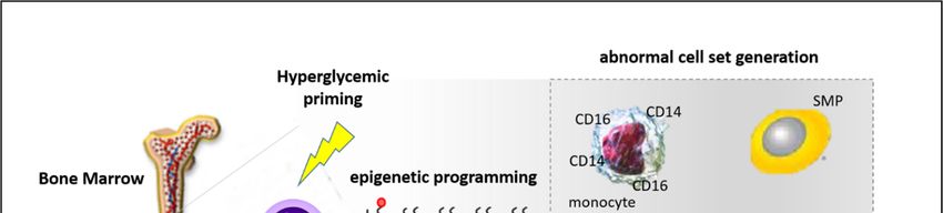
Int. J. Mol. Sci. 2020, 21, 3864 2 of 22
both victims and perpetrators in DM and diabetic complications, respectively [5]. HSPC alterations are
attributable to BM dysfunction, which is another target organ damaged by diabetic complications [6].
Speculatively, the defects of HSPCs could represent an epiphenomenon of BM impairment that become
a central housekeeper of an organism’s health [7]. This review deals with the modulatory effects of the
diabetic milieu on BM function, providing evidence of the mechanisms involved in BM dysfunction
and alteration of differentiation programs of HSPCs. Finally, a section is dedicated to the recent
advances in pharmacological management of diabetes, discussing the potential protective effects of
new glucose-lowering agents on BM and HSPCs.
2. Endothelial Progenitor Cells: A Multifaceted Definition and a Unique Recognized Role in
Diabetic Complications
CD34 is a transmembrane glycoprotein that is expressed by BM-derived HSPCs [8]. HSPCs are
responsible for the generation of all adult blood cells, including specialized cells that control immune
function, homeostasis balance, response to microorganisms, and inflammation [9].
Since 1968, HSPC transplantation therapy has been successfully used for the treatment of various
malignant human blood disorders because of their ability to reconstitute all of the hemato-lymphoid
system [10]. The vast majority of HSPCs resides in specialized niches within the BM, whereas
a small number of stem and progenitor cells normally traffic throughout the body. However,
the mechanisms underlying this trafficking as well as the physiological significance of their migration
have remained relatively obscure. The current hypothesis is that the continual egression from the
BM niche, extravasation into different tissues, and then their re-ingression into the BM, processes that
are regulated by circadian rhythms [11], have the role of patrolling peripheral organs, with the aim
of maintaining the immune responsiveness, tissue homeostasis, and regeneration. The continuous
information exchange from the periphery to the BM, mediated by HSPCs and other factors, grants,
beside a constant BM niche remodeling, a timely and efficient response to stressors. Indeed, different
injury paradigms, such as hemorrhagic shock, inflammation, stroke, infection, and ischemia, are known
to increase the circulating pool of HSPCs [12,13], even if the contribution of these cells to tissue repair
and regeneration is still under investigation [14]. In 1997, Asahara and colleagues pioneered over
20 years of research in stem cell biology and regenerative medicine, which is still focused on the
identification, isolation, and functional characterization of circulating BM-derived progenitors with
vasculogenic properties [15]. Asahara, by identifying BM-derived circulating CD34- kinase insert
domain receptor (KDR)-positive cells capable of incorporating into regenerating host vessels and
contributing to vascular repair, challenged for the first time, the dogma that vasculogenesis is restricted
to embryogenesis. These cells were named endothelial progenitor cells (EPCs). Since this pioneering
work, nowadays, the search term “endothelial progenitor cells” in PubMed returns over 84,900 clinical,
preclinical, and basic research articles, often controversial, in which these cells play the leading
role (reviewed in [16–18]). The lack of consensus relies on two main reasons: The heterogeneous
population of cells originated from the BM and the different methodological approaches used for
their identification and characterization, such as surface marker antigens, cell culture, and clonal
proliferation techniques [19–21]. So far, from a simplistic but usefulness point of view, two major
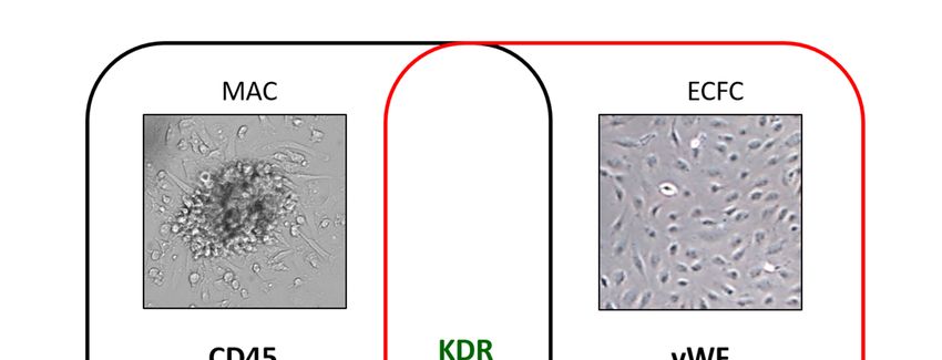
EPC cell populations have been isolated by culture techniques from peripheral blood: The myeloid
angiogenic cells (MACs, also called circulating angiogenic cells (CACs), pro-angiogenic hematopoietic
cells (PACs) or “early EPCs”) and the endothelial colony-forming cells (ECFCs or outgrowth endothelial
cells or “late EPCs) [18]. Both populations, although profoundly different in surface marker antigens and
functions [21], share KDR, CD31, CD34, CD105, lectin, and acetylated low-density lipoprotein (acLDL)
markers (Figure 1), and are thought to synergistically cooperate in the re-vascularization processes.
MACs, without changing their lineage fate and integrating into the remodeled endothelium [22,23],
support endothelial repair and regeneration through paracrine factors, whereas ECFCs, an endothelial
cell type characterized by potent intrinsic clonal proliferative potential, structurally contribute to de
novo blood vessel formation in vivo [24,25]. Considering tissue re-vascularization as a two-steps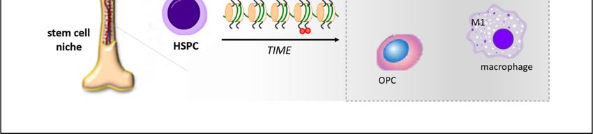
Int. J. Mol. Sci. 2020, 21, 3864                                                                                    3 of 22

Int. J. Mol. Sci. 2020, 21, x FOR PEER REVIEW                                                                      3 of 22
proces, it has been hypothesized that MACs are first actively attracted into the damaged tissue, where,
by the release
instruct ECFCof   soluble factors,
                proliferation   and ECFCs   are recruited,
                                     migration               hypothetically
                                                 to the injury                 either
                                                                 site to restore  the from   the circulation
                                                                                      endothelial    integrityoroflocal
                                                                                                                    the
vessel wall.  Next,  MACs     instruct  ECFC   proliferation   and   migration    to the  injury
vascular wall [26,27]. The ECFCs, isolated from adult human peripheral blood and cord blood,      site to restore   the
endothelial  integrity of the   vascular  wall [26,27].   The  ECFCs,    isolated  from   adult  human
display a clonal phenotype in vitro and cell surface markers undistinguishable from non-proliferative     peripheral
blood andendothelial
vascular    cord blood, cells.
                         display   a clonal
                                This   rare phenotype     in vitrowhose
                                            cell population,        and cell
                                                                           BMsurface
                                                                                 originmarkers
                                                                                          is stillundistinguishable
                                                                                                   largely debated
from  non-proliferative   vascular   endothelial   cells.  This rare  cell population,    whose    BM
[25,28,29], does not express progenitor cell markers and shows different functions and transcriptomic   origin is still
largely debated  [25,28,29],  does  not  express  progenitor   cell markers   and   shows   different
features compared with mature endothelial cells [30]. However, though ECFCs are widely accepted        functions   and
transcriptomic
as putative EPCs features
                   in vitro,compared     with existence
                             their effective  mature endothelial
                                                          and function cells
                                                                          in [30].  However,
                                                                             vivo are   elusive.though ECFCs are
widely accepted as putative EPCs in vitro, their effective existence and function in vivo are elusive.

      Figure 1. Cluster differentiation (CD) and other markers that identify myeloid angiogenic cells (MACs),
      black left
      Figure  1. circle,
                  Clusteranddifferentiation
                              endothelial colony
                                             (CD) forming   cellsmarkers
                                                    and other     (ECFCs),that
                                                                            red identify
                                                                                right circle. The markers
                                                                                          myeloid          shared
                                                                                                    angiogenic     by
                                                                                                                cells
      both cell black
      (MACs),    populations    are and
                       left circle, in the center. Cluster
                                         endothelial colonydomain
                                                             forming(CD);
                                                                       cellsCXC  motif,red
                                                                             (ECFCs),    receptor 4 (CXCR4);
                                                                                           right circle.      kinase
                                                                                                         The markers
      insert domain     receptor   (KDR);  acetylated  low-density   lipoprotein   (acLDL);   Von
      shared by both cell populations are in the center. Cluster domain (CD); CXC motif, receptor Willebrand   factor
                                                                                                                    4
      (vWF);  endothelial   nitric  oxide synthase  (eNOS);  Neuropilin-1    (NRP-1).
      (CXCR4); kinase insert domain receptor (KDR); acetylated low-density lipoprotein (acLDL); Von
     Willebrand factor (vWF); endothelial nitric oxide synthase (eNOS); Neuropilin-1 (NRP-1).
2.1. Circulating HSPCs: Current Concept and Implication in Health and Disease
      Despite theHSPCs:
2.1. Circulating   20 years of research
                          Current       efforts
                                   Concept and dedicated
                                                Implicationtointhe identification
                                                                Health and Diseaseof EPCs, the impossibility of
sharply separating EPCs from hematopoietic cells, either progenitor or myeloid lineage-committed,
      Despite the 20 years of research efforts dedicated to the identification of EPCs, the impossibility
is so far evident because of their overlapping phenotypes [31,32]. Today, many research groups are now
of sharply separating EPCs from hematopoietic cells, either                 progenitor or myeloid lineage-
inclined to identify circulating EPCs with the more generic CD34+ /CD133+ HSPC population because
committed, is so far evident because of their overlapping phenotypes [31,32]. Today, many research
it is the ancestor of all EPC phenotypes. The qualitative and quantitative depletion of circulating
groups are now inclined to identify circulating EPCs with the more generic CD34+/CD133+ HSPC
HSPCs, which encompasses progenitor subsets with multiple lineage commitment, has been shown to
population because it is the ancestor of all EPC phenotypes. The qualitative and quantitative
be independently associated with the risk of mortality in patients with coronary artery disease [33],
depletion of circulating HSPCs, which encompasses progenitor subsets with multiple lineage
renal disease [34], as well as with the severity of pathologies, such as Alzheimer’s [35] and cognitive
commitment, has been shown to be independently associated with the risk of mortality in patients
impairment [36]. Such epidemiological observations derived from different cohorts of patients clearly
with coronary artery disease [33], renal disease [34], as well as with the severity of pathologies, such
suggest an important role of circulating HSPCs in the physiological mechanisms of endogenous
as Alzheimer’s [35] and cognitive impairment [36]. Such epidemiological observations derived from
tissue regeneration. This hypothesis is further supported by numerous preclinical and clinical studies
different cohorts of patients clearly suggest an important role of circulating HSPCs in the
showing the ability of these cells to repopulate non-hematopoietic tissues, including the kidney, heart,
physiological mechanisms of endogenous tissue regeneration. This hypothesis is further supported
and lungs [37–41], as well as their efficacy, although controversial regarding the exact definition and
by numerous preclinical and clinical studies showing the ability of these cells to repopulate non-
function of such cells, in the treatment of cardiovascular disease, peripheral arterial disease, and critical
hematopoietic tissues, including the kidney, heart, and lungs [37–41], as well as their efficacy,
limb ischemia in humans [42–44].
although controversial regarding the exact definition and function of such cells, in the treatment of
cardiovascular disease, peripheral arterial disease, and critical limb ischemia in humans [42–44].

2.2. Circulating HSPCs and Diabetes: A Strong and Intertwined Relationship
Int. J. Mol. Sci. 2020, 21, 3864 4 of 22
2.2. Circulating HSPCs and Diabetes: A Strong and Intertwined Relationship
Following EPCs’ identification, HSPCs are now considered integral components of the mechanisms
involved in the maintenance of endothelial integrity, vascular homeostasis, and endogenous tissue
regeneration in general. DM induces micro- and macro-angiopathies that over time lead to long-term
damage and failure of various organ and systems. So far, numerous molecular mechanisms and
pathways have been described to translate hyperglycemia, the hallmark of DM, into tissue damage [45].
However, dramatically, the same mechanisms seem to also be involved in impairment of the endogenous
vascular reparative system, including stem cells. Considering that cell turnover is essential to face
the physiological rate of cell senescence and death, it is easy to predict that in the subjects in whom
intrinsic repair processes are defective, tissue damage can worsen. Under this prospective, the DM
has been considered a disease of impaired damage control [46], which would explain the two- to
three-fold increase of cardiovascular risk in this patient category. The discovery that diabetes affects
stem/progenitor cell biology opened an important research field in diabetology, where stem cell
dysfunction is at the center of the pathophysiological mechanisms of diabetic complications [47].
A consistent body of studies has documented the reduction and dysfunction in T2DM and T1DM
of pro-angiogenic HSPCs (at the time still termed EPCs) [48–52]. These alterations, interpreted in
view of a regenerative role, have been mechanistically considered to contribute to the onset and
progression of vascular disease [53]. Importantly, a careful analysis of the same clinical studies reveals
that pro-angiogenic HSPC alterations follow the natural history of diabetes, from its development
to the later stage. This peculiarity suggests that the reduction of circulating pro-angiogenic HSPC
numbers may also represent a biomarker of adverse cardiovascular events. Consistently, reduced levels
of circulating pro-angiogenic HSPCs (EPCs) have already been associated with endothelial dysfunction,
cardiovascular events, and with the pathogenesis of stroke in different cohorts of patients [54–57].
A recent meta-analysis by Rigato et al. showed that the numerical pauperization of circulating
progenitors, in particular CD34+ /CD133+ HSPCs, was associated with a 2-fold increased risk of future
cardiovascular events and cardiovascular death in patients with suspected coronary artery disease,
acute coronary syndrome, previous stroke, or in patients without acute events but with cardiovascular
risk factors [58]. To note, the reduction of the same circulating cell population has recently been proven
to be an efficient clinical-grade-independent biomarker in macro- and microvascular disease, capable of
predicting long-term adverse cardiovascular outcomes in T2DM patients [59,60]. Though the molecular
mechanisms mediating numerical and functional alteration of CD34+ /CD133+ HSPCs in diabetes are
still largely obscure (reviewed in [61]), BM is emerging as the original core of HSPC dysfunction.
To better clarify the mechanisms underpinning diabetic BM dysfunction, it is appropriate to
introduce the complex cellular and molecular network regulating HSPC niche function.
3. Healthy BM
BM is the major hematopoietic organ and lymphoid tissue responsible for the production of
erythrocytes, granulocytes, monocytes, lymphocytes, and platelets; moreover, it also represents the
largest reserve of HSPCs in an adult organism [62]. Within the BM, HSPCs reside in a specialized
microenvironment called the niche [63]. The BM niche, defined for the first time by Richard Shofield in
1978 [64], is an anatomical compartment composed by stromal cells, mesenchymal cells, osteoblasts,
endothelial cells, reticular cells, and the extracellular matrix. The continuous molecular crosstalk
between HSPCs and the other niche cellular constituents regulates HSC migration, quiescence, and
differentiation [63,65,66].
So far, two distinct niches have been identified: Endosteal niche, lining the bone surface, and the
vascular niche located around the sinusoidal endothelium. The two compartments are not separated
but dynamically regulated and interrelated. Endosteal HSPCs are thought to mature during their
relocation to the vascular niche; however, it is still unclear if endosteal and vascular HSPCs are mutually
exchangeable [62]. The HSPC localization within the niches tightly correlates with the proliferative and
maturation status of the cells. Indeed, HSPCs characterized by a high self-renewal capacity are localizedInt. J. Mol. Sci. 2020, 21, 3864 5 of 22
near the endosteum, and as they mature, they move towards vascular sinusoids (vascular niche) [63,67],
which play a pivotal role in progenitor cell egression from the BM to the circulation [68]. Osteoblasts of
the endostal niche, which is further composed of a heterogeneous cell population, including fibroblasts
and endothelial cells, regulate HSPC proliferation and quiescence [69]. There is evidence that osteoblast
lineage cells are required to maintain the ‘endosteal stem cell niche’ as their expansion by genetic or
pharmacologic means results in a concurrent expansion of HSPCs, whereas their ablation leads to
HSPC loss [70]. In addition, osteoblasts also play a key role in HSPCs’ mobilization because they are a
source of stromal cell-derived factor 1 (SDF-1/CXCL12 (CXC motif, ligand 12)), even if the interaction
of this chemokine with its cognate receptor, CXCR4 (CXC motif, receptor 4) is not the only mechanism
regulating HSPC trafficking in the BM. In this regard, numerous studies reported a very complex
picture of the mechanisms regulating the homeostasis and mobilization of HSPCs that comprise
not only endothelial cells and mesenchymal stromal progenitor cells but also mature hematopoietic
neutrophils and macrophages. A recent review detailed the role that each of these cells of the HSPC
niche play in contributing to mobilization [71]. Osteoblasts are also able to regulate HSPC survival,
quiescence, and differentiation by producing factors, such as angiopoietin-1 [72] and thrombopoietin,
capable of directly binding the HSPCs [73]. Stromal cells, on the other hand, are able to retain the
HSPC in the BM by interacting between the vascular cell adhesion molecule 1 (VCAM-1) to the very
late antigen-4 (VLA-4) antigen present on the HSPCs [74]. The maturation of the HSPCs is regulated
by the Notch signal through interaction with osteoblasts [75].
The arterial vessels enter into the BM through the foramina nutricia and then divide into arterioles,
capillaries, and sinusoids. Sinusoids, where the vascular niche is localized, consist of fenestrated
endothelium and, to a lesser extent, pericytes, forming a permeable barrier for HSPC passage into
the circulation [76]. While HSPCs located at the endosteum are more quiescent and have a greater
self-renewal capacity due to a variety of cytokines, adhesion molecules, and hypoxia, HSPCs located
close to the sinusoid endothelium (vascular niche) have reduced self-renewal capacity and cycle
more rapidly due to higher oxygen levels. The cells of the vascular niche communicate with cells
of the endosteal niche, and the subtle balance of factors from these sub-compartments governs the
behavior of the HSPCs [77]. The vascular niche is involved in the proliferation and differentiation
of HSPCs thanks to the presence of hormones, growth factors, oxygen, and nutrients from the
peripheral circulation [78,79]. Moreover, the presence of different cell types, such as endothelial cells,
mesenchymal cells, perivascular stromal cells (e.g., CXCL12-abundant reticular-CAR+ , nestin+ , and
leptin+ receptor-Lepr+ cells), and Schwann cells [80–82], associated with HSPCs grant a fine-tuned
regulation of stem cell quiescence and maintenance [83,84].
Overall, both endosteal and vascular niches support hematopoiesis and mobilization through
paracrine signaling and physical interactions. Moreover, BM vascular cells direct the regular
trafficking of HSPCs to the systemic circulation and back to the BM. Importantly, both niches
are highly innervated [67]. Indeed, both niches are also finely regulated at the neuronal level by the
sympathetic nervous system (SNS), which acts on perivascular cells through β3-adrenergic receptors.
These signals that regulate CXCL12 expression in BM stromal cells allow HSPCs’ mobilization through
circadian rhythms [85]. For example, nestin+ cells expressing the beta-adrenergic receptors, following
noradrenergic stimuli, are able to downregulate the retention signals, allowing HSPC mobilization [78].
Similarly, granulocyte colony stimulating factor (G-CSF)-mediated HSPC mobilization is the result
of SNS signaling on perivascular cells [86]. The SNS also acts directly on the HSPCs by the binding
of adrenergic neurotransmitters (epinephrine and norepinephrine) and dopamine on β2-adrenergic
receptors of HSPCs, which act as chemoattractants [87].
In homeostatic conditions, the number of HSPCs that physiologically leave the BM niche and
reach the peripheral organs through the circulation to maintain tissue integrity is low. However,
this number rapidly increases following tissue injury [62]. The mobilization of HSPC from the BM
after injury is a delicate process that involves not only chemokine gradients and SNS activation [88,89]
but also mediators, such as matrix metalloproteinases type 2 and 9; cathepsin K, which is secreted byInt. J. Mol. Sci. 2020, 21, 3864 6 of 22
osteoclasts; and elastases, produced by neutrophils [90]; all these proteases have the task of cutting the
interactions between HSPCs and their niche microenvironment [62].
4. Diabetic BM
BM is now recognized as a target organ for chronic diabetic complications, in which defects, such as
mobilopathy, neuropathy, and microangiopathy, are the main pathologic manifestations [76]. Profound
alterations in gene expression and cytokine signaling induced by the diabetic microenvironment are
the basis of changes in the BM cellular component, such as increased adipogenesis and decreased
osteoblastogenesis [91].
Preclinical and clinical studies have implicated reactive oxygen species (ROS) overproduction,
endoplasmic reticulum stress alterations, and advanced glycation end products (AGEs) in the abrogation
of proangiogenic pathways involved in the restoration of perfusion and regeneration of ischemic
tissue in DM. To these should also be added, for the sake of completeness, the marked impairment
of BM-derived HSPC mobilization, recruitment, and pro-angiogenic differentiation [50,90,92–95].
These defects could depend on (i) reduced cell survival in the circulatory stream, (ii) altered homing
out of the vessels, (iii) BM dysfunction with insufficient and less viable stem cell release, or (iv) a
combination of all [7].
4.1. Mobilopathy
Mobilopathy is defined as the inefficient release of HSPCs from BM due to an altered gradient of
chemokines both within the BM and between the BM and the periphery [67]. This functional defect
can be partly explained by CXCL12/CXCR4 axis dysregulation [96]. Under homeostatic conditions,
both the stem cell factor (SCF (also referred as KIT-ligand)/c-Kit (CD117) axis, and the binding between
CXCL12, mainly produced by BM stromal cells, and CXCR4/CXCR7 receptors expressed on HSPCs
keep the cells anchored to the niche. In response to peripheral ischemia, the decline of BM CXCL12
levels is the key signal of HSPC mobilization. In diabetes, however, CXCL12 in the BM does not
decrease following ischemia, leading to an inefficient mobilization of HSPCs [97]. In this regard,
Albiero and co-workers found an excess of proinflammatory M1 macrophages in the BM of a T1DM
mice model. Moreover, they also found that oncostatin M (OSM) was the long-sought soluble factor
released by macrophages that sustained CXCL12 expression by mesenchymal stem/stromal cells, which
was no longer downregulated even after G-CSF stimulation. A few years later, the same authors also
demonstrated that the oncostatin M (OSM)-p66Shc pathway mechanistically linked HSPC mobilopathy
to excessive myelopoiesis [98,99].
In the ischemic tissue, the transcription factor hypoxia-inducible factor-1 (HIF-1) regulates
CXCL12 gene expression in endothelial cells, which is directly proportional to reduced oxygen tension.
HIF-1-induced CXCL12 expression increases the adhesion, migration, and homing of circulating
CXCR4-positive progenitor cells to the ischemic site [100]. Diabetes is known to upregulate dipeptidyl
peptidase-4 (DDP-4) expression, an enzyme involved in CXCL12 degradation, in the peripheral tissue.
This DDP4-dependent reduction of CXCL12 levels results in the dampening of HSPC recruitment with
a consequent angiogenesis deficit, as demonstrated in the heart of rats with chronic heart failure [101].
In the BM, on the other hand, DM inadequately upregulates DPP-4 on CD34+ cells, which is required
for the mobilizing effect of G-CSF [102]. Nevertheless, DDP-4 inhibition was shown to promote
HSPC mobilization by protecting CXCL12 from inactivation [103] and enzymatic degradation [104] in
T2DM patients.
In addition, endothelial nitric oxide synthase/nitric oxide (eNOS/NO) pathway activation,
fundamental for HSPC mobilization, is also compromised in diabetes, as demonstrated in a murine
model of diabetes, where the eNOS enzyme was decoupled [105] and its phosphorylation rate
reduced [106].Int. J. Mol. Sci. 2020, 21, 3864 7 of 22
4.2. Inflammation
The increase of proinflammatory cytokines, such as interleukin-3 (IL-3), IL-10, IL-1β, tumor
necrosis factor-α (TNF-α), and monocyte generation has been observed in the BM of a T1DM mice
model [107]. Moreover, the downregulation of insulin-like growth factor 1, insulin-like growth
factor-binding protein 5, osteoprotegerin, and vascular endothelial growth factor (VEGF) n the BM
plasma of T1DM mice was associated with reduced Bmi1expression, a gene involved in senescence
protection, and with an impaired HSPC repopulation of BM [108]. Hyperglycemia is also able to induce
a small but specific BM cell subpopulation, identified by proinsulin production, that expresses TNF-α,
a cytokine implicated in numerous diabetic complications, including diabetic peripheral neuropathy
(DPN) [109]. This latter finding represents the first emerging evidence of active HSPC contribution to
the pathogenesis of DPN.
4.3. Neuropathy
Sensory neuropathy is a common complication of DM characterized by damage of the sensory
nerves. This causes an altered pain perception and impairs the activation of healing mechanisms after
injury. Under physiological conditions, peripheral sensory neurons release, after damage, nociceptive
factors, such as substance P (SP), which exert local and systemic actions, including recruitment to the
site of damage of BM and circulating HSPCs expressing the neurokinin 1 receptor (NK1R), the main
SP receptor. Recent studies demonstrated that sensory neuropathy can occur in the bone marrow of
T2DM patients and mice. The rarefaction of nociceptive fibers in the diabetic BM was associated with
an impairment in the neurokinin gradient between the BM, peripheral blood, and peripheral tissue,
accompanied by a depressed recruitment of NK1R-HSPCs to the site of injury [110,111].
It is known that SNS is prominently involved in BM niche function [88] and stem cell trafficking
is regulated by catecholaminergic neurotransmitters [87]; therefore, diabetic autonomic neuropathy
(DAN) may also impact the BM. Albiero et al. demonstrated that BM denervation in an experimental
model of diabetes (streptozotocin-induced and ob/ob mice) was mediated by p66Shc upregulation and
that impaired mobilization relied on sirtuin 1 (Sirt1) dysregulation [112]. In this complex scenario,
Lucas et al. showed that G-CSF is able to directly increase the sympathetic tone. Peripheral sympathetic
nerve neurons express the G-CSF receptor and its stimulation with G-CSF reduced norepinephrine
reuptake significantly, suggesting that G-CSF potentiates the sympathetic tone by increasing the
norepinephrine availability. Their data suggest that the blockade of norepinephrine reuptake may
be a novel therapeutic target to increase the stem cell yield in DAN patients [113]. Using a rat model
of T2DM, Busik et al. demonstrated that BM neuropathy preceded the development of diabetic
complications. They observed a reduced number of nerve endings in the BM of diabetic rats that
coincided with a numerical increase of pro-angiogenic HSPCs within the BM and decrease of their
circulating levels. In addition, denervation was also accompanied by a loss of HSPC circadian release,
suggesting that inherent sympathetic denervation can alter the circadian peripheral clock. Overall,
both DM-induced denervation and circadian clock impairment led to diminished reparative capacity,
eventually resulting in the development of diabetic retinopathy [114].
4.4. Microangiopathy
Diabetes induces microvascular remodeling with a negative consequence for BM homeostasis.
Oikawa et al. demonstrated that cultured endothelial cells from the BM of T1DM mice showed
increased levels of oxidative stress, senescence, reduced migratory and network formation capacities,
and increased permeability. BM endothelial cell dysfunction, in which numerous mechanisms,
including increased expression and activity of Ras homolog family member A (RhoA) and reduced Akt
phosphorylation/activity, are involved, is responsible for reduced BM perfusion and consequent HSPC
loss. In addition, HSPC depletion induced by BM microangiopathy was associated with increased
oxidative stress, DNA damage, and apoptosis of the stem cells [6,115].Int. J. Mol. Sci. 2020, 21, 3864 8 of 22
4.5. Oxidative Stress
The reactive oxygen species (ROS) gradient is a fundamental signaling mechanism controlling
functional niche compartmentalization of stem cells in the BM. The precious low replicating HSPCs,
necessary for BM repopulation, reside in the “low ROS zones”, ideal for the maintenance of quiescence
and retention, whereas the “high ROS zone” adjacent to the marrow vasculature has the function
of facilitating stem cell maturation [6]. However, under pathologic conditions, such as diabetes,
excessive ROS production [116] might jeopardize stem cell viability by promoting DNA damage and
senescence [6,62]. Numerous mechanisms might account for increased oxidative stress in HSPCs,
including BM hypoperfusion and hyperglycemia, both recognized as strong inducers of ROS generation
by mitochondrial complex III, and exposure to ROS produced by other cell sources, such as endothelial
and stromal cells of the niche [6,117,118]. In this context, Src homology/collagen (Shc) adaptor protein
66 (p66Shc), a gene that regulates the apoptotic responses to oxidative stress, plays a fundamental role.
Di Stefano et al. showed that in vitro hyperglycemia-induced upregulation of p66Shc protein in HSPCs
derived from mouse BM correlated with increased mitochondrial ROS production and a marked
reduction of pro-angiogenic commitment of the cells. Conversely, p66Shc knockout BM-derived
HSPCs were insensitive to high glucose exposure. Importantly, the high glucose resistance of p66Shc
knockout BM-derived HSPCs was prevented by nitric oxide (NO) synthase inhibition, suggesting
that the reduction of NO bioavailability induced by ROS was the underlying mechanism involved in
pro-angiogenic HSPC dysfunction [119].
5. Evidence of Diabetes-Induced HSPC Programming and Pathobiological Implications
The BM harbors immature progenitor cell populations characterized by high plasticity and
multiple cell lineages’ commitment, not limited to the hematopoietic system [120,121]. As previously
reported, the stem cell niche has an important role in HSPC homeostasis [122,123]. This suggests
that BM niche alterations induced by the diabetic milieu might have profound implications in HSPC
biology, for example, by redirecting differentiation toward harmful cell populations that rather than
protecting, promote the progression of diabetic complications. In this regard, numerous studies have
reported the existence of BM-derived circulating progenitors with smooth muscle (smooth muscle
progenitors, SMPs) [124,125] and calcifying phenotypes (osteoprogenitor cells, OPCs) [126,127], in DM
patients. These cells, hypothetical “side products” of differentiation drift, witness the fact that diabetes
not only reduces the number of vasculoprotective cells but also promotes the generation of cells with
anti-angiogenic [128] and pro-fibrotic properties [129], with clear implications in diabetic micro- and
macro-angiopathies’ development. In addition to mobilization defects of pro-angiogenic HSPCs and
generation of aberrant progenitor cell populations, DM patients tend to have an elevation of peripheral
inflammatory monocytes [130–132], an alteration in macrophage polarization [133], as well as increased
levels of circulating inflammatory cytokines [134]. Terenzi and colleagues recently demonstrated
the incontrovertible reduction of circulating proangiogenic progenitors and the increased frequency
of circulating proinflammatory cells in TD2M patients [135]. Cumulatively, this unbalance, which
culminates in a reduction of the intrinsic vascular homeostatic capacity and in the establishment of a
low-grade inflammatory status [136], might be the main driver in atherosclerosis development and
the increased risk of CVD in DM patients [137,138]. Loomans et al. for the first time hypothesized
that hyperglycemia could modulate HSPC function. He demonstrated that hyperglycemia altered
the differentiation fate of BM precursor cells, reducing the potential to generate vascular regenerative
cells and favoring the development of proinflammatory cells [139]. After this publication, other
studies based on the murine model of diabetes further substantiated and mechanistically detailed this
finding [107,140,141].
Recent evidence supports the concept that epigenetic mechanisms may participate in the
differentiation drift of HSPCs into more inflammatory cell populations within the BM. Epigenetic
modifications have been documented in inflammation [142], and diabetic conditions are known to elicit
epigenetic changes in a variety of cell types, including HSPCs [143,144]. In this regard, we recentlyInt. J. Mol. Sci. 2020, 21, 3864 9 of 22
Int. J. Mol. Sci. 2020, 21, x FOR PEER REVIEW 9 of 22
promoter of the IL-12 gene in BM progenitors and this epigenetic signature is passed down to
demonstrated
macrophagesthat[146].
a hyperglycemic
Similar findings,condition induced
even if in different inhibitory
genes histone settings,
and experimental modifications
have been and DNA
methylation of the CXCR4 promoter in cord blood-derived CD34 + stem cells. These epigenetic
described by other authors [147,148]. In particular, Yan et al. observed in T2DM mice DNA
alterations, also identified
methyltransferase in BM-derived CD34
1 (DNMT1)-dependent + stem cells of
dysregulation of T2DM
genes patients,
related to were associated with a
macrophage
functional impairment of the CXCR4/CXCL12 axis [145]. progenitor
differentiation in HSPCs that was carried down through This study, cells to terminally
which differentiated
demonstrates a direct impact
cells [148]. This study supports the concept that the gene expression pattern of terminally
of hyperglycemia exposure on the epigenetic make-up of stem cell promoters, further supports the
differentiated immune cells might be epigenetically “preprogrammed” by hyperglycemia at the
hypothesis that epigenetic changes are likely additional mechanisms by which diabetes promotes the
HSPC level and may potentially be responsible for the prolonged and intrinsic inflammatory status
generation of inflammatory
of diabetics [138] (Figure BM-derived
2). monocytes and macrophages in diabetic patients, contributing
to enhanced CVD.
Thus, In a mouse
epigenetic changes model of insulincausative
can be considered resistance, Gallagher
factors contributing and colleagues
to the found that a
differentiation
drift of
repressive HSPCsmethylation
histone in DM. Whether this H3K27me3,
mark, alteration in DM is potentially
is decreased reversible
at the promoteris notoffully
the clear;
IL-12 gene in
however, there
BM progenitors andisthis
growing body of signature
epigenetic evidence regarding
is passed thedown
use of epigenetic drugs to restore/improve
to macrophages [146]. Similar findings,
HSPC function. Multiple studies have reported that chemical modifiers of DNA demethylation, such
even if in different genes and experimental settings, have been described by other authors [147,148].
as 5-azacytidine, or histone deaacetylase inhibitors, such as trichostatin A, valproic acid, or
In particular, Yan et al.
methyltransferase EZH2 observed
inhibitors,inenzymes
T2DM that mice DNA repressive
establish methyltransferase
H3K27me3 marks,1 (DNMT1)-dependent
have been
dysregulation
shown to not only enhance both the plasticity and function of progenitor cells but also carried
of genes related to macrophage differentiation in HSPCs that was change the down
cell through
progenitor cells
fate by to terminally
chromatin differentiated
remodeling [149,150].cells [148]. This
Interestingly, thestudy supports
inhibition the concept that
of bromodomain and the gene
extraterminal
expression pattern ofdomain (BET) differentiated
terminally proteins by the food drug
immune cellsadministration (FDA)-approved
might be epigenetically drug
“preprogrammed”
apabetalone (RVX-208) has been shown to modulate coagulation, vascular inflammation [151,152],
by hyperglycemia at the HSPC level and may potentially be responsible for the prolonged and intrinsic
and innate/adaptive immune responses, suggesting its potential use in the epigenetic treatment of
inflammatory status of diabetics [138] (Figure 2).
chronic diabetic inflammation and vascular complications [153].
FigureFigure 2. Schematic
2. Schematic representation of
representation of hyperglycemia-induced
hyperglycemia-induced programming of hematopoietc
programming stem
of hematopoietc stem
progenitor cells (HSPCs) at the bone marrow level. Diabetic milieu promotes epigenetic changes in
progenitor cells (HSPCs) at the bone marrow level. Diabetic milieu promotes epigenetic changes
HSPCs that result in the abnormal expansion of cells with inflammatory and pro-atherosclerotic
in HSPCs that result in the abnormal expansion of cells with inflammatory and pro-atherosclerotic
features, such as intermediate monocytes (CD14+CD16+), M1 macrophages, osteoprogenitor cells
features, such
(OPCs), andassmooth
intermediate monocytes
muscle progenitors (CD14+CD16+), M1 macrophages, osteoprogenitor cells
(SMPs).
(OPCs), and smooth muscle progenitors (SMPs).
6. The Protective Role of Glucose-Lowering Drugs on the HSPC Level and Inflammatory Profile
Thus,Patients
epigenetic changes can be considered causative factors contributing to the differentiation drift
with T2DM are at high risk of developing ASCVD, the leading cause of morbidity and
of HSPCs in DM.
mortality Whether
in these this[154].
patients alteration
Recentin DM is potentially
guidelines reversible
indicate that people withis not
T2DMfully
andclear; however, there
established
is growing
ASCVD, body of evidence
besides adequate regarding the use
glucose control, of epigenetic
should be managed drugs to restore/improve
with glucose-lowering drugs HSPC
proven function.
to studies
Multiple reduce major adverse cardiovascular
have reported events and/or
that chemical modifiers cardiovascular
of DNA mortality
demethylation, such([155,156],
as 5-azacytidine,
http://www.siditalia.it/clinica/standard-di-cura-amd-sid). Nevertheless, the mechanisms
or histone deaacetylase inhibitors, such as trichostatin A, valproic acid, or methyltransferase by which EZH2
these drugs exert pleiotropic cardiovascular effects are not completely understood.
inhibitors, enzymes that establish repressive H3K27me3 marks, have been shown to not only enhance
It is well known that HSPC dysfunction and systemic chronic inflammation are very common
both the plasticity
features in T2DMand function
patients, of reports
and some progenitor cells shown
have already but alsothat change
metformin the
cancell fatereverse
partially by chromatin
remodeling [149,150]. Interestingly, the inhibition of bromodomain and extraterminal domain (BET)
proteins by the food drug administration (FDA)-approved drug apabetalone (RVX-208) has been shown
to modulate coagulation, vascular inflammation [151,152], and innate/adaptive immune responses,
suggesting its potential use in the epigenetic treatment of chronic diabetic inflammation and vascular
complications [153].Int. J. Mol. Sci. 2020, 21, 3864 10 of 22
6. The Protective Role of Glucose-Lowering Drugs on the HSPC Level and Inflammatory Profile
Patients with T2DM are at high risk of developing ASCVD, the leading cause of morbidity
and mortality in these patients [154]. Recent guidelines indicate that people with T2DM and
established ASCVD, besides adequate glucose control, should be managed with glucose-lowering
drugs proven to reduce major adverse cardiovascular events and/or cardiovascular mortality ([155,156],
http://www.siditalia.it/clinica/standard-di-cura-amd-sid). Nevertheless, the mechanisms by which
these drugs exert pleiotropic cardiovascular effects are not completely understood.
It is well known that HSPC dysfunction and systemic chronic inflammation are very common
features in T2DM patients, and some reports have already shown that metformin can partially reverse
these processes [157,158]. Accordingly, a number of studies have been performed (or are still ongoing)
to evaluate whether therapeutic approaches with other glucose-lowering drugs, including peroxisome
proliferator-activated receptor gamma (PPAR-γ) agonists, dipeptidyl peptidase 4 (DPP-4) inhibitors,
glucagon-like peptide 1 (GLP-1) receptor agonists, and sodium-glucose transport protein 2 (SGLT2)
inhibitors, may reduce or reverse these pathophysiologic factors in T2DM patients.
PPAR-γ agonists, also known as thiazolidinediones, were the first investigated. Several
randomized and non-randomized clinical trials using PPAR-γ agonists in T2DM patients have
shown positive results in terms of the endothelial function and inflammatory profile [159–164]. As an
example, the largest randomized clinical trial by Esposito et al. [162], cumulatively enrolling 110 newly
diagnosed T2DM patients, proved the superiority of pioglitazone versus metformin in improving
the imbalance between endothelial damage (i.e., the endothelial microparticles expressing CD31)
and the endothelial repair capacity (i.e., the endothelial progenitors expressing CD34/KDR). As for
inflammation and oxidative stress, in a study on T2DM patients naïve to therapy, pioglitazone after
16 weeks significantly reduced CRP and E-selectin levels from baseline and in comparison to metformin,
whereas no difference was observed for nitrotyrosine (a marker of oxidative stress) between the two
treatments [165]. Interestingly, a prospective randomized controlled study that compared pioglitazone
and rosiglitazone with the best standard of care showed that both treatments significantly decrease
serum VEGF levels compared to controls (p = 0.00) and reduced thee serum TNF-α concentration,
with a statistical significance for the pioglitazone group only (p = 0.01) [166]. Cumulatively, these results
provide solid evidence for the anti-inflammatory and cardioprotective effect of PPAR-γ therapy and
pioglitazone, which so far represents a valid therapeutic strategy in T2DM patients with established
ASCVD ([155], http://www.siditalia.it/clinica/standard-di-cura-amd-sid).
As for DPP-4 inhibitors, sitagliptin and saxagliptin are the most widely investigated along
with vildagliptin in this setting [104,167,168]. However, results are mixed. For example, the EDGE
study (Effectiveness of Diabetes control with vildaGliptin and vildagliptin/mEtformin) revealed that
12 weeks of sitagliptin treatment increased circulating CD34+ cells (p = 0.03) but did not change
inflammatory markers (i.e., high-sensitivity CRP and pentraxin-3) and oxidative stress markers (i.e.,
malondialdehyde-modified low-density lipoprotein and urine 8-hydroxy-20 -deoxyguanosine) [169].
Other investigators observed similar biological effects with saxagliptin for 12 weeks and vildagliptin
for 12 months, respectively [167,168]. Conversely, other investigators did not find differences
regarding both EPC functionality and the inflammatory profile in patients treated with different DPP-4
inhibitors [170–173]. In addition, a very recent network meta-analysis demonstrated the superiority of
SGLT-2 inhibitors and GLP-1 agonists versus DPP-4 inhibitors in preventing cardiovascular events and
mortality in this setting of patients [174].
Likewise, GLP-1 receptor agonists have been proposed for their protective role on vascular
endothelium and the immune system [175–177]. Wei et al. [176] enrolled 31 newly diagnosed T2DM
patients receiving lifestyle modifications plus incremental doses of exenatide (10 µg/day for 1 month
and 20 µg/day for 2 months) or lifestyle modifications alone. This study showed that exenatide
treatment significantly improved the endothelial function of coronary arteries by measuring the
coronary flow velocity reserve (CFVR) and the system inflammation status by reducing the circulating
levels of vascular adhesion molecules (i.e., soluble intercellular and vascular adhesion molecule-1).Int. J. Mol. Sci. 2020, 21, 3864 11 of 22
Similar results were also reported in other head-to-head comparison studies. For example, it was
shown that exenatide and metformin treatments can equally improve endothelial dysfunction and
inflammation [178], even in a pre-diabetes setting [179].
However, it is worth noting that overall, these trials did not adopt a placebo-controlled group.
In addition to exenatide, the impact of liraglutide in T2DM patients is under investigation [180,181]
but available data are still limited. A parallel-group study of liraglutide and glargine therapy showed
a reduced deterioration of endothelial function for the group receiving liraglutide compared with
controls as measured by flow-mediated dilation. However, this difference was not significant (5.7%
to 5.4% and 6.7% to 5.7%, respectively) [181]. In a recent prospective randomized open-label trial,
the administration of liraglutide and dulaglutide for 24 weeks produced the same antioxidant effect as
demonstrated by improvements in the diacron-reactive oxygen metabolite and reactive hyperemia
index [180]. However, this was an open-label study with a small sample size (n = 22). More definitive
indications will arise from the ongoing clinical trials testing the role of semaglutide (NCT04126603)
and liraglutide (NCT02686177) in regulating vascular integrity and angiogenesis.
More recently, investigators have focused on the novel drug class of SGLT-2 inhibitors [182,183].
Specifically, in the DEFENCE trial (dapagliflozin effectiveness on vascular endothelial function and
glycemic control), Shigiyama et al. [184] compared the effect of dapaglifozin plus metformin and
metformin alone in 80 early stage T2DM patients. At the end of the 16-week treatment period,
the authors showed that the dapaglifozin add-on therapy compared to metformin-alone therapy
significantly improves the flow-mediated dilation in those patients having HbA1c≥7.0% at baseline
(1.05 ± 2.59 versus −0.94 ± 2.39; p < 0.05) and reduces urine 8-hydroxy-20 -deoxyguanosin, a clinical
marker of oxidative stress (−0.6 ± 1.8 versus 1.1 ± 2.2; p < 0.001). In contrast, the EMBLEM trial
(Effect of Empagliflozin on Endothelial Function in Cardiovascular High Risk Diabetes Mellitus) [185],
in which a total of 117 adults with T2DM and established ASCVD were randomized to receive either
empagliflozin 10 mg daily or placebo for 24 weeks, did not find differences in the primary endpoint
(i.e., the change in the reactive hyperemia peripheral arterial tonometry index). Further insights will
emerge from the ongoing randomized parallel-group trials. For example, the “Role of Canagliflozin on
CD34+ Cells in Patients With Type 2 Diabetes” trial (NCT02964585) is currently recruiting patients
with T2DM to study, as the primary endpoint, the gene expression and functional changes of CD34+
EPC. Other secondary endpoints, including serum endothelial inflammatory markers (hs-CRP, IL-6,
and TNF-alpha), will also be investigated.
Interestingly, clinical trials are ongoing, and aim to evaluate the combination of glucose-lowering
drugs possessing complementary and synergistic effects, such as saxagliptin plus dapaglifozin
(NCT03660683) or empagliflozin plus liraglutide (NCT03878706).
7. Concluding Remarks
It is now clear that the BM is a target organ for chronic diabetic complications and HSPCs play
an active role in the development of multiple organ vascular complications. The realization that DM
may not only impair HSPC mobilization from the BM but also program the differentiation towards
progenitors with defective vasculotrophic functions and excessive proinflammatory, pro-calcific,
and fibrotic properties, with direct effects in the pathogenesis of diabetic complications, opens a
new and exciting chapter of investigation in the diabetes field. From a therapeutic standpoint,
the achievement of good glycemic control without glucose variability remains the best and primary
way of preventing diabetic complications. However, a better understanding of the cellular and
epigenetic mechanisms involved, as inciting events, in the differentiation drift of BM stem/progenitor
cells towards deleterious cell populations in diabetes will help to identify new treatments to slow
down or reverse diabetic complications.Int. J. Mol. Sci. 2020, 21, 3864 12 of 22
Author Contributions: M.C.V.: Literature review, collection, conception, design and manuscript writing, final
approval of the manuscript. E.G.: Literature review collection and assembly of data, manuscript writing. B.B.:
collection and assembly of data, manuscript writing. S.G.: manuscript editing. G.P.: manuscript editing.
All authors have read and agreed to the published version of the manuscript.
Funding: This study was supported by Italian Ministry of Health, Ricerca Corrente (RC 2019).
Conflicts of Interest: The authors declare no conflict of interest.
References
1. De Matheus, A.S.M.; Tannus, L.R.M.; Cobas, R.A.; Palma, C.C.S.; Negrato, C.A.; de Gomes, M.B. Impact of
diabetes on cardiovascular disease: An update. Int. J. Hypertens. 2013, 2013, 653789. [CrossRef] [PubMed]
2. Miller, R.G.; Mahajan, H.D.; Costacou, T.; Sekikawa, A.; Anderson, S.J.; Orchard, T.J. A Contemporary
Estimate of Total Mortality and Cardiovascular Disease Risk in Young Adults With Type 1 Diabetes:
The Pittsburgh Epidemiology of Diabetes Complications Study. Diabetes Care 2016, 39, 2296–2303. [CrossRef]
[PubMed]
3. Kanter, J.E.; Bornfeldt, K.E. Inflammation and diabetes-accelerated atherosclerosis: Myeloid cell mediators.
Trends Endocrinol. Metab. 2013, 24, 137–144. [CrossRef] [PubMed]
4. Patel, J.; Donovan, P.; Khosrotehrani, K. Concise Review: Functional Definition of Endothelial Progenitor
Cells: A Molecular Perspective. Stem Cells Transl. Med. 2016, 5, 1302–1306. [CrossRef] [PubMed]
5. Kojima, H.; Kim, J.; Chan, L. Emerging roles of hematopoietic cells in the pathobiology of diabetic
complications. Trends Endocrinol. Metab. 2014, 25, 178–187. [CrossRef]
6. Oikawa, A.; Siragusa, M.; Quaini, F.; Mangialardi, G.; Katare, R.G.; Caporali, A.; van Buul, J.D.;
van Alphen, F.P.J.; Graiani, G.; Spinetti, G.; et al. Diabetes mellitus induces bone marrow microangiopathy.
Arterioscler. Thromb. Vasc. Biol. 2010, 30, 498–508. [CrossRef]
7. Fadini, G.P.; Ciciliot, S.; Albiero, M. Concise Review: Perspectives and Clinical Implications of Bone Marrow
and Circulating Stem Cell Defects in Diabetes. Stem Cells 2017, 35, 106–116. [CrossRef]
8. Satterthwaite, A.B.; Burn, T.C.; Le Beau, M.M.; Tenen, D.G. Structure of the gene encoding CD34, a human
hematopoietic stem cell antigen. Genomics 1992, 12, 788–794. [CrossRef]
9. Chotinantakul, K.; Leeanansaksiri, W. Hematopoietic stem cell development, niches, and signaling pathways.
Bone Marrow Res. 2012, 2012, 270425. [CrossRef]
10. Gatti, R.A.; Meuwissen, H.J.; Allen, H.D.; Hong, R.; Good, R.A. Immunological reconstitution of sex-linked
lymphopenic immunological deficiency. Lancet (Lond. Engl.) 1968, 2, 1366–1369. [CrossRef]
11. Casanova-Acebes, M.; Pitaval, C.; Weiss, L.A.; Nombela-Arrieta, C.; Chèvre, R.; A-González, N.; Kunisaki, Y.;
Zhang, D.; van Rooijen, N.; Silberstein, L.E.; et al. Rhythmic modulation of the hematopoietic niche through
neutrophil clearance. Cell 2013, 153, 1025–1035. [CrossRef] [PubMed]
12. Badami, C.D.; Livingston, D.H.; Sifri, Z.C.; Caputo, F.J.; Bonilla, L.; Mohr, A.M.; Deitch, E.A. Hematopoietic
progenitor cells mobilize to the site of injury after trauma and hemorrhagic shock in rats. J. Trauma 2007, 63,
596–600. [CrossRef] [PubMed]
13. Dutta, P.; Sager, H.B.; Stengel, K.R.; Naxerova, K.; Courties, G.; Saez, B.; Silberstein, L.; Heidt, T.; Sebas, M.;
Sun, Y.; et al. Myocardial Infarction Activates CCR2(+) Hematopoietic Stem and Progenitor Cells. Cell Stem Cell
2015, 16, 477–487. [CrossRef] [PubMed]
14. Porada, C.D.; Atala, A.J.; Almeida-Porada, G. The hematopoietic system in the context of regenerative
medicine. Methods 2016, 99, 44–61. [CrossRef]
15. Asahara, T.; Murohara, T.; Sullivan, A.; Silver, M.; van der Zee, R.; Li, T.; Witzenbichler, B.; Schatteman, G.;
Isner, J.M. Isolation of putative progenitor endothelial cells for angiogenesis. Science 1997, 275, 964–967.
[CrossRef]
16. Steinmetz, M.; Nickenig, G.; Werner, N. Endothelial-regenerating cells: An expanding universe. Hypertension
(Dallas, Tex. 1979) 2010, 55, 593–599. [CrossRef]
17. Basile, D.P.; Yoder, M.C. Circulating and tissue resident endothelial progenitor cells. J. Cell. Physiol. 2014,
229, 10–16. [CrossRef]
18. Medina, R.J.; Barber, C.L.; Sabatier, F.; Dignat-George, F.; Melero-Martin, J.M.; Khosrotehrani, K.; Ohneda, O.;
Randi, A.M.; Chan, J.K.Y.; Yamaguchi, T.; et al. Endothelial Progenitors: A Consensus Statement on
Nomenclature. Stem Cells Transl. Med. 2017, 6, 1316–1320. [CrossRef]You can also read