PDS5A and PDS5B in Cohesin Function and Human Disease - MDPI

Page created by Scott Sandoval
 
CONTINUE READING
PDS5A and PDS5B in Cohesin Function and Human Disease - MDPI
International Journal of
               Molecular Sciences

Review
PDS5A and PDS5B in Cohesin Function and Human Disease
Nenggang Zhang                , Luiza E. Coutinho and Debananda Pati *

                                          Department of Pediatrics, Texas Children’s Cancer Center, Baylor College of Medicine, 1102 Bates Street,
                                          Houston, TX 77030, USA; nzhang@bcm.edu (N.Z.); luizaecoutinho@gmail.com (L.E.C.)
                                          * Correspondence: pati@bcm.edu; Tel.: +1-832-824-4575; Fax: +1-832-825-4651

                                          Abstract: Precocious dissociation of sisters 5 (PDS5) is an associate protein of cohesin that is conserved
                                          from yeast to humans. It acts as a regulator of the cohesin complex and plays important roles in
                                          various cellular processes, such as sister chromatid cohesion, DNA damage repair, gene transcription,
                                          and DNA replication. Vertebrates have two paralogs of PDS5, PDS5A and PDS5B, which have
                                          redundant and unique roles in regulating cohesin functions. Herein, we discuss the molecular
                                          characteristics and functions of PDS5, as well as the effects of its mutations in the development of
                                          diseases and their relevance for novel therapeutic strategies.

                                          Keywords: PDS5A; PDS5B; cohesin; cancer; cohesinopathy

                                          1. Introduction
                                                Cohesin plays essential roles in a variety of cellular processes, including sister chro-
                                          matid cohesion, repair of damaged DNA, gene transcription, DNA replication, centrosome
         
                                          biogenesis, and maintenance of genome integrity [1–10]. It is a conserved multiprotein
                                   complex with three core subunits—two structural maintenance of chromosomes (SMC)
Citation: Zhang, N.; Coutinho, L.E.;
                                          proteins (SMC1 and SMC3) and an α-kleisin (RAD21/MCD1/SCC1)—forming an annular
Pati, D. PDS5A and PDS5B in Cohesin       trimer [8,11–13]. Both SMC1 and SMC3 fold into intramolecular antiparallel coiled coils
Function and Human Disease. Int. J.       with an ATPase-containing head domain on one end and a hinge domain on the other end.
Mol. Sci. 2021, 22, 5868. https://        The ATPase-containing head is formed with the N- and C-termini of each SMC molecule,
doi.org/10.3390/ijms22115868              and the hinge domain is from its middle region [14]. SMC1 and SMC3 join together through
                                          their hinge domains, creating a V-shaped heterodimer with the ATPase-containing heads at
Academic Editor: Lidia Larizza            the distal end of the two coiled-coils arms [14–16]. The ATPase-containing head domains
                                          of SMC1 and SMC3 together form a composite ABC-like ATPase that consists of Walker
Received: 30 April 2021                   A and Walker B motifs of one head and the Signature motif of the opposite head. Two
Accepted: 22 May 2021                     molecules of ATP can bind to the composite ABC-like ATPase [14,17]. The C-terminus of
Published: 30 May 2021                    RAD21 binds to the ATPase-containing head of SMC1, and the N-terminus of RAD21 binds
                                          to the coiled coil next to the ATPase-containing head of SMC3, resulting in the formation
Publisher’s Note: MDPI stays neutral      of a tripartite ring [14,18]. Besides those three core subunits, cohesin-associated proteins,
with regard to jurisdictional claims in   such as SCC3, PDS5, and WAPL, bind to the cohesin core complex via RAD21 [19–23].
published maps and institutional affil-
                                                Cohesin functions in both mitotic and meiotic cells, but some subunits of the co-
iations.
                                          hesin in these two types of cells are different. In mammal, SMC1A, SMC3, RAD21, and
                                          STAG1/STAG2 are cohesin subunits found in mitotic cells, which form two types of co-
                                          hesin complex because of two STAG proteins (STAG1/STAG2). In meiotic cells, SMC1B,
                                          Rad21/REC8/RAD21L, and STAG3 are the components of cohesin complex, which form
Copyright: © 2021 by the authors.         three different cohesins because there are three different α-kleisins [24].
Licensee MDPI, Basel, Switzerland.              In dividing cells, cohesin complexes are loaded to chromatin at telophase by a cohesin-
This article is an open access article    loading complex that consists of NIPBL/MAU2 heterodimer in humans and SCC2/SCC4
distributed under the terms and           in yeast. NIPBL and SCC2, as well as MAU2 and SCC4 are orthologous proteins. Cohesin
conditions of the Creative Commons
                                          dynamically associates with chromatin until the S phase, when DNA is replicated. The
Attribution (CC BY) license (https://
                                          sister chromatids are held together by the cohesin complexes, and sister chromatid cohesion
creativecommons.org/licenses/by/
                                          is generated after SMC3 is acetylated by cohesin acetyltransferases (CoATs) (Eco1/Ctf7
4.0/).

Int. J. Mol. Sci. 2021, 22, 5868. https://doi.org/10.3390/ijms22115868                                            https://www.mdpi.com/journal/ijms
PDS5A and PDS5B in Cohesin Function and Human Disease - MDPI
Int. J. Mol. Sci. 2021, 22, 5868                                                                                            2 of 21

                                   in yeast or ESCO1/2 in human) [25–28]. PDS5 plays important roles in the generation,
                                   maintenance, and resolution of sister chromatid cohesion. In vertebrates, PDS5, sororin,
                                   and WAPL form a cohesin-regulatory complex, in which sororin and WAPL compete to
                                   bind to a specific site on PDS5 to regulate the association of cohesin on chromatin [23,29–34].
                                   The cohesin-regulatory complex regulates the association of cohesin with chromosomes
                                   either positively or negatively depending on which protein binds to the site on PDS5. PDS5-
                                   sororin complex maintains sister chromatid cohesion from the S phase to G2, whereas
                                   PDS5-WAPL complex dislodges cohesin from chromatin. After WAPL displaces sororin
                                   that is phosphorylated by kinases, such as Cdk1/Cyclin B and Plk1 in the prophase, a PDS5-
                                   WAPL complex is formed, which then plays a role in releasing cohesins from chromosome
                                   arms. The cohesins on centromeres and residual arm cohesins are removed by separase
                                   through cleaving RAD21 on the transition of metaphase and anaphase, resulting in the
                                   sister chromatids being segregated into daughter cells [35–40].
                                         There are two paralogs of PDS5, PDS5A and PDS5B, in vertebrates. Limited studies
                                   indicate that they have common and specific roles in cell growth and development. Al-
                                   though PDS5 plays important roles in regulating functions of the cohesin complex, it has
                                   not attracted much attention in the cohesin field. Herein, we review the protein character-
                                   istics of PDS5 and its role in regulating cohesin functions in mitotic cells, as well as the
                                   implication in human disease.

                                   2. Identification and Characteristics of PDS5
                                         Cohesin is a versatile protein complex that is involved in various cellular processes by
                                   associating different regulatory partners. In efforts to identify more cohesin-interacting
                                   proteins that are required to elucidate the mechanisms on sister chromatid cohesion and
                                   condensation, SPO76 gene cloned from Sordaria macrospora [41] and bimD from Aspergillus
                                   nidulans [42] drew the attention of researchers in the cohesin biology field [43,44] for
                                   their roles in mitotic and meiotic chromosome segregation [42,45]. Genetic and functional
                                   analyses indicated that PDS5 is a cohesin-associated protein and is essential for the co-
                                   hesin to function. It is homologous to Spo76p and BIMD and conserved from fungi to
                                   human [43–45].
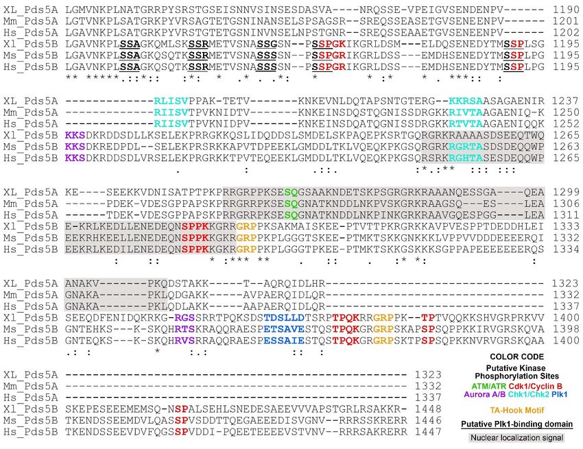
                                         PDS5 proteins contain more than 20 HEAT repeats (each is composed of two alpha
                                   helices linked by a short loop), forming two clusters separated by a helical insert domain
                                   (HID) (Figure 1) [44,46–48]. Multiple HEAT repeats form extended superhelical structures
                                   and can function as scaffolds to facilitate the assembly of other molecular components.
                                   Similar to PDS5, cohesin subunit SCC3 (STAG1, STAG2) and cohesin-associated proteins,
                                   such as SCC2, WAPL, and separase, also consist of HEAT repeats. [20,21,49–55].
                                         Full-length PDS5 proteins from Lachancea thermotolerans, Saccharomyces cerevisiae, and
                                   humans are comprised of 1292aa, 1277aa, and 1347aa, respectively. Crystal structures
                                   for truncated L. thermotolerans PDS5 (45-1221aa), S. cerevisiae Pds5 (1-701aa), and human
                                   PDS5B (21-1121aa) have been resolved (Figure 1A,B) [46,56,57]. Although yeast and human
                                   PDS5 have only approximately 20% identity at the amino acid residue level, they are
                                   conserved at the structural level. Both yeast Pds5 and human PDS5B are composed of
                                   alpha helical coils and look like a hook, with the stem or spine consisting of the HEAT
                                   repeats of the N-terminus and the hook consisting of the HEAT repeats of the middle and
                                   C-termini (Figure 1A,B) [46,56]. The hook tip bends back and contacts the middle of the
                                   spine, forming a ring-like space approximately 10Å in diameter [56]. Most of the HEAT
                                   repeats are on the spine [56,57]. The size of the hook structure is approximately 150Å [56].
PDS5A and PDS5B in Cohesin Function and Human Disease - MDPI
Int. J. Mol. Sci. 2021, 22, 5868                                                                                                                  3 of 21
Int. J. Mol. Sci. 2021, 22, x FOR PEER REVIEW                                                                                                    3 of 21

                                                                        Crystal structure
     Figure 1. Structure and characteristics of the PDS5 protein. Crystal         structure images
                                                                                              images of
                                                                                                      of (A) L. thermotolerans
                                                                                                         (A) L.  thermotolerans Pds5
                                                                                                                                Pds5 (PDB
                                                                                                                                      (PDB ID:
                                                                                                                                             ID:
     5F0O) and
             and (B)
                   (B)human
                       humanPDS5BPDS5B(PDB
                                        (PDBID:ID: 5HDT)
                                                 5HDT)     were
                                                         were     generated
                                                              generated       by using
                                                                          by using         NCBI’s
                                                                                      NCBI’s       web-based
                                                                                               web-based         3D structure
                                                                                                           3D structure        viewer
                                                                                                                          viewer  iCn3D.iCn3D.
                                                                                                                                          PDS5
     PDS5   is a hook-like
     is a hook-like   moleculemolecule  consisting
                                 consisting of HEAT ofrepeats
                                                       HEAT (green
                                                               repeatssticks)
                                                                        (greenwith
                                                                                 sticks)  with
                                                                                      helical   helical
                                                                                              insert    insert(HID,
                                                                                                     domain     domain   (HID,
                                                                                                                      golden    golden
                                                                                                                             sticks)     sticks)
                                                                                                                                     extruding
     extruding    on  one  side of the hook. Binding   of Scc1/Rad21   (pink  line)  to L. thermotolerans  Pds5   is shown  in
     on one side of the hook. Binding of Scc1/Rad21 (pink line) to L. thermotolerans Pds5 is shown in (A). Binding of WAPL (red(A). Binding   of
     WAPL     (red  line) to the C-terminus  of human   PDS5B    and IP6  to the  bottom    of PDS5B  hook-like    structure are
     line) to the C-terminus of human PDS5B and IP6 to the bottom of PDS5B hook-like structure are shown in (B). (C) Schematic   shown   in (B).
     (C) Schematic drawing shows PDS5 molecular features. The relative site is based on human PDS5B. Hrk1 interacting motif
     drawing shows PDS5 molecular features. The relative site is based on human PDS5B. Hrk1 interacting motif (HIM) on PDS5
     (HIM) on PDS5 N-terminus interacts with the PDS5 interacting motif (PIM) on HrK1, WAPL, and sororin. RAD21 and IP6
     N-terminus interacts with the PDS5 interacting motif (PIM) on HrK1, WAPL, and sororin. RAD21 and IP6 interact with
     interact with PDS5 in the middle region. Nuclear localization signal (NLS) is on the C-termini of PDS5A and PDS5B (Refer
     PDS5
     to     in the
        Figure   2). middle region. Nuclear localization signal (NLS) is on the C-termini of PDS5A and PDS5B (Refer to Figure 2).

                                            The PDS5 protein has several interesting features at the sequence level. The N-
                                           The PDS5 protein has several interesting features at the sequence level. The N-termi-
                                    terminus of PDS5 has a conserved serine/threonine-protein kinase Haspin/Hrk1-interacting
                                    nus of PDS5 has a conserved serine/threonine-protein kinase Haspin/Hrk1-interacting
                                    motif (HIM) (A-P-D/E-A-P) that interacts with a PDS5-interacting motif (PIM) (K/P-S/T-
                                    motif (HIM) (A-P-D/E-A-P) that interacts with a PDS5-interacting motif (PIM) (K/P-S/T-
                                    Y-S/T/G-K/R) found on WAPL and Hrk1 from yeast to human, as well as yeast Eso1
                                    Y-S/T/G-K/R) found on WAPL and Hrk1 from yeast to human, as well as yeast Eso1 of the
                                    of the Schizosaccharomyces group and sororin in vertebrates (Figure 1C) [30,46]. Human
                                    Schizosaccharomyces group and sororin in vertebrates (Figure 1C) [30,46]. Human PDS5B
                                    PDS5B binds to WAPL at the region of 1-33aa and to sororin at the region of 131–171aa.
                                    binds to WAPL at the region of 1-33aa and to sororin at the region of 131–171aa. The PIM
                                    The PIM on both WAPL and sororin contains a YSR motif (with the consensus of [K/R]
                                    on both WAPL and sororin contains a YSR motif (with the consensus of [K/R] [S/T]YSR)
                                    [S/T]YSR) that is conserved in vertebrates [46]. It has been reported that the region of
                                    that is conserved in vertebrates [46]. It has been reported that the region of the YSR motif
                                    the YSR motif on sororin interacts with cohesin complex, but to which cohesin-subunit
                                    on   sororin interacts
                                    or associated       proteinwith      cohesin
                                                                    remains     to becomplex,
                                                                                       identifiedbut[33,34,58].
                                                                                                       to which Mutations
                                                                                                                      cohesin-subunit
                                                                                                                                  of the orYSR associated
                                                                                                                                                  motif in
                                    protein    remains     to  be  identified    [33,34,58].  Mutations     of  the
                                    WAPL or sororin abolished their binding to purified PDS5B, suggesting that the YSRYSR  motif  in  WAPL      or sororin
                                                                                                                                                     motif
                                    abolished      their  binding      to  purified    PDS5B,    suggesting       that  the YSR
                                    is crucial for WAPL and sororin to interact with PDS5 directly [46]. Interestingly, the FGF    motif   is  crucial  for
                                    WAPL
                                    motif onand      sororin
                                                 sororin   andto  twointeract    with FGF
                                                                        of the three    PDS5motifs
                                                                                               directly    [46]. Interestingly,
                                                                                                      on WAPL                       the FGFofmotif
                                                                                                                     located downstream                 on
                                                                                                                                                  the YSR
                                    sororin    and   two    of  the  three   FGF    motifs
                                    motif are also important to bind to PDS5 [33,34,58].    on  WAPL      located     downstream      of  the  YSR   motif
                                    are also    important
                                            Structural         to bindoftohuman
                                                          analysis           PDS5 [33,34,58].
                                                                                      PDS5B reveals that inositol hexakisphosphate (IP6)
                                           Structural     analysis     of
                                    binds to the bottom of the PDS5 hook    human     PDS5B     reveals
                                                                                          (Figure   1B,C)that[46].inositol  hexakisphosphate
                                                                                                                     The basic   amino acid residues (IP6)
                                    binds    to the the
                                    with which        bottom      of the PDS5
                                                          IP6 interacts            hook (Figures
                                                                             are positively          1B,C)
                                                                                               charged     and[46].   The basic
                                                                                                                 conserved       amino
                                                                                                                               from   yeastacid
                                                                                                                                              to residues
                                                                                                                                                  humans.
                                    with    whichof
                                    Mutations        thethose
                                                          IP6 interacts
                                                                 amino acid  are positively   charged
                                                                                  residues cause     PDS5 anddeficiency
                                                                                                                 conservedinfrom      yeastbinding
                                                                                                                                cohesin       to humans.
                                                                                                                                                      [46],
                                    Mutations       of those     amino    acid    residues   cause   PDS5     deficiency
                                    implying that IP6 may contribute to maintaining the proper structure of PDS5 in order   in  cohesin    binding    [46],
                                    implying      that IP6with
                                    for it to interact       maycohesin.
                                                                     contribute IP6toand
                                                                                      maintaining
                                                                                          other higher the inositol
                                                                                                            proper structure      of PDS5 in
                                                                                                                       polyphosphates,           order
                                                                                                                                              such      for
                                                                                                                                                    as IP5
                                    it to  interact   with   cohesin.     IP6  and   other  higher    inositol   polyphosphates,
                                    and IP4, are abundant lipid-derived metabolites in eukaryotic cells and can function as            such    as IP5  and
                                    IP4,    are abundant
                                    structural     cofactors for lipid-derived       metabolites
                                                                      enzymes, receptors,       andinprotein
                                                                                                         eukaryotic      cells [59–61].
                                                                                                                 complexes      and canAlthough
                                                                                                                                             functionIP6 as
                                    structural
                                    has not been   cofactors
                                                       reported  for in
                                                                      enzymes,
                                                                        the crystalreceptors,
                                                                                       structureand   protein
                                                                                                   study         complexes
                                                                                                            of yeast           [59–61].
                                                                                                                        Pds5 [56],         Although IP6
                                                                                                                                     the abundance       of
                                    has
                                    IP6 not    been reported
                                          in eukaryotic      cells inandthethecrystal  structureofstudy
                                                                                conservation        the PDS5of yeast    Pds5 suggest
                                                                                                                   structure  [56], thethat
                                                                                                                                          abundance
                                                                                                                                                IP6 mightof
                                    IP6
                                    alsoinbind
                                             eukaryotic
                                                  to PDS5cellsfromand     theorganisms
                                                                      other     conservation
                                                                                           andof    the PDS5 in
                                                                                                 participate       structure  suggest interactions.
                                                                                                                      protein-protein    that IP6 might
                                    also bind
                                            YeasttoPds5
                                                     PDS5     from other
                                                           interacts     withorganisms      and participate
                                                                                the N-terminus       of Scc1/Rad21in protein-protein
                                                                                                                          (101-122aa in  interactions.
                                                                                                                                            human, 120-
                                    141aaYeast      Pds5L.interacts
                                             in yeast                    with the
                                                             thermotolerans)       atN-terminus
                                                                                      the openingofofScc1/Rad21          (101-122aa
                                                                                                          the hook (Figure             in human, This
                                                                                                                                 1A) [46,56,57].      120-
                                    141aa    in yeast L.region
                                    Pds5-interacting         thermotolerans)
                                                                      on Scc1 is at   the opening
                                                                                   located             of the hook
                                                                                            just downstream             (Figure
                                                                                                                    of the domain 1A)where
                                                                                                                                        [46,56,57].   This
                                                                                                                                               Scc1 binds
                                    to SMC3 [18]. Scc1
                                    Pds5-interacting            bindson
                                                            region      to Pds5
                                                                            Scc1 isand  is wedged
                                                                                      located          between the spine
                                                                                                just downstream          of theand   the hook,
                                                                                                                                 domain      wherecausing
                                                                                                                                                      Scc1
                                    the opening of the hook [56].
Int. J. Mol. Sci. 2021, 22, x FOR PEER REVIEW                                                                                                     4 of 21

Int. J. Mol. Sci. 2021, 22, 5868                                                                                                                   4 of 21

                                      binds to SMC3 [18]. Scc1 binds to Pds5 and is wedged between the spine and the hook,
                                      causing the opening of the hook [56].
                                                                        paralogs of the PDS5 protein, PDS5A and PDS5B. In humans,
                                            Vertebrates have two paralogs
                                      PDS5A isislocated
                                      PDS5A        locatedon onthe
                                                                 the  small
                                                                    small  armarm  of chromosome
                                                                                of chromosome    4, at4,position
                                                                                                          at position   14 (4p14),
                                                                                                                   14 (4p14), and hasand36 has  36
                                                                                                                                           exons.
                                      exons. PDS5B
                                      PDS5B   is on the is long
                                                           on thearmlong
                                                                       of arm  of chromosome
                                                                          chromosome             13, at position
                                                                                         13, at position            13.1 (13q13.1),
                                                                                                           13.1 (13q13.1),   and hasand    has 38
                                                                                                                                        38 exons.
                                      PDS5A    and PDS5B
                                      exons. PDS5A             consistconsist
                                                         and PDS5B       of 1337aa   and 1447aa,
                                                                                of 1337aa         respectively,
                                                                                            and 1447aa,             with anwith
                                                                                                           respectively,      overall  sequence
                                                                                                                                   an overall  se-
                                      identity of   70%.   The  N-termini    of PDS5A   and   PDS5B   (1-1200aa)     have
                                      quence identity of 70%. The N-termini of PDS5A and PDS5B (1-1200aa) have approxi-    approximately     72%
                                      sequence
                                      mately 72%  identity,
                                                      sequencewhereas     thewhereas
                                                                  identity,    C-termini
                                                                                       the(from   1201aa
                                                                                            C-termini       to the
                                                                                                         (from       end) to
                                                                                                                  1201aa   arethe
                                                                                                                                predominantly
                                                                                                                                   end) are pre-
                                      different
                                      dominantly(sharing     only(sharing
                                                      different    less thanonly
                                                                              28%less
                                                                                    identity).
                                                                                       than 28%Therefore,   it isTherefore,
                                                                                                   identity).     possible that
                                                                                                                             it isthe N-termini
                                                                                                                                    possible that
                                      determine
                                      the N-termini their  commontheir
                                                        determine      function,
                                                                            commonwhereas   the C-termini
                                                                                      function,  whereas the  differentiate   their roles intheir
                                                                                                                  C-termini differentiate     the
                                      cell
                                      rolescycle.
                                            in the cell cycle.

      Figure
      Figure 2.2. C-terminal
                   C-terminal alignment
                                alignment ofof PDS5A
                                               PDS5A and and PDS5B
                                                              PDS5B from
                                                                      from frog
                                                                            frog (Xenopus),
                                                                                  (Xenopus), mouse
                                                                                               mouse and
                                                                                                       and human.
                                                                                                             human. The
                                                                                                                      The following
                                                                                                                           following putative
                                                                                                                                          putative
      regulatory features
      regulatory    featuresare
                              arehighlighted
                                  highlightedinincolor:
                                                   color:ATM/ATR
                                                          ATM/ATRphosphorylation
                                                                      phosphorylationmotifmotif(S/T)-Q
                                                                                                 (S/T)-Q [62];
                                                                                                           [62];Aurora
                                                                                                                AuroraA/BA/B phosphorylation
                                                                                                                             phosphorylation
      motif (R/K)
      motif  (R/K)1-3 -X-(S/T)-(^P) where X is any amino acid, and ^P is not proline [63–65]; Cdk1/Cyclin B phosphorylation motif
                    1-3 -X-(S/T)-(ˆP) where X is any amino acid, and ˆP is not proline [63–65]; Cdk1/Cyclin B phosphorylation motif
      minimum      consensus
      minimum consensus sequencesequence(S/T)-P,
                                           (S/T)-P,full
                                                    fullconsensus
                                                          consensussequence
                                                                     sequence(S/T)-P-X-(K/R)
                                                                                 (S/T)-P-X-(K/R) [66];
                                                                                                   [66]; Chk1/Chk2
                                                                                                          Chk1/Chk2 phosphorylation
                                                                                                                      phosphorylation motif  motif
      (Φ/B)-(X/B)-(R/K)-X-X-(S/T)-Φ where B is a basic amino acid and Φ is a hydrophobic amino acid [67]; Plk1 phosphorylation
      (Φ/B)-(X/B)-(R/K)-X-X-(S/T)-Φ where B is a basic amino acid and Φ is a hydrophobic amino acid [67]; Plk1 phosphorylation
      motif (D/E)-X-(S/T)-Φ-X-(D/E). Plk1 binding domain (PBD) S-(pS/pT)-(P/X) where pS is a phosphorylated serine, pT is a
      motif (D/E)-X-(S/T)-Φ-X-(D/E). Plk1 binding domain (PBD) S-(pS/pT)-(P/X) where pS is a phosphorylated serine, pT is a
      phosphorylated threonine [68]. PBD is underlined. AT-hook motif GRP is highlighted in orange [69]. Nuclear localization
      phosphorylated      threonine
      signals are predicated         [68].
                                 using     PBDMapper
                                        cNLS    is underlined.
                                                         [70] andAT-hook
                                                                  shaded inmotif
                                                                             gray.GRP
                                                                                    Theisalignment
                                                                                          highlightedwas inperformed
                                                                                                            orange [69].using
                                                                                                                         Nuclear
                                                                                                                              Clustallocalization
                                                                                                                                          O (1.2.4)
      signals  are predicated   using cNLS   Mapper   [70] and  shaded  in gray. The alignment    was  performed   using  Clustal   O
      [71]. Invariant, conserved and semi-conserved residues are indicated by an asterisk (*), colon (:) and period (.), respectively. (1.2.4) [71].
      Invariant,   conserved
      The abbreviations      areand
                                 XL, semi-conserved
                                     Xenopus laevis; Mm,residues  are indicated
                                                             Mus musculus;        by an sapiens.
                                                                             Hs, Homo    asteriskProtein
                                                                                                  (*), colon  (:) and period
                                                                                                           sequences  are from (.),NCBI
                                                                                                                                    respectively.
                                                                                                                                           and the
      The abbreviations
      access                are XL, Xenopus
              reference numbers               laevis; Mm, Mus
                                     are NP_001090063.1          musculus;NP_001089658.1
                                                            (XL-PDS5A);     Hs, Homo sapiens.    Protein sequences
                                                                                              (XL-PDS5B),             are from (Mm-PDS5A),
                                                                                                              NP_001074790.1     NCBI and the
      NP_780519.3
      access  reference(Mm-PDS5B),
                          numbers areNP_001093869.1
                                         NP_001090063.1(Hs-PDS5A),
                                                            (XL-PDS5A);NP_055847.1      (Hs-PDS5B).
                                                                           NP_001089658.1     (XL-PDS5B), NP_001074790.1 (Mm-PDS5A),
      NP_780519.3 (Mm-PDS5B), NP_001093869.1 (Hs-PDS5A), NP_055847.1 (Hs-PDS5B).
                                            Several interesting features were revealed after close examination of the C-termini of
                                      PDS5ASeveral   interesting
                                               and PDS5B     fromfeatures   were revealed
                                                                   frog (Xenopus), mouse, after close examination
                                                                                          and human                of the 2).
                                                                                                       sequences (Figure  C-termini
                                                                                                                              (1) Nu-
                                      of PDS5A
                                      clear       and PDS5B
                                             localization       from
                                                           signals  arefrog (Xenopus),
                                                                         located on themouse, and human
                                                                                        C-terminus        sequencesin(Figure
                                                                                                     and conserved      PDS5A  2).and
                                                                                                                                   (1)
                                      Nuclear localization signals are located on the C-terminus and conserved in PDS5A and
Int. J. Mol. Sci. 2021, 22, 5868                                                                                           5 of 21

                                   PDS5B from different species. (2) PDS5A has two putative Chk1/Chk2 phosphorylation
                                   sites on its C-terminus, which are conserved in Xenopus, mice, and humans. Although
                                   one of the Chk1/Chk2 putative phosphorylation sites is also found on PDS5B in mice and
                                   humans, it is mutated in Xenopus PDS5B (Figure 2). (3) PDS5A has one putative ATM/ATR
                                   phosphorylation site, which does not present on PDS5B. (4) PDS5B has two putative Aurora
                                   A/B phosphorylation sites, six Cdk1/cyclin B putative phosphorylation sites, and one
                                   putative Plk1 phosphorylation site, as well as five Plk1 putative binding sites [S-(pS/pT)-P]
                                   (Figure 2). Phosphorylation of many of the sites have been detected in cells treated with
                                   UV or IR [72].
                                        Two AT-hook motifs are present in PDS5B (Figure 2) [73–76]. The core amino acid
                                   residues of the AT-hook motif is GRP [69]. Proteins containing AT-hook motifs can bind to
                                   the minor groove of adenine-thymine (AT)-rich DNA. The optimal sequences for AT-hook
                                   proteins to bind are (TATT)n or (AATA)n repeat with AA(T/A)T at its center [77]. PDS5B
                                   can bind to DNA, and mutations of AT hook motifs abolish the binding [75]. However, the
                                   results from a study to identify which part of PDS5B contributes to the binding to DNA
                                   indicate that the DNA-binding property of PDS5B is not from the AT-hook-containing
                                   C-terminus [76]. It is possible that the reason PDS5B AT-hook motifs fail to bind to DNA is
                                   because the DNA used in this study lacks AT repeats. In summary, PDS5A and PDS5B are
                                   highly conserved proteins except for their C-termini, suggesting that the common functions
                                   of PDS5A and PDS5B may be directed through their N-termini, whereas their C-termini
                                   play regulatory roles corresponding to their specific functions.

                                   3. PDS5 Functions
                                   3.1. Role in Sister Chromatid Cohesion and Separation
                                        PDS5 regulates cohesin functions by promoting the establishment, maintenance, and
                                   resolution of sister chromatid cohesion. It achieves these roles, sometimes contradictory in
                                   nature (promotion and resolution of cohesion), by partnering with other cohesin regulatory
                                   proteins including Eco1/Esco1/Esco2, sororin and WAPL. PDS5 is recruited to the cohesin
                                   complex by binding to a small domain on RAD21 (α-kleisin), located immediately distal to
                                   the N-terminal helices where RAD21 binds to SMC3 [22,23,46,56,57,78]. PDS5 promotes the
                                   establishment of sister chromatid cohesion by helping acetyltransferase (Eco1 in budding
                                   yeast; Eso1 in fission yeast; ESCO1 and ESCO2 in vertebrates) acetylate SMC3 during DNA
                                   replication at the S phase [26,28,78–80]. The binding of acetyltransferase to PDS5 is required
                                   to achieve the acetylation of SMC3. The HIM domain on the N-terminus of PDS5 and
                                   the PIM domain on fission yeast acetyltransferase Eso1 are responsible for the interaction
                                   of the two molecules [30]. However, except in the phyla Schizosaccharomyces, the PIM
                                   domain is not conserved [30]. Instead, PDS5 binds to the region containing amino residues
                                   263–344 on human ESCO1, which is conserved among vertebrates [81]. The region on PDS5
                                   that interacts with ESCO1 is on neither the N-terminus (1-200aa) nor the C-terminus [81],
                                   indicating the mechanisms of interactions between PDS5 and acetyltransferase in fission
                                   yeasts and vertebrates are different, although the functionality is conserved. The exact
                                   mechanisms whereby PDS5 promotes Eco1 in budding yeast and ESCO2 in vertebrates to
                                   acetylate SMC3 remain to be determined.
                                        Once sister chromatid cohesion is established, the association of PDS5 to cohesin is
                                   required for the maintenance of cohesion through the G2/M phase, in which PDS5 prevents
                                   the de-acetylation of SMC3 [78] by the class I histone deacetylase Hos1 in yeast [25,82,83]
                                   and HDAC8 in mammalian cells [84]. Sororin competing with WAPL to bind to PDS5
                                   is required for the maintenance of cohesion in metazoan [33,34,85,86]. Besides the PIM
                                   domain on sororin and WAPL that interacts with the HIM domain on PDS5, FGF motifs on
                                   sororin and WAPL are also important for their binding to PDS5 [33,34].
                                        One FGF motif is present and conserved in sororin orthologs across different taxa of
                                   metazoan, whereas three FGF motifs are found in WAPL of vertebrates (not in yeast and
                                   fly WAPL) [87]. In sororin the FGF motif is close to the C-terminus of PIM, while in WAPL
                                   only one of the three FGF motifs is located proximately to PIM. The FGF motif is essential
Int. J. Mol. Sci. 2021, 22, x FOR PEER REVIEW                                                                                                             6 of 21
Int. J. Mol. Sci. 2021, 22, 5868                                                                                                                           6 of 21

                                     only one of the three FGF motifs is located proximately to PIM. The FGF motif is essential
                                     for sororin
                                     for sororin and and WAPL
                                                            WAPL to   to bind
                                                                         bind to
                                                                               to PDS5,
                                                                                   PDS5, although
                                                                                            although therethere isis controversy
                                                                                                                     controversy in   in which
                                                                                                                                          which of of the
                                                                                                                                                       the three
                                                                                                                                                             three
                                     FGF   motifs      on  WAPL      is  more   important      for  WAPL      to
                                     FGF motifs on WAPL is more important for WAPL to interact with PDS5 [46,87]. interact   with    PDS5    [46,87].
                                           Interestingly, yeasts
                                           Interestingly,        yeasts dodo not
                                                                              not have
                                                                                    have sororin
                                                                                            sororin [33],
                                                                                                       [33], and
                                                                                                              and yeast
                                                                                                                     yeast Wpl1
                                                                                                                             Wpl1 lacks
                                                                                                                                      lacks most
                                                                                                                                              most of of the
                                                                                                                                                           the N-
                                                                                                                                                                N-
                                     terminus found
                                     terminus      found in  in vertebrate
                                                                 vertebrate WAPL
                                                                              WAPL [87]. [87]. Considering
                                                                                                Considering the    the competitive
                                                                                                                        competitive binding
                                                                                                                                         binding of  of Sororin
                                                                                                                                                         Sororin
                                     and WAPL
                                     and  WAPL to      to PDS5
                                                          PDS5 and and the
                                                                         the antagonistic
                                                                             antagonistic effects
                                                                                               effects ofof sororin
                                                                                                            sororin and and WAPL
                                                                                                                               WAPL in  in sister
                                                                                                                                            sister chromatid
                                                                                                                                                     chromatid
                                     cohesion,      it  has  been    proposed     that    sororin    and   WAPL       have
                                     cohesion, it has been proposed that sororin and WAPL have functionally co-evolved in     functionally      co-evolved       in
                                     vertebrates
                                     vertebrates [34]. [34].
                                           WAPL and
                                           WAPL         and PDS5
                                                              PDS5 form
                                                                      form aa complex
                                                                               complex that that is
                                                                                                  is responsible
                                                                                                      responsible for  for unloading
                                                                                                                            unloading the  the cohesin
                                                                                                                                                 cohesin com-com-
                                     plexes from
                                              from chromosomes
                                                         chromosomesduring  duringthe   thecell cycle,
                                                                                             cell  cycle,including
                                                                                                            including  G1,G1,S, G2S, and
                                                                                                                                     G2 andprophase      in met-
                                                                                                                                                  prophase      in
                                     azoan. It is important
                                     metazoan.                       for the
                                                       It is important      fordynamics
                                                                                 the dynamics of cohesin    associating
                                                                                                      of cohesin              with chromosomes
                                                                                                                      associating      with chromosomes   during
                                     cell processes,
                                     during                such as gene
                                               cell processes,        suchtranscription,        DNA replication,
                                                                             as gene transcription,                      and DNA and
                                                                                                              DNA replication,          damage DNA  repair.
                                                                                                                                                        damage De-
                                     pletion Depletion
                                     repair.   of either WAPL           or PDS5
                                                                of either   WAPL  prolongs
                                                                                      or PDS5   theprolongs
                                                                                                     binding the of cohesins
                                                                                                                      bindingtoofchromosomes
                                                                                                                                      cohesins to chromo-[32,88].
                                     When the
                                     somes          cell cycle
                                              [32,88].     Whenprogresses        to the
                                                                    the cell cycle        M phase,tocohesion
                                                                                      progresses                     must be
                                                                                                          the M phase,           resolved
                                                                                                                             cohesion     mustin order     for the
                                                                                                                                                   be resolved
                                     sister
                                     in     chromatids
                                        order    for the sisterto bechromatids
                                                                      segregated intoto betwo    daughterinto
                                                                                             segregated        cells.  In yeast,
                                                                                                                     two   daughterthe Wpl1-Pds5
                                                                                                                                         cells. In yeast,cohesin
                                                                                                                                                               the
                                     unloading complex
                                     Wpl1-Pds5                    seems not to
                                                       cohesin unloading            function
                                                                                 complex         on the
                                                                                              seems    notremoval
                                                                                                             to functionof cohesin
                                                                                                                              on the from
                                                                                                                                       removal sister
                                                                                                                                                    of chroma-
                                                                                                                                                        cohesin
                                     tids atsister
                                     from      the chromatids
                                                      M phase because  at the Mallphase
                                                                                    cohesins     associated
                                                                                           because               with associated
                                                                                                       all cohesins      chromosomes    withare     cleaved by
                                                                                                                                               chromosomes
                                     are cleaved
                                     separase          by separase
                                                  during                duringof
                                                              the transition     the   transitionto
                                                                                    metaphase        ofanaphase
                                                                                                         metaphase       to anaphase [89,90].
                                                                                                                      [89,90].
                                           However, sister chromatid cohesion in metazoan            metazoan cells is     is resolved
                                                                                                                               resolved in in two
                                                                                                                                               two steps:
                                                                                                                                                      steps: the
                                                                                                                                                               the
                                     prophase step and the anaphase step [36,91]. There are two types of cohesin complexes,
                                     SMC1-SMC3-RAD21-STAG1 (STAG1-cohesin)        (STAG1-cohesin)      and SMC1-SMC3-RAD21-STAG2
                                                                                                                and       SMC1-SMC3-RAD21-STAG2        (STAG2-
                                     cohesin),    in vertebrate
                                     (STAG2-cohesin),                mitotic cells.
                                                                in vertebrate          PDS5A
                                                                                  mitotic        and
                                                                                             cells.     PDS5B
                                                                                                     PDS5A        canPDS5B
                                                                                                                and     associatecanwith    eitherwith
                                                                                                                                      associate      of the   two
                                                                                                                                                            either
                                     cohesins,
                                     of the twoforming
                                                     cohesins,  a total  of four
                                                                   forming        complexes
                                                                               a total   of four(STAG1/2-cohesin-PDS5A/B)
                                                                                                    complexes (STAG1/2-cohesin-PDS5A/B)       that contribute that
                                     to the sistertochromatid
                                     contribute                        cohesion atcohesion
                                                         the sister chromatid         differentatregions
                                                                                                     differentof regions
                                                                                                                   the chromosome         (Figure 3) (Figure
                                                                                                                             of the chromosome            [92,93].
                                     All four complexes
                                     3) [92,93].     All fourcontribute
                                                                   complexes   to contribute
                                                                                   the chromosomalto the arm       cohesion, whereas
                                                                                                            chromosomal                      STAG1-cohesin-
                                                                                                                                  arm cohesion,         whereas
                                     PDS5A      and     STAG1-cohesin-PDS5B              are  responsible
                                     STAG1-cohesin-PDS5A and STAG1-cohesin-PDS5B are responsible for telomere  for  telomere      cohesion.     Only STAG2- cohe-
                                     cohesin-PDS5B          is responsible for centromere
                                     sion. Only STAG2-cohesin-PDS5B                   is responsible cohesion     [93].
                                                                                                          for centromere        cohesion [93].

      Figure 3.
      Figure  3. Stepwise cohesin removal from chromosomes
                                                         chromosomes in    in vertebrates.
                                                                              vertebrates. PDS5A
                                                                                             PDS5A and PDS5B bind to STAG1-cohesin or        or
      STAG2-cohesin,forming
      STAG2-cohesin,     formingfourfourdifferent
                                         different   types
                                                  types   of of complexes,
                                                             complexes,      to execute
                                                                          to execute    their
                                                                                     their     functions
                                                                                           functions       in establishment,
                                                                                                      in the  the establishment, maintenance,
                                                                                                                             maintenance,  and
      and resolution
      resolution        of sister
                 of sister        chromatid
                            chromatid          cohesion
                                        cohesion           at S,
                                                   at S, G2,  andG2,
                                                                   Mand   M phases,
                                                                      phases,         respectively.
                                                                               respectively. All fourAll  four PDS5-associated
                                                                                                      PDS5-associated    cohesinscohesins con-
                                                                                                                                  contribute to
      tribute to the chromosomal arm cohesion. The telomere regions contain PDS5A- and PDS5B-associated STAG1-cohesin
      the chromosomal arm cohesion. The telomere regions contain PDS5A- and PDS5B-associated STAG1-cohesin specifically,
      specifically, whereas the centromeres have STAG2-cohesin-PDS5B only. At prophase, WAPL displaces phosphorylated
      whereas the centromeres have STAG2-cohesin-PDS5B only. At prophase, WAPL displaces phosphorylated sororin and
      sororin and binds to PDS5 to release the cohesins on the arm and telomere. At the transition of metaphase and anaphase,
      binds to PDS5
      activated        to release
                 separase   cleavesthe cohesins
                                     cohesin      on the
                                               subunit      arm and
                                                         Rad21,       telomere.
                                                                  resulting       At theon
                                                                             in cohesin  transition  of metaphase
                                                                                            the centromere            and anaphase,
                                                                                                              and residual           activated
                                                                                                                            cohesin on the arm
      separase  cleaves   cohesin   subunit   Rad21,   resulting   in cohesin   on the centromere   and
      being released. Therefore, the sister chromatids are free to be segregated into two daughter cells.  residual  cohesin on the arm  being
      released. Therefore, the sister chromatids are free to be segregated into two daughter cells.
                                          In mitosis, sororin is phosphorylated at prophase and displaced by WAPL, which
                                          In mitosis, sororin is phosphorylated at prophase and displaced by WAPL, which then
                                     then binds to PDS5 to release most of the cohesin from the chromosomal arms
                                     binds to PDS5 to release most of the cohesin from the chromosomal arms [9,33,34,94,95].
                                     [9,33,34,94,95]. The remaining cohesins from the chromosomal arms and all cohesins on
                                     The remaining cohesins from the chromosomal arms and all cohesins on centromeres,
                                     centromeres, which are protected by shugoshin from the PDS5-WAPL mechanism (Sgol1,
                                     which are protected by shugoshin from the PDS5-WAPL mechanism (Sgol1, Sgol2), are
                                     destroyed by separase at the onset of anaphase [36,40,91]. Due to the spatial localization
Int. J. Mol. Sci. 2021, 22, 5868                                                                                            7 of 21

                                   of PDS5A-cohesin and PDS5B-cohesin, PDS5A and PDS5B are removed together with
                                   their binding cohesins from chromosomal arms at the prophase, whereas centromere
                                   PDS5B-cohesin is released at the metaphase and anaphase transition [96]. It suggests that
                                   the specificity of telomeric cohesion is determined by STAG1-cohesin, whereas that of
                                   centromeric cohesion is determined by STAG2-cohesin and PDS5B.

                                   3.2. Role in DNA Repair and Homologous Recombination
                                         DNA damage that is caused by extracellular and intracellular DNA-damaging agents
                                   and events occurs frequently during the cell cycle. Two distinct mechanisms are used
                                   in eukaryote cells to repair DNA double-strand breaks (DSB): the non-homologous end-
                                   joining (NHEJ) pathway and the homologous recombination (HR) pathway. The NHEJ
                                   pathway connects the two free ends of damaged DNA, which often results in the loss of
                                   genetic information because it cannot recover the lost nucleotides or epigenetic modification
                                   at the break site. To restore the original sequence information, the HR pathway utilizes the
                                   sister chromatid as a template to faithfully repair the damaged DNA. Which pathway to
                                   use to repair DNA DSB depends on the phase of the cell cycle when the damage occurs [97].
                                   If DNA damage happens at the G1 phase, it is repaired by the NHEJ pathway because no
                                   homologous chromosome can be used as a template. If DSB occurs at the S phase, both
                                   NHEJ and HR pathways are operational depending on the location where the DNA has
                                   been replicated or not. If DNA breaks at the G2-phase, the HR pathway is the predominant
                                   method of repair [98].
                                         The function of cohesin in the repair of DSBs is conserved from yeast to humans [99–103].
                                   Cohesin plays at least three important roles in DSB repair: transcription suppression at
                                   damage sites, DNA damage-induced cohesion (DI-cohesion), and checkpoint activation.
                                   In human cells, once DNA DSB is detected by DNA-damage sensors (ATM, ATR, MRE11,
                                   RAD50, NBS1, etc.) at S and G2/M phases, cohesins are recruited to re-enforce the cohesion
                                   genome-wide [5]. At the breaking site, cohesin complexes are recruited in a Mre11/Rad50-
                                   dependent manner [6] and cohesin subunits SMC1 and SMC3 are phosphorylated in an
                                   ATM- and NBS1-dependent fashion [104–107]. The DNA damage-induced cohesion not
                                   only enforces the sister chromatid cohesion, but also facilitates the recruitment of other
                                   checkpoint proteins, such as the mediator protein 53BP1 [108], to the DNA-break sites to
                                   activate the checkpoints. Cohesin is also required to suppress transcription at DNA DSB in
                                   the entire interphase [109]. Although there is no sister chromatid cohesion in the G1 phase
                                   because DNA replication has not yet occurred, cohesin is essential for the phosphorylation
                                   and activation of Chk2, suggesting that the roles of cohesin in checkpoint function and
                                   sister chromatid cohesion are independent [108]. Although cohesin has been found to
                                   activate DNA checkpoints in higher eukaryotes, this pathway has not been confirmed in
                                   yeast [3]. Besides cohesin itself, cohesin-regulating factors that function on cohesin loading,
                                   cohesion establishment, or cohesion maintenance also play important roles in repairing the
                                   damaged DNA [80,86,100,110–114]. As an associated protein, PDS5 plays a crucial role in
                                   repair of damaged DNA.
                                         In S. pombe, Pds5 is important for DSB repair during meiotic recombination. pds5
                                   mutant cells are more sensitive to DNA damage than the wild type cells [115]. DSB
                                   repair is dependent on PDS5, as pds5 null cells could not fully repair the damage [116]. In
                                   addition, Pds5 is responsible for pairing homologues and limiting chromosome compaction
                                   in meiosis [116]. The requirement of PDS5 for DSB repair is similar in other organisms. In
                                   the plant Arabidopsis thaliana, mutations in the AtPDS5 gene caused dysfunctions in DNA-
                                   damage response and DSB repair, as well as a reduction of homologous recombination [117].
                                   In Drosophila, PDS5 forming a complex with Brca2 mobilizes persisting meiotic DNA
                                   double-strand breaks to the nuclear envelope for repair [118].
                                         Cohesin suppresses gene transcription at the region of the DNA-damage site, but
                                   depletion of PDS5B fails to suppress transcription, similar to other cohesin core subunits
                                   SMC3, RAD21, and STAG2, suggesting the function of cohesin in transcription suppression
                                   is disrupted when PDS5B is absent. Interestingly, depletion of PDS5A or STAG1 does not
Int. J. Mol. Sci. 2021, 22, 5868                                                                                          8 of 21

                                   affect gene transcription [109], implying PDS5B and STAG2-cohesin play a critical role in
                                   transcription suppression at chromatin regions with damaged DNA. Mechanistic study
                                   indicates that PDS5B is recruited to DNA-damage sites before the HR mediator protein
                                   Rad51. Depletion of PDS5B reduces another HR mediator protein, PALB2, recruitment to
                                   the damage site, whereas knockdown of PALB2 does not affect accumulation of PDS5B on
                                   the damage site [76]. In an in vitro interaction assay using purified proteins from the sf9
                                   protein expression system, PDS5B directly interacts with Rad51, PALB2, and BRCA2 [76].
                                   In addition, PDS5B does not bind dsDNA but can bind diverse HR intermediates and
                                   preferentially binds displacement-loop (D-loop) structures over ssDNA, splayed arms (SA),
                                   and holiday junction (HJ) [76].
                                         To repair the DSB, the dsDNA is required to be resected and the newly formed ssDNA
                                   is protected by replication protein A (RPA). Prior to strand invasion, RPA must be removed
                                   from the single strand tail to allow formation of the RAD51 filament to promote the
                                   presynaptic filament assembly. Such function is known to be accomplished by BRCA2
                                   and PALB2 recombination mediators [119,120]. PDS5B can also displace RPA and facilitate
                                   RAD51 binding to ssDNA, as well as stimulate RAD51-mediated D-loop formation [76],
                                   suggesting that PDS5B, similar to PALB2 and BRCA2, functions as a mediator of HR, a
                                   fail-safe mechanism in case of a malfunction of the canonical PALB2/BRCA2 pathway.
                                         Cohesin and its loading complex subunit NIPBL are involved in NHEJ during im-
                                   munoglobulin (Ig) class switch recombination (CSR), whereas the unloading protein WAPL
                                   is not important in this process [121]. Interestingly, knockdown or knockout of PDS5B does
                                   not affect NHEJ during regular dsDNA-break repair or CSR [76], implying that PDS5B
                                   functions only in HR. Whether or not PDS5A plays a role in DNA DSB similar to PDS5B
                                   remains unclear. The study of the PDS50 s role in protecting the stalled replication forks
                                   indicates that both PDS5A and PDS5B can recruit fork-protective factors WRNIP1, RAD51
                                   recombinase, and BRCA2 to protect the nascent DNA and keep the integrity of stalled
                                   replication forks [32]. Because those proteins also function in DNA DSB repair, it is likely
                                   that PDS5A and its functional paralog PDS5B may have a similar role in DSB repair.

                                   3.3. Role in Gene Expression and Chromatin Architecture
                                         Besides its main role in sister chromatid cohesion and DNA repair, cohesin also orga-
                                   nizes interphase chromatin, regulating gene expression and chromatin architecture [122].
                                   Depleting PDS5 causes loss of stability in cohesin-dependent loops, which are an important
                                   mechanism of gene regulation [88,123]. In S. cerevisiae, inactivation of PDS5 leads to an
                                   increase in the sizes of the DNA loops [123]. Mutations in Pds5 in Drosophila also altered
                                   its regular gene-expression mechanism [124]. In the fruit fly, Pds5 null mutation increases
                                   the number of nicks on the edges of the wings by decreasing the expression of the cut
                                   gene, whereas Pds5 N-terminal truncated mutation increases cut expression and decreases
                                   wing nicks [124]. Because cohesins serve as physical barriers to loop growth, removal of
                                   PDS5 relieves cohesin’s restrictions. Thus, PDS5-depleted cells have increased long-range
                                   contacts, as loops can expand for longer distances [123,125]. However, whether the effect
                                   of PDS5 on gene expression is direct or indirect through its functions on cohesin is not very
                                   well defined.
                                         Eukaryotic genomes are spatially organized into compartments, topologically as-
                                   sociation domains (TAD), and loops to facilitate the chromosome-structure compaction
                                   and gene regulation [126–128]. Cohesin and CCCTC binding factor (CTCF) are the major
                                   players in defining TAD and loops [88,129–136]. CTCF, a zinc finger DNA-binding protein,
                                   functions in various aspects of gene regulation, such as serving as an enhancer-blocking
                                   transcriptional insulator [137,138] and facilitating enhancer–promoter interactions [139]. In
                                   mammalian genomes, approximately 90% of cohesin is localized to the binding sites for
                                   CTCF in chromosome arms [140,141]. It has been hypothesized that cohesin functions as a
                                   loop extruder factor, which partners with the CTCF that defines the directional boundaries
                                   of chromatin to generate chromatin loops [142,143], and accumulating evidence supports
                                   this loop-extruding hypothesis [88,128,136,144,145].
Int. J. Mol. Sci. 2021, 22, 5868                                                                                         9 of 21

                                        WAPL and PDS5 play crucial roles in forming TAD and loops via cohesin and CTCF.
                                   When WAPL is depleted, cohesin cannot be properly released from DNA, resulting in mild
                                   compaction of chromatin and accumulation of cohesin in axial structures (“vermicelli”) [39].
                                   Knockdown of PDS5A or PDS5B alone has little effect on the chromatin’s compaction and
                                   cohesin’s localization, and co-depletion of WAPL with either PDS5A or PDS5B does not
                                   enhance the chromatin’s morphology or the vermicelli’s phenotype compared with the
                                   depletion of WAPL alone [88]. However, the severity of chromatin’s compaction and
                                   vermicelli increases with co-depletion of PDS5A and PDS5B [46,88], and even more when
                                   WAPL, PDS5A, and PDS5B are depleted simultaneously [88]. These findings suggest
                                   that PDS5 proteins regulate cohesin’s localization and chromatin’s compaction, and the
                                   functions of PDS5A and PDS5B in these processes are redundant [88].
                                        WAPL and PDS5 form a cohesin-releasing complex that plays a crucial role in the
                                   dissociation of cohesin from chromatin [29,39,146–149]. The loop-extruding hypothesis
                                   predicts that depletion of WAPL and PDS5 leads to a longer association of cohesin with
                                   chromatin and larger chromatin loops, which is consistent with the findings of cohesin
                                   accumulated in vermicelli and compacted chromatin [39,46,88]. Further study shows that
                                   chromatin-interaction patterns are different between depletions of WAPL and PDS5, sug-
                                   gesting that WAPL functions through reducing residence time of cohesin on chromatin,
                                   whereas PDS5 functions through stopping loop extrusion once it recognizes the boundary el-
                                   ement CTCF [88]. Thus, PDS5 probably regulates gene transcription via cohesin-modulated
                                   enhancer-promoter interaction.

                                   4. Animal Models and Effects of PDS5 Proteins in Development
                                         Two sets of PDS5A and PDS5B knockout mouse models have been reported [74,75,93]
                                   (Table 1). For easy discussion, they are called Zhang’s model and Carretero’s model, respec-
                                   tively. In Zhang’s model, PDS5A-deficient mice were generated using gene-trap with an
                                   insertion of the β-geo transgene in the second intron and disrupting the normal splicing
                                   of downstream of Pds5A exon 2 [75], whereas Pds5B knockout mice were developed by
                                   homologous recombination, in which the coding region of the second exon of Pds5B is
                                   replaced by a β-gal [74]. In Carretero’s model, conditional knockout mice of Pds5A and
                                   Pds5B were generated with loxp flanking exon 6 of Pds5A or exon 4-5 of Pds5B, respec-
                                   tively. Excisions of the targeted exons were achieved by crossing with mice ubiquitously
                                   expressing the Cre recombinase (CMV-Cre). Elimination of these exons leads to premature
                                   termination of translation and complete absence of the PDS5 protein [93].
                                         In Zhang’s model, Pds5A+/ − , Pds5B+/ − , and Pds5A+/ − ;Pds5B+/ − mice are viable and
                                   fertile [75] (Table 1). PDS5A null animals die perinatally and are smaller in size compared
                                   to the Pds5A-WT and heterozygous newborn pups. Pds5A−/ − mice display cervical ribs
                                   and abnormal ossification patterns and also have a higher incidence of cleft palate than
                                   do the heterozygous counterparts [75]. Deficiency in PDS5A in general led to growth
                                   retardation and cleft palate and displayed an atypical pattern of skeleton. PDS5A-deficient
                                   animals also had defects in renal development, which could contribute to their perinatal
                                   death. However, PDS5A null animals have usual sympathetic neuronal projections, which
                                   shows that PDS5A is not required for the development of the central nervous system [75].
                                         Approximately 75% of Pds5B−/ − mice were born alive, but they all died soon after
                                   birth, not surviving after P1. PDS5B deficiency in newborns led to growth retardations,
                                   abnormal skeleton, and cleft palate. However, in contrast to Pds5A−/ − mice, Pds5B mu-
                                   tants displayed abnormality in the development of the peripheral nervous system and in
                                   sympathetic innervation [74,75].
Int. J. Mol. Sci. 2021, 22, 5868                                                                                                                        10 of 21

                             Table 1. Comparison of PDS5A and PDS5B functions in mouse models and cell lines.

                                                Genotype/Phenotype/Functions                                      Note                     References
                                                     Pds5A+/− : viable, fertile
                                                                                                                                            [74,75,93]
                                                     Pds5B+/− : viable, fertile
                                                                                                   PDS5A null has more severe effect
                                     Pds5A−/− : embryonic lethality a /perinatal lethality b
                                                                                                   on embryonic development than             [75,93]
                                     Pds5B−/− : embryonic lethality a /perinatal lethality b             does PDS5B null.a
                                                Pds5A+/− ;Pds5B+/− : viable, fertile                                                           [75]
                                            Pds5A−/− ;Pds5B+/− : embryonic lethality
      Mouse Models                                                                                                                             [75]
                                            Pds5A+/− ;Pds5B−/− : embryonic lethality
                                   Common developmental abnormalities found in Pds5A−/−
                                       and Pds5B−/− mice: including cleft palate, skeletal
                                    patterning defects, growth retardation, congenital heart       PDS5A is not required for meiosis
                                   defects and delayed migration of enteric neuron precursors.          or primordial germ
                                         Specific developmental abnormalities found in                   cell development.                   [74,75]
                                                  Pds5A−/− mice: renal agenesis.
                                     Pds5B− /− mice: abnormalities in the superior cervical
                                    ganglia (SCG) and its projections to target organs; severe
                                   depletion of primordial germ cells in the testes and ovaries.
                                                                                                   Pds5A null has more pronounced
                                           Pds5A−/− MEFs grow slow than WT cells.
                                                                                                   defect on cell proliferate than does        [93]
                                           Pds5B−/− MEFs grow slow than WT cells.
                                                                                                              PDS5B null.
                                                                                                      Suggesting both PDSA and
                                     Pds5A−/− MEFs increase chromosome-bound cohesin.
                                                                                                        PDS5B are required in                  [93]
                                     Pds5B−/− MEFs increase chromosome-bound cohesin.
                                                                                                         cohesin unloading.
                                             PDS5A: required for SMC3 acetylation.
                                                                                                                                               [93]
                                             PDS5B: required for SMC3 acetylation.
                                   PDS5A: required for the recruitment of WAPL and sororin.
                                                                                                                                               [93]
                                   PDS5B: required for the recruitment of WAPL and sororin.
                                       PDS5A: chromosome arm and telomere cohesion.
                                                                                                                                               [93]
                                       PDS5B: chromosome arm and telomere cohesion
         MEF Cells
                                    PDS5B functions in centromeric cohesion and promotes
                                                                                                         PDS5A has no role in
                                      ESCO2, sororin, Aurora B to localize to pericentric                                                      [93]
                                                                                                         centromeric cohesion.
                                                  heterochromatin (PCH)
                                                    PDS5B null: aneuploidy                                                                     [93]
                                               PDS5A: replication fork protection.
                                                                                                                                               [32]
                                               PDS5B: replication fork protection.
                                       Pds5A−/− ;Pds5B−/− : decrease cohesin mobility in           PDS5A null or PDS5B null barely
                                                                                                                                               [32]
                                     G0-arrested MEFs. Also observed in HeLa cells [46,88]            affects cohesin dynamics.
                                                                                                    PDS5A null or PDS5B null does
                                     Pds5A−/− ;Pds5B−/− : reduce replication fork velocity             not affect the fork velocity.
                                   compared with their WT counterparts. Also confirmed in          Reducing cohesin restores the fork          [32]
                                                          HeLa cells.                               velocity in Hela cells with PDS5
                                                                                                              knock down.
                                      PDS5A and PDS5B recruit and/or stabilize the fork
                                   protection complex (Brca2, Rad51, WRNIP1 etc.) at stalled                                                   [32]
                                               forks possibly via cohesin in HeLa.
                                     PDS5A and PDS5B protect nascent DNA strands from
                                                                                                                                             [32,150]
                                                  MRE11 Degradation.
         Cell Lines                                                                                Both PDS5A and PDS5B regulate          Ouyang, 2016
                                   PDS5A and PDS5B depletion facilitates DNA compaction
                                                                                                        cohesin dynamics and              1979/id; Wutz,
                                                       in HeLa.
                                                                                                       chromatin compaction.               2017 1982/id
                                      PDS5A and PDS5B interact with sororin and WAPL                                                       [33,146,147]
                                   PDS5A: cannot repress transcription during DNA damage
                                                                                                                                              [109]
                                      PDS5B: repress transcription during DNA damage
                                                                            a   [93], b [75].
Int. J. Mol. Sci. 2021, 22, 5868                                                                                         11 of 21

                                         The abnormalities between the Pds5A and Pds5B knockout mice are very similar to
                                   each other, which indicates PDS5A and PDS5B can play redundant roles in embryonic
                                   development, and likely PDS5A can compensate for loss of PDS5B and vice versa [75]. Both
                                   homologues are required for cardiac development, and their lack causes congenital heart
                                   defects. Both are also important for palatogenesis and skeletal patterning [74,75]. However,
                                   PDS5 deficiency did not affect mice’s brains, showing that their role in the cohesin complex
                                   does not affect the development of the CNS [74,75].
                                         Compound heterozygotes animals were obtained by crossing Pds5A+/ − with Pds5B+/ −
                                   mice, which were then crossed to generate double homozygous or compound homozygous-
                                   heterozygous mice [75]. Depletion of both PDS5 proteins (double homozygous mice) led to
                                   early embryonic death at E9.5. Mice lacking one of the two PDS5 proteins and heterozy-
                                   gous allele of the other (homozygous-heterozygous mice) had growth retardation, and this
                                   depletion led to midgestational lethality in E11.5 and E12.5 for Pds5A+/− ;Pds5B−/− and
                                   Pds5A−/ − ;Pds5B+/ − mice, respectively. Both Pds5A−/ − ;Pds5B+/ − and Pds5A+/ − ;Pds5B−/ −
                                   mice had many cardiac dysfunctions, such as truncus arteriosus, a single joint for the atri-
                                   oventricular canal, a thin compact ventricular myocardium, and dilated atria [75]. However,
                                   Pds5A−/ − ;Pds5B+/ − embryos had lens agenesis more often, whereas Pds5A+/ − ;Pds5B−/ −
                                   animals displayed hypoplastic lenses. Because expression of Pds5A is much higher than
                                   that of Pds5B in head surface ectoderm at E9.5, a stage when the lens is beginning to form,
                                   it suggests that formation of lenses depends on PDS5A. Both PDS5 proteins are important
                                   for development of the enteric nervous system, but lack of PDS5B leads to more severe
                                   outcomes than deficiency of PDS5A [75]. Thus, different dosages of the genes have an
                                   important outcome in mice, as they display distinct phenotypes. Loss of either PDS5A or
                                   PDS5B leads to lethality shortly after birth, whereas lack of three alleles causes embryonic
                                   death. As it is not possible to generate Pds5A;Pds5B double homozygous, it is implicit that
                                   PDS5 function is required for early embryogenesis [75].
                                         Interestingly, the findings in embryonic defects of PDS5A-null and PDS5B-null mice
                                   in Zhang’s model are not consistent with those in Carretero’s model (Table 1). Carretero
                                   et al. observed that Pds5A and Pds5B are non-redundant, and both are essential to fulfill
                                   embryonic development because knockout of either Pds5A or Pds5B caused embryonic
                                   lethality [93]. In addition, analysis of mouse embryonic fibroblasts (MEFs) lacking PDS5A,
                                   PDS5B, or both proteins revealed that PDS5A and PDS5B have different functions in sister
                                   chromatid cohesion. Both PDS5A and PDS5B contribute to telomere and arm cohesion,
                                   whereas centromeric cohesion relies specifically on PDS5B [93]. Moreover, MEFs and
                                   embryonic hepatocytes lacking PDS5B display loss of sister chromatid cohesion and ane-
                                   uploidy [93], but this finding is contradictory to the result of Zhang’s model, in which
                                   MEFs lacking PDS5B show normal sister chromatid cohesion [74]. More detailed studies
                                   are required to resolve these discrepancies between these two models.

                                   5. Cohesinopathies
                                         Mutations in genes of the cohesin complex and its regulators are important genetic
                                   factors that can contribute to rare human diseases with developmental phenotypes, which
                                   is collectively known as cohesinopathies [84,151–156]. Mutations in cohesin and its associate
                                   proteins are often linked to the development of Cornelia de Lange syndrome (CdLS).
                                   The syndrome is rare and known by the phenotypes including craniofacial features (low
                                   anterior hairline, synophrys, arched thick eyebrows, long eyelashes, thin upper vermilion,
                                   and downturned corners of the mouth, etc.), growth retardation, intellectual disability,
                                   myopia, congenital heart defects, limb and organ defects, abnormal skeleton pattern,
                                   cleft palate. [156,157]. Some patients have abnormal behaviors, such as autism spectrum
                                   disorder, self-injury, and stereotypic movements [156]. Some characterized molecular
                                   features in individuals with CdLS include mutations in the subunit of cohesin-loading
                                   complex, NIPBL, and in cohesin-structural subunits, SMC1A and SMC3. At least half of
                                   the CdLS cases have loss-of-function mutations in NIPBL, which implies that defects in
                                   cohesin are highly associated with CdLS [152,153,155,156,158].
Int. J. Mol. Sci. 2021, 22, 5868                                                                                             12 of 21

                                        PDS5A and PDS5B knockout mice display most of the characteristics of CdLS [74,75].
                                   However, cases of CdLS that are caused by PDS5 mutations are rare. A study sequenc-
                                   ing the genes of PDS5B and PDS5A from 114 CdLS patients who had no mutations in
                                   NIPBL, SMC1A, and SMC3 identified a missense mutation (R1292Q) on PDS5B from one
                                   patient [75]. This missense mutation occurred at the invariant core sequence (GRP) of the
                                   AT-hook DNA binding motif (Figure 2). Further study revealed that this mutation was
                                   inherited from the proband’s father, who had a consanguineous marriage. In this family,
                                   three of the four children have CdLS; two of the three children with CdLS were confirmed
                                   to bear the R1292Q mutation and whether the third one had the PDS5B mutation was
                                   unknown because he was deceased before this mutation was identified. The father and the
                                   fourth child do not show CdLS, although they have the R1292Q mutation on PDS5B. The
                                   mother does not have this mutation and CdLS. It suggests that R1292Q mutation alone does
                                   not cause CdLS. Interestingly, besides the mutated PDS5B, sequencing analysis showed
                                   that the other allele of PDS5B in the two affected siblings was the same, which is different
                                   from the PDS5B allele that the unaffected sibling inherited [75]. It implies that R1292Q is
                                   transmitted in a recessive manner in this family, and it required another hit for the mutated
                                   PDS5B to show CdLS in a person. The secret might hide in the maternal chromosomes,
                                   which remains to be revealed.

                                   6. Cancer
                                        Because PDS5 plays important roles in chromatin architecture and gene regulation, it
                                   is not surprising that dysregulation of PDS5 expression (deletion, up-regulation, down-
                                   regulation, mutation, etc.) could link to initiation and development of tumors. Abnormal
                                   expression of PDS5 has been found in a variety of cancers, such as prostate, cervical, head
                                   and neck, and esophageal [73,159–163]. PDS5B has been suggested as a tumor suppressor
                                   because its expression is reduced or lost in many cancer types [164]. PDS5B coordinates the
                                   differentiation of stem cells, and knockdown of PDS5B causes the failure of differentiation
                                   and results in immature proliferative cells that are similar to the cancer-initiation cells [165].
                                        Immunohistochemistry analysis indicates that nearly 47% of breast tumors tested had
                                   reduced expression of PDS5B and the frequency of low PDS5B expression is significantly
                                   correlated with histological grade (i.e., the higher the grade of cancers, the lower the
                                   expression of PDS5B) [166]. Low expression of PDS5B is also correlated with the ER-
                                   negative tumors (64.7%), especially with basal-like/triple-negative tumors (67%). Those
                                   findings were confirmed by another study using microarray data, indicating that reduced
                                   expression of PDS5B in tumors is significantly correlated with the basal-like phenotype,
                                   when compared with HER2-positive and luminal subtypes [166]. A common mechanism
                                   that regulates gene expression is epigenetic modification. The methylation of CpG on
                                   PDS5B promoter is found to be inversely correlated to the expression of mRNA [166],
                                   suggesting that promoter methylation may be a mechanism for PDS5B silencing in tumors
                                   with normal copy numbers. Because PDS5 is essential for the HR during DNA repair,
                                   loss of expression could sensitize tumor cells to agents that cause DNA damage. When
                                   patients with ER-negative breast cancer were treated with adjuvant anthracycline-based
                                   chemotherapy, those with low expression of PDS5B in their tumors had a statistically
                                   significant longer metastasis-free survival or disease-free survival compared to those with
                                   high expression of PDS5B [166]. The association of reduced expression of PDS5B with
                                   increase of survival rates is also observed in patients with pancreatic cancer [167]. Because
                                   anthracycline is a DNA-intercalating agent and causes DNA damage, the DNA-repair
                                   mechanism is probably weakened in tumor cells with low expression of PDS5B. The
                                   correlation between low expression of PDS5B levels and better survival rates is also found
                                   in patients with ovarian cancer [76].
                                        A mechanism whereby PDS5B depletion promotes proliferation of cancer cells is
                                   possible through the IL-6/STAT3/cyclin D axis [168]. Depletion of PDS5B in cancer cells
                                   induces secretion of interleukin-6 (IL-6), which binds to its receptor and in turn activates
                                   intracellular STAT3. The activated STAT3 (pSTAT3) dimerizes and are translocated to the
Int. J. Mol. Sci. 2021, 22, 5868                                                                                          13 of 21

                                   nucleus where it, together with other transcription factors, binds to the promoter of cyclin
                                   D1 and enhances the expression of cyclin D1 [168]. Cyclin D1 in turn binds to Cdk4/6,
                                   which drives the progression of cell cycle from G1 to S phase. This information suggests
                                   targeting IL-6/STAT3/cyclin D axis in cancers with low PDS5B might provide a novel
                                   therapeutic approach [168].
                                        Another mechanism to reduce PDS5 by tumor cells is through microRNA. miRNA-223
                                   is overexpressed in numerous cancers and is proposed to be used as a diagnostic/prognostic
                                   marker in cancers such as pediatric lymphoblastic T cell lymphoma, lung cancer, gastroe-
                                   sophageal adenocarcinoma, oral cancer, and pancreatic cancer [169–177]. Recently, miR-223
                                   was found to target PDS5B mRNA [178]. miR-223 causes the reduction of PDS5B protein
                                   levels and promotes the growth of pancreatic cancer cells, whereas increase of PDS5B by
                                   overexpressing PDS5B or miRNA-223 inhibitor inhibits cancer cell growth [178].
                                        Gene mutations are common in tumors with loss of PDS5 functions. Loss of PDS5B
                                   was found in gastric and colorectal cancers, which commonly result from frameshift
                                   mutations or high microsatellite instability [164]. Although the mechanisms that lead to the
                                   reduction/loss of PDS5 expression may be different for different tumors, the goal seems to
                                   be the same (i.e., tumor cells try to eliminate the suppressing property of PDS5 for their
                                   growth). This finding creates a caveat for the tumor cells that are vulnerable to cancer
                                   treatments, such as radiation and chemotherapy. As a result, low expression of PDS5B
                                   levels is correlated with better survival in patients with breast and ovarian cancer [76,166].
                                        Currently most studies are focused on the status of PDS5B on tumors. PDS5A may
                                   also play a critical role in initiation and development of cancer. A study of the paired
                                   sets of human tumor and normal tissues revealed that the PDS5A mRNA and protein
                                   levels were significantly low in primary tumor tissues as compared to the corresponding
                                   normal breast and kidney tissues. Overexpression of PDS5A in Cos-1 monkey kidney cells
                                   and MDA-MB 231 human breast cancer cells results in accumulation of G1-phase cells
                                   and increase of cell death [179]. Interestingly, compared with matched normal tissues,
                                   up-regulation of PDS5A in some tumors has also been reported [180,181]. PDS5A is signif-
                                   icantly overexpressed in human gliomas, and its overexpression is correlated positively
                                   with the tumor grade [180]. PDS5A is reported to be up-regulated in esophagus, stomach,
                                   and liver tumors tested in each paired set of normal and tumor tissues. Overexpression of
                                   PDS5A in 293T and three nasopharyngeal cell lines promotes cell proliferation and clone
                                   formation, whereas knockdown of PDS5A inhibits cell growth [181]. The mechanism by
                                   which PDS5A overexpression promotes tumor growth remains to be revealed.
                                        PDS5A and PDS5B have their unique and redundant functions in the cell cycle,
                                   and depletion of both proteins will cause severe defects or cell death [75,93,96,182]. The
                                   redundant function of PDS5A and PDS5B provides a possibility to develop a therapeutic
                                   approach by using a synthetic lethal strategy. When a tumor lacks PDS5B, depletion of
                                   PDS5A is expected to inhibit the tumor’s growth, and vice versa. Similar strategies are
                                   currently being explored for STAG1 and STAG2, two paralogs of cohesin subunits, and
                                   shows the depletion of STAG1 selectively kills SATG2-deficient cancer cells [183–186].

                                   7. Conclusions
                                         PDS5 is a versatile protein regulating a variety of cell processes by partnering with
                                   different proteins such as acetyltransferase, WAPL, sororin, Hrk1, and BRCA2. Most
                                   of the roles that PDS5 plays are plausibly via regulating the association of cohesin with
                                   chromosomes. Whether or not PDS5 can function independent of cohesin remains unclear.
                                   In vertebrates, the roles of PDS5 are extended by harboring two homologs, PDS5A and
                                   PDS5B. They play redundant and specific functions in temporal and spatial manners. Both
                                   cell lines and animal models with knockout or overexpression of PDS5A/B are critical
                                   tools in defining their redundant and unique functions in cell growth and development.
                                   Identification of disease-specific mutations is both important for the mechanistic study
                                   and therapy.
You can also read