Fundamental in vitro 3D human skin equivalent tool development for assessing biological safety and biocompatibility - towards alternative for ...
←
→
Page content transcription
If your browser does not render page correctly, please read the page content below
4open 2021, 4, 1
Ó A. Idrees et al., Published by EDP Sciences, 2021
https://doi.org/10.1051/fopen/2021001

                                                                                                                         Available online at:
                                                                                                                        www.4open-sciences.org

    RESEARCH ARTICLE

Fundamental in vitro 3D human skin equivalent tool development
for assessing biological safety and biocompatibility – towards
alternative for animal experiments
Ayesha Idrees1,2,3, Inge Schmitz4, Alice Zoso1,5, Dierk Gruhn3, Sandra Pacharra3, Siegfried Shah3,
                                                            ,                          ,
Gianluca Ciardelli1, Richard Viebahn2,3, Valeria Chiono1,5,* **, and Jochen Salber2,3,* **
1
  Department of Mechanical and Aerospace Engineering (DIMEAS), Politecnico di Torino, 10129 Turin, Italy
2
  Universitätsklinikum Knappschaftskrankenhaus Bochum, Department of Surgery, Hospital of the RUHR-University,
  44892 Bochum, Germany
3
  Department of Experimental Surgery, Centre for Clinical Research, RUHR-University, 44801 Bochum, Germany
4
  Institute of Pathology, RUHR University, 44789 Bochum, Germany
5
  Interuniversity Centre for the Promotion of the 3Rs Principles in Teaching and Research, Centro 3R, 56122 Pisa, Italy

            Received 17 December 2020, Accepted 3 February 2021

            Abstract – Nowadays, human skin constructs (HSCs) are required for biomaterials, pharmaceuticals and
            cosmetics in vitro testing and for the development of complex skin wound therapeutics. In vitro three-
            dimensional (3D) dermal-epidermal based interfollicular, full-thickness, human skin equivalent (HSE) was here
            developed, recapitulating skin morphogenesis, epidermal differentiation, ultra-structure, tissue architecture,
            and barrier function properties of human skin. Different 3D cell culture conditions were tested to optimize
            HSE maturation, using various commercially available serum/animal component-free and/or fully defined
            media, and air-liquid interface (ALI) culture. Optimized culture conditions allowed the production of HSE
            by culturing normal human dermal fibroblasts (NHDFs) for 5–7 days in CELLnTEC-Prime Fibroblast
            (CnT-PR-F) medium and then culturing normal human epidermal keratinocytes (NHEKs) for 3 days in
            CELLnTEC-Prime Epithelial culture (CnT-PR) medium on them. Co-culture was then submerged overnight
            in CELLnTEC-Prime-3D barrier (CnT-PR-3D) medium to stimulate cell-cell contact formation and finally
            placed at ALI for 15–20 days using CnT-PR-3D medium. Histological analysis revealed uniform distribution
            of NHDFs in the dermal layer and their typical elongated morphology with filopodia. Epidermal compartment
            showed a multi-layered structure, consisting of stratum basale, spinosum, granulosum, and corneum. NHDFs
            and keratinocytes of basal layer were positive for the proliferation marker Kiel 67 (Ki-67) demonstrating their
            active state of proliferation. The presence of typical epidermal tissue proteins (keratins, laminins, filaggrin,
            loricin, involucrin, and b-tubulin) at their correct anatomical position was verified by immunohistochemistry
            (IHC). Moreover, transmission electron microscopy (TEM) analyses revealed basement membrane with lamina
            lucida, lamina densa, hemidesmosomes and anchoring fibers. The epidermal layers showed abundant intracel-
            lular keratin filaments, desmosomes, and tight junction between keratinocytes. Scanning electron microscopy
            (SEM) analyses showed the interwoven network of collagen fibers with embedded NHDFs and adjacent
            stratified epidermis up to the stratum corneum similar to native human skin. HSE physiological static contact
            angle confirmed the barrier function. The developed HSE represents a fundamental in vitro tool to assess bio-
            compatibility of biomaterials, pharmacotoxicity, safety and effectiveness of cosmetics, as well as to investigate
            skin biology, skin disease pathogenesis, wound healing, and skin infection.

            Keywords: 3D, Actin, Animal model, Biocompatibility, Biomaterials, Collagen, Culture, Dermal,
            Dermatoblasts, Development, ECM, Electron microscopy, Extracellular matrix, Effectiveness, Engineering,
            Epidermal, Equivalent, Fibroblasts, Flg, Human skin equivalent, HSE, Inv, iPSC, Keratinocytes, KGF, Lam,
            Layer, Lor, Maturation, Medium, Microenvironment, Organoid, NHDF, NHEK, Pharmacotoxicity,
            Proliferation, Regenerative medicine, SE, Signaling, TEM, Tissue engineering, Wound healing

* Corresponding authors: valeria.chiono@polito.it; jochen.salber@rub.de
** These authors have contributed equally to this work and share senior authorship.
This is an Open Access article distributed under the terms of the Creative Commons Attribution License (https://creativecommons.org/licenses/by/4.0),
              which permits unrestricted use, distribution, and reproduction in any medium, provided the original work is properly cited.
2                                             A. Idrees et al.: 4open 2021, 4, 1

Introduction                                                     been made on differentiating mouse and human induced
                                                                 pluripotent stem cells (iPSCs) in fully functional skin cells
    Human skin constructs (HSCs) are three-dimensional           [23], and human skin organoids have been developed [24],
(3D) in vitro tissue-engineered human skin, which may find        these models completely submerged in medium are not able
application in regenerative medicine and as in vitro models      to recapitulate the physiological air-liquid interface environ-
for fundamental research and industrial uses, for example        ment or may still contain a heterogenous cell population
for cytotoxicity analysis, and testing new therapeutic           with undifferentiated stem cells [24, 25].
approaches. In advanced 3D in vitro systems, keratinocytes           Advances in tissue engineering, cell transplantation, and
are able to develop well-ordered epidermis and basement          gene therapy allow to obtain pure human cell cultures with
membrane features [1–3], to closely recapitulate the charac-     high quality. However, the standardization of the proce-
teristics of native human skin (NHS). On the other side,         dures, especially for proliferative keratinocytes preparation
fibroblasts have an important role in enhancing the               as well as for their maintenance is a major obstacle. Addi-
keratinocytes resistance towards toxic compounds [4]. This       tionally, the use of serum and other medium components
suggests that a single compartment skin model may not be         is associated with batch-to-batch variations [26]. Animal-
predictive for in vitro toxicological studies.                   free compounds and recombinant human growth factors
    As HSCs are physiologically more similar to the NHS,         could represent a valuable alternative [27].
they offer advanced 3D testing systems as an alternative             In this study, 3D cell culture conditions (3D-CCs) and
to animals for drugs and cosmetics evaluation, irritancy         culture at air-liquid interface (ALI) were optimized to
and toxicity testing, wound healing studies, cancer research,    develop full-thickness human skin equivalent (HSE) using
skin infection studies, and research on other skin diseases      human primary cells. To achieve this aim, a gradual shift
[5–7]. Human and animal skin (e.g. mouse) have different         towards the use of serum- and animal component-free cell
structures, functionalities and responsiveness. Murine epi-      culture media was performed to optimize HSE development
dermis is quite thin composed of only three layers with a        to obtain thus dermal-epidermal constructs closely mimick-
high turnover rate and present a cutaneous muscle layer,         ing human skin.
while human epidermis is thick, composed of six to ten               The HSE was based on a dermal compartment consist-
layers but lacks the muscle layer [8]. Moreover, mouse skin      ing of fibroblasts embedded in collagen type I (Col. I) and a
is effectively able to regenerate after wounding, while          multi-layered, well differentiated epidermal compartment.
human skin damage may lead to hypertrophic scar (keloids)        Unlike epidermal or dermal skin models, full-thickness skin
formation [9].                                                   models comprising both dermal and epidermal compart-
    Replacing low- to non-transferable in vivo animal            ments are advantageous. Firstly, fibroblasts secrete growth
experiments and in vitro 3D animal skin models with              factors (e.g. keratinocyte growth factor, KGF) which
in vitro 3D skin models constructed from human cells is in       directly influence the growth and differentiation of ker-
line with the European Union regulations, 7th amendment          atinocytes [3, 28, 29]. Soluble factors secreted by dermal
(Dir. 2003/15/EC) of the “Cosmetics Directive” (76/768/          fibroblasts are diffused to the overlying epidermis, influenc-
EEC) which made obligatory to replace animal trials for          ing the keratinocytes to induce the synthesis of basement
cutaneous resorption with reliable in vitro tests by the year    membrane proteins (i.e. laminin, collagen type IV, perlecan
2009 [10], leading to the development of the 3Rs principle       and nidogen) [1, 3, 30–32]. On the other hand, keratinocytes
“Replacement, Reduction and Refinement” [11].                     by secreting interleukin-1 (IL-1), also directly influence the
    Extracellular matrix (ECM) is the mechanical support         proliferation of fibroblasts [29]. Moreover, the interaction
and main component of 3D tissue microenvironment. The            between these two cell types is also highly important in
ECM also serves as a lead structure for diffusible molecules,    the formation of the basement membrane as a part of
including growth factors, which bind to ECM macro-               dermal-epidermal junction (DEJ) [33, 34]. Thus, the recip-
molecules and regulate inter- and intracellular communica-       rocal interaction works as a double-paracrine mechanism
tion via signalling pathways. It plays a key role in             regulating each cell type [35, 36]. This cell-cell communica-
physiological tissue homeostasis, growth and differentiation,    tion is mediated by hormones, growth factors, cytokines
but in pathological processes as well. Furthermore, the          and other signalling molecules that are secreted by a cell
mutual interactions between ECM and cells by mechan-             and act on cells in its immediate vicinity and vice versa.
otransduction, result in specific cell surface receptor expres-       The goal of this work was to develop an open-source and
sion (e.g. integrins) [12], metabolic functions, cell            well-reproducible 3D HSC as in vitro model of human full
proliferative potential, ECM production and release of key       skin, that can be helpful for preclinical testing, in compli-
regulators [13–20]. 2D cell cultures fail to reproduce these     ance with the 3Rs principle.
features and result in non-predictive outcomes mainly due
to forcing cells to adapt to an artificial flat 2D surface [21].
Cells grown in 3D environment may replicate the relevant         Materials and methods
complexity and dynamicity of the in vivo microenvironment.       Cell source and maintenance
    Up to now, studies on in vitro engineered human
dermal-epidermal based skin models have been limited,                Primary cells including normal human dermal fibrob-
while simple but poorly predictive epidermal models have         lasts (NHDF; CD90 positive) and normal human epidermal
been widely investigated [22]. Although progresses have          keratinocytes (NHEK; Cytokeratin positive) were obtained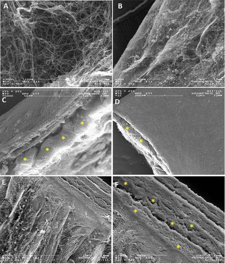
A. Idrees et al.: 4open 2021, 4, 1                                              3

from PromoCell. The company guaranteed the following             to allow hydrogel remodelling by the embedded cells
requested specifications: Same batch, single adult donor,         (Figs. 2A and 2B).
chest region, and passage number P2 after thawing.
    NHDF and NHEK were maintained in CnT-Prime                   Development of epidermis onto human dermal
fibroblast medium (CnT-PR-F, CELLnTECH) and CnT-                  constructs
Prime epithelial culture medium (CnT-PR, CELLnTECH)
respectively under the physiological culture conditions               Media was removed from HDC 20 min before NHEK
(37 °C, 5% CO2), and sub-cultured using DetachKit-Promo-         seeding. NHEK were resuspended (400,000 cells for a sur-
Cell HEPES BSS (2-[4-(2-hydroxyethyl)piperazine-1-yl]            face area of dermal construct of 12 mm diameter) in a small
ethanesulfonic acid buffered saline solution); 0.04% Tryp-       volume of 50 lL KGM2 (3D-CC-I and II), CnT-PR (3D-
sin/0.03% EDTA (ethylenediaminetetraacetic acid); and            CC-III) or CnT-PR-FTAL (3D-CC-IV) in the center and
TNS (Trypsin Neutralizing Solution) containing 0.05%             plated onto the matrix for cultures epithelialization. Plates
trypsin inhibitor from soybean/0.1% bovine serum albumin.        were not moved for 15 min to allow keratinocytes to initiate
Collagen type I (Col. I) from rat tail tendons, 5 mg/mL          adhesion and then placed at 37 °C for 30–60 min without
stock concentration was purchased from Ibidi.                    medium for complete adhesion. Depending on the selected
    Different media were used to optimize the development        culture conditions 3D-CC-I–IV, the plates were fed with
of HSE: i) CnT-PR-F (CnT-Prime fibroblast medium,                 the appropriate media KGM2, CnT-PR (Medium B) or
CELLnTECH) named as Medium A; ii) CnT-PR (CnT-                   CnT-PR-FTAL as shown in Table 2. The Transwell inserts
Prime epithelial culture medium, CELLnTECH) named                were placed in 12 well cell culture plate (Corning 3513),
as Medium B; iii) CnT-PR-3D (CnT-Prime-3D barrier                thus providing medium volumes of ~0.3 mL inside and of
medium, CELLnTECH) named as Medium C; iv) CnT-                   ~1.7 mL outside the insert. Medium was changed daily by
PR-FTAL (CnT-Prime-full thickness airlift medium,                discarding first medium from outside the insert and then
CELLnTECH); v) KGM2 (Keratinocyte Growth Medium                  from inside; fresh medium was then added initially inside
2, i.e. Basal medium supplemented with Bovine Pituitary          the insert and then outside. Cultures were maintained in
Extract 0.004 mL/mL, human EGF 0.015 ng/mL, human                submerged conditions for 3–4 days to allow complete cover-
Insulin 5 lg/mL, Hydrocortisone 0.33 lg/mL, Epinephrine          age to form a monolayer. In case of culture condition 3D-
0.39 lg/mL, Transferrin 10 lg/mL, CaCl2 0.06 mM; Pro-            CC-II KGM2 was replaced with CnT-PR-3D (Medium C)
moCell); vi) High calcium KGM2 (KGM2 + 1.2 mM CaCl2,             and for 3D-CC-III Medium B was replaced with Medium
Sigma); vii) FGM2 is a low serum media (2% v/v) that con-        C (Tab. 2). For both culture conditions cultures were kept
tains: 0.02 mL/mL foetal calf serum (FCS), 1 ng/mL basic         submerged overnight (15–16 h) to allow cells to form inter-
fibroblasts growth factor (bFGF) (recombinant human),             cellular adhesion structures. Finally, the medium was aspi-
5 lg/mL Insulin (recombinant human) (PromoCell).                 rated from inside the insert to allow ALI culture for 20 days
                                                                 (Figs. 2C–2E). During ALI, medium was changed 3 times/
Preparation of Human Skin Equivalents (HSEs)                     week using a minimum volume of 1 mL.
                                                                      Different 3D cell culture conditions (namely 3D-CC-I,
   The 3D HSEs were generated by successively fabricat-          -II, -III, -IV) based on different types of commercially avail-
ing dermal and epidermal layers as summarized in                 able media were tested to optimize HSE development. The
Figure 1A.                                                       details of the 3D-CC are reported in Table 2. The test set-
Preparation of Human Dermal Constructs (HDCs)                    up is described as timeline in Figure 1B.
                                                                      In this study, the same batch (single donor, chest
    Gelation of Col. I solution was performed in 10 media       region) and passage number (P2 after thawing) of fibrob-
(M199, Sigma) supplemented with L-glutamine and sodium           lasts and keratinocytes were used for optimizing the HSE
bicarbonate (NaHCO3), resulting in a final Col. I concentra-      development under different 3D culture conditions, thus
tion of 1.5 mg/mL (containing a salt concentration of 1 in      eliminating the variability associated with batch-to-batch
final mixture with pH of 7.2–7.4) (Tab. 1)                        variation.
    HDCs were prepared by fabricating acellular (200 lL)
and cell containing layers (400 lL) of Col. I matrix on polye-   Morphological analysis
ster membrane of 12 well inserts (Corning 3460, 0.4 lm pore
size, 12 mm diameter, 1.12 cm2 surface area) to obtain 5 mm          Harvested HSE tissue was rinsed in PBS twice, fixed in
thick HDCs. A thin layer of acellular collagen served as a       4% buffered paraformaldehyde (PFA, pH 6.8 – 7.0) for
substrate for the cellular collagen, preventing the cellular     4 h, dehydrated through increasing gradient series of etha-
collagen from complete contraction from the insert mem-          nol (50%, 70%, 80%, 90%, 95%,100%, 100%, 100%; each for
brane. Actively dividing mitotic cells (5  104 NHDF cells       5 min), cleared in xylol (three times, each for 5 min), and
per 12 mm diameter insert to obtain the human in vitro der-      infiltrated in paraffin using Leica TP1020 Semi-enclosed
mal construct) were embedded in Col. I solution and added        Benchtop Tissue Processor (over a total period of 12 h).
on the top of the previously deposited and gelled acellular      The processed tissue was paraffin embedded at right orien-
Col. I layer (room temperature, 20 min). After gelation of       tation using Leica EG1160 Embedding Centre. The sample
cellularized matrix (37 °C for 30 min), fresh cell culture       was thinly sliced (~5 lm) using Leica RM 2155 microtome.
medium (Medium A) was added and incubated for 5–7 days           Sections were dryed at 37 °C overnight. Morphological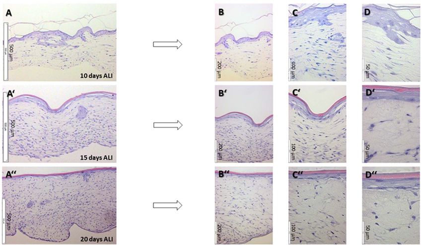
4                                                 A. Idrees et al.: 4open 2021, 4, 1

Fig. 1. (A) Schematic overview of methodology for the development of HSE. It starts with the embedding of human dermal
fibroblasts in collagen I matrix to contract and remodel the gel to form dermal layer. Then the keratinocytes are seeded on the top to
form a monolayer and the culture is raised to ALI under 3D cell culture conditions allowing the keratinocytes to differentiate and form
epidermal layers. (B) Timeline for the development of HSE.

Table 1. Guidelines for construction of collagen I (1.5 mg/mL) based dermal layer.

Constituents                    Calculated vol. of each constituent (lL)                Calculated vol. of each constituent (lL)
                           Total vol. of acellular layer = 200 lL per insert         Total vol. of cellular layer = 400 lL per insert
10 M199 (Sigma)       20 lL                                                     40 lL
                       (10% of final vol. of 200 lL results in 1 M199)           (10% of final vol. of 400 lL results in 1 M199)
200 mM Glutamine       0.68 lL                                                   1.36 lL
(Biosciences)          Formula: M1·V1 = M2·V2 (200 mM                           Formula: M1·V1 = M2·V2 (200 mM  V1 = 0.68
                       V1 = 0.68  200 lL) (This 0.68 lL vol. results in 0.68    400 lL) (This 1.36 lL vol. results in 0.68 mM Glutamine
                       mM Glutamine in final                                      in final 400 lL vol. of cellular layer)
                       200 lL vol. of acellular layer)
ddH2O                  113.32 lL                                                 176.64 lL
                       (vol. calculated in the end)                              (vol. calculated in the end)
7.5% of NaHCO3         6 lL                                                      12 lL
(Sigma)                (This volume is 10% of the volume of collagen I, that     (This is an optimized volume as 10% of the volume of
                       results in a final mixture pH of 7.2–7.4 – optimized)      collagen I, that results in a final mixture pH of 7.2–7.4)
5 mg/mL Collagen I     60 lL                                                     120 lL
(Ibidi)                (This volume makes final conc.                             (This volume makes final conc. of 1.5 mg/mL in final
                       of 1.5 mg/mL in final volume)                              volume)
NHDF (PromoCell)       N/A                                                       50 lL
                                                                                 (This volume should contain
                                                                                 5  104 NHDF per insert)
Total vol.             200 lL                                                    400 lL

analysis was performed on formalin-fixed paraffin-                       visualized by applying F-actin/nuclei staining. This was
embedded sections through Haematoxylin and Eosin                       performed using Phalloidin/DAPI stain from Promokine
(H&E) staining following a standard protocol [37]. The                 following a standard protocol [47]. The fluorescence micro-
H&E stained slides were examined with Olympus BX51                     scopy and Z-stack imaging was performed using Olympus
light microscope. NHDF embedded in Col. I (Fig. 1A) were               XM10 with cellSense Standard software.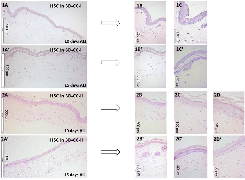
A. Idrees et al.: 4open 2021, 4, 1                                                5

Fig. 2. Macroscopic view of Human Dermal Construct (HDC) – upper panel: (A) View of experimental set-up of Human Dermal
Construct on Day-01 with NHDFs are embedded in collagen I matrix and incubated to allow gel remodeling. (B) Macroscopic top view
of contracted collagen on Day-07 retaining its circular shape. Macroscopic view of HSE – lower panel: (C) Top view of experimental
set-up of HSE development during incubation at ALI showing the white epidermal layer over the dermal layer. (D) Side view of HSE
containing insert at the time of tissue harvesting after 20 days at ALI, showing a thickness of ~2 mm. (E) Top view of harvested HSE
at the end of ALI period (20 days) retaining the size and diameter of initial mould (12 mm in diameter).

Table 2. Experimental set-up details of different 3D cell culture conditions (3D-CCs).

Step duration       Description of 3D culture                3D-CC-I             3D-CC-II          3D-CC-III          3D-CC-IV
                    condition
5–7 days            HDC feeding                               FGM2                 FGM2            CnT-PR-F           CnT-PR-F
                                                                                                  (Medium A)         (Medium A)
3–4 days            Epidermal monolayer                       KGM2                 KGM2             CnT-PR          CnT-PR-FTAL
                    formation (onto HDC); feeding                                                 (Medium B)
15–16 h             Epidermal monolayer                          –              CnT-PR-3D         CnT-PR-3D                –
                    formation (onto HDC); feeding                               (Medium C)        (Medium C)
10/15/20 days       HSC culture at ALI for                1.2 mM CaCl2          CnT-PR-3D         CnT-PR-3D         CnT-PR-FTAL
                    10, 15, and/or 20 days              containing KGM2         (Medium C)        (Medium C)
Abbreviations: CnT-PR-F named as Medium A; CnT-PR named as Medium B; CnT-PR-3D named as Medium C.

Immunophenotypic analysis                                               Immunophenotypic analysis of epidermal markers was
                                                                     performed on cryosections. Specifications on the primary
    HSE was rinsed in phosphate buffered saline (PBS)                antibodies’ concentrations used in this study are described
twice, treated with 2M sucrose solution (Fisher Chemical)            in Table 3. Secondary antibodies used are the following:
at 4 °C for at least 1 h and then infiltrated in OCT (Opti-           Alexa Fluor 488 Goat anti-Rabbit IgG (H + L) (Abcam),
mal Cutting Temperature) embedding medium (VWR) for                  Alexa Fluor 594 Donkey anti-Mouse IgG (H+L) (Abcam),
20–30 min at room temperature (RT). The tissue was                   Alexa Fluor 488 Donkey anti-Rabbit IgG (H+L)
embedded in OCT by gradual freezing in liquid nitrogen               (ThermoFischer), Alexa Fluor 568 Goat anti-Mouse IgG
vapor, stored at 80 °C and cryo-sectioned (~10 lm) using            (H + L) (ThermoFischer). Negative controls were
Microm HM 550 Cryostat.                                              performed using only secondary antibodies. Cell nuclei were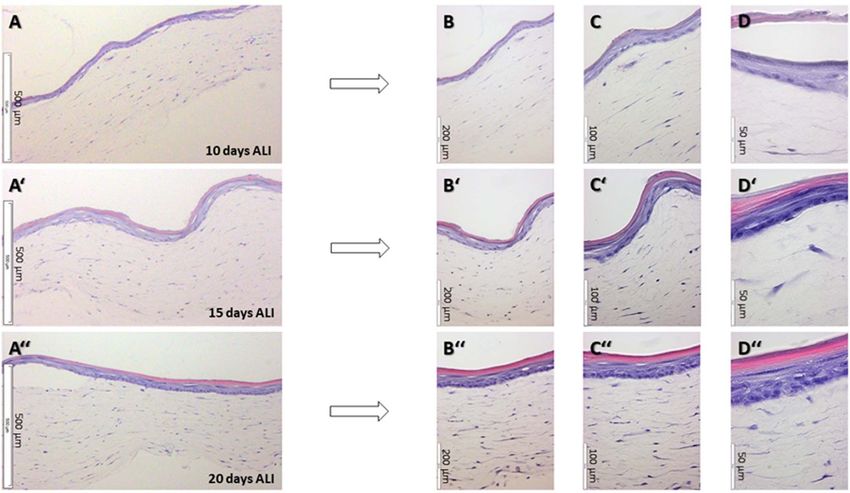
6                                             A. Idrees et al.: 4open 2021, 4, 1

Table 3. Primary antibodies used for immunostaining of HSE tissue cryosections.

Primary antibody details and source                                     Dilution                                     Clone-No.
Ki-67 (Rb, MA) – Cell signal                                            1:300                                         D2H10
Laminin 5 (Rb, PA) – Abcam                                              1:500                                            –
Keratin 14 (Ms, MA) – Abcam                                             1:100                                         LL002
Keratin 10 (Rb, MA) – Abcam                                             1:500                                       EP1607IHCY
Filaggrin (Rb, PA) – Abcam                                              1:200                                            –
b-Tubulin (Ms, MA) – Sigma                                              1:400                                        EPR16774
Loricrin (Rb, PA) – Abcam                                               1:200                                            –
Keratin 16 (Rb, PA) – Abcam                                     1:100 & 2° Ab as 1:200                                   –
PA = polyclonal antibodies; MA = monoclonal antibodies; Rb = rabbit; Ms = mouse; 2° Ab = secondary antibodies.

Table 4. Details of reagents required for Immunostaining.

Reagent Type               Reagent constitution                                     Reagent Quantity (calculated)
Wash buffer                TBS + 0.025% Triton X-100                                250 lL (0.25 mL) Triton X-100 in 1000 mL
                                                                                    TBS
Permeabilization reagent   0.2% Triton X-100 in PBS                                 20 lL (0.02 mL) Triton X-100 in 10 mL TBS
Fixative                   4% paraformaldehyde in PBS                               10 mL of 16% PFA in 30 mL PBS
Antibody diluent buffer    TBS + 1% BSA                                             0.1 g BSA in 10 mL TBS
Blocking Buffer            10% normal serum + 1% BSA + TBS                          1mL normal serum + 0.1 g BSA + 9 mL TBS
                           For double staining: 5% normal serum of species in
                           which first secondary antibodies were raised + 5%
                           normal serum of species in which second type of
                           secondary antibodies was raised + 1% BSA + TBS
                           Note: Goat and donkey serum 60 mg/mL – Jackson
                           Immuno Research
Buffers                    1 TBS (10 TBS comprised of 500 mM tris                 100 mL of 10 in 900 mL distilled deionized
                           base + 1.5 M NaCl + HCl to set pH to 7.4); 1 PBS        H2O
BSA is bovine serum albumin, TBS is tris-buffered saline, PBS is phosphate-buffered saline.

counterstained with DAPI (40 ,6-diamidino-2-phenylindole)         HSE, technical replicates and N = 2, number of indepen-
(1.5 lg/mL, VECTASHIELD, Vector labs). Immunohisto-               dent experiments) by dividing the Ki-67 positive nuclei
chemical (IHC) analysis was performed in accordance with          (green) by total number of nuclei (blue). The equation
the standard protocol from Abcam, with primary antibod-           applied is as follows: [N(Ki-67 positive cell nuclei)/
ies’ concentrations optimized for HSE (Tab. 3). Details of        Ntotal(R(DAPI-stained cell nuclei, Ki-67 positive cells))]
reagents used in IHC staining are provided in Table 4.            100 = % (Ki-67 positive cells).
Cryosections were fixed in methanol-free 4% PFA (Thermo
Scientific) for 10 min then rinsed in PBS (2 times for 5 min).
Sections were then permeabilized in 0.2% Triton X-100 for         Electron microscopy analyses: SEM and TEM
10 min then rinsed in wash buffer (TBS, tris-buffered saline,
0.025% Triton X-100). Sections were blocked in blocking               Samples were fixed in 2.5% glutaraldehyde for 1 h,
buffer (10% normal serum from the same secondary                  washed in PBS (3 times for 5 min), incubated with 1%
antibody species, 1% BSA, Bovine Serum Albumin, in                osmium tetraoxide for 15 min, followed by PBS washing
TBS) for 2 h at room temperature. Slides were drained             (3 times for 5 min). Then, samples were dehydrated in an
and treated with primary antibody diluted in antibody dilu-       increasing gradient series of ethanol (30%-5 min, 50%-
ent buffer (1% BSA in TBS) and incubated in a humidified           5 min, 70%-overnight at 4 °C, 96%-5 min for 3 times,
chamber overnight at 4 °C. Fluorophore-conjugated sec-            100%-15 min for 3 times) and submerged in polypropylene
ondary antibody diluted in antibody diluent buffer were           oxide (PO) for 15 min before inclusion in epoxy resin-based
applied and incubated for 1 h at room temperature, pro-           embedding medium (Epoxy Embedding Medium kit
tected from light. Finally, sections were rinsed in TBS           cat.-no. 45359, Sigma-Aldrich) with PO (1:1) for 1 h [38].
(3 times for 5 min) and mounted using VECTASHIELD                 Thereafter, samples were embedded in the above mentioned
mounting medium. The immunostained slides were exam-              medium in the right orientation and incubated at 80 °C for
ined with fluorescent microscope (Olympus XM10 with cell-          8 h. Tissue blocks were ultrasectioned (~40 nm) using Leica
Sense Standard software).                                         Ultracut UCT Ultramicrotome, contrasted with Leica
    The percentage of Ki-67 positive dermal fibroblasts and        ultrastain 2 (3% lead citrate) and Leica ultrastain (0.5%
basal keratinocytes was calculated from three IHC images          uranyl acetate), and imaged with JEM-2100 TEM at an
(n = 3 means three cryosectioned slices from a single             accelerating voltage of 200 kV.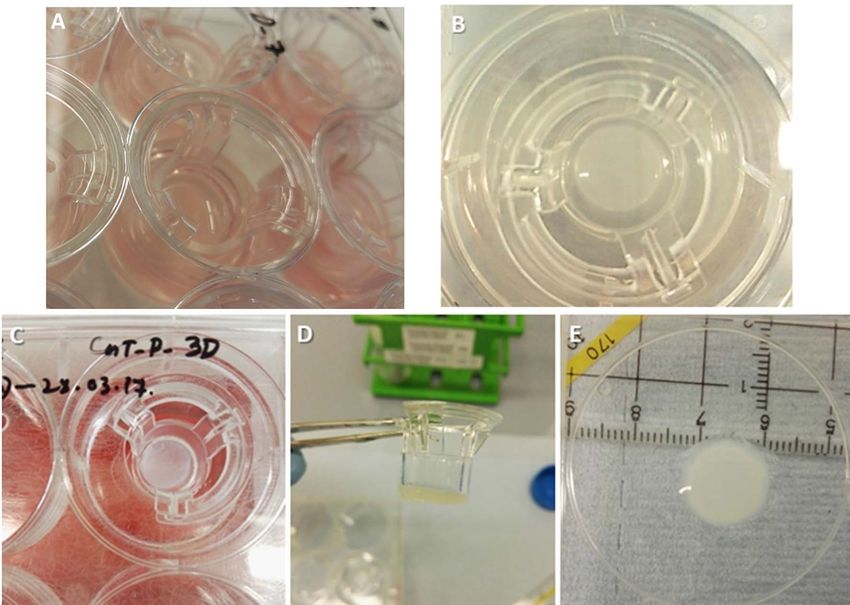
A. Idrees et al.: 4open 2021, 4, 1                                            7

     For scanning electron microscopy (SEM), samples were       conditions, it is noticeable that the stratum corneum (SC)
fixed and dehydrated as described above. Then, samples           in the case of 3D-CC-II forms a clearer or more coherent
were placed in BAL-TEC CPD 030 Critical Point Dryer             layer. Furthermore, organization of epidermal layers under
and dryed with liquid CO2 by short consecutive immersions       3D-CC-II significantly improved when air-lift (ALI) culture
(3 times for 10 min). Then, samples were mounted onto           phase was extended to 15 days (Fig. 3, 2A0 –2D0 ).
electrically conductive, double sided adhesive carbon discs         Furthermore, compared to 3D-CC-IV (based on
(Leit C tabs) and gold coated using Edwards S150B Sput-         CnT-FTAL, Fig. 4), 3D-CC-III (based on CnT-PR-3D
ter Coater under Argon gas (101 mbar) at voltage of            (Medium C), Fig. 5) demonstrated an improved differenti-
15 mA for 6 min to form 40 nm thick gold (Au) coating.          ated epidermal formation.
Coated samples were examined with SEM DSM 982 Gem-                  Among the tested conditions, improved morphogenesis
ini, Zeiss. Multiple SEM images were taken at random areas      results were obtained under 3D-CC-III and at ALI for
of different samples at 15 kV accelerating voltage. The         20 days, resulting in constructs better mimicking epithelial
diameter of the collagen fibers was estimated as an average      differentiation found in healthy human skin. Hence, such
value from multiple TEM images.                                 HSC was termed as human skin equivalent (HSE)
                                                                (Figs. 5A00 –5D00 ). The epidermal part showed improved dif-
                                                                ferentiated layers of keratinocytes namely stratum basale
Wettability analysis
                                                                (SB), spinosum (SS), granulosum (SG), and corneum (SC)
    Static contact angle measurements were performed to         (Fig. 6A), more comparable with its in vivo counterpart
investigate wettability of HSE by sessile-drop method,          (Fig. 6B).
using custom-built contact angle goniometer instrument
under ambient conditions. An amount of 5 lL MilliQ water        IHC analysis of differentiation markers on HSE
was carefully placed on dry HSE prepared in two different           IHC characterization of HSE (Figs. 7.1A and 7.1B)
experiments (N = 2; Number of independent experiments);         showed the presence of specific epidermal differentia-
Multiple images were recorded (shown as an example in           tion markers. Cytokeratin-14 (K14) was expressed by
Supplementary Fig. 1S) and analyzed for 10 drops per            epithelial cells in the SB (Fig. 7.1A), while the suprabasal
HSE surface (N = 2; n = 10).                                    layers displayed the presence of Cytokeratin-10 (K10)
                                                                (Fig. 7.1B). Cells in basal layer were found positive for Ki-
Statistical analysis                                            67 (highlighted by the arrows), demonstrating an active
                                                                state of proliferation (Fig. 7.2E). After counting, HSE dis-
    All experiments, where not explicitly described other-      played 10.5 ± 0.8% (p-value < 0.05) Ki-67 positive basal
wise, were performed independently in duplicate (N = 2,         keratinocytes. The terminal differentiation of epidermis
number of independent experiments) and 3 technical repli-       was demonstrated by the spotted expression (highlighted
cates were made and examined per experiment (n = 3).            by arrows) of filaggrin (Flg) in the SG (Fig. 7.1C). Loricrin
Standard deviations were given where appropriate and            (Lor), a major protein component of cornified envelope and
expressed as mean ± SD (standard deviation). For statisti-      marker of terminally differentiated epidermal cells, also
cal analysis, GraphPad Prism 5.00.288 (Inc., San Diego,         showed spotted expression in SC (highlighted by arrows in
CA, USA) was used to evaluate the significance of the dif-       Fig. 7.2F). Involucrin (Inv) was displayed in upper spinous,
ferences in experimental data. Significance between groups       sub-corneal, and corneal layers (Fig. 7.2G). Laminin 5
was considered for p < 0.05.                                    (Lam 5) was expressed at DEJ as a thin continuous line
                                                                (Fig. 7.1D) suggesting stable epidermal-dermal interaction.
                                                                Beta-tubulin as a component of the cytoskeletal micro-
Results                                                         tubules was mainly found as a continuous seam in the SB
    The development of dermal-epidermal based HSE               area, where differentiating and proliferating NHEK are
was optimized to obtain well-differentiated epidermis onto      located, and in the NHDF in the HDC area (Fig. 7.2E).
HDC. To this purpose, different 3D-CCs based on cell
culture media and the parameter of ALI (Tab. 2) were            Comparison of immunoreactivity among
applied showing significant impact on tissue morphogenesis.      different 3D-CCs

Tissue morphogenesis of HSCs under                                  IHC analysis for cytokeratins showed that K14 and K10
different 3D-CCs                                                were expressed by epithelial cells in SB (Fig. 8A) and supra-
                                                                basal layers (Fig. 8B) of HSC obtained under 3D-CC-III
    Figure 3 shows histological images of HSCs obtained         and at ALI for 15 days. However, K14 and K10 expression
using 3D-CC-I and 3D-CC-II, respectively. Two structurally      was lower than for HSE obtained under 3D-CC-III and at
distinct compartments epidermis and dermis were present.        ALI for 20 days (Figs. 7.1A and 7.1B).
Epidermis was slightly better organized in 3D-CC-II (Figs. 3,       Similarly, IHC analysis for cytokeratins was performed
2A–2D, 2A0 –2D0 ) than in 3D-CC-I (Figs 3; 1A–1C,               on HSC obtained from 3D-CC-IV at ALI for 20 days.
1A0 –1C0 ), which on the contrary displayed larger gaps         Results showed that K14 and K10 were expressed by
between the layers. When comparing both culture                 epithelial cells in the SB (Fig. 9A) and suprabasal layers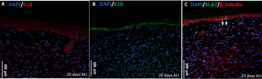
8                                               A. Idrees et al.: 4open 2021, 4, 1

Fig. 3. Histological cross sections of HSC using the 3D culture conditions 3D-CC-I (upper panels 1A–C and 1A0 –C0 ) and 3D–CC-II
(lower panels 2A–D and 2A0 –D0 ); N = 2, n = 3. Upper panels (1A, 1B, 1C) and (1A0 , 1B0 , 1C0 ) showed H&E-stained histological
microscopy images of HSCs at different magnification elevated to the air-liquid interface (ALI) for 10 days and 15 days, respectively.
Lower panels (2A, 2B, 2C, 2D) and (2A0 , 2B0 , 2C0 , 2D0 ) show H&E-stained histological microscopy images of HSCs at different
magnification elevated to the air-liquid interface (ALI) for 10 days and 15 days, respectively.

(Fig. 9B), respectively. However, K14 and K10 expression            and attaching them into dense basal lamina through
was found comparatively lower than for HSE (3D-CC-III,              anchoring filaments. On the other side, Figures 10E and
at ALI for 20 days) (Figs. 7.1A and 7.1B), while Ki-67              10H show an abundance of keratins and tonofilaments in
was found similarly expressed (Fig. 9C) as in HSE                   keratinocytes, and the presence of tight junctions and lipid
(Fig. 7.2E).                                                        droplets in SG. TEM imaging finally also demonstrated the
                                                                    presence of desmosomes as spot-like connections for cell-to-
Ultrastructure (TEM) of HSE                                         cell adhesion of keratinocytes (Figs. 10F–10H).
    TEM was used to investigate the ultrastructure of HSE.          Tissue architecture (SEM) of HSE
Regarding the dermal compartment, TEM analysis con-
firmed the presence of embedded fibroblasts in a network                  Histological analyses highlighted the typical elongated
of collagen- and immature elastin-based ECM (Fig. 10D).             morphology of NHDF (Fig. 6A). SEM images confirmed
Collagen fibrils combine with each other to form collagen            the presence of embedded fibroblasts, with dendritic exten-
fibers. In HSE, collagen formed both fibers and fibrils                sions lying along dense matrix of collagen fibers (Fig. 11B).
(Figs. 10A–10D) with the characteristic banded pattern              ECM of the dermal compartment of HSE demonstrated the
with a period of ~65 nm [39, 40]. Elastin was found depos-          typical interwoven network morphology and architecture
ited on collagen fibrils or microfibrils in an amorphous              (Fig. 11A).
immature form (Fig. 10C) [41].                                          The average diameters of microfibril, collagen fibril, and
    Figures 10A and 10B also show hemidesmosomes con-               collagen fiber calculated from multiple TEM images (n = 3)
necting basal cells through tonofibrils/keratin filaments             were 26 ± 21 nm, 65 ± 57 nm, and 157 ± 10 nm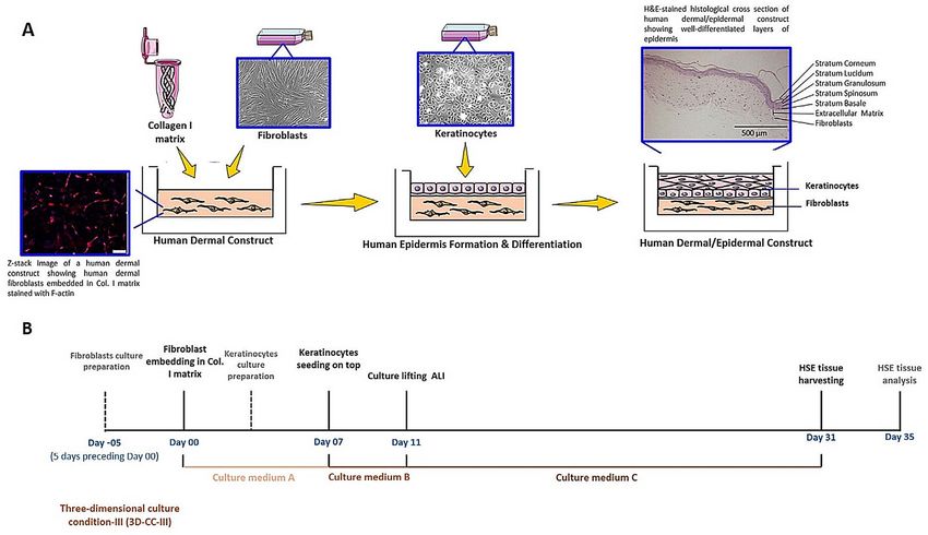
A. Idrees et al.: 4open 2021, 4, 1                                                      9

Fig. 4. Histological cross sections of HSC using the 3d culture conditions 3D-CC-IV; N = 2, n = 3. Upper (A, B, C, D), middle (A0 , B0 ,
C0 , D0 ), and lower panels (A00 , B00 , C00 , D00 ) (images at different magnifications) showed H&E-stained histological micrographs of HSCs
raised for 10 days, 15 days, and 20 days at ALI, respectively. The longer the ALI cultivation period, the better a structured
stratification of the epidermis from stratum basale to stratum corneum can be recognised (cf. labelling in Fig. 6).

Fig. 5. Histological cross sections of HSC using 3D culture condition 3D-CC-III; N = 2, n = 3. Upper (A, B, C, D), middle (A0 , B0 , C0 ,
D0 ), and lower (A00 , B00 , C00 , D00 ) panels (images at different magnifications) showed H&E-stained histological micrographs of HSCs
raised for 10 days, 15 days and 20 days at ALI, respectively. The longer the ALI cultivation period, the better a structured
stratification of the epidermis from stratum basale to stratum corneum can be recognised (cf. labelling in Fig. 6).
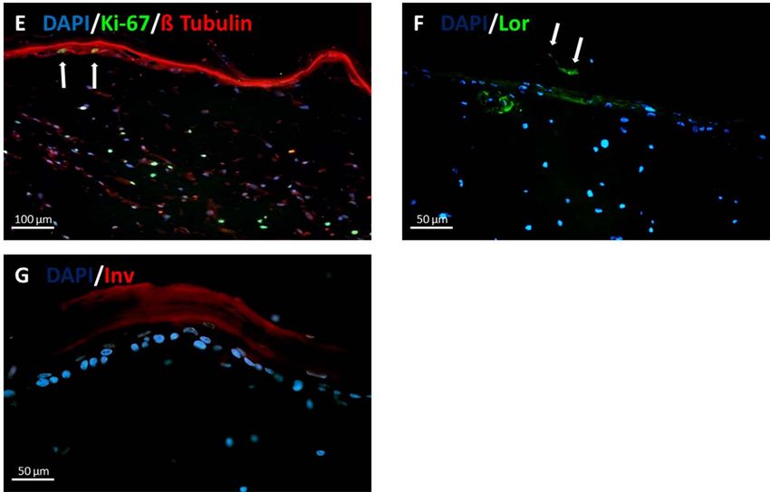
10                                             A. Idrees et al.: 4open 2021, 4, 1

Fig. 6. (a) HSC as HSE. H&E-stained histological image of HSC obtained from 3D-CC-III (cf. Fig. 5C00 ) was able to best recapitulate
epidermal differentiation, morphogenesis and organization similarly as found in (b) NHS (origin: human leg; histology: different
staining protocol) and thus named as HSE. HSE showed two structurally distinct layers of skin the outer epidermal layer, and the
underlying thicker dermal layer. The epidermal part showed well differentiated layers of keratinocytes namely stratum basale,
spinosum, granulosum, and corneum. The staining differences between HSE and NHS are due to a different H&E protocol used to
stain NHS at the Institute of Pathology, Ruhr University Bochum, Germany, where the NHS tissue was obtained.

Fig. 7.1. Immunotypic analysis of HSE. IHC characterization of HSE showed immunolabelling for specific epidermal differentiation
markers namely (A) Keratin 14 (K14) red, (B) Keratin 10 (K10) green, (C) Filaggrin (Flg) green (indicated by white arrows), and
(D) Laminin 5 (Lam 5) green. Cell nuclei were counterstained with DAPI (in blue); N = 2, n = 3.
A. Idrees et al.: 4open 2021, 4, 1                                                11

Fig. 7.2. Further immunotypic analysis of HSE. (E) Detection of proliferative keratinocytes in the area of the stratum basale of the
epidermis (white arrows) and of division-active fibroblasts in the dermis by using the proliferation marker Ki-67 green, further
evidence for specific epidermal differentiation was provided by the detection of (F) Loricrin (Lor) green, and (G) Involucrin (Inv).
Cell nuclei were counterstained with DAPI (in blue) and microtubules in cytoplasm were shown in red using b Tubulin staing (E);
N = 2, n = 3.

respectively. Fibril-associated molecules on the surface of         82.5 ± 8.9° that was highly comparable (difference not
collagen fibrils play an important role in controlling interac-      significant at p-value < 0.05) to the value (90.0 ± 5.1°)
tions between the fibrils, and thus the relative frequency of        reported for NHS [45]. This result is in agreement with
collagen bundles’ diameters. ECM showed thinner collagen            results obtained by TEM and SEM analysis (Figs 10 and
fibers than mature collagen bundles, suggesting an imma-             11) demonstrating that SC had formed, providing protec-
ture state of the dermal compartment [42, 43].                      tion with tight cell junctions (corneo-desmosomes), lipids
    SEM images also showed adjacent stratified epidermal             sealing intercellular spaces, and the presence of keratin
cells when approaching SC (Figs. 11C–11F). NHEK of                  filaments (including filaggrin and degradation products)
basal and suprabasal epidermal layers showed a typical              (Figs. 7.1C, 7.2F, 7.2G).
morphology and were continuously organized (Fig. 11C).
When keratinocytes are cultured in vitro to develop 3D              Dermal compartment of HSE
tissue, one of the main issues is the limited longevity of
                                                                        IHC was performed for laminin expression in dermal
keratinocytes due to the long culture time at ALI (e.g.
                                                                    compartment of HSE and was detected as fainted spots
2–3 weeks). The surface layers of developed epidermis
                                                                    around the fibroblasts (Fig. 12). Moreover, the percentage
become very thick because of their inability to desquamate
                                                                    of Ki-67 positive dermal fibroblasts calculated was
causing the compression of the lower layers, as observed in
                                                                    24.7 ± 7.8%, indicating their proliferative potential.
SEM results (Figs. 11C–11D). In vivo, the normal desqua-
mation occurs unnoticeably: mature keratinocytes move
from SB to SG in 14 days, then they are shed off [44].
                                                                    Discussion
                                                                    Development of dermal layer of HSE
Barrier function properties of HSE
                                                                        Connective tissue has an important role as a substrate
   An important interest associated with well-developed             for the growth of epidermal tissue. Particularly ECM
SC is the skin barrier function. This functionality was eval-       proteins affect skin tissue phenotype [46]. For instance,
uated by measuring the surface wettability of HSE that is           the use of decellularized dermis, e.g. AlloDerm (a commer-
highly related with physiological surface properties of skin.       cially available cadaveric dermis for clinical applications)
The static Water Contact Angle (WCA) value of HSE was               favors the fast assembly of basement membrane [33]. The
12 A. Idrees et al.: 4open 2021, 4, 1
Fig. 8. Immunotypic analysis of HSC using 3D-CC-III at 15 days of ALI. IHC analysis of HSE showed immunolabelling for specific
epidermal differentiation markers, namely (A) Keratin 14 (K14) red, and (B) Keratin 10 (K10) green. Cell nuclei were shown in blue
by using DAPI staining; N = 2, n = 3.
Fig. 9. Immunotypic analysis of HSC using 3D-CC-IV. IHC analysis of HSE showed immunolabelling for specific epidermal
differentiation markers namely (A) Keratin 14 (K14) red, (B) Keratin 10 (K10) green, and (C) Ki-67 green. Cell nuclei were
counterstained with DAPI (in blue) and microtubule in cytoplasm keratinocytes and fibroblasts are shown in red using b Tubulin
staining (C). Exemplary presentation of the quantification of Ki-67 positive cells: [N(Ki-67 positive cell nuclei)/Ntotal(R(DAPI-stained
cell nuclei, Ki-67 positive cells))] 100 = % (Ki-67 positive cells), [(2/19) 100 = 10.5% (in relation to the image section being
viewed); N = 2, n = 3.
contracted collagen gel used in this work might result in a construct in submerged culture conditions using CnT-PR-
less intact basement membrane, however it greatly sup- 3D medium.
ported keratinocyte growth and differentiation.
In this work, a 3D functional HSE was prepared Epidermal differentiation of HSE
by successively fabricating the dermal and epidermal
compartments (Fig. 1). Optimal culture conditions for the HDC was previously developed based on Col. I from rat
engineering of a biomimetic HSE were: (i) NHDF tail tendons, mimicking skin ECM thanks to its irregular
(~8 104 cells/mL) culture within Col. I gel for 5–7 days fibrils with in vivo-like structure [47]. The development of
in CnT-PR-F medium; ii) NHEK (8 106 cells/mL) HSE with dermal-epidermal compartments has been
seeding on the top of dermal layer and culture in submerged improved by using commercially available primary cells
conditions for 3 days using CnT-PR medium; iii) sub- and ready-to-use media provided according to Good
merged culture overnight in CnT-PR-3D medium to stimu- Laboratory Practice (GLP) and Good Manufacturing
late the formation of cell-cell contacts; iv) culture of Practice (GMP) criteria to form a well-differentiated
epidermal layer at ALI for 15–20 days while keeping dermal epidermis accessible to everyone. To this purpose, the effectA. Idrees et al.: 4open 2021, 4, 1 13 Fig. 10. Ultrastructure analysis of HSE. TEM images (A, B) revealed dermal-epidermal junction (DEJ) that separates epidermis from the dermis through basement membrane consisting of lamina lucida (LL) and lamina densa (LD). LD was connected with the collagen matrix by loops of anchoring fibrils (black arrows). The hemidesmosomes (white arrows) were connected with tonofibrils (Tf) in keratinocytes. Collagen fibers were shown as black dotted arrows. TEM images (C, D) demonstrated collagen fibrils (Col. I) with their characteristic banded pattern (white arrows). Microfibrils or fibrillins (black arrows) were also secreted by fibroblasts, forming a scaffold that was detected near collagen fibrils. Amorphous or “immature” elastin was secreted and aggregated (asterisks) on microfibrils. Collagen fibers (black dotted arrow) appeared when collagen fibrils combined to form fibers. Normal Human Dermal Fibroblasts (NHDF) were also seen as embedded in ECM. In TEM images (E, F, G, H) epidermis showed tight junction (TJ) between the keratinocytes. Keratinocytes showed abundant intracellular filament of keratins (K) and upper epidermal layers showed lipid droplets (L). Tonofibrils (Tf) in keratinocytes (NHEK) were made of tonofilaments (keratin intermediate filaments), that appear converging to form spot like connections or desmosomes (Des or white arrows); N = 2, n = 3.
14                                              A. Idrees et al.: 4open 2021, 4, 1

Fig. 11. SEM analysis of HSE. (A, B) SEM images of dermal compartment of HSE showed ECM with a typical interwoven network
morphology and architecture with embedded dermal fibroblasts lying along dense matrix of collagen fibers. (C, D, E, F) Epidermal
layers of skin were increasingly flattened as they move to the surface to form SC. The inner layers of keratinocytes (C) were continuous
with less flattened morphology. Keratinocytes of the individual layers are marked with yellow asterisk; N = 2, n = 3.

of different 3D-CCs (Tab. 2) on tissue morphogenesis was             in the higher cell layers) is achieved due to air-lift [49].
analyzed.                                                            ALI also helps in achieving the proper lipid profile (acylglu-
    It is known that calcium activates intracellular signaling       cosylceramides and acylceramides) of SC [50], as the rate of
able to initiate epidermal differentiation [48]. For this rea-       trans-epidermal water loss influences lipid biosynthesis [51].
son, media constituted or commercially available for 3D cul-         Moreover, cells cultured at ALI are not in direct contact
ture at ALI were supplemented with a high calcium                    with cell culture medium components, such as retinoids,
concentration (1.2 mM). The role of ALI in epidermal dif-            that strongly inhibit epidermal differentiation [52].
ferentiation has been attributed to the formation of calcium             Growth factors have also an important role in epidermal
gradient in the epidermal compartment (due to water loss             differentiation. The traditional culture media used to
A. Idrees et al.: 4open 2021, 4, 1 15
ALI period was optimized by comparing the tissue mor-
phogenesis of HSCs obtained from different air-lift periods
of 10, 15, and 20 days.
KGM2 (PromoCell), a serum-free medium containing
0.06 mM CaCl2 was used to culture keratinocytes at
submerged conditions (3D-CC-I) and was then supple-
mented with CaCl2 to achieve a concentration of 1.2 mM
during air-lift phase.
CnT-Prime media are fully-defined, animal component-
free and furthermore without cholera toxin. The basal CnT
medium, such as CnT-PR medium is supplemented with
CaCl2 (0.07 mM concentration), progenitor cell targeted
(PCT) growth factors able to extend cell longevity, and
co-factors able to improve growth factor binding to
membrane-bound receptors. On the other hand, the CnT
prime media for 3D culture, such as CnT-PR-3D and
CnT-FTAL, contain elevated levels of calcium ions
(1.2 mM), while they do not contain PCT growth factors,
Fig. 12. ECM proteins in dermal compartment. IHC analysis of
or other proliferative factors that retard differentiation,
HSE showed immunolabelling for one of the ECM proteins
laminin in green. Cell nuclei are shown in blue (DAPI staining);
thus providing maximal stimulus to differentiation and
N = 2, n = 3. increased barrier function.
Figure 3 shows histological images of HSCs obtained
using 3D-CC-I and 3D-CC-II. Epidermis was comparatively
expand biopsies from human amniotic membrane include better organized in 3D-CC-II compared to 3D-CC-I,
hydrocortisone, triiodothyronine, adenine, and cholera toxin which instead displayed larger gaps between the layers
[53–56]. Hydrocortisone has significant role for maintaining (Figs. 3, 1A–1C, 1A0 –C0 ). Among the examples of abnormal
epithelial colonies and keratinocyte proliferation [57]. or pathological epidermal conditions, formation of cell
Triiodothyronine, being a supplement hormone, has been nests in dermis depicts malignant behavior of melanocytes
shown to play a beneficial role in keratinocyte cultivation [68], as it happens in early tumor invasion in vivo with
under minimal levels of fetal calf serum [58]. Cholera toxin, the melanoma cells getting into dermis and invading the
a protein secreted by Vibrio cholerae that causes cholera basement membrane [69]. Another example is the formation
infection (watery diarrhea) has been shown to highly stimu- of thickened scaly skin plaques due to hyperplasia, which is
late colony growth of cultured human epidermal ker- an indication of psoriatic skin and is histologically detected
atinocytes through increased levels of intracellular cyclic through spongiform micro-pustules and elongated rete
adenosine monophosphate [59]. Adenine also has an effect ridges [70]. The presence of wide interlayer spaces is one
on epithelial cells by improving their colony forming ability of the abnormalities of skin appearance, to be avoided
[60–62]. Among the several stimuli inducing keratinocyte through improved tissue morphogenesis, in order to mimic
differentiation, ascorbic acid has been reported to cause an healthy skin structure.
increased expression of the genes encoding cornified envel- Under 3D-CC-II (Figs. 3, 2A–2D, 2A0 –2D0 ), the epider-
ope proteins. The pro-differentiating effects of ascorbic acid mal layers were better organized after 15 days of air-lift
follow a similar signaling pathway as the ones mediated by compared to 10 days condition. Exposure to ALI enabled
calcium. However ascorbic acid-associated differentiation keratinocytes to differentiate and recapitulate the morpho-
is also accompanied by an enhanced ascorbate transport logical as well as biochemical processes of native human
and a prevention of hydrophilic antioxidants depletion [63, epidermis [71, 72]. Hence, cell cultures for extended periods
64]. at ALI comparatively demonstrated improved morphologi-
Additionally, in the optimization of 3D-CCs for the cal and biochemical differentiation, structural organization,
development of HSE, the commonly used self-constituted and stratification (Figs. 3, 2A0 –2D0 ).
cornification media for 3D skin culture are not animal Figures 4 and 5 show histological images of HSCs
component-free. In fact, they contain bovine calf serum obtained from 3D-CC-IV (based on CnT-FTAL) and 3D-
(BCS) or fetal calf serum (FCS) and CaCl2, along with CC-III (based on CnT-PR-3D), respectively. CnT-PR-3D
the above mentioned vital elements [65, 66]. Besides causing medium is recommended for the development of 3D “epider-
challenging safety issues for clinical applications, serum mal model” at air-lift phase, while CnT-FTAL medium is
presence at ALI has been reported to impair the terminal recommended to be used during the air-lift phase of
differentiation of keratinocytes [67]. “dermal-epidermal full-thickness skin model” to promote
In this work, a gradual shift towards the use of animal ECM secretion by fibroblasts. The work showed better
component-free cell culture media to optimize HSE develop- organized and differentiated epidermal tissue, after 20 days
ment was performed. To this aim, the effects of four differ- culture at air-lift, under 3D-CC-III using CnT-PR-3D
ent 3D-CCs (-I, -II, -III, -IV) on tissue morphogenesis were medium, rather than under 3D-CC-IV, using CnT-FTAL
analyzed (Tab. 2). medium.16 A. Idrees et al.: 4open 2021, 4, 1
Protein distribution in HSE complex that initiates hemidesmosome formation in the
basal lamina, and has also an important role in accelerating
The sequential expression of cytokeratins allowed to the assembly of basement membranes as well as enhancing
follow epidermal differentiation. the recovery of damaged skin [81]. Laminin 5 expression at
In the skin, K5 and K14 are expressed in the mitotically DEJ demonstrated a stable epidermal-dermal interaction
active cells in SB. When these cells leave the SB and enter (Fig. 7.1D).
the differentiation program, they become postmitotic, Moreover, HSE revealed the typical ultrastructure
downregulating K14 mRNAs expression and switching to features, tissue architecture, and physiological barrier func-
expression of another set of keratins from K5/K14 to supra- tion properties of human skin. In Figures 10A and 10B, the
basal keratins K1/K10 in the SS. Such changes in keratin connection between epidermis and DEJ interface was
expression pattern (and keratin pair expression) are partic- demonstrated by the presence of hemidesmosomes, connect-
ularly important as they confer precise functional require- ing basal cells through tonofibrils/keratin filaments and
ments to epidermal keratinocytes [73]. The expression of attaching them into dense basal lamina through anchoring
differentiation-specific keratins causes keratin filament filaments. Basal lamina is one of the two layers of basement
network reorganization resulting in denser bundle forma- membrane and is further divided into lamina lucida (LL)
tion [74]. In more detail, cells from inner layers of the epider- and lamina densa (LD) [67]. Regarding the connection of
mis have small keratins (46–58 kDa), while cells from the dermis through DEJ, loops of fibrils perpendicularly
outer layers contain large keratins (63–67 kDa) [75]. These arranged to basal lamina have been reported to anchor into
changes in keratin composition and synthesis occur during DEJ. They are called oxytalan elastic fibers and their pres-
the course of terminal differentiation. Keratins on the outer- ence suggests a strong cohesion between DEJ and dermal
most layer have to be synthesized earlier, in order to be compartment [41].
post-translationally processed during the final phases of On the other side, in Figures 10E–10H, the epider-
differentiation, considering there is no synthesis in SC mal compartment showed an abundance of keratins and
[75]. In our model, K14 and K10 keratins, are shown to tonofilaments in keratinocytes, tight junctions, and lipid
be respectively expressed in basal and suprabasal layers droplets in SG. In vivo, SG contains lamellar bodies and
(Figs. 7.1A, 7.1B), as support of the differentiation pro- lipid-containing vesicles (as observed in Fig. 10F) that
gramme described above. Ki-67, a cellular marker for prolif- secrete lipid content into the connection between SG and
eration, is present during all active phasesf of the cell cycle SC [82].
and is absent in quiescent (G0) cells. The Ki-67 antigen The matured outermost layer (SC) is a cornified envel-
could be exclusively detected within the cell nucleus, which ope around bundled keratin filaments which is adherent to
demonstrated a proliferative state of basal keratinocytes in the lipid envelope contributed by the SG, resulting in an
HSE (Fig. 7.2E). Moreover, HSE displayed 10.5% Ki-67 impermeable covering protecting the lower layers [83].
positive basal keratinocytes, comparable to the in vivo pro- Results also demonstrated the presence of desmosomes as
liferation rate of 10–12% measured by Ki-67 expression in spot-like connections for cell-to-cell adhesion of ker-
the stratum basale of NHS [76]. atinocytes, which in vivo enable skin to bear intense
Cells in SS also produce involucrin, a cornified envelope mechanical stresses.
component, and transglutaminase-K, the enzyme responsi- Altogether, these findings demonstrated complex ECM
ble for crosslinking of involucrin in SC [77, 78]. SG has elec- network organization (even if still immature) of HSE der-
tron-dense keratohyalin granules containing profilaggrin mal compartment, epidermal differentiation, and DEJ
(precursor of filaggrin), and the smaller granules contain maturation.
loricrin, that is a major constituent of cornified envelope
[79, 80]. During terminal differentiation of epithelial cells, Protein expression of HSCs under different 3D-CCs
filaggrin is post-translationally produced from large profilag-
grin precursor protein. Filaggrin is a filament-aggregating Immunotypic analysis for K14 and K10 (Fig. 8 com-
protein, that connects keratin fibers by promoting disul- pared to Figs. 7.1A, 7.1B) demonstrated differences in epi-
fide-bond formation among the intermediate filaments. dermal differentiation under 3D-CC-III at air-lift for “15
The terminal differentiation of epidermis was demonstrated days” vs. “20 days”. K14 and K10 expression was lower at
by the expression of Involucrin, loricrin, and filaggrin air-lift for “20 days” than for “15 days”. These findings indi-
(Figs. 7.1C and 7.2F, 7.2G). cated the positive role of “longer air-lift phase” on tissue
The stable attachment of the epidermis to the dermis morphogenesis and differentiation.
was shown by DEJ presence, suggesting the formation of Immunotypic analysis for K14, K10, and b-Tubulin
basement membrane. The epidermal basal cells connected (Fig. 9 compared to Figs. 7.1A, 7.1B, and 7.2E) demon-
to papillary dermis through anchoring filaments of strated the differences in epidermal differentiation in
hemidesmosomes. The proteins within the anchoring com- HSC under 3D-CC-IV vs. 3D-CC-III. K14, K10, and
plex provided links to both the intracellular cytoskeletal b-Tubulin expression was lower under 3D-CC-IV than under
keratins in keratinocytes and connective tissue proteins of 3D-CC-III, confirming that the culture conditions had an
the papillary dermis. Laminins, a family of ECM glycopro- impact on tissue morphogenesis, on protein expression, and
teins are major non-collagenous components of basement on microtubules degradation and reorganisation in corneo-
membranes. Laminin 5 is a key component of this anchoring cytes of SC [84].A. Idrees et al.: 4open 2021, 4, 1 17
Normal and abnormal expression of epidermal markers is (DESS). In the case of extensive full-thickness skin defects
highly related with the physiological and pathological condi- (FTSD), as can occur in burns, avulsion injuries, septic skin
tions of skin. For example, a “skin blistering disorder” necrosis or iatrogenic defects, autologous split-thickness or
namely bullous congenital ichthyosiform erythroderma full-thickness skin transplantation is nevertheless currently
(BCIE) or epidermolytic hyperkeratosis (EHK) is caused the gold standard among surgical defect coverage concepts.
by defects in KRT10 gene and histologically seen as diffused Healthy skin (usually in the form of split skin) is removed
epidermolytic degeneration of lower spinous layer in epider- from one part of the patient’s body, creating a secondary
mis [85]. Other “skin blistering conditions” involving defect, and transplanted onto the defect site. CUTISS AG
intraepidermal epidermolysis bullosa named epidermolysis (a Swiss Company) has developed a method or process to
bullosa simplex Dowling-Meara type (DM-EBS), Weber- generate an autologous DESS with the product name
Cockayne type (WC-EBS), Koebner type (K-EBS), and denovoSkin™ from a single skin biopsy. This DESS is a
autosomal recessive (AREBS) are caused by defects in personalised bio-engineered skin substitute that has now
KRT14 [86]. Defects in loricrin cause a skin condition with been registered as an Advanced Therapeutic Medicinal
“erythematous plaques” named as progressive symmetric Product (ATMP) and is being tested in initial clinical trials
erythrokeratodermia (PSEK) and Vohwinkel syndrome (phase 1 and 2b). To the best of our knowledge, technolog-
with ichthyosis (VSI) [87]. Therefore, the right level of pro- ical details have not been published at present.
tein expression at correct anatomical location is important At present, it is not foreseeable whether such a medical
for defining the morphological and functional characteristics product will be affordable for any health system. The HSE
of skin tissue. In our model, morphological analysis of histo- developed as part of this work is a first step towards
logical cross sections (Fig. 6A) and IHC results (Figs. 7.1, establishing an open-source platform that is accessible to
7.2) confirmed the proper selection of HSC obtained from everyone, can be reproduced in any cell culture laboratory
“3D-CC-III” at air-lift for “20 days” as “HSE” for a good in the world and can be further developed for various
differentiation toward full-thickness skin phenotype. applications in research and clinical settings.
Dermal fibroblasts of HSE Conclusions
Dermal ECM contains many components secreted by A dermal-epidermal organotypic model of human skin
dermal fibroblasts including, collagen, elastin, laminin, equivalent (HSE) was developed by optimizing the 3D cell
fibronectin, and hyaluronic acid. Results showed that culture conditions, in order to: (1) guide the cells towards
human dermal fibroblasts actively produced laminin the formation of a well-differentiated and fully functional
(Fig. 12). Hence, cells provided a physiological environment epidermal tissue and (2) ensure the correct cross-talk signals
by depositing ECM molecules. between dermis and epidermis. Histological results of
Another important aspect in 3D cultures is the presence HSE showed characteristic multi-layered epidermis with
of a homeostatic equilibrium that depends on collagen well-differentiated layers, while immunostaining analysis
remodeling as well as on proportion of proliferative vs. supported the expression of dermal and epidermal markers
non-proliferative dermal fibroblasts [88, 89]. Collagen at accurate anatomical locations. TEM and SEM revealed
remodeling equilibrium can be calculated as degree of colla- ultrastructure features and tissue architecture, respectively.
gen assembly indicating the assembled fraction of collagen Static WCA analysis demonstrated the barrier function of
network. In this study, IHC analysis allowed measurement HSE.
of dermal fibroblast immunoreactivity for Ki-67 as an indi- The here developed HSE may represent a suitable
cation of their active proliferation status. The percentage of in vitro model for investigating pathophysiological events
Ki-67 positive dermal fibroblasts was 24.7 ± 7.8%, similar involving dermis-epidermis interactions. In this regard,
to values reported in the literature for an endogenous HSE also provides a reliable pre-clinical testing platform
human dermal equivalent (24.9 ± 6.2%) [45], thus suggest- for the in vitro screening of novel biomaterials, therapeutics
ing a similar proliferative potential. or cosmetics. Alternatively, HSE could be used as allogenic
The proliferative behavior of fibroblasts explains the or autologous skin graft (depending on the cell source) to
non-contraction behavior of HSE during epidermis forma- fulfill the demand for skin graft, e.g. in the treatment of
tion. It was experimentally observed that HSE retained burnt patients.
its size and shape during epidermal development on the This model represents a open-source, fundamental
top of HDC (Figs. 2B and 2E). basic construct with potentialities for further improvements
by the substitution of the here still used rat tail collagen
Clinical applications of HSE type I by human recombinant Col. I and by enclosure
of advanced skin features: for example, the addition of
There are currently some commercially available Langerhans cells would help this model to serve as a tool
temporary or permanent skin substitutes with approval for studying skin immunological reactions, while the
for medical use in patients. A distinction is made between introduction of skin appendages would help to conduct more
epidermal, dermal and dermal-epidermal skin substitutes reliable drugs/cosmetics penetration studies.You can also read